Review

Dentin Hypersensitivity: Etiology, Diagnosis and Contemporary Therapeutic Approaches—A Review in Literature

by
Dimitrios Dionysopoulos
1,*,
Olga Gerasimidou
1 and
Charis Beltes
2
1
Department of Operative Dentistry, Faculty of Dentistry, School of Health Sciences, Aristotle University of Thessaloniki, 54124 Thessaloniki, Greece
2
Department of Endodontics, Faculty of Dentistry, School of Health Sciences, Aristotle University of Thessaloniki, 54124 Thessaloniki, Greece
*
Author to whom correspondence should be addressed.
Appl. Sci. 2023, 13(21), 11632; https://doi.org/10.3390/app132111632
Submission received: 3 September 2023 / Revised: 19 October 2023 / Accepted: 23 October 2023 / Published: 24 October 2023

Abstract

:
The aim of this review was to provide dentists with comprehensive information regarding dentin hypersensitivity. This includes presenting its etiology, outlining the process of diagnosis, discussing clinical management strategies, and exploring technical approaches aimed at alleviating sensitivity. Dentin hypersensitivity is characterized by distinctive short, sharp pain arising from exposed cervical dentin in response to various external stimuli. The etiological factors cause two specific changes in teeth. First, the dentin surface must be exposed and denuded, which requires the loss of enamel or gingival recession combined with the loss of cementum. The second condition is the opening of the dentin tubules to allow the sensory mechanisms in the pulpal area following stimulation of the dentin surface. The accurate diagnosis of dentin hypersensitivity before receiving therapies is critical for successful treatment. The diagnosis of the disease starts through investigating the medical history of the patient and examination. In the examination, some techniques such as air stream and water rinsing are used in order to simulate the stimulating factors and to determine the degree of pain of the patient. Numerous materials and methods have been proposed to reduce or alleviate sensitivity. These materials typically exert their effects by either sealing dentin tubules or disrupting the transmission of nerve impulses. Generally, the primary approach in addressing dentin hypersensitivity involves the utilization of toothpaste containing potassium salts and fluoride. Newly introduced materials and in-office methods for treating dentin hypersensitivity include bioactive glasses, iontophoresis, CPP-ACP, and lasers.

1. Introduction

Dentin hypersensitivity (DH) is characterized by brief and intense pain, occurring in teeth with exposed cervical dentin. This pain is triggered by different external stimuli, including thermal changes, cold air evaporation, tactile pressures (e.g., probing), electrical sensations, osmotic influences, or chemical exposures. Importantly, this pain cannot be attributed to any other dental pathology, defect, or disease [1,2,3,4].
A widely accepted theory that explains the sensation of pain resulting from external stimuli is the “hydrodynamic theory”, initially proposed by Brannstrom et al. [5]. This theory suggests that the movement of fluids within dentin tubules is responsible for the sensation of pain [5,6]. To induce DH, two surface changes must occur on the tooth: the exposure and removal of dentin due to receding gums, coupled with the loss of cementum, and the enlargement of dentin tubules [7,8]. Gum recession and dentin exposure can arise from factors such as excessive or incorrect brushing techniques, as well as periodontal disease [7].
Accurately diagnosing DH requires a substantial amount of time, as it involves obtaining a comprehensive dental history and relies on the process of eliminating other comparable conditions. When symptoms of DH are present, it is imperative to conduct a differential diagnosis in order to distinguish it from other forms of orofacial pain, such as pulp inflammation, periodontal discomfort, tooth cracking, microleakage, and atypical odontalgia, among others [7,8].
Although DH is most frequently observed in premolars and canines, all teeth and surfaces are susceptible to experiencing hypersensitivity. The prevalence of DH has been documented to vary significantly across various studies, even reaching up to 100%. This variability arises from the diverse population groups studied and variations in assessment methodologies [9].
Despite the numerous methods proposed for treating DH, a definitive solution to the issue has yet to be identified. Clinical studies evaluating the efficacy of different treatment approaches yield contradictory results [10,11,12]. This discrepancy stems from the fact that all these studies assess pain intensity, a parameter that proves notably challenging to evaluate due to its inherently subjective and intricate nature [13].
Therefore, the objective of this literature review was to furnish dental practitioners with comprehensive information concerning DH. This includes outlining its etiology, diagnostic methods, and treatment options, while also addressing the effectiveness of the approaches employed to manage DH.

2. Etiology of Dentin Hypersensitivity

Based on the hydrodynamic theory of dentin sensitivity, thermal, osmotic, tactile (from probe pressure), and drying-induced evaporation stimuli transmit sensations to the pulp nerves through the dentin tubules. These tubules contain fluid, originating from pulp fluids. Dentin hypersensitivity typically occurs when external stimuli are applied to the surface of exposed dentin with open tubules. Different stimuli lead to rapid fluid movement within the tubules, creating pressure changes that activate pressure receptors located close to the pulp. This activation results in immediate acute pain [14]. Unlike other stimuli, heat causes a gentle movement of the dentinal tubule fluid toward the pulp, leading to a milder activation of the pressure receptors and consequently causing less pain [14].
It has been discovered that clinically hypersensitive teeth exhibit an exposed dentin tubule count approximately eight times higher and a dentin tubule diameter around two times larger in comparison to non-sensitive teeth [15,16]. This discrepancy in dentin tubule diameter between hypersensitive and non-sensitive teeth appears to hold clinical significance [14]. Two prerequisites are necessary for DH to occur on a tooth’s surface. Firstly, there must be exposure and removal of dentin, which can result from enamel loss or a combination of receding gums and the loss of root cementum. Secondly, the dentinal tubules must become accessible, allowing external stimuli to activate the sensory mechanism present on the dentin surface [6].
Gum recession and dentin exposure can be attributed to excessive brushing, improper brushing techniques, and periodontal disease, often arising after either surgical or non-surgical treatment [14]. While regular brushing typically does not lead to substantial enamel loss, the combination of acid erosion from acidic foods and beverages with brushing can result in significant tooth tissue loss across all tooth surfaces, with a particular impact on cervical regions [17]. During the removal of tartar from root surfaces, it is common for scalers to inadvertently eliminate a thin layer of cementum (20–40 μm), giving rise to the creation of a smear layer containing blood and saliva that covers the underlying dentin [18].
Pashley and Tay (2012) [18] demonstrated that dentin tubule fluid gradually permeates the smear layer and moistens the dentin surface. However, the smear layer presents significant resistance (86%) to the outward flow of fluids through the dentin [18,19]. Generally, the presence of the smear layer offers patients protection against dentin sensitivity, as it effectively seals the dentin tubules, even more efficiently than the resin infiltrations formed by bonding agents [20,21]. This smear layer, however, does not persist on the dentin surface for an extended period. Over approximately 7–10 days, microbes accumulate on it, forming a biofilm that produces acids capable of dissolving the smear layer [20]. The loss of the smear layer leads to increased dentin permeability, resulting in the emergence of dentin sensitivity. In some instances, the level of sensitivity can escalate further, progressing to the state of hypersensitivity from the prior condition of simple dentin sensitivity.

3. Pain Mechanism of Dentin Hypersensitivity

While considerable knowledge has been acquired regarding DH since its initial documentation, there remains a deficiency in clinical evidence-based research, particularly concerning the underlying pain mechanisms, which remain incompletely understood [22]. The theory that continues to hold the most widespread acceptance posits that sensitive dentin arises from the stimulus-triggered fluid flow within the dentin tubules, leading to the subsequent activation of nociceptors at the border between the pulp and dentin [23]. It is believed that intradental myelinated A-β fibers and some A-δ fibers respond to stimuli that displace the fluid within the dentin tubules, culminating in the distinct, brief, and sharp pain experienced in cases of DH [24].
Despite the fact that the inner diameter of dentin tubules measures about 1 µm, their hydrodynamic behavior, influenced by collagen fibers and calcium phosphate salts within them, resembles that of tubules with a functional diameter of less than 0.1 µm [21]. Consequently, while microbial penetration through the dentin tubules into the pulp is unlikely, microbial byproducts, including endotoxins and exotoxins, can readily traverse the tubules and reach the pulp [25,26]. In cases where toxins induce localized inflammation of the pulp, an escalation in pulpal pressure occurs, consequently increasing fluid outflow. This process ultimately leads to increased sensitivity of the pulp nerves beyond normal levels. Furthermore, localized pulp inflammation can prompt the proliferation of nerve endings located close to the dentin tubules, intensifying the overall sensitivity of the pulpo–dental complex [21,27].
The proliferation of nerve endings is triggered by inflammation mediators, such as histamine, bradykinin, prostaglandins, neuropeptides, and others. These mediators induce the proliferation of fibroblasts, aiming to restore the damaged collagen within the pulp’s connective tissue due to inflammation [28,29,30]. Furthermore, the cell membrane receptors on the newly formed nerve branches identify microbial antigens and inflammatory byproducts. This recognition activates mechanisms for generating new protein factors that subsequently stimulate sodium channels. As a result, these channels are further stimulated, leading to an increased sensitivity of the pulp [31].
It has been noted that DH can, in certain instances, gradually subside without external therapeutic intervention, often following a period of exacerbation [32,33]. Experimental studies conducted in vivo by Pashley et al. [30] offered insight into this phenomenon. When cavities were created in dog teeth and dentin permeability was measured hourly over an 8 h span, a decline of 15% in permeability occurred each hour in living teeth, in contrast to pulpless teeth [33]. The researchers deduced that this decrease in permeability resulted from the release of plasma proteins from the pulp into the fluid of the dentin tubules, leading to a reduction in tubule diameter. The significance of fibrinogen, the largest plasma protein, in reducing dentin permeability after cavity preparation was also experimentally confirmed in dogs [34]. Additionally, other researchers discovered that immunoglobulins, such as IgG, can induce a gradual reduction in dentin permeability over time [35].
Consequently, microbial toxins originating from saliva and plaque have the capability to infiltrate the pulp, prompting pulp irritation, elevating pulp pressure, stimulating new neuron growth, and increasing nerve sensitivity [21]. These toxins are believed to underlie the sustained presence of DH within a subset of patients who initially experienced it. It is plausible that this persistence of hypersensitivity is linked to multiple episodes of localized pulpitis in these patients, which eventually resolves through pulpal eschar formation [21]. The escharated tissue lacks vasculature, potentially hindering the delivery of sizable proteins into the dentin tubules, unlike the norm for a healthy pulp. Furthermore, the reaction of the pulp to microbial toxins might not be uniform across all patients [21].

4. Diagnosis of Dentin Hypersensitivity

4.1. Clinical Examination

The accurate diagnosis of DH demands a substantial amount of time, primarily because it involves obtaining a thorough dental history and relies on the process of eliminating other analogous conditions. In the course of the clinical examination, patients are questioned about discomfort experienced when consuming hot, cold, iced, acidic, or soft foods and beverages. Additionally, patients are inquired about whether symptoms manifest during brushing or emerge subsequent to a dental restoration procedure. In instances of pain, patients are further queried about specific attributes of the pain, encompassing its location, character, intensity, timing, and the particular stimulus that triggers it [36].
Subsequently, the patient’s personal habits (including the consumption of acidic foods and beverages, as well as excessive brushing) and prior dental interventions (such as teeth scaling, other periodontal treatments, bleaching, and fillings) are assessed. Furthermore, the presence of corrosion, receding gums, and the exposure of cervical dentin is examined. As long as symptoms of DH are present, it becomes essential to differentiate and diagnose other forms of orofacial pain, such as pulp inflammation, periodontal discomfort, tooth fractures, microleakage, atypical odontalgia, and more [36]. Following this step, a specific clinical assessment of DH is conducted by gently probing the exposed dentin in the mesial–distal direction. To confirm the diagnostic outcome, a second stimulus is typically employed, often utilizing an air stream from an air syringe [4,37].
Once the diagnosis is confirmed, patients are provided with instructions to mitigate predisposing factors such as acidic foods and drinks, as well as incorrect brushing techniques [38,39]. Treatment then commences with agents that alleviate sensitivity, either at the dentist’s office or through at-home methods [14]. Individuals suffering from DH often recognize a diminished quality of oral health compared to the general population. Employing a questionnaire specifically focused on the impact of DH on patients’ quality of life would significantly enhance the disease diagnosis process within routine dental practice.
Regrettably, the two most widely recognized and validated questionnaires employed in various clinical studies focusing on oral health consist of a minimum of 48 questions each [40,41]. Given the extensive nature of these questionnaires and their practical challenges, there have been recommendations to employ shorter versions consisting of around 12 to 14 questions, referred to as the Oral Health Impact Profile −14 and the General Oral Health Assessment Index [42,43].

4.2. Assessment and Recording of Dentin Hypersensitivity

When addressing DH, it is often necessary to document the severity of symptoms or the effectiveness of implemented treatments. The assessment of DH in clinical studies should always be regarded as subjective and reliant on the individual and distinct responses of each patient to various stimuli [36,44]. The perception of pain stemming from a stimulus on the exposed dentin surface is influenced by a range of different factors unique to each patient, encompassing psychological, cultural, emotional, and socio-economic elements [36].
Patients’ responses tend to be more pronounced when exposed to sudden stimuli in contrast to anticipated ones. Furthermore, it is acknowledged that interactions with healthcare providers can trigger psychological effects in patients, potentially giving rise to phenomena such as the placebo, nocebo, and Hawthorne effects [36,45,46]. In particular, the placebo effect constitutes a multifaceted psychophysiological reaction caused by the administration of a sham treatment. Essentially, patients experience an improvement in their condition after interacting with their dentist and expecting positive outcomes following medical advice contributes to symptom amelioration [45,47]. Conversely, the nocebo effect, which operates through negative expectations, can induce adverse side effects even within placebo treatments, shifting symptoms from positive to negative [48]. Currently, it is believed that, under normal circumstances, individuals are subject to varying degrees of susceptibility to the placebo effect, making it challenging to overlook or categorize those who respond positively.
The Hawthorne effect represents another psychological facet in the patient–dentist interaction, wherein individuals alter or enhance aspects of their behavior in response to the perception of being observed [46]. These factors may provide insight into the scarcity of robust evidence or the presence of contradictory outcomes in clinical studies concerning the efficacy of different treatment options [49]. The potential for quantitatively assessing patients’ sensitivity to external stimuli on the exposed dentin surface could offer a more precise determination of the problem’s extent and assist in evaluating various treatment approaches in a more objective and dependable manner.
Two distinct approaches for evaluating dentin sensitivity have been outlined. Firstly, sensitivity can be assessed based on the strength of the stimulus required to induce pain (stimulus-based assessment), and secondly, a subjective evaluation of the pain provoked by a specified stimulus is conducted (response-based assessment) [3,50]. Response-based methods gauge the immediacy of pain following the application of a consistent, dependable, and repeatable stimulus. Normal stimuli such as probe pressure (tactile stimulus), cold, and evaporative air are frequently employed due to their easy control and reproducibility [51,52]. Given that dentin sensitivity can vary in response to different stimuli, it is recommended to employ a minimum of two distinct stimuli during the examination to validate the outcome [3,53]. Among these stimuli, probe (tactile) stimulation has been observed to be less effective than thermal and drying stimuli. When applying various stimuli, it is advisable to commence with the least intense stimulus initially [44,50].

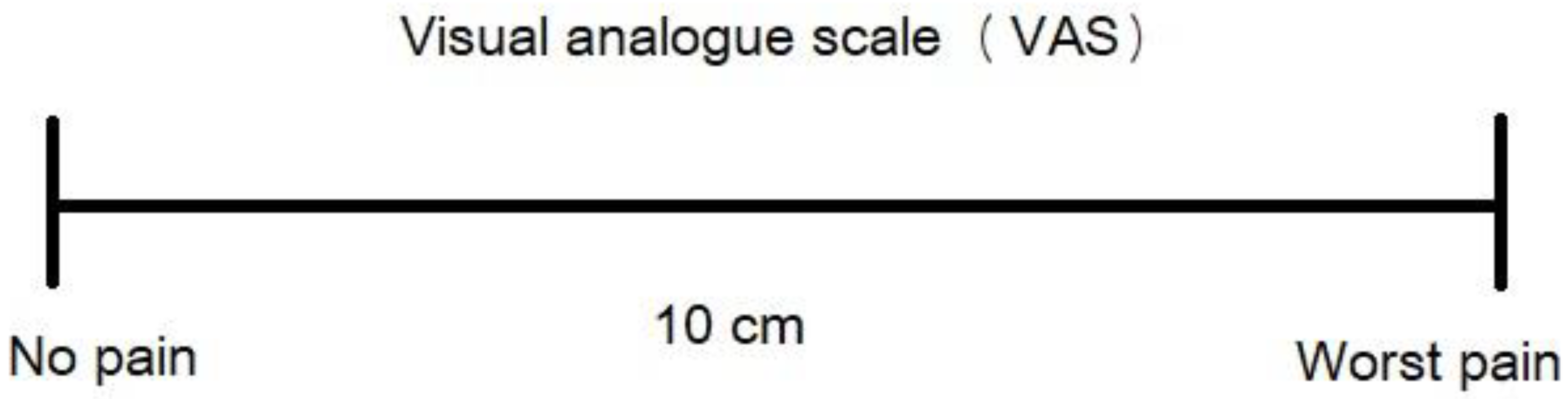
5. Dentin Hypersensitivity Assessment Methods

5.1. Response-Based Methods

Within response-based methods, the stimulus remains constant while the patient’s response varies. An illustrative example of this approach involves employing an air current for a specific duration. During this method, adjacent teeth are isolated using the examiner’s fingers, and a stream of air with a pressure of 60 ± 5 psi and a temperature of 21 ± 1 °C is directed perpendicularly to the tooth under examination from a distance of 1 cm for a duration of 1 s. Immediately following the stimulus, the patient rates the pain by visually recording it on an analog table (visual analog scale—VAS), which features gradations spanning from no pain to the most intense pain (Figure 1) [54]. Alternatively, another method entails using a pain scale wherein the patient verbally describes the pain intensity [55]. Utilizing VAS for pain recording offers the advantage of being more accurately portrayed the various levels of pain intensity.
Schiff’s scale of sensitivity to cold air, frequently introduced, comprises four distinct gradations as follows [51,56,57,58]:
  • 0—The patient displays no response to the air stimulus.
  • 1—The patient reacts to the air stimulus without requesting it to cease.
  • 2—The patient reacts to the air stimulus and either requests it to stop or moves away from it.
  • 3—The patient responds to the air stimulus, deeming it painful, and explicitly asks for it to stop.

5.2. Stimulus-Based Methods

In stimulus-based investigations of dentin sensitivity, the stimulus is adjusted with varying levels of intensity. One frequently used approach in this category involves utilizing a probe that is adjusted to exert forces on the tooth in increments of 10 g, employing an electronic device for control [51,56,58]. In this method, the patient is directed to indicate the point at which they initially experience pain subsequent to the probe stimulation, and the applied force is recorded. While thermal and electrical devices have been previously used to generate increasing stimulus intensities, concerns have arisen regarding their efficacy [59]. Drawbacks of methods hinging on stimulus intensity include the potential for repeated stimuli to alter sensitivity and the possibility that the expectation of pain linked to the stimulus could influence the degree of response. Additionally, the time-intensive nature of these approaches leads to a reduction in the number of teeth that can be examined within a given session [59].

6. Therapeutic Approaches of Dentin Hypersensitivity

The treatment of patients with DH entails a structured series of steps, comprising six key points [2]:
  • Accurate diagnosis subsequent to recording the patient’s dental history and initial clinical examination.
  • Identification of etiological and predisposing factors, with particular attention to dietary and brushing habits and their potential impact on erosion and abrasion.
  • Differential diagnosis to rule out other conditions presenting similar pain symptoms.
  • Therapeutic intervention for any coexisting conditions manifesting symptoms similar to DH.
  • Removal or mitigation of etiological and predisposing factors, accompanied by guidance on appropriate dietary habits and oral hygiene practices.
  • Dispensing patient instructions or implementing office-based treatments in accordance with the patient’s requirements.
Addressing the potential etiological factors contributing to DH requires comprehensive patient education. The patient should be instructed in proper brushing techniques, advised against using abrasive toothpaste (such as whitening dentifrices) [60] and firm toothbrushes, cautioned against excessive brushing force and advised to abstain from brushing for at least one hour after consuming acidic foods or beverages. Acidic substances, whether from dietary intake or gastric reflux, have been identified as triggers or exacerbators of DH [61]. Following the consumption of acidic foods or beverages, the consumption of alkaline drinks like milk or neutral options such as water is recommended. Practicing proper oral hygiene can also play a substantial role in maintaining healthy periodontal tissues and preventing gum recession, which can lead to root exposure and potentially trigger DH [61].

6.1. At-Home Therapeutic Treatments of Dentin Hypersensitivity

For at-home management of DH, patients receive guidance to utilize specialized toothpastes and mouthwashes designed to effectively alleviate sensitivity [62,63,64]. These approaches function by either impeding nerve impulse transmission or blocking dentin tubules (Table 1). If DH persists for 2 to 4 weeks after utilizing specialized toothpaste or mouthwashes, patients should consider progressing to the second phase of treatment, which involves professional treatment at the dentist’s office.

6.1.1. Potassium Salts

Many DH-reducing toothpastes contain potassium salts such as potassium chloride (KCl), potassium citrate (K3C6H5O7), and potassium nitrate (KNO3) [65]. Through penetration into dentin tubules, these salts numb nerve fibers, consequently diminishing tooth sensitivity. More specifically, they can increase the pain threshold of nerve fibers in response to specific stimuli [66,67]. Toothpastes containing KNO3 and fluoride have demonstrated positive impacts on DH reduction [66,68,69,70]. For optimal outcomes, these toothpastes should be used in conjunction with soft toothbrushes and minimal water, two times daily following tooth brushing for at least 14 days [66]. Moreover, mouthwashes containing potassium oxalate (K2C2O4), potassium nitrate, and fluoride are also deployed for daily use, significantly contributing to DH alleviation [71].

6.1.2. Sodium Fluoride

The most common fluorine compound in toothpastes is sodium fluoride (NaF). Fluoride helps to deposit minerals, such as calcium and phosphate, onto the dentin surface, which can help strengthen and harden it. Toothpastes incorporating NaF and calcium phosphate [Ca3(PO4)2], aimed at dentin remineralization, also notably mitigate DH. When fluoride ions come into contact with the tooth’s surface, they can block the exposed dentin tubules [72].

6.1.3. Stannous Fluoride

Stannous fluoride (SnF2) has shown remarkable effectiveness in sealing dentin tubules, which can be attributed to the strong attraction of Sn ions to the mineral surfaces of tooth hard tissues [73]. It creates a protective layer on the tooth’s surface by establishing bonds with exposed tissues. More specifically, tin ions (Sn+2) become part of the near-surface layer of the tooth, penetrating to a depth of around 20 μm and providing extended protection, guarding the underlying surface against further external stimuli. Moreover, the bond between F and Ca+2 resulting from the application of various fluoride salts is less robust compared to the bond established on tooth surfaces treated with SnF2 [74]. In a previous study [75], it was observed that after 8 weeks of using a toothpaste containing 0.454% SnF2, the dentin surfaces displayed effective coating, with the open dentin tubules being sealed. In contrast to the control toothpaste, the experimental toothpaste showed a significant reduction in tooth sensitivity, underscoring its notable efficacy. In addition, in a recent investigation, it was reported that the ability of a SnF2 gel to occlude dentin tubules reached 99.5% [76]. Nonetheless, despite its effectiveness, SnF2 presents some disadvantages, such as a metallic taste, the potential for tooth staining, and the possibility of diminished taste sensation with prolonged use [74].

6.1.4. Arginine

Another category comprises toothpastes containing the amino acid arginine (C6H14N4O2), recognized for their substantial DH reduction potential and anticariogenic effects [77,78,79,80]. These formulations, containing 8% arginine (arginine bicarbonate), calcium carbonate (CaCO3), and 1450 ppm fluoride, establish an alkaline environment that leads to reduced calcium and phosphorus levels on the dentin surface and within the dentin tubules. The arginine works by interacting with calcium ions to create a calcium-rich layer that can occlude the dentin tubules. One notable benefit of arginine-based toothpaste is its potential for long-lasting relief [81].

6.1.5. Nano-Hydroxyapatite

Currently, toothpastes containing nano-hydroxyapatite (n-HA) have emerged delivering positive effects in DH reduction [82,83,84]. Nano-hydroxyapatite is a form of calcium phosphate that is structurally similar to the hydroxyapatite found in natural hard tooth tissues. When applied to the tooth surface, n-HA can mimic the natural mineral composition of the tooth structure, which helps to replenish lost mineral content in areas where dentin is exposed. By strengthening and remineralizing the tooth, n-HA can reduce the sensitivity caused by the exposure of dentin tubules [85,86]. Furthermore, mouthwashes containing n-HA, and fluoride are also deployed for daily use, significantly contributing to DH reduction [87,88].

6.1.6. Bioactive Glasses

Additionally, toothpastes containing bioactive glasses in combination with fluoride have been suggested to alleviate the symptoms of DH. Bioactive glasses can effectively occlude or block the dentin tubules. When applied to the tooth surface, bioactive glass particles can penetrate the tubules and form a physical barrier within them, as well as attract tooth structural ions (Ca+2 and PO4−3) and promote the formation of hydroxyapatite crystals [76,81,89].

6.1.7. Casein Phosphopeptide-Amorphous Calcium Phosphate (CPP-ACP)

A recently introduced dentin remineralizing agent derived from milk proteins is known as Recaldent™. This product incorporates phosphopeptides and plays a role in retaining and stabilizing amorphous calcium phosphate. CPP-ACP possesses the capability to remineralize initial subsurface enamel lesions [90]. Additionally, it has been asserted that CPP-ACP significantly contributes to both the prevention and treatment of DH. The mechanism by which CPP-ACP operates involves a series of processes that collectively contribute to its beneficial effects on tooth surfaces. Firstly, CPP-ACP enhances the availability of binding sites on the tooth surface for Ca, which results in a reduction of the continuous diffusion of Ca. Moreover, ACP functions as a buffer for free calcium and phosphate ions found in dentin, helping to maintain a state of supersaturation. This state is crucial in preventing demineralization and promoting the remineralization of the tooth’s structure [91]. Another reason for the positive outcomes of CPP-ACP is that its application facilitates the formation of a crystalline layer that covers the openings of dentin tubules. This protective layer acts as a barrier against external stimuli, providing additional protection for the tooth surface [92]. However, this layer is not entirely uniform; it exhibits attached irregularities and appears as clusters of structures that fill the interprismatic spaces while partially covering the tubules [76].

6.1.8. Strontium Chloride

Strontium chloride (SrCl2), usually at a concentration of 10%, operates in a manner reminiscent of calcium within the human body [93]. It has the potential to act as a substitute for apatite biomineralization, as has been described in previous reports [94]. Within the oral environment, Sr+2, along with Ca+2 and other divalent cations sharing a similar charge/size ratio as calcium, can seamlessly integrate into the hydroxyapatite structure [95]. The scientific literature also highlights that this active element stimulates dentinogenesis in human dental pulp stem cells. This stimulation, as it has been demonstrated in previous in vitro studies, may foster their proliferation, differentiation, and mineralization, leading to the formation of tertiary dentin [96].
One of the key attributes of SrCl2 is its capacity to create intricate strontium phosphate compounds, thus exerting a chemical influence on dentin [93]. A previous report [97] affirmed the formation of a Ca and Sr apatite denoted as Ca6Sr4(PO4)6(OH)2, which emerges through the replacement of intracrystalline Ca in apatite with Sr. Furthermore, the affinity of strontium salts toward dentin remains significant due to the increased permeability of dentin and the likelihood of absorption in organic connective tissues as well as odontoblast processes [98]. This propensity leads to protein precipitation and the formation of a sealing film [99], potentially capable of restricting the transmission of external stimuli within the structure through a tubular occlusive mechanism.

6.2. In-Office Therapeutic Treatments of Dentin Hypersensitivity

Numerous products and methods have been suggested for addressing DH in dental practice, and these have been assessed through various studies, yielding divergent outcomes. While in-office treatments can provide immediate relief, long-term management of DH may require additional at-home measures. The agents employed within the dental practice for DH treatment are categorized based on their mode of action into two groups (Table 2):
  • Those that hinder nerve impulse transmission;
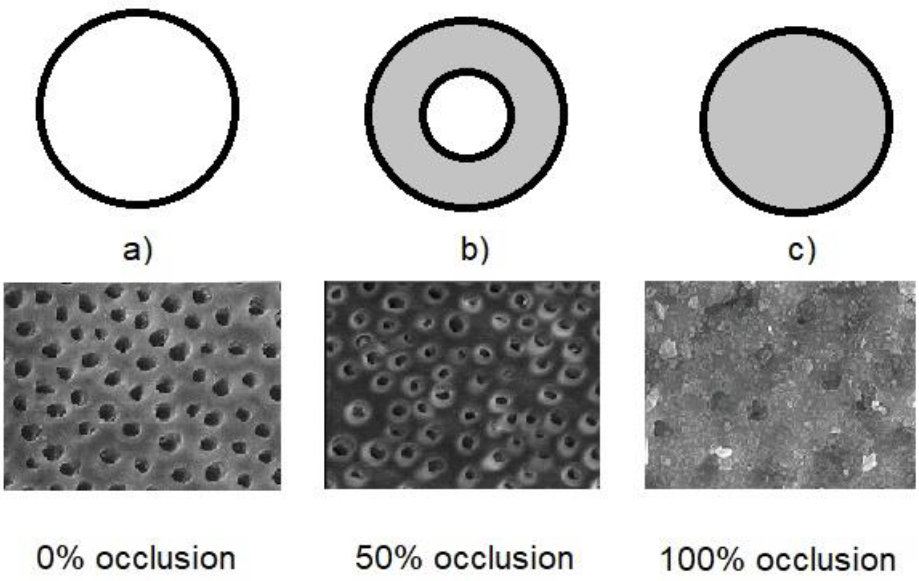
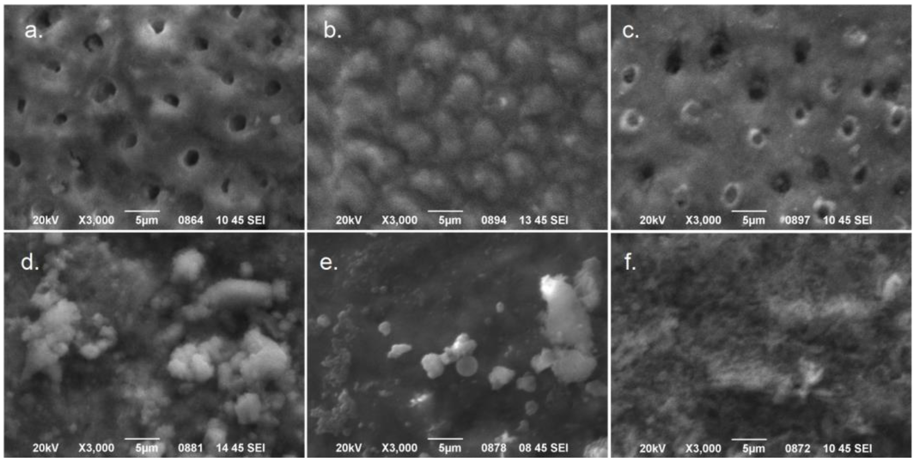
  • Those that occlude dentin tubules (Figure 2).
Table 2. The in-office therapeutic treatments for dentin hypersensitivity.
In-Office Treatments of Dentin Hypersensitivity
Active AgentsFormMechanism of ActionDuration of Therapy
Potassium saltsGelImpeding nerve impulse transmission2–3 times during a two-week period
Low-level laser irradiation (LLLT)IrradiationImpeding nerve impulse transmissionCase-sensitive
Sodium fluorideVarnish or gelOcclusion of dentin tubulesOnce
Silver diamine fluoride (SDF)SolutionOcclusion of dentin tubulesOnce
Adhesive agentsSolutionOcclusion of dentin tubulesOnce
Bioactive glass powderAir abrasionOcclusion of dentin tubulesOnce
Portland cementPasteOcclusion of dentin tubulesOnce
Oxalate saltsGel or solutionOcclusion of dentin tubulesOnce
Laser irradiationIrradiationOcclusion of dentin tubulesOnce
a. Therapeutic agents that hinder the transmission of the nerve impulses
Potassium salts (most common KNO3) and low-level laser therapies (LLLT) belong to this category.

6.2.1. Potassium Salts Gels

Potassium salts-containing products for in-office treatments are marketed mainly in the form of gels. In particular, potassium-based agents facilitate the elevation of potassium ion (K+) concentration in nerve endings, thereby diminishing the nerve’s capacity to transmit sensory stimuli and modifying its action potential [2,100]. When these salts are applied to the exposed dentin, they increase the concentration of K+ in the tubules and surrounding tissues, influencing the normal depolarization and repolarization processes of nerve cells [101]. More specifically, nerve cells rely on the movement of various ions, including Na+ and K+, to generate and transmit electrical signals, known as action potentials. When a nerve is stimulated, Na+ ions rush into the cell, causing depolarization, which is a change in the electrical potential of the cell membrane. This depolarization triggers the transmission of the nerve impulse along the nerve fiber. Potassium ions play a crucial role in repolarizing the nerve cell after the action potential has passed. They move out of the cell, helping to restore the cell’s resting electrical state. However, when potassium salts are present in high concentrations, as in the case of potassium-based agents used for treating DH, the movement of K+ becomes disrupted. The elevated concentration of K+ outside the nerve cells interferes with their ability to repolarize efficiently. This results in a dampened or altered response to sensory stimuli. Essentially, the nerve cell becomes less responsive to subsequent stimuli, reducing its capacity to transmit pain signals caused by external triggers like temperature changes or mechanical pressure. This mechanism contributes to the reduction of DH by impeding the transmission of nerve impulses associated with pain sensation [102].

6.2.2. Low-Level Laser Therapy

Low-level laser therapy (LLLT), also known as cold laser therapy or photobiomodulation therapy has been explored as a potential treatment for DH. Various laser wavelengths have been investigated for this purpose, with some of the commonly used wavelengths falling within the red (630–690 nm) and near-infrared (810–980 nm) spectrum. LLLT can engage with the dental pulp, resulting in increased odontoblastic activity, which leads to the production of tertiary dentin and a reduction in the lumen size of dentin tubules. Such lasers can also induce cell activity, trigger anti-inflammatory responses, and generate analgesic effects [103,104,105]. LLLT is dose-dependent; therefore, it is generally used in sequential appointments with a time interval. There are various clinical protocols that dental practitioners can follow. Most of the studies found that pain levels were significantly reduced 3 months after the therapies [106].
LLLT is believed to reduce pain during DH through various biological mechanisms. While the exact processes are still being studied, here are some proposed ways LLLT might work to alleviate pain in this context [107,108,109]:
  • Increased blood flow and microcirculation: LLLT has been shown to promote vasodilation and improve microcirculation in the treated area. This enhanced blood flow can facilitate the delivery of nutrients and oxygen to the tissues, aiding in their healing and reducing inflammation. Improved circulation can also help remove waste products and inflammatory mediators that contribute to pain.
  • Stimulation of cellular activity: LLLT is thought to stimulate cellular activity, including the activation of mitochondria, the cell’s energy-producing organelles. This increased cellular energy can accelerate tissue repair and regeneration, promoting healing and reducing pain.
  • Anti-inflammatory effects: LLLT is known to have anti-inflammatory effects by modulating immune responses and reducing the release of inflammatory cytokines. This can lead to a decrease in inflammation, which often accompanies pain in conditions like DH.
  • Neurological modulation: LLLT may influence nerve function and sensitivity. It could modulate nerve conduction and reduce the transmission of pain signals. Additionally, LLLT might affect nerve endings and receptors, altering their response to stimuli.
  • Endorphin release: LLLT might trigger the release of endorphins, which are natural pain-relieving chemicals produced by the body. Endorphins can help block pain signals and promote a sense of well-being.
  • Stimulation of dentin remineralization: LLLT might encourage the remineralization of dentin, potentially sealing or occluding the tubules and reducing their sensitivity.
It is important to note that the effectiveness of LLLT for DH can vary among individuals and might depend on factors such as the specific laser parameters used, the duration and frequency of treatment, and the underlying causes of hypersensitivity.
b. Therapeutic agents that occlude the dentin tubules

6.2.3. Sodium Fluoride

The impact of fluorine compounds on DH reduction has been extensively investigated [110,111,112]. Fluorine compounds lead to a reduction in calcium fluoride (CaF2) crystal presence within dentin tubules, subsequently decreasing dentin permeability [113]. These crystals are almost insoluble in saliva. The 2% sodium fluoride (NaF) varnish commonly employed in dental practice induces a reduction in calcium salts; however, these are readily eliminated from the dentin surface by saliva and brushing. To address this challenge, an acidified formulation, namely acidulated phosphate fluoride (APF 1.23%, 12,300 ppm F, pH 3.2) is available as solution, gel, or thixotropic gel, and as foam, is employed to prompt the reduction of salts deep within dentin tubules [65]. If fluoroapatite forms after the treatment, it exhibits improved stability against the effects of saliva, brushing, and dietary substances [114]. Fluorine compounds can also be employed in conjunction with iontophoresis, a method that employs an electric current to enhance ion diffusion [4,115]. Products containing fluorine compounds for in-office use are applied to exposed dentin usually in varnish forms.

6.2.4. Silver Diamine Fluoride

Silver diamine fluoride (SDF) is a topical medicament used in dentistry to treat various dental issues, including DH. When SDF comes into contact with the tooth surface, it reacts with the hydroxyapatite in the dentin and enamel, forming silver fluoride (AgF) and silver chloride (AgCl) compounds. These compounds can occlude the tubules present in the dentin [116], preventing the external stimuli from reaching the nerve endings, thus reducing sensitivity [117]. It is important to note that while SDF can be highly effective in managing DH [118], it may cause temporary discoloration of the treated area. The darkening of the tooth surface occurs due to the formation of silver compounds and for this reason, it has been suggested to be accompanied by potassium iodide (KI) [119]. This potential aesthetic concern should be discussed with patients before using SDF (38%) as a treatment option. Consequently, SDF is usually considered for use in cases where conventional treatments for DH, such as desensitizing toothpaste or fluoride varnishes, have not provided adequate relief.

6.2.5. Adhesive Agents

In contrast to other agents that reduce DH, adhesive agents exert a prolonged or even permanent effect. When combined with resin composites, they create an occlusion of the dentin tubules, yielding a hybrid zone [68]. In the past, older adhesive agents formed this hybrid zone through the removal of the smear layer and etching of the dentin surface, allowing for deep resin penetration into the dentin tubules [120]. However, newer bonding agents alter the smear layer and integrate it into the hybrid zone. It is assumed that these modern adhesive agents can effectively address and act preventively against DH. An illustrative instance of an adhesive agent utilized to mitigate DH is Gluma (Heraeus, Hanau, Germany; Kulzer), which comprises hydroxyethyl methacrylate (HEMA, Amsterdam, The Netherlands), glutaraldehyde, and fluoride. Glutaraldehyde prompts protein coagulation within the dentin tubules, while HEMA penetrates the tubules and blocks them. Clinical studies have substantiated Gluma’s significant efficacy in DH reduction [121].

6.2.6. Air Abrasion with Bioactive Glass Particles

Bioactive glasses have demonstrated a significant role in promoting dentin remineralization [110,121]. Their principal constituent, silicon (Si), functions as a nucleation site for calcium and phosphorus ions. Scanning electron microscopy (SEM) studies have revealed that the application of this bioactive substance leads to the formation of an apatite layer, ultimately occluding dentin tubules [122,123].
Bioactive glasses represent specialized biomaterials comprised of glass that are notable for their tendency to react on their surfaces. When these glasses come into contact with the surface of teeth and interact with saliva in acidic conditions, they can dissolve. This dissolution releases calcium and phosphate ions while also raising the pH levels [124]. This series of actions set off a chain of chemical responses that result in the formation of a layer made up of hydroxycarbonate apatite [125]. This apatite layer shares similarities with the mineral hydroxyapatite, which is commonly found in dentin. Both substances can create a chemical bond, promoting a stronger attachment of the glass particles to the dentin surface [126]. It is important to note that the complete development of this apatite layer takes some time, often spanning multiple hours. This gradual formation of reparative dentin, characterized by its low permeability, hinders the diffusion of harmful substances from dentin tubules toward the dental pulp. As a result, it restricts the movement of dentin fluid, offering a long-lasting solution for tooth sensitivity [127].
A commonly used variety of bioactive glass in dentistry is known as Bioglass 45S5, which is a calcium sodium-phospho-silicate compound sold under the trade name NovaMin®. In general, glasses are amorphous solids lacking a defined crystal structure, primarily composed of silica-based elements with minor additives. However, Bioglass 45S5 diverges from this standard by featuring lower levels of silica and higher concentrations of calcium and phosphorus compared to typical glasses [128]. A previous study demonstrated that using a powder of Bioglass 45S5 for air abrasion can effectively block the dentin tubules (>99%) [76].

6.2.7. Portland Cement

Portland cement, specifically a type called “white Portland cement”, has been explored as a potential treatment for DH. Similarly, to bioactive glasses, Portland cement, primarily comprised of Si, has shown substantial potential in treating DH. Its attributes facilitate the occlusion of dentin tubules through the remineralization of dentin [115]. White Portland cement has fine particles that can infiltrate and occlude these dentin tubules. It contains calcium silicates, which can release calcium and hydroxide ions when they come into contact with moisture, contributing to the remineralization of the dentin, and making the tooth structure stronger and more resilient [129]. It is important to note that while white Portland cement has been investigated as a treatment for DH, it is not a standard or widely accepted treatment in dentistry.

6.2.8. Oxalate Salts

Oxalates have been identified as agents capable of occluding dentin tubules, potentially leading to a reduction in dentinal permeability of up to 98% [130,131]. The application of 28% potassium oxalate can facilitate the formation of calcium oxalate within the dentin tubules. For enhanced efficacy, dentin etching can be employed in conjunction with oxalates [131]. Specifically, oxalate salts, such as potassium oxalate and ferric oxalate [132], are believed to work through a mechanism of precipitation and formation of crystals within the dentin tubules [133]. Oxalate salts have the ability to react with Ca+2 present in the dentin structure. Indeed, when applied to the exposed dentin surface, these salts can react with the Ca+2 in the tubules and surrounding dentin, leading to the formation of insoluble calcium oxalate crystals. The formation of calcium oxalate crystals within the dentin tubules creates physical barriers, which can partially or completely block the dentin tubules [132,133]. It is important for patients to be aware that oxalate treatments might cause tooth discoloration or lead to the formation of insoluble deposits on the teeth. As such, the potential aesthetic concerns associated with these treatments should be discussed with patients before their use.

6.2.9. Laser Irradiation Treatments

According to the results of various studies, the effectiveness of the laser in the treatment of DH varies significantly and ranges between 5–100% [134,135,136]. Patients often experience immediate relief from DH after laser treatment. The results depend on the wavelength of the laser and the setting parameters, such as application time, mode of irradiation (pulsed or continuous), and average power [137,138,139].
Various laser wavelengths have been utilized for the reduction of the DH symptoms. More specifically, diodes (810–980 nm) [139,140,141], neodymium: yttrium–aluminum–garnet—Nd:YAG (1064 nm) [142,143], erbium: yttrium–aluminum–garnet—Er:YAG (2940 nm) [138,144], erbium, chromium: yttrium–scandium–gallium–garnet—Er,Cr:YSGG (2780 nm) [76,145], and carbon dioxide—CO2 (10,600 nm) [146,147] lasers have been employed against DH.
The mechanism of action of lasers has not been definitively determined, but multiple theories exist regarding the effects of laser exposure on dentin. The most widely accepted one suggests that it involves the occlusion or sealing of dentin tubules through a process of dentin melting and subsequent recrystallization [148]. When addressing tooth sensitivity, it is crucial for the laser beam to avoid ablation of the treated dentin surface and instead induce only structural and chemical transformations. Ablation of dentin can lead to a surface that lacks a smear layer, has open dentin tubules, and exhibits uneven textures, which is an undesirable outcome for the procedure [149]. Furthermore, using laser settings exceeding 0.75 W may result in the formation of charred areas, making such protocols unsuitable for addressing DH [150]. There have been suggestions that employing lower power settings might initiate the dissipation of dentinal fluid. This, in turn, could lead to a reduction in dentin permeability, ultimately resulting in a decrease in dentinal pain [148]. Representative SEM images of the rate of occlusion after various treatments for DH are presented in Figure 3 [73]. It is important to mention that DH cannot lead to necrosis of the pulp like it could in cases of extensive tooth restorations [151].

7. Conclusions

Although numerous methods have been suggested for addressing DH, a definitive solution to the problem remains elusive. The outcomes derived from clinical studies assessing the efficacy of diverse treatment approaches for DH often yield conflicting results. One plausible explanation for this disparity is that assessing objective pain intensity proves challenging due to the inherently subjective and intricate nature of pain. Hence, the effective therapeutic management of DH should rest upon accurate diagnosis alongside robust measurement and evaluation. The development of new materials and techniques (at-home and in-office) to treat DH has led to the possibility of alleviating the symptoms in many clinical cases. This literature review focuses on the most important contemporary information on this topic to help dental clinicians stay aware of the latest developments in the treatment of DH.

Author Contributions

Conceptualization, D.D.; methodology, D.D., O.G. and C.B.; validation, C.B. and O.G.; formal analysis, D.D.; investigation, D.D. and C.B.; data curation, D.D.; writing—original draft preparation, D.D.; writing—review and editing, O.G. and C.B.; supervision, D.D. All authors have read and agreed to the published version of the manuscript.

Funding

This research received no external funding.

Institutional Review Board Statement

Not applicable.

Informed Consent Statement

Not applicable.

Data Availability Statement

The study does not report any data.

Conflicts of Interest

The authors declare no conflict of interest.

References

  1. Dowell, P.; Addy, Μ.; Dummer, Ρ. Dentine hypersensitivity: Aetiology, differential diagnosis and management. Br. Dent. J. 1985, 158, 97–98. [Google Scholar] [CrossRef] [PubMed]
  2. Canadian Advisory Board on Dentin Hypersensitivity. Consensus-based recommendations for the diagnosis and management of dentin hypersensitivity. J. Can. Dent. Assoc. 2003, 69, 221–226. [Google Scholar]
  3. Holland, G.R.; Narhi, M.N.; Addy, M.; Gangarosa, L.; Orchardson, R. Guidelines for the design and conduct of clinical trials on dentine hypersensitivity. J. Clin. Periodontol. 1997, 24, 808–813. [Google Scholar] [CrossRef] [PubMed]
  4. Porto, I.C.; Andrade, A.K.; Montes, M.A. Diagnosis and treatment of dentinal hypersensitivity. J. Oral Sci. 2009, 51, 323–332. [Google Scholar] [CrossRef]
  5. Brännström, M. Etiology of dentin hypersensitivity. Proc. Finn. Dent. Soc. 1992, 88 (Suppl. 1), 7–14. [Google Scholar] [PubMed]
  6. Ciucchi, B.; Bouillaguet, S.; Holz, J.; Pashley, D.H. Dentinal fluid dynamics in human teeth, in vivo. J. Endod. 1995, 21, 191–194. [Google Scholar] [CrossRef]
  7. Gernhardt, C.R. How valid and applicable are current diagnostic criteria and assessment methods for dentin hypersensitivity? An overview. Clin. Oral Investig. 2013, 17 (Suppl. 1), S31–S40. [Google Scholar] [CrossRef]
  8. West, N.X. Dentine hypersensitivity: Preventive and therapeutic approaches to treatment. Periodontology 2000, 48, 31–41. [Google Scholar] [CrossRef]
  9. Trushkowsky, R.D.; Oquendo, A. Treatment of dentin hypersensitivity. Dent. Clin. N. Am. 2011, 55, 599–608. [Google Scholar] [CrossRef] [PubMed]
  10. Bak, S.; Kim, J.Y.; Chung, B.; Kim, J.; Cho, M.J.; Ha, W.; Lee, K.H.; Jung, Y. A randomized controlled clinical evaluation of desensitization efficacy of a newly developed toothpaste with highly stabilized SnF2. Am. J. Dent. 2023, 36, 183–187. [Google Scholar]
  11. Jang, J.H.; Oh, S.; Kim, H.J.; Kim, D.S. A randomized clinical trial for comparing the efficacy of desensitizing toothpastes on the relief of dentin hypersensitivity. Sci. Rep. 2023, 13, 5271. [Google Scholar] [CrossRef] [PubMed]
  12. Arshad, S.; Zaidi, S.J.A.; Farooqui, W.A. Comparative efficacy of BioMin-F, Colgate Sensitive Pro-relief and Sensodyne Rapid Action in relieving dentin hypersensitivity: A randomized controlled trial. BMC Oral Health 2021, 21, 498. [Google Scholar] [CrossRef] [PubMed]
  13. West, N.X. Dentine hypersensitivity. Monogr. Oral Sci. 2006, 20, 173–189. [Google Scholar] [PubMed]
  14. Addy, M.; West, Ν. Etiology, mechanisms, and management of dentine hypersensitivity. Curr. Opin. Periodontol. 1994, 1, 71–77. [Google Scholar]
  15. Absi, E.G.; Addy, M.; Adams, D. Dentine hypersensitivity. A study of the patency of dentinal tubules in sensitive and non-sensitive cervical dentine. J. Clin. Periodontol. 1987, 14, 280–284. [Google Scholar] [CrossRef]
  16. Absi, E.G.; Addy, M.; Adams, D. Dentine hypersensitivity. The development and evaluation of a replica technique to study sensitive and non-sensitive cervical dentine. J. Clin. Periodontol. 1989, 16, 190–195. [Google Scholar] [CrossRef]
  17. Zero, D.T.; Lussi, A. Erosion—chemical and biological factors of importance to the dental practitioner. Int. Dent. J. 2005, 55, 285–290. [Google Scholar] [CrossRef]
  18. Pashley, D.H.; Tay, F.R. Pulpodentin complex. In Seltzer and Bender’s Dental Pulp, 2nd ed.; Hargreaves, K.M., Goodis, H.E., Tay, F.R., Eds.; Quintessence Publishing Co.: Chicago, IL, USA, 2012; pp. 47–66. [Google Scholar]
  19. Pashley, D.H.; Livingston, M.J.; Greenhill, J.D. Regional resistances to fluid flow in human dentine in vitro. Arch. Oral Biol. 1978, 23, 807–810. [Google Scholar] [CrossRef]
  20. Pashley, D.H. How can sensitive dentine become hypersensitive and can it be reversed? J. Dent. 2013, 41, S49–S55. [Google Scholar] [CrossRef]
  21. Carrilho, M.R.; Tay, F.R.; Sword, J.; Donnelly, A.; Agee, K.A.; Nishitani, Y.; Sadek, F.T.; Carvalho, R.M.; Pashley, D.H. Dentine sealing provided by smear layer/smear plugs vs. adhesive resins/resin tags. Eur. J. Oral Sci. 2007, 115, 321–329. [Google Scholar] [CrossRef]
  22. West, N.X.; Lussi, A.; Seong, J.; Hellwig, E. Dentin hypersensitivity: Pain mechanisms and aetiology of exposed cervical dentin. Clin. Oral Investig. 2013, 17 (Suppl. 1), S9–S19. [Google Scholar] [CrossRef] [PubMed]
  23. Nähri, M.; Jyväsjärvi, E.; Virtannen, A. Role of intradental A and C- type fibres in dental pain mechanisms. Proc. Finn. Dent. Soc. 1992, 88 (Suppl. 1), 507–516. [Google Scholar]
  24. Matthews, B.; Andrew, D.; Wanachantararak, S. Biology of the Dental Pulp with Special Reference to Its Vasculature and Innervation. In Tooth Wear and Sensitivity; Embery, G., Edgar, W.M., Orchardson, R., Addy, M., Eds.; Taylor and Francis: London, UK, 2000; pp. 39–51. [Google Scholar]
  25. Michelich, V.; Schuster, G.S.; Pashley, D.H. Bacterial penetration of human dentin in vitro. J. Dent. Res. 1980, 59, 1398–1403. [Google Scholar] [CrossRef]
  26. Pashley, D.H.; Matthew, W.G. The effects of outward forced corrective flow on inward diffusion in human dentin in vitro. Arch. Oral Biol. 1993, 38, 577–582. [Google Scholar] [CrossRef]
  27. Kerns, D.G.; Scheidt, M.J.; Pashley, D.H.; Homer, J.A.; Strong, S.L.; Van Dyke, T.E. Dentinal tubule occlusion and root hypersensitivity. J. Periodontol. 1991, 62, 421–428. [Google Scholar] [CrossRef]
  28. Caviedes-Bucheli, J.; Munoz, H.R.; Azuero-Holguin, M.M.; Ulate, E. Neuropeptides in dental pulp: The silent protagonists. J. Endod. 2008, 34, 773–788. [Google Scholar] [CrossRef] [PubMed]
  29. Byers, M.; Närhi, M.V.O. Dental injury models: Experimental tools for understanding neuroinflammatory nociceptor functions. Crit. Rev. Oral Biol. Med. 1999, 10, 4–39. [Google Scholar] [CrossRef] [PubMed]
  30. Byers, M.; Henry, M.A.; Närhi, M.V.O. Dental innervation and its response to tooth injury. In Seltzer and Bender’s Dental Pulp, 2nd ed.; Hargreaves, K.M., Goodis, H.E., Tay, F.R., Eds.; Quintessence Publishing Co.: Chicago, IL, USA, 2012; pp. 133–157. [Google Scholar]
  31. Renton, T.; Yiangou, Y.; Plumpton, C.; Tate, S.; Bountra, C.; Anand, P. Sodium channel Nav1.8 immunoreactivity in painful human dental pulp. BMC Oral Health 2005, 5, 5. [Google Scholar] [CrossRef]
  32. Lundy, T.; Stanley, H.R. Correlation and pulpal histopathology and clinical symptoms in human teeth subjected to experimental irritation. Oral Surg. Oral Med. Oral Pathol. 1969, 27, 187–201. [Google Scholar] [CrossRef]
  33. Pashley, D.H.; Kepler, E.E.; Williams, E.C.; Okabe, A. Progressive decrease in dentine permeability following cavity preparation. Arch. Oral Biol. 1983, 28, 853–858. [Google Scholar] [CrossRef]
  34. Pashley, D.H.; Galloway, S.E.; Stewart, F.P. Effects of fibrinogen in vivo on dentin permeability in the dog. Arch. Oral Biol. 1984, 29, 725–728. [Google Scholar] [CrossRef] [PubMed]
  35. Hahn, C.L.; Overton, B. The effects of immunoglobulins on the convective permeability of human dentine in vitro. Arch. Oral Biol. 1997, 42, 835–843. [Google Scholar] [CrossRef]
  36. Gillam, D.G.; Orchardson, R.; Närhi, M.V.O.; Kontturi-Närhi, V. Present and future methods for the evaluation of pain associated with dentine hypersensitivity. In Tooth Wear and Sensitivity; Addy, M., Embery, G., Edgar, W.M., Orchardson, R., Eds.; Martin Dunitz Ltd.: London, UK, 2000; pp. 283–497. [Google Scholar]
  37. Miglani, S.; Aggarwal, V.; Ahuja, Β. Dentin hypersensitivity: Recent trends in management. J. Conserv. Dent. 2010, 13, 218–224. [Google Scholar] [CrossRef]
  38. O’Toole, S.; Bartlett, D. The relationship between dentine hypersensitivity, dietary acid intake and erosive tooth wear. J. Dent. 2017, 67, 84–87. [Google Scholar] [CrossRef]
  39. Bamise, C.T.; Olusile, A.O.; Oginni, A.O. An analysis of the etiological and predisposing factors related to dentin hypersensitivity. J. Contemp. Dent. Pract. 2008, 9, 52–59. [Google Scholar]
  40. Boiko, O.V.; Baker, S.R.; Gibson, B.J.; Locker, D.; Sufi, F.; Barlow, A.P.S.; Robinson, P.G. Construction and validation of the quality of life measure for dentine hypersensitivity (DHEQ). J. Clin. Periodontol. 2010, 37, 973–980. [Google Scholar] [CrossRef]
  41. Slade, G.D.; Spencer, A.J. Development and evaluation of the Oral Health Impact Profile. Community Dent. Health 1994, 11, 3–11. [Google Scholar] [PubMed]
  42. John, M.T.; Miglioretti, D.L.; LeResche, L.; Koepsell, T.D.; Hujoel, P.; Micheelis, W. German short forms of the Oral Health Impact Profile. Community Dent. Oral Epidemiol. 2006, 34, 277–288. [Google Scholar] [CrossRef]
  43. Hassel, A.J.; Rolko, C.; Koke, U.; Leisen, J.; Rammelsberg, Ρ. A German version of the GOHAI. Community Dent. Oral Epidemiol. 2008, 36, 34–42. [Google Scholar] [CrossRef] [PubMed]
  44. Gillam, D.G.; Newman, H.N. Assessment of pain in cervical dentinal sensitivity studies. A review. J. Clin. Periodontol. 1993, 20, 383–394. [Google Scholar] [CrossRef]
  45. Ernst, Ε. Placebo: New insights into an old enigma. Drug. Discov. Today 2007, 12, 413–418. [Google Scholar] [CrossRef] [PubMed]
  46. McCarney, R.; Warner, J.; Iliffe, S.; van Haselen, R.; Griffin, M.; Fisher, P. The Hawthorne Effect: A randomized, controlled trial. BMC Med. Res. Μethodol. 2007, 7, 30. [Google Scholar] [CrossRef]
  47. Addy, M.; West, N.X.; Barlow, A.; Smith, S. Dentine hypersensitivity: Is there both stimulus and placebo responses in clinical trials? Int. J. Dent. Hyg. 2007, 5, 53–59. [Google Scholar] [CrossRef]
  48. Barsky, A.J.; Saintfort, R.; Rogers, M.P.; Borus, J.F. Nonspecific medication side effects and the nocebo phenomenon. JAMA 2002, 287, 622–627. [Google Scholar] [CrossRef] [PubMed]
  49. Cunha-Cruz, J.; Stout, J.R.; Heaton, L.J.; Wataha, J.C. Dentin hypersensitivity and oxalates: A systematic review. J. Dent. Res. 2011, 90, 304–310. [Google Scholar] [CrossRef]
  50. Gillam, D.G.; Aris, A.; Bulman, J.S.; Newman, H.N.; Ley, F. Dentine hypersensitivity in subjects recruited for clinical trials: Clinical evaluation prevalence and intra-oral distribution. J. Oral Rehabil. 2002, 29, 226–231. [Google Scholar] [CrossRef]
  51. Schiff, T.; Delgado, E.; Zhang, Y.P.; Cummins, D.; DeVizio, W.; Mateo, L.R. Clinical evaluation of the efficacy of an in-office desensitizing paste containing 8% arginine and calcium carbonate in providing instant and lasting relief of dentin hypersensitivity. Am. J. Dent. 2009, 22, 8A–15A. [Google Scholar]
  52. Kobler, A.; Kuss, O.; Schaller, H.G.; Gernhardt, C.R. Clinical effectiveness of a strontium chloride-containing desensitizing agent over 6 months: A randomized, double-blind, placebo-controlled study. Quintessence Int. 2008, 39, 321–325. [Google Scholar] [PubMed]
  53. Orchardson, R.; Coffins, W.J. Thresholds of hypersensitive teeth to 2 forms of controlled stimulation. J. Clin. Periodontol. 1987, 14, 68–73. [Google Scholar] [CrossRef] [PubMed]
  54. Hicks, C.L.; von Baeyer, C.L.; Spafford, P.A.; van Korlaar, I.; Goodenough, Β. The Faces Pain Scale-Revised: Toward a common metric in pediatric pain measurement. Pain 2001, 93, 173–183. [Google Scholar] [CrossRef] [PubMed]
  55. Melzack, R. The short-form McGill Pain Questionnaire. Pain 1987, 30, 191–197. [Google Scholar] [CrossRef]
  56. Schiff, T.; Delgado, E.; Zhang, Y.P.; DeVizio, W.; Cummins, D.; Mateo, L.R. The clinical effect of a single direct topical application of a dentifrice containing 8.0% arginine, calcium carbonate, and 1450 ppm fluoride on dentin hypersensitivity: The use of a cotton swab applicator versus the use of a fingertip. J. Clin. Dent. 2009, 20, 131–136. [Google Scholar]
  57. Petrou, I.; Heu, R.; Stranick, M.; Lavender, S.; Zaidel, L.; Cummins, D.; Sullivan, R.J.; Hsueh, C.; Gimzewski, J.K. A breakthrough therapy for dentin hypersensitivity: How dental products containing 8% arginine and calcium carbonate work to deliver effective relief of sensitive teeth. J. Clin. Dent. 2009, 20, 23–31. [Google Scholar] [PubMed]
  58. Hamlin, D.; Milliams, K.P.; Delgado, E.; Zhang, Y.P.; DeVizio, W.; Mateo, L.R. Clinical evaluation of the efficacy of a desensitizing paste containing 8% arginine and calcium carbonate for the in-office relief of dentin hypersensitivity associated with dental prophylaxis. Am. J. Dent. 2009, 22, 16A–20A. [Google Scholar] [PubMed]
  59. Walline, B.W.; Wagner, J.G.; Marx, D.B.; Reinhardt, R.A. Comparison of methods for measuring root and mucogingival sensitivity. Oral Surg. Oral Med. Oral Pathol. Oral Radiol. 2000, 90, 641–646. [Google Scholar] [CrossRef]
  60. Dionysopoulos, D.; Papageorgiou, S.; Papadopoulos, C.; Davidopoulou, S.; Konstantinidis, A.; Tolidis, K. Effect of whitening toothpastes with different active agents on the abrasive wear of dentin following tooth brushing simulation. J. Funct. Biomater. 2023, 14, 268. [Google Scholar] [CrossRef]
  61. Eisenburger, M.; Addy, M. Erosion and attrition of human enamel in vitro part I: Interaction effects. J. Dent. 2002, 30, 341–347. [Google Scholar] [CrossRef] [PubMed]
  62. Amaechi, B.T.; Mathews, S.M.; Mensinkai, P.K. Effects of theobromine-containing toothpaste on dentin tubule occlusion in situ. Clin. Oral Investig. 2015, 19, 109–116. [Google Scholar] [CrossRef] [PubMed]
  63. Zhong, Y.; Liu, J.; Li, X.; Yin, W.; He, Τ.; Hu, D.; Liao, Y.; Yao, X.; Wang, Y. Effect of a novel bioactive glass-ceramic on dental tubule occlusion: An in vitro study. Aust. Dent. J. 2015, 60, 96–103. [Google Scholar] [CrossRef]
  64. Magno, M.B.; Nascimento, G.C.; Da Penha, N.K.; Pessoa, O.F.; Loretto, B.C.; Maia, L.C. Difference in effectiveness between strontium acetate and arginine-based toothpastes to relieve dentin hypersensitivity. A systematic review. Am. J. Dent. 2015, 28, 40–44. [Google Scholar]
  65. Cummins, D. Recent advances in dentin hypersensitivity: Clinically proven treatments for instant and lasting sensitivity relief. Am. J. Dent. 2010, 23, 3A–13A. [Google Scholar]
  66. Kopycka-Kedzierawski, D.T.; Meyerowitz, C.; Litaker, M.S.; Heft, M.W.; Tasgaonkar, N.; Day, M.R.; Porter-Williams, A.; Gordan, V.V.; Yardic, R.L.; Lawhorn, T.M.; et al. National Dental PBRN Collaborative Group. Management of dentin hypersensitivity by practitioners in The National Dental Practice-Based Research Network. J. Am. Dent. Assoc. 2017, 148, 728–736. [Google Scholar] [CrossRef]
  67. Peacock, J.M.; Orchardson, R. Effects of potassium ions on action potential conduction in A- and C-fibers of rat spinal nerves. J. Dent. Res. 1995, 74, 634–641. [Google Scholar] [CrossRef] [PubMed]
  68. Orchardson, R.; Gillam, D.G. Managing dentin hypersensitivity. J. Am. Dent. Assoc. 2006, 137, 990–998. [Google Scholar] [CrossRef]
  69. Aranha, A.C.; Pimenta, L.A.; Marchi, G.M. Clinical evaluation of desensitizing treatments for cervical dentin hypersensitivity. Braz. Oral Res. 2009, 23, 333–339. [Google Scholar] [CrossRef]
  70. Chu, C.H.; Lo, E.C.M. Dentin hypersensitivity: A review. Hong Kong Dent. J. 2010, 7, 15–22. [Google Scholar]
  71. Sharma, D.; McGuire, J.A.; Gallob, J.T.; Amini, P. Randomized clinical efficacy trial of potassium oxalate mouthrinse in relieving dentinal sensitivity. J. Dent. 2013, 41 (Suppl. 4), S40–S48. [Google Scholar] [CrossRef] [PubMed]
  72. Sowinski, J.A.; Battista, G.W.; Petrone, M.E.; Chaknis, P.; Zhang, Y.P.; DeVizio, W.; Volpe, A.R.; Proskin, H.M. A new desensitizing dentifrice-an 8-week clinical investigation. Compend. Contin. Educ. Dent. Suppl. 2000, 27, 11–16. [Google Scholar]
  73. Faller, R.V.; Eversole, S.L. Protective effects of SnF2—Part III. Mechanism of barrier layer attachment. Int. Dent. J. 2014, 64, 16–21. [Google Scholar] [CrossRef]
  74. Faller, R.V.; Eversole, S.L.; Tzeghai, G.E. Enamel protection: A comparison of marketed dentifrice performance against dental erosion. Am. J. Dent. 2011, 24, 205–210. [Google Scholar]
  75. Hines, D.; Xu, S.; Stranick, M.; Lavender, S.; Pilch, S.; Zhang, Y.P.; Sullivan, R.; Montesani, L.; Montesani, L.; Mateo, L.R.; et al. Effect of a stannous fluoride toothpaste on dentinal hypersensitivity: In vitro and clinical evaluation. J. Am. Dent. Assoc. 2019, 150, S47–S59. [Google Scholar] [CrossRef] [PubMed]
  76. Papazisi, N.; Dionysopoulos, D.; Naka, O.; Strakas, D.; Davidopoulou, S.; Tolidis, K. Efficiency of various tubular occlusion agents in human dentin after in-office tooth bleaching. J. Funct. Biomater. 2023, 14, 430. [Google Scholar] [CrossRef] [PubMed]
  77. Yan, B.; Yi, J.; Li, Y.; Chen, Y.; Shi, Z. Arginine-containing toothpastes for dentin hypersensitivity: Systematic review and meta-analysis. Quintessence Int. 2013, 44, 709–723. [Google Scholar] [PubMed]
  78. França, I.L.; Sallum, E.A.; Do Vale, H.F.; Casati, M.Z.; Sallum, A.W.; Stewart, B. Efficacy of a combined in office/home-use desensitizing system containing 8% arginine and calcium carbonate in reducing dentin hypersensitivity: An 8 week randomized clinical study. Am. J. Dent. 2015, 28, 45–50. [Google Scholar] [PubMed]
  79. Bae, J.H.; Kim, Y.K.; Myung, S.K. Desensitizing toothpaste versus placebo for dentine hypersensitivity: A systematic review and meta-analysis. J. Clin. Periodontol. 2015, 42, 131–141. [Google Scholar] [CrossRef] [PubMed]
  80. Dionysopoulos, D. New strategies for caries prevention. The role of alkali production in dental biofilms. ARC J. Dent. Sci. 2016, 1, 19–21. [Google Scholar]
  81. Champaiboon, C.; Ongphichetmetha, N.; Lertpimonchai, A. Bioactive glass and arginine dentifrices reduce root sensitivity during daily activities following non-surgical periodontal therapy: A randomized controlled trial. J. Periodontol. 2023; online ahead of print. [Google Scholar] [CrossRef] [PubMed]
  82. Low, S.B.; Allen, E.P.; Kontogiorgos, E.D. Reduction in dental hypersensitivity with nano-hydroxyapatite, potassium nitrate, sodium monoflurophosphate and antioxidans. Open Dent. J. 2015, 9, 92–97. [Google Scholar] [CrossRef]
  83. Gopinath, N.M.; John, J.; Nagappan, N.; Prabhu, S.; Kumar, E.S. Evaluation of dentifrice containing nano-hydroxyapatite for dentinal hypersensitivity: A randomized controlled trial. J. Int. Oral Health 2015, 7, 118–122. [Google Scholar]
  84. Vano, M.; Derchi, G.; Barone, A.; Covani, U. Effectiveness of nano-hydroxypatite toothpaste in reducing dentin hypersensitivity: A double-blind randomized controlled trial. Quintessence Int. 2014, 45, 703–711. [Google Scholar]
  85. Yuan, P.; Liu, S.; Lv, Y.; Liu, W.; Ma, W.; Xu, P. Effect of a dentifrice containing different particle sizes of hydroxyapatite on dentin tubule occlusion and aqueous Cr (VI) sorption. Int. J. Nanomed. 2019, 14, 5243–5256. [Google Scholar] [CrossRef]
  86. de Melo Alencar, C.; de Paula, B.L.F.; Guanipa Ortiz, M.I.; Baraúna Magno, M.; Martins Silva, C.; Cople Maia, L. Clinical efficacy of nano-hydroxyapatite in dentin hypersensitivity: A systematic review and meta-analysis. J. Dent. 2019, 82, 11–21. [Google Scholar] [CrossRef] [PubMed]
  87. Hill, R.G.; Chen, X.; Gillam, D.G. In vitro ability of a novel nanohydroxyapatite oral rinse to occlude dentine tubules. Int. J. Dent. 2015, 2015, 153284. [Google Scholar] [CrossRef] [PubMed]
  88. Lin, X.; Xie, F.; Ma, X.; Hao, Y.; Qin, H.; Long, J. Fabrication and characterization of dendrimer-functionalized nano-hydroxyapatite and its application in dentin tubule occlusion. J. Biomater. Sci. Polym. Ed. 2017, 28, 846–863. [Google Scholar] [CrossRef] [PubMed]
  89. Petrović, D.; Galić, D.; Seifert, D.; Lešić, N.; Smolić, M. Evaluation of bioactive glass treatment for dentin hypersensitivity: A systematic review. Biomedicines 2023, 11, 1992. [Google Scholar] [CrossRef]
  90. Cai, F.; Shen, P.; Morgan, M.V.; Reynolds, E.C. Remineralization of enamel subsurface lesions in situ by sugar-free lozenges containing casein phosphopeptide-amorphous calcium phosphate. Aust. Dent. J. 2003, 48, 240–243. [Google Scholar] [CrossRef]
  91. Ranjitkar, S.; Kaidonis, J.A.; Richards, L.C.; Townsend, G.C. The effect of CPPACP on enamel wear under severe erosive conditions. Arch. Oral Biol. 2009, 54, 527–532. [Google Scholar] [CrossRef]
  92. Poggio, C.; Lombardini, M.; Dagna, A.; Chiesa, M.; Bianchi, S. Protective effect on enamel demineralization of a CPP-ACP paste: An AFM in vitro study. J. Dent. 2009, 37, 949–954. [Google Scholar] [CrossRef]
  93. Pompeu, D.dS.; de Paula, B.L.F.; Barros, A.P.O.; Nunes, S.C.; Carneiro, A.M.P.; Araujo, J.L.N.; Silva, C.M. Combination of strontium chloride and photobiomodulation in the control of tooth sensitivity post-bleaching: A split-mouth randomized clinical trial. PLoS ONE 2021, 16, e0250501. [Google Scholar] [CrossRef]
  94. Durgalakshmi, D.; Ajay Rakkesh, R.; Kesavan, M.; Ganapathy, S.; Ajithkumar, T.G.; Karthikeyan, S.; Balakumar, S. Highly reactive crystalline-phase-embedded strontium-bioactive nanorods for multimodal bioactive applications. Biomater. Sci. 2018, 6, 1764–1776. [Google Scholar] [CrossRef]
  95. Saeki, K.; Marshall, G.W.; Gansky, S.A.; Parkinson, C.R.; Marshall, S.J. Strontium effects on root dentin tubule occlusion and nanomechanical properties. Dent. Mater. 2016, 32, 240–251. [Google Scholar] [CrossRef] [PubMed]
  96. Huang, M.; Hill, R.G.; Rawlinson, S.C. Strontium (Sr) elicits odontogenic differentiation of human dental pulp stem cells (hDPSCs): A therapeutic role for Sr in dentine repair? Acta Biomater. 2016, 38, 201–211. [Google Scholar] [CrossRef] [PubMed]
  97. Dedhiya, M.G.; Young, F.; Higuchi, W.I. Mechanism for the retardation of the acid dissolution rate of hydroxapatite by strontium. J. Dent. Res. 1973, 52, 1097–1109. [Google Scholar] [CrossRef]
  98. Ross, M.R. Hypersensitive teeth: Effect of strontium chloride in a compatible dentifrice. J. Periodontol. 1961, 32, 49–53. [Google Scholar] [CrossRef]
  99. Majji, P.; Murthy, K.R. Clinical efficacy of four interventions in the reduction of dentinal hypersensitivity: A 2-month study. Indian J. Dent. Res. 2016, 27, 477–482. [Google Scholar] [CrossRef] [PubMed]
  100. Lochaiwatana, Y.; Poolthong, S.; Hirata, I.; Okazaki, M.; Swasdison, S.; Vongsavan, N. The synthesis and characterization of a novel potassium chloride fluoridated hydroxyapatite varnish for treating dentin hypersensitivity. Dent. Mater. J. 2015, 34, 31–40. [Google Scholar] [CrossRef] [PubMed]
  101. Bamise, C.T.; Esan, T.A. Mechanisms and treatment approaches of dentine hypersensitivity: A literature review. Oral Health Prev. Dent. 2011, 9, 353–367. [Google Scholar]
  102. Orchardson, R.; Gillam, D.G. The efficacy of potassium salts as agents for treating dentin hypersensitivity. J. Orofac. Pain 2000, 14, 9–19. [Google Scholar]
  103. Papadopoulou, A.; Vourtsa, G.; Tolidis, K.; Koliniotou-Koumpia, E.; Gerasimou, P.; Strakas, D.; Kouros, P. Clinical evaluation of a fluoride gel, a low-level laser, and a resin varnish at the treatment of dentin hypersensitivity. Lasers Dent. Sci. 2019, 3, 129–135. [Google Scholar] [CrossRef]
  104. Lopes, A.O.; de Paula Eduardo, C.; Aranha, A.C.C. Evaluation of different treatment protocols for dentin hypersensitivity: An 18-month randomized clinical trial. Lasers Med. Sci. 2017, 32, 1023–1030. [Google Scholar] [CrossRef]
  105. Tabibzadeh, Z.; Fekrazad, R.; Esmaeelnejad, A.; Shadkar, M.M.; Khalili Sadrabad, Z.; Ghojazadeh, M. Effect of combined application of high- and low-intensity lasers on dentin hypersensitivity: A randomized clinical trial. J. Dent. Res. Dent. Clin. Dent.Prospects 2018, 12, 49–55. [Google Scholar] [CrossRef]
  106. Tolentino, A.B.; Zeola, L.F.; Fernandes, M.R.U.; Pannuti, C.M.; Soares, P.V.; Aranha, A.C.C. Photobiomodulation therapy and 3% potassium nitrate gel as treatment of cervical dentin hypersensitivity: A randomized clinical trial. Clin. Oral Investig. 2022, 26, 6985–6993. [Google Scholar] [CrossRef] [PubMed]
  107. Pandey, R.; Koppolu, P.; Kalakonda, B.; Lakshmi, B.V.; Mishra, A.; Reddy, P.K.; Bollepalli, A.C. Treatment of dentinal hypersensitivity using low-level laser therapy and 5% potassium nitrate: A randomized, controlled, three arm parallel clinical study. Int. J. Appl. Basic Med. Res. 2017, 7, 63–66. [Google Scholar] [CrossRef] [PubMed]
  108. Shan, Z.; Ji, J.; McGrath, C.; Gu, M.; Yang, Y. Effects of low-level light therapy on dentin hypersensitivity: A systematic review and meta-analysis. Clin. Oral Investig. 2021, 25, 6571–6595. [Google Scholar] [CrossRef] [PubMed]
  109. Machado, A.C.; Viana, Í.E.L.; Farias-Neto, A.M.; Braga, M.M.; de Paula Eduardo, C.; de Freitas, P.M.; Aranha, A.C.C. Is photobiomodulation (PBM) effective for the treatment of dentin hypersensitivity? A systematic review. Lasers Med. Sci. 2018, 33, 745–753. [Google Scholar] [CrossRef] [PubMed]
  110. Shah, F.A. Fluoride-containing bioactive glasses: Glass design, structure, bioactivity, cellular interactions and recent developments. Mater. Sci. Eng. C Mater. Biol. Appl. 2016, 58, 1279–1289. [Google Scholar] [CrossRef]
  111. Han, L.; Okiji, T. Dentin tubule occluding ability of dentin desensitizers. Am. J. Dent. 2015, 28, 90–94. [Google Scholar] [PubMed]
  112. Antoniazzi, R.P.; Machado, M.E.; Grellmann, A.P.; Santos, R.C.; Zanatta, F.B. Effectiveness of a desensitizing agent for topical and home use for dentin hypersensitivity: A randomized clinical trial. Am. J. Dent. 2014, 27, 251–257. [Google Scholar]
  113. Chen, C.L.; Parolia, A.; Pau, A.; Celerino de Moraes Porto, I.C. Comparative evaluation of effectiveness of desensitizing agents in dentine tubule occlusion using scanning electron microscopy. Aust. Dent. J. 2015, 60, 65–72. [Google Scholar] [CrossRef]
  114. Markowitz, K. A new treatment alternative for sensitive teeth: A desensitizing oral rinse. J. Dent. 2013, 41 (Suppl. 1), S1–S11. [Google Scholar] [CrossRef]
  115. Chaiwat, A.; Chunhacheevachaloke, E.; Kidkhunthod, P.; Pakawanit, P.; Ajcharanukul, O. Enamel remineralization and crystallization after fluoride iontophoresis. J. Dent. Res. 2023, 102, 402–411. [Google Scholar] [CrossRef]
  116. Chan, A.K.Y.; Tsang, Y.C.; Jiang, C.M.; Leung, K.C.M.; Lo, E.C.M.; Chu, C.H. Treating hypersensitivity in older adults with silver diamine fluoride: A randomised clinical trial. J. Dent. 2023, 136, 104616. [Google Scholar] [CrossRef]
  117. Kiesow, A.; Menzel, M.; Lippert, F.; Tanzer, J.M.; Milgrom, P. Dentin tubule occlusion by a 38% silver diamine fluoride gel: An in vitro investigation. BDJ Open 2022, 8, 1. [Google Scholar] [CrossRef] [PubMed]
  118. Piovesan, É.T.A.; Alves, J.B.; Ribeiro, C.D.P.V.; Massignan, C.; Bezerra, A.C.B.; Leal, S.C. Is silver diamine fluoride effective in reducing dentin hypersensitivity? A systematic review. J. Dent. Res. Dent. Clin. Dent. Prospects 2023, 17, 63–70. [Google Scholar] [CrossRef]
  119. Craig, G.G.; Knight, G.M.; McIntyre, J.M. Clinical evaluation of diamine silver fluoride/potassium iodide as a dentine desensitizing agent. A pilot study. Aust. Dent. J. 2012, 57, 308–311. [Google Scholar] [CrossRef] [PubMed]
  120. Duran, I.; Sengun, A. The long-term effectiveness of five current desensitizing products on cervical dentine sensitivity. J. Oral Rehabil. 2004, 31, 351–356. [Google Scholar] [CrossRef]
  121. Dondi dall’Orologio, G.; Lone, A.; Finger, W.J. Clinical evaluation of the role of glutardialdehyde in a one-bottle adhesive. Am. J. Dent. 2002, 15, 330–334. [Google Scholar]
  122. Forsback, A.P.; Areva, S.; Salonen, J.I. Mineralization of dentin induced by treatment with bioactive glass S53P4 in vitro. Acta Odontol. Scand. 2004, 62, 14–20. [Google Scholar] [CrossRef]
  123. Vollenweider, M.; Brunner, T.J.; Knecht, S.; Grass, R.N.; Zehnder, M.; Imfeld, T.; Stark, W.J. Remineralization of human dentin using ultrafine bioactive glass particles. Acta Biomater. 2007, 3, 936–943. [Google Scholar] [CrossRef] [PubMed]
  124. Bakry, A.S.; Marghalani, H.Y.; Amin, O.A.; Tagami, J. The effect of a bioglass paste on enamel exposed to erosive challenge. J. Dent. 2014, 42, 1458–1463. [Google Scholar] [CrossRef]
  125. Karaoulani, K.; Dionysopoulos, D.; Tolidis, K.; Kouros, P.; Konstantinidis, A.; Hill, R. Effect of air-abrasion pretreatment with three bioactive materials on enamel susceptibility to erosion by artificial gastric juice. Dent. Mater. 2022, 38, 1218–1231. [Google Scholar] [CrossRef]
  126. Lopez, T.C.; Diniz, I.M.; Ferreira, L.S.; Marchi, J.; Borges, R.; de Cara, S.P.; D’Almeida-Couto, R.; Marques, M.M. Bioactive glass plus laser phototherapy as promise candidates for dentine hypersensitivity treatment. J. Biomed. Mater. Res. B Appl. Biomater. 2017, 105, 107–116. [Google Scholar] [CrossRef] [PubMed]
  127. Chen, Q.; Thompson, I.; Boccaccini, A. 45S5 Bioglass®-derived glass–ceramic scaffolds for bone tissue engineering. Biomaterials 2006, 27, 2414–2425. [Google Scholar] [CrossRef]
  128. Behzadi, S.; Mohammadi, Y.; Rezaei-Soufi, L.; Farmany, A. Occlusion effects of bioactive glass and hydroxyapatite on dentinal tubules: A systematic review. Clin. Oral Investig. 2022, 26, 6061–6078. [Google Scholar] [CrossRef]
  129. Gandolfi, M.G.; Farascioni, S.; Pashley, D.H.; Gasparotto, G.; Prati, C. Calcium silicate coating derived from Portland cement as treatment for hypersensitive dentine. J. Dent. 2008, 36, 565–578. [Google Scholar] [CrossRef] [PubMed]
  130. Pillon, F.L.; Romani, I.G.; Schmidt, E.R. Effect of a 3% potassium oxalate topical application on dentinal hypersensitivity after subgingival scaling and root planning. J. Periodontol. 2004, 75, 1461–1464. [Google Scholar] [CrossRef] [PubMed]
  131. Vieira, A.H.; Passos, V.F.; de Assis, J.S.; Mendonça, J.S.; Santiago, S.L. Clinical evaluation of a 3% potassium oxalate gel and a GaAlAs laser for the treatment of dentinal hypersensitivity. Photomed. Laser Surg. 2009, 27, 807–812. [Google Scholar] [CrossRef] [PubMed]
  132. Pashley, D.H.; Andringa, H.J.; Eichmiller, F. Effects of ferric and aluminum oxalates on dentin permeability. Am. J. Dent. 1991, 4, 123–126. [Google Scholar] [PubMed]
  133. Peacock, J.M.; Orchardson, R. Action potential conduction block of nerves in vitro by potassium citrate, potassium tartrate and potassium oxalate. J. Clin. Periodontol. 1999, 26, 33–37. [Google Scholar] [CrossRef] [PubMed]
  134. Bader, J.; Balevi, B.; Farsai, P.; Flores-Mir, C.; Gunsolley, J.; Mathews, D.; Vig, K.; Zahrowsky, J. Laser may reduce pain arising from dentin hypersensitivity. J. Am. Dent. Assoc. 2014, 145, e1–e2. [Google Scholar] [CrossRef]
  135. Lopes, A.O.; Eduardo Cde, P.; Aranha, A.C. Clinical evaluation of low-power laser and a desensitizing agent on dentin hypersensitivity. Lasers Med. Sci. 2015, 30, 823–829. [Google Scholar] [CrossRef]
  136. Yilmaz, H.G.; Bayindir, H. Clinical and scanning electron microscopy of the Er,Cr:YSGG laser therapy for treating dentin hypersensitivity: A short-term, randomized, controlled study. J. Oral Rehabil. 2014, 41, 392–398. [Google Scholar] [CrossRef] [PubMed]
  137. Lee, S.Y.; Jung, H.I.; Jung, B.Y.; Cho, Y.S.; Kwon, H.K.; Kim, B.I. Desensitizing efficacy of nano-carbonate apatite dentifrice and Er,Cr:YSGG laser: A randomized clinical trial. Photomed. Laser Surg. 2015, 33, 9–14. [Google Scholar] [CrossRef]
  138. Tunar, O.L.; Gürsoy, H.; Çakar, G.; Kuru, B.; Ipci, S.D.; Yilmaz, S. Evaluation of the effect of Er:YAG laser and desensitizing part containing 8% arginine and calcium carbonate and their combinations on human dentine tubules: A scanning electron microscopic analysis. Photomed. Laser Surg. 2014, 32, 540–545. [Google Scholar] [CrossRef]
  139. He, S.; Wang, Y.; Li, X.; Hu, D. Effectiveness of laser ther¬apy and topical desensitising agents in treating dentine hypersensitivity: A systematic review. J. Oral Rehabil. 2011, 38, 348–358. [Google Scholar] [CrossRef] [PubMed]
  140. Meng, Y.; Huang, F.; Wang, S.; Huang, X.; Lu, Y.; Li, Y.; Dong, Y.; Pei, D. Evaluation of dentinal tubule occlusion and pulp tissue response after using 980-nm diode laser for dentin hypersensitivity treatment. Clin. Oral Investig. 2023, 27, 4843–4854. [Google Scholar] [CrossRef]
  141. Khoubrouypak, Z.; Hasani Tabatabaei, M.; Chiniforush, N.; Moradi, Z. Evaluation of the effects of 810 nm diode laser alone and in combination with Gluma© and chromophore on dentinal tubule occlusion: A scanning electron microscopic analysis. J. Lasers Med. Sci. 2020, 11, 268–273. [Google Scholar] [CrossRef] [PubMed]
  142. Solati, M.; Fekrazad, R.; Vahdatinia, F.; Farmany, A.; Farhadian, M.; Hakimiha, N. Dentinal tubule blockage using nanobioglass in the presence of diode (980 nm) and Nd:YAG lasers: An in vitro study. Clin. Oral Investig. 2022, 26, 2975–2981. [Google Scholar] [CrossRef]
  143. Landmayer, K.; da Silva, J.C.V.; Anhesini, B.H.; Iatarola, B.O.; Aranha, A.C.C.; Francisconi-Dos-Rios, L.F. Effect of Nd:YAG laser irradiation, used as a desensitizing strategy, on bond strength to simulated hypersensitive dentin. Clin. Oral Investig. 2022, 26, 4109–4116. [Google Scholar] [CrossRef]
  144. Zhuang, H.; Liang, Y.; Xiang, S.; Li, H.; Dai, X.; Zhao, W. Dentinal tubule occlusion using Er:YAG Laser: An in vitro study. J. Appl. Oral Sci. 2021, 29, e20200266. [Google Scholar] [CrossRef]
  145. Forouzande, M.; Rezaei-Soufi, L.; Yarmohammadi, E.; Ganje-Khosravi, M.; Fekrazad, R.; Farhadian, M.; Farmany, A. Effect of sodium fluoride varnish, Gluma, and Er,Cr:YSGG laser in dentin hypersensitivity treatment: A 6-month clinical trial. Lasers Med. Sci. 2022, 37, 2989–2997. [Google Scholar] [CrossRef]
  146. Gholami, G.A.; Fekrazad, R.; Esmaiel-Nejad, A.; Kalhori, K.A. An evaluation of the occluding effects of Er;Cr:YSGG, Nd:YAG, CO₂ and diode lasers on dentinal tubules: A scanning electron microscope in vitro study. Photomed. Laser Surg. 2011, 29, 115–121. [Google Scholar] [CrossRef] [PubMed]
  147. Belal, M.H.; Yassin, A. A comparative evaluation of CO2 and erbium-doped yttrium aluminium garnet laser therapy in the management of dentin hypersensitivity and assessment of mineral content. J. Periodontal. Implant Sci. 2014, 44, 227–234. [Google Scholar] [CrossRef] [PubMed]
  148. Schwarz, F.; Arweiler, N.; Georg, T.; Reich, E. Desensitizing effects of an Er:YAG laser on hypersensitive dentine. J. Clin. Periodontol. 2002, 29, 211–215. [Google Scholar] [CrossRef] [PubMed]
  149. Aranha, A.C.; Eduardo Cde, P. Effects of Er:YAG and Er,Cr:YSGG lasers on dentine hypersensitivity. Short-term clinical evaluation. Lasers Med. Sci. 2012, 27, 813–818. [Google Scholar] [CrossRef]
  150. Aranha, A.C.; Eduardo Cde, P. In vitro effects of Er,Cr:YSGG laser on dentine hypersensitivity. Dentine permeability and scanning electron microscopy analysis. Lasers Med. Sci. 2012, 27, 827–834. [Google Scholar] [CrossRef] [PubMed]
  151. Femiano, F.; Femiano, L.; Grassia, V.; D’Apuzzo, F.; Nucci, L.; Femiano, R. Evaluation of pulp necrosis of a tooth with extensive restoration in absence of microleakages. J. Biol. Regul. Homeost. Agents 2019, 33, 1941–1944. [Google Scholar] [PubMed]
Figure 1. The visual analog scale that is used for assessment of dentin hypersensitivity.
Applsci 13 11632 g001
Figure 2. Occlusion of the orifices of the dentin tubules after treatments for dentin hypersensitivity. The rate of occlusion is measured by the reduction of the diameter of the tubules (%). (a) 0% reduction, (b) 50% reduction, and (c) 100% reduction of the diameter of the tubules.
Applsci 13 11632 g002
Figure 3. Representative SEM photomicrographs showing the rate of occlusion of the dentin tubules after various treatments for dentin hypersensitivity (×3000 magnification). (a). Untreated dentin; (b). SnF2 treatment; (c). CPP-ACPF treatment; (d). calcium phospho-fluoro-silicate glass treatment; (e): Bioglass 45S5 treatment; and (f): Er,Cr:YSGG laser treatment [73].
Applsci 13 11632 g003
Table 1. The at-home therapeutic treatments for dentin hypersensitivity.
At-Home Treatments for Dentin Hypersensitivity
Active AgentsFormMechanism of ActionDuration of Therapy
Potassium saltsToothpaste–mouthwashImpeding nerve impulse transmission2 times daily for
2–4 weeks
Sodium fluorideToothpaste–mouthwashOcclusion of dentin tubules2 times daily for
2–4 weeks
Stannous fluorideToothpaste–gelOcclusion of dentin tubules2 times daily up to
2 weeks
ArginineToothpaste–mouthwashOcclusion of dentin tubules2 times daily for
2–4 weeks
Hydroxyapatite or nano-hydroxyapatiteToothpaste–mouthwashOcclusion of dentin tubules2 times daily for
2–4 weeks
Bioactive glassToothpaste–mouthwashOcclusion of dentin tubules2 times daily for
2–4 weeks
CPP-ACPToothpaste–mousseOcclusion of dentin tubules2 times daily for
2–4 weeks
Strontium chlorideToothpaste–mouthwashOcclusion of dentin tubules2 times daily for
2–4 weeks
Disclaimer/Publisher’s Note: The statements, opinions and data contained in all publications are solely those of the individual author(s) and contributor(s) and not of MDPI and/or the editor(s). MDPI and/or the editor(s) disclaim responsibility for any injury to people or property resulting from any ideas, methods, instructions or products referred to in the content.

