Article

Effects of Rosemary Extract on C2C12 Myoblast Differentiation and 5-Aminoimidazole-4-carboxamide Ribonucleoside (AICAR)-Induced Muscle Cell Atrophy

1
Department of Pharmacy, College of Pharmacy, Pusan National University, Busan 46241, Republic of Korea
2
KINDS BIOTIX, Inc., Hanam 12951, Republic of Korea
3
Division of Basic Sciences, College of Liberal Studies, Dong-Eui University, Busan 47340, Republic of Korea
4
Department of Biochemistry, College of Korean Medicine, Dong-Eui University, Busan 47227, Republic of Korea
*
Authors to whom correspondence should be addressed.
Appl. Sci. 2023, 13(2), 986; https://doi.org/10.3390/app13020986
Submission received: 25 November 2022 / Revised: 3 January 2023 / Accepted: 8 January 2023 / Published: 11 January 2023
(This article belongs to the Special Issue Functional Food and Chronic Disease II)

Abstract

:
Sarcopenia is an aging-related disease that involves the gradual loss of muscle mass and function. However, no suitable therapeutic drugs are currently available. Accordingly, the purpose of the present study was to evaluate the effectiveness of rosemary extract (RE) in inducing myotube differentiation and inhibiting 5-aminoimidazole-4-carboxamide ribonucleoside (AICAR)-induced muscle atrophy in mouse C2C12 cells. Morphological changes associated with the onset of RE-induced differentiation were evaluated by measuring myotube diameter, and the expression of proteins related to muscle differentiation and atrophy was measured using western blot analysis. Treatment with RE increased myotube thickness and the expression of the myogenic differentiation 1 (MyoD) and myogenin proteins. The effect of RE treatment on 5′-adenosine monophosphate-activated protein kinase (AMPK), acetyl-CoA carboxylase (ACC), MyoD, myogenin, muscle atrophy factors forkhead box O3a (FoxO3a), MAFbx/atrogin-1, and muscle RING finger-1 (MuRF-1) protein expression in AICAR-induced muscle-atrophied C2C12 cells was evaluated using western blot analysis. Treatment with RE reduced FoxO3a, MAFbx/atrogin-1, and MuRF-1 expression and significantly increased MyoD and myogenin expression. These findings suggest that RE has the potential to be used as an active ingredient in sarcopenia treatments.

1. Introduction

Sarcopenia is a chronic condition that develops during the natural process of aging. However, although its clinical significance has been discussed for a long time, it has recently been officially classified as a disease. In 2016, the USA included sarcopenia in the ICD-10-CM, which is a global classification of diseases published by the World Health Organization, and assigned the disease a specific cod (M62.84) [1]. Sarcopenia was also included in the eighth revision of the Korean Classification of Diseases (KCD) and was assigned an ICD-10-CM code (M62.5) [2]. Sarcopenia is defined as persistent muscle atrophy characterized by a slow loss of skeletal muscle mass and function, physical disability, poor quality of life, and death [3]. Sarcopenia is generally diagnosed by measuring muscle mass, strength, and function. Regular exercise is the best treatment for muscle atrophy [4]. As no drugs have been approved to date, new therapeutic agents must be developed to prevent sarcopenia.
Rosemary (Rosmarinus officinalis L., Lamiaceae), which is native to the Mediterranean region, has many culinary and medicinal uses. To date, there are more than 20 varieties of rosemary, and the herb has been reported to exert a variety of physiological effects, including the improvement of memory [5], antioxidant status [6], inflammation [7], osteoporosis [8], as well as promoting the differentiation of mesenchymal stem cells into osteoblasts and adipocytes [9] and hypertrophy in human skeletal muscle cells [10]. Rosemary contains a variety of phytochemicals, including rosmarinic acid, carnosol, carnosic acid, oleanolic acid, and ursolic acid (UA), which itself has been reported to exhibit physiological activities, including cytotoxicity to tumor cells [11], anticancer effects [12], and hypouricemic [13]. Many research results have been reported on the effects of various phytochemicals in rosemary on skeletal muscle cells. Rosmarinic acid increases glucose uptake in skeletal muscle cells and activates 5′-adenosine monophosphate-activated protein kinase (AMPK) [14]. Carnosol attenuated C2C12 myotube atrophy induced by tumor-derived exosomal miR-183-5p via inhibition of Smad3 pathway activation and maintenance of mitochondrial respiration [15]. Carnosic acid weakens insulin resistance induced by free fatty acids in muscle cells and adipocytes [16]. Ursolic acid ameliorates indoxyl sulfate-induced disorder of mitochondrial biogenesis in C2C12 cells [17]. Together, UA and leucine have been reported to potentiate the differentiation of C2C12 murine myoblasts through the mTOR signaling pathway [18]. Indeed, an ethanol extract of loquat (Eriobotrya japonica), which contained UA, was reported to prevent dexamethasone-induced muscle atrophy by inhibiting the muscle degradation pathway in Sprague Dawley rats [19], and loquat leaf extract has been reported to enhance both myogenic differentiation and muscle function in aged Sprague Dawley rats [20] and healthy human adults [21]. Therefore, it is necessary to study the inhibitory effects of ethanolic rosemary extracts on sarcopenia.
Skeletal muscle differentiation is a strictly regulated process involving the specification of mesodermal precursors in myoblasts and subsequent differentiation and fusion into multinucleated myotubes. During the process of muscle differentiation, myogenic regulatory factors (MRFs), including the basic helix-loop-helix (bHLH) transcription factors MyoD, Myf5, myogenin, and MRF4, play important roles in muscle formation [21]. In particular, MyoD is essential to the formation of skeletal muscle and plays unique roles in the development of epaxial and hypaxial muscle [22,23,24].
AMPK is an energy-sensing and signaling protein that, when activated, increases ATP production by stimulating glucose uptake and fatty acid oxidation and simultaneously inhibits ATP-consuming processes such as protein synthesis. In the skeletal muscles of both rodents and humans, AMPK is strongly activated in skeletal muscles by repeated muscle contraction [25] and exercise [26,27,28], and in many tissues, including skeletal muscle, AMPK is activated by 5-aminoimidazole-4-carboxamide ribonucleoside (AICAR) [28,29,30]. The 26S proteasome degrades proteins that are tagged for destruction through the covalent attachment of ubiquitin chains, which is catalyzed by three enzymes (E1, E2, and E3). The E3 enzymes include multiple ubiquitin ligases, each of which is specific for the degradation of particular proteins. In skeletal muscle, the E3 enzymes MAFbx/atrogin-1 and muscle RING finger-1 (MuRF-1) play important roles in the proteasomal protein breakdown that occurs during muscle cell atrophy. The expression of the atrophy-related genes MAFbx/atrogin-1 and MuRF-1 is regulated by forkhead box (FoxO) transcription factors, especially FoxO3a, which is activated by AMPK and promotes skeletal muscle cell atrophy [31].
In order to investigate the possibility of using RE as a functional material by suppressing aging-related muscle cell atrophy, the present study evaluated the effects of RE on myoblast differentiation and AICAR-induced muscle cell atrophy in C2C12 myoblasts. As far as I know, this is the first report on the induction of differentiation of C2C12 myoblasts using RE.

2. Materials and Methods

2.1. Chemicals and Reagents

RE was provided by KINDS BIOTIX Inc. (Hanam, Republic of Korea). Whole rosemary leaves were authenticated by Min Hye Yang (Ph.D. in Pharmacognosy, Department of Pharmacy, College of Pharmacy, Pusan National University, Busan, Republic of Korea). A voucher specimen (PNU-0039) was deposited at the College of Pharmacy, Pusan National University. RE was extracted with 20% ethanol and contained UA (25.31% w/w) (Supplementary Table S1). The RE stock solution (100 mg/mL) was prepared using dimethyl sulfoxide (DMSO; Generay Biotech, Shanghai, China), stored at −20 °C, and then diluted in cell culture medium with a final DMSO concentration of 0.1% (v/v) so as not to inhibit cell growth. 3-(4,5-Dimethylthiazol-2-yl)-2,5-dipheny tetrazolium bromide (MTT) was obtained from Invitrogen (Waltham, MA, USA), and AICAR was obtained from Sigma-Aldrich Chemical Co. (St. Louis, MO, USA). Antibodies against MyoD (SC-398608), myogenin (SC-52903), FoxO3a (SC-48348), MuRF-1 (SC-377460), ACC (SC-137104), AMPK (SC-74461), and MAFbx/atrogin-1 (SC-166806) were purchased from Santa Cruz Biotechnology (Dallas, TX, USA), and antibodies against p-FoxO3a (9465s), p-ACC (3661s), and p-AMPK (2535s) were purchased from Cell Signaling Technology (Danvers, MA, USA). β-Actin (BS6007M) was purchased from Bioworld Technology, Inc. (Nanjing, China). Dulbecco’s modified Eagle’s medium (DMEM) was purchased from Welgene Inc. (Daegu, Republic of Korea), and horse serum (HS) was obtained from Invitrogen (Grand Island, NY, USA). Fetal bovine serum (FBS) and penicillin–streptomycin were purchased from HyClone (Logan, UT, USA).

2.2. Cell Culture and Induction of Differentiation

Murine C2C12 myoblasts were obtained from the American Type Culture Collection (ATCC; Manassas, VA, USA). The cells were cultured in growth medium (DMEN with 10% FBS and 1% penicillin-streptomycin; 37 °C, 95% relative humidity, 5% CO2) and subcultured at 80–90% confluence. To induce the differentiation of myoblasts into myotubes, C2C12 cells were seeded in six-well plates (1.0 × 10⁵ cells/well) that contained DMEM with 10% FBS and grown to 70–80% confluence (37 °C, 24 h), after which the medium was replaced with differentiation medium (DMEM with 2% HS), which was subsequently replaced every 2 d. After 4 d in differentiation medium, 90% of the cells had fused into myotubes, and cells cultured in differentiation medium for 4–6 d were used for further experiments.

2.3. Cell Viability Assay

Cell viability was evaluated by the MTT assay to measure the metabolic activity of mitochondria in living cells. In the MTT assay, yellow tetrazolium MTT is reduced to purple MTT formazan by mitochondrial enzymes. C2C12 myoblasts were seeded into six-well plates (1.0 × 10⁵ cells/well). After 24 h, the cells were incubated (37 °C) in medium that contained different concentrations of RE (0–200 μg/mL) or AICAR (0–2 mM). After another 24 h, MTT (0.5 mg/mL) was added to the culture medium in the dark. Absorbance at 540 nm was measured using a microplate reader (Molecular Devices, San Jose, CA, USA), and relative cell viability was calculated as follows: (treated group/control group) × 100%.

2.4. Measurement of Myotube Diameter

Myotube cultures were photographed using a phase-contrast microscope (Carl Zeiss, Oberkochen, Germany; 200× magnification) for 4–6 d following the induction of differentiation. Four fields were chosen randomly, and the diameters of 150 myotubes were measured using ImageJ (version 4.16; National Institutes of Health, Bethesda, MD, USA). The mean diameter of each myotube was calculated from three short axis measurements taken along each myotube’s length.

2.5. Western Blot Analysis

Both RE- and AICAR-treated cells were harvested after treatment using a cell scraper, incubated in lysis buffer [50 mM Tris-HCl (pH 7.5), 1% Nonidet P-40, 40 mM β-glycerol phosphate, 120 mM NaCl, 25 mM sodium fluoride, 0.1 mM sodium orthovanadate, 1 mM phenylmethylsulfonyl fluoride, 2 mM microcystin-LR, and 1 mM benzamidine] for 30 min at 4 °C, and centrifuged at 13,000× g for 30 min. The protein contents of the resulting supernatants were quantified using protein assay reagents (Bio-Rad, Hercules, CA, USA). Equal amounts of quantified proteins were denatured by boiling at 100 °C for 5 min in sample buffer (Bio-Rad). Proteins in the supernatants were separated via 8–15% sodium dodecyl sulfate-polyacrylamide gel electrophoresis and transferred to polyvinylidene difluoride membranes. The membranes were then blocked using 5% non-fat dry milk in Tris-buffered saline with Tween-20 (1 h, room temperature, with shaking). The blocked membranes were washed three times with TBS (10 min each), incubated overnight (4 °C) in TBS-T containing desired primary antibodies, and incubated for 1 h with horseradish peroxidase-conjugated secondary antibodies (Santa Cruz Biotechnology). After washing, protein bands on the membranes were visualized using an enhanced chemiluminescence detection system (GE Healthcare Life Sciences, Chicago, IL, USA).

2.6. Statistical Analysis

All results are presented as the mean ± standard deviation (SD) values of at least three independent experiments. The significance of differences between treatment groups (p < 0.05) was evaluated by one-way analysis of variance (ANOVA), followed by Turkey’s test, using GraphPad Prism (version 5.0; GraphPad Software).

3. Results

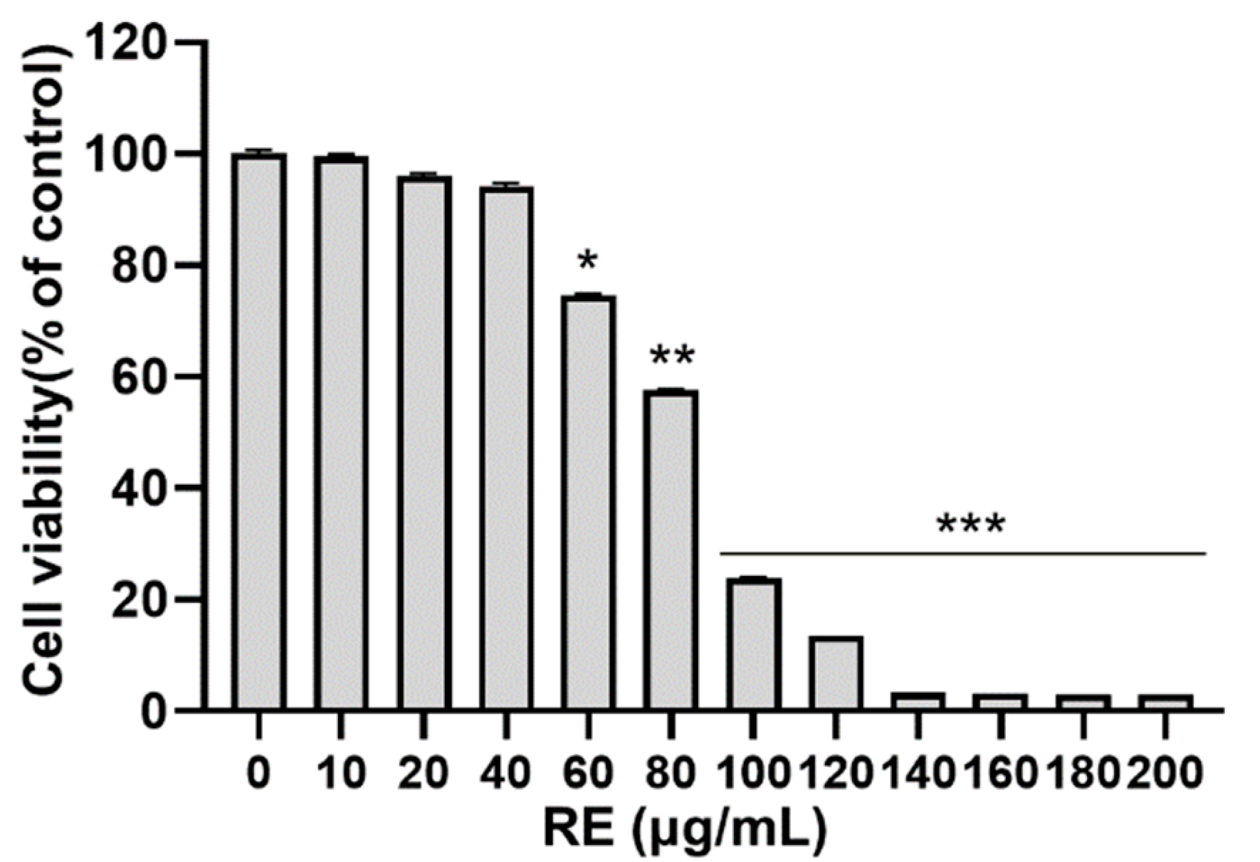
3.1. Effects of RE on Cytotoxicity in C2C12 Myoblasts

To evaluate the cytotoxicity of RE to murine C2C12 myoblasts, cell viability was examined using the MTT assay. Treatment with RE did not affect cell viability up to a concentration of 40 μg/mL (Figure 1). However, treatment with concentrations greater than 40 μg/mL reduced cell viability. Therefore, further experiments were conducted using a RE concentration of 0–40 μg/mL.

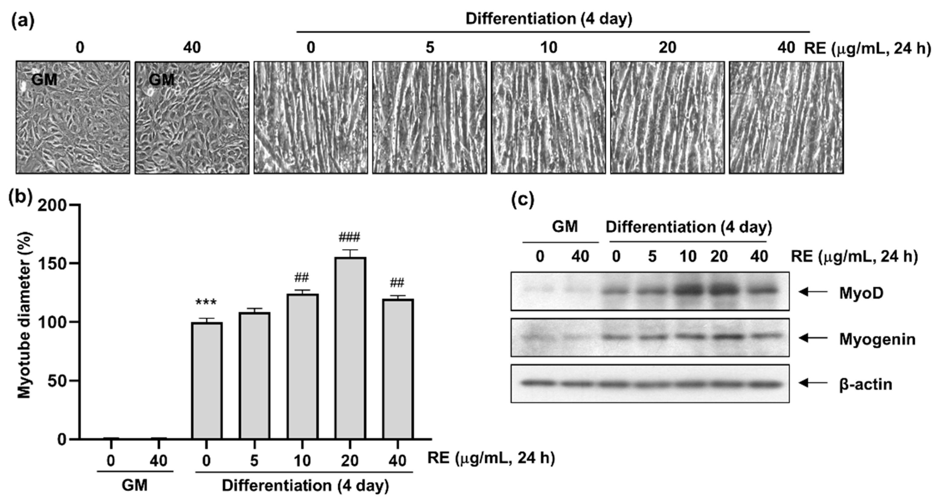
3.2. Effects of RE on C2C12 Myotube Differentiation

To evaluate the effect of RE on myotube differentiation, C2C12 myoblasts cultured in growth medium were subsequently cultured in differentiation-inducing medium in the presence or absence of RE, and the diameters of C2C12 myotubes under different treatments were measured after 4 d. Treatment with RE increased C2C12 myotube diameter in a concentration-dependent manner (Figure 2a), and myotube length was increased by promoting myotube differentiation capacity. In addition, myotube diameter and length exhibited a positive relationship. However, under treatment with 40 μg/mL RE, the C2C12 myoblasts failed to differentiate, and the cobblestone shape of the original C2C12 myoblasts was maintained. The positive effect of RE on myotube diameter was concentration-dependent up to 20 μg/mL RE (Figure 2b).
Next, the effects of RE treatment on the expression of MRF-related proteins were evaluated using western blot analysis. Treatment with RE had a positive and concentration-dependent effect on the expression of the differentiation markers MyoD and myogenin up to 20 μg/mL RE (Figure 2c). Furthermore, neither MyoD nor myogenin were expressed at substantial levels in cells that failed to differentiate (Figure 2c). Based on these observations, the experiment confirmed that RE induces the differentiation of C2C12 myoblasts into myotubes.

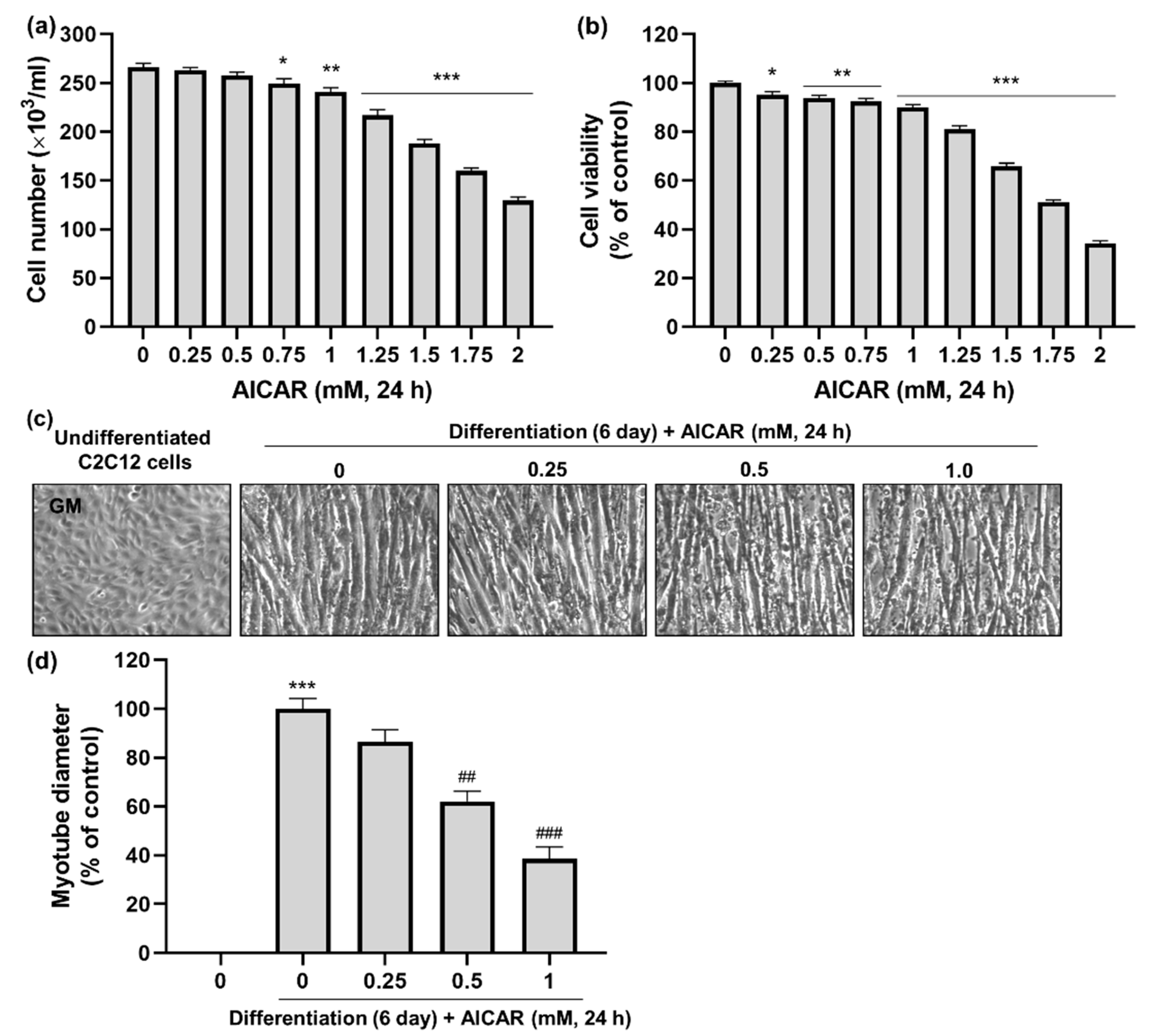
3.3. Effects of AICAR on C2C12 Myotube Atrophy

To evaluate the hypertrophic effects of RE on muscle cell atrophy, a muscle cell atrophy model was established using AICAR (an AMPK activator) to induce muscle cell atrophy. Prior to the experiment, the cytotoxicity of AICAR on C2C12 myoblasts and myotubes was determined using cell counting (Figure 3a) and the MTT assay (Figure 3b). Treatment with AICAR negatively affected both cell number and viability in a concentration-dependent manner (Figure 3a,b). Based on this, the maximum concentration of AICAR was set at 1 mM for the next experiment.
Next, the morphology and diameter of C2C12 myotubes were measured to evaluate the effect of muscle cell atrophy. Non-differentiated C2C12 myoblasts exhibited a cobblestone-shaped morphology, whereas cells that had undergone differentiation were observed as elongated myotubes (Figure 3c). Treatment with AICAR reduced myotube diameter in a concentration-dependent manner (Figure 3d). Therefore, AICAR treatment was used to establish a muscle cell atrophy model to investigate the effects of RE.

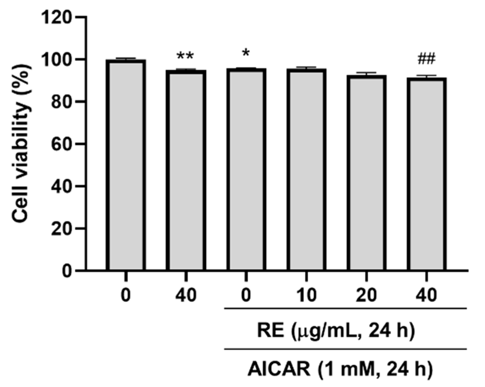
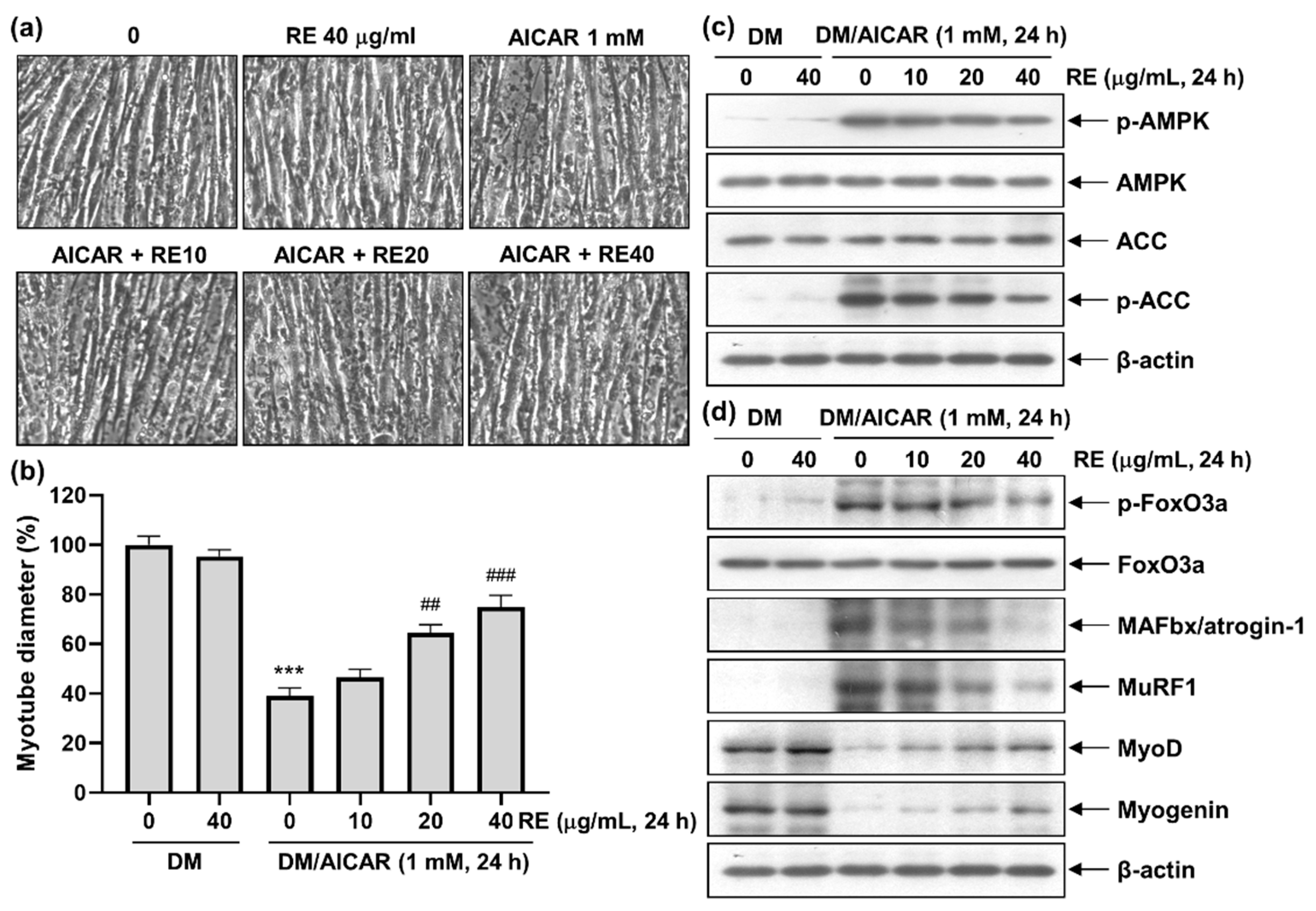
3.4. Effect of RE on AICAR-Induced C2C12 Myotube Atrophy
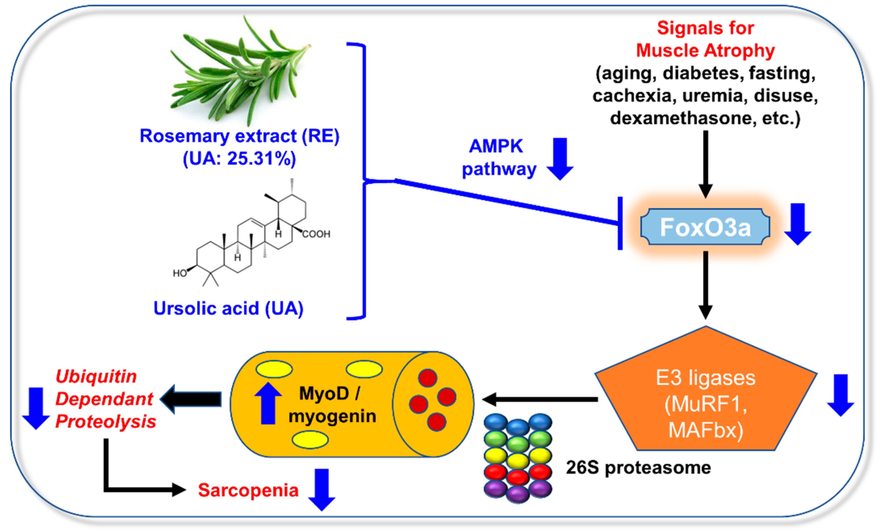
Before evaluating whether RE promoted hypertrophy in AICAR-induced C2C12 myotube atrophy, the cytotoxicity of simultaneous RE and AICAR treatment was evaluated using maximum RE and AICAR concentrations (40 μg/mL and 1 mM, respectively). After no serious cytotoxicity was observed, RE concentrations of up to 40 μg/mL were used (Figure 4). C2C12 myoblasts were cultured in differentiation medium for 6 d, and the resulting myotubes were treated with 1 mM AICAR for 24 h to induce muscle cell atrophy. Then, various concentrations of RE were added, and the inhibition of muscle cell atrophy and promotion of hypertrophy were evaluated after 24 h.
The C2C12 myoblasts clearly differentiated into myotubes when cultured in differentiation medium, with or without 40 μg/mL RE (Figure 5a), and clearly exhibited muscle cell atrophy when treated with 1 mM AICAR. However, treatment with RE reduced AICAR-induced muscle cell atrophy in a concentration-dependent manner and resulted in muscle cell hypertrophy (Figure 5a). Treatment with AICAR alone reduced myotube diameter by ~60% when compared with vehicle-treated control myotubes (Figure 5b). In addition, myotube diameter increased with increasing RE concentration, reaching 70–80% the diameter of vehicle-treated control myotubes at 40 μg/mL RE (Figure 5b).
The relationship between these morphological changes and the expression of muscle differentiation factors was investigated using western blot analysis. Expression levels of phosphorylated AMPK and phosphorylated ACC, which are related to muscle differentiation, increased during AICAR treatment but decreased in response to treatment with RE (Figure 5c). In addition, expression levels of muscle degradation-related transcription factors (p-FoxO3a, MAFbx/atrogin-1, and MuRF1) were increased by AICAR treatment but reduced by RE treatment (Figure 5d). Moreover, when myotubes, in which muscle cell atrophy occurred, were treated, expression levels of the muscle differentiation factors MyoD and myogenin gradually increased (Figure 5d).

4. Discussion

Skeletal muscles are involved in movement, respiration, and the maintenance of homeostasis in the body. Thus, the loss of skeletal muscle and function can be fatal. However, muscle atrophy and sarcopenia cannot be cured using current technology, and regular exercise is the only effective strategy for maintaining muscle mass and suppressing sarcopenia. Therefore, continuous prevention and more effective pharmaceutical agents are needed. In addition, it would be more useful to consume active ingredients that prevent sarcopenia daily. The RE used in the present study induced myotube differentiation, inhibited AICAR-induced muscle cell atrophy, and further promoted hypertrophy, thereby exhibiting its potential for use as a functional substance for preventing sarcopenia.
Skeletal muscles are involved in the regeneration of muscle fibers that contain mechanical, chemical, or degenerative lesions through activation, differentiation, and proliferation [32,33]. The differentiation of myoblasts into myotubes is essential for the formation of muscle fibers involved in the development and regeneration of skeletal muscles [34]. These monocyte myoblasts proliferate, differentiate, and then fuse with existing muscle fibers to form multinucleated myotubes and myofibers. Similar proliferation and differentiation of muscle cells occur during development and postnatal birth [35]. Therefore, the promotion of myoblast proliferation and differentiation and the induction of myotube hypertrophy should benefit muscle regeneration and the maintenance of muscle mass.
Recent studies have reported that muscle atrophy occurs as a result of increased proteolysis, which is, in turn, caused by activation of the ubiquitin-proteasome pathway [36,37,38]. Indeed, the muscle-specific ubiquitin ligases MAFbx/atrogin-1 and MuRF-1 are expressed early in the muscle atrophy process and are directly involved in muscle protein degradation [37,38]. In addition, when muscle atrophy is induced, muscle-specific transcription factors that are involved in myogenic differentiation (e.g., MyoD and myogenin) are downregulated in response to muscle-specific gene expression [39,40]. In addition, it has been reported that carnosol, a rosemary leaf extract, can also induce skeletal muscle hypertrophy by inhibiting the ubiquitin-proteasome system-dependent protein degradation pathway via suppression of E3 ubiquitin ligase MuRF-1 [10].
The injection of AICAR into mice increased FoxO1 and FoxO3 expression, and the AICAR-induced upregulation of FoxO1 was not affected by the suppression of AMPKα2 [41]. The treatment of C2C12 myotubes with AICAR has been reported to induce both protein breakdown and the upregulation of FOXO, MAFbx/atrogin-1, MuRF-1, and two other FoxO target genes, namely microtubule-associated protein 1A/1B-light chain 3 (LC3) and Bnip3 [42]. Meanwhile, AMPK phosphorylates FoxO3a at a site known to activate the transcription factors, thereby inducing generalized protein degradation; however, this may not necessarily affect the localization of FoxO3a to the nucleus. Treatment with AICAR also increases the binding of FoxO3 to MuRF-1 and atrogin-1 promoters [43].
The regulation of FoxO3a activity is possible by very complex mechanisms such as phosphorylation, acetylation, and methylation [44]. In particular, phosphorylation modification regulates FoxO3a activity via a cytoplasmic-nuclear shuttle mechanism. FOXO3a is targeted for phosphorylation by numerous protein kinases, and phosphorylation leads to two opposing results. The first is a signaling pathway that causes a negative effect on FoxO3a activity. Phosphorylation through AKT/GSK/ERK/IKK after stimulation of insulin and growth factors increases the cytoplasmic retention of FoxO3a and further induces the degradation of FoxO3a. The second is a signaling system that causes a positive effect on FoxO3a activity. In the presence of oxidant/nutrient stress, phosphorylation occurs through JNK/MST1/AMPK, promotes nuclear translocation, and increases the activity of FoxO3a. According to our experiments, the phosphorylation of FoxO3a was increased when AICAR, an AMPK activator, was treated on C2C12 cells (Figure 5d). Moreover, when RE was treated to the cells, the phosphorylation was reduced. Therefore, the down-regulation of E3 ligase (MuRF1 and MAFbx) activity was caused by inhibition of AMPK activity and inhibition of FoxO3a nuclear translocation, which were not investigated in our experiment (Figure 6).
In the present study, AICAR-induced muscle cell atrophy was confirmed through the activation of AMPK and FoxO3a, and the atrophy-inhibiting effects of RE treatment were confirmed by the observation of increased muscle hypertrophy. To study more specific mechanisms, additional studies related to the interaction between the AMPK pathway and its down-stream transcription factors are needed. In addition, further investigation of other myogenic factors (e.g., MyoD and myogenin) is also warranted.

5. Conclusions

In the present study, RE treatment was observed to promote muscle differentiation by increasing the expression of the myoblast differentiation factors MyoD and myogenin and to inhibit muscle cell atrophy by downregulating the transcription factors FoxO3, MAFbx/atrogin-1, and MuRF1 (Figure 6). Additional animal studies are needed to elucidate the specific mechanisms underlying the action of RE and the efficacy of RE in suppressing sarcopenia.

Supplementary Materials

The following supporting information can be downloaded at: https://www.mdpi.com/article/10.3390/app13020986/s1, Table S1: Certificate of Analysis.

Author Contributions

Writing—original draft preparation and formal analysis, J.H.L.; data curation, J.H.L., J.Y.J. and Y.H.K.; resources, S.H.L.; supervision, C.P.; project administration, Y.H.C. and N.D.K.; funding acquisition, N.D.K. All authors have read and agreed to the published version of the manuscript.

Funding

This study was supported by a National Research Foundation of Korea (NRF) grant funded by the Korean government (MSIT) (No. 2021R1F1A1051265) and the Basic Research Program through the National Research Foundation of Korea (NRF) funded by the Ministry of Education (2018R1D1A1B07044648) (N.D.K.). This research was supported by the Basic Science Research Program through the National Research Foundation of Korea (NRF), funded by the Ministry of Education (2022R1A6A3A01085858) (J.Y.J.).

Institutional Review Board Statement

Not applicable.

Informed Consent Statement

Not applicable.

Data Availability Statement

All data presented in this study are available on request from the corresponding author.

Conflicts of Interest

The authors declare no conflict of interest.

References

  1. Anker, S.D.; Morley, J.E.; von Haehling, S. Welcome to the ICD-10 code for sarcopenia. J. Cachexia Sarcopenia Muscle 2016, 7, 512–514. [Google Scholar] [CrossRef]
  2. Yoo, J.I.; Kim, J.T.; Park, C.H.; Cha, Y. Diagnosis and management of sarcopenia after hip fracture surgery: Current concept review. Hip Pelvis 2022, 34, 1–9. [Google Scholar] [CrossRef]
  3. Dao, T.; Green, A.E.; Kim, Y.A.; Bae, S.J.; Ha, K.T.; Gariani, K.; Lee, M.R.; Menzies, K.J.; Ryu, D. Sarcopenia and muscle aging: A brief overview. Endocrinol. Metab. 2020, 35, 716–732. [Google Scholar] [CrossRef]
  4. Dhillon, R.J.; Hasni, S. Pathogenesis and management of sarcopenia. Clin. Geriatr. Med. 2017, 33, 17–26. [Google Scholar] [CrossRef] [Green Version]
  5. Farr, S.A.; Niehoff, M.L.; Ceddia, M.A.; Herrlinger, K.A.; Lewis, B.J.; Feng, S.; Welleford, A.; Butterfield, D.A.; Morley, J.E. Effect of botanical extracts containing carnosic acid or rosmarinic acid on learning and memory in SAMP8 mice. Physiol. Behav. 2016, 165, 328–338. [Google Scholar] [CrossRef] [Green Version]
  6. Ho, C.T.; Wang, M.; Wei, G.J.; Huang, T.C.; Huang, M.T. Chemistry and antioxidative factors in rosemary and sage. Biofactors 2000, 13, 161–166. [Google Scholar] [CrossRef] [PubMed]
  7. Rocha, J.; Eduardo-Figueira, M.; Barateiro, A.; Fernandes, A.; Brites, D.; Bronze, R.; Duarte, C.M.; Serra, A.T.; Pinto, R.; Freitas, M.; et al. Anti-inflammatory effect of rosmarinic acid and an extract of Rosmarinus officinalis in rat models of local and systemic inflammation. Basic Clin. Pharmacol. Toxicol. 2015, 116, 398–413. [Google Scholar] [CrossRef] [PubMed]
  8. Elbahnasawy, A.S.; Valeeva, E.R.; El-Sayed, E.M.; Rakhimov, I.I. The impact of thyme and rosemary on prevention of osteoporosis in Rats. J. Nutr. Metab. 2019, 2019, 1431384. [Google Scholar] [CrossRef] [PubMed] [Green Version]
  9. Takahashi, A.; Dohi, H.; Egashira, Y.; Hirai, S. Erucic acid derived from rosemary regulates differentiation of mesenchymal stem cells into osteoblasts/adipocytes via suppression of peroxisome proliferator-activated receptor gamma transcriptional activity. Phytother. Res. 2020, 34, 1358–1366. [Google Scholar] [CrossRef]
  10. Morel, S.; Hugon, G.; Vitou, M.; Védère, M.; Fons, F.; Rapior, S.; Saint, N.; Carnac, G. A bioassay-guided fractionation of rosemary leaf extract identifies carnosol as a major hypertrophy inducer in human skeletal muscle cells. Nutrients 2021, 13, 4190. [Google Scholar] [CrossRef]
  11. Kahnt, M.; Fischer Nee Heller, L.; Al-Harrasi, A.; Csuk, R. Ethylenediamine derived carboxamides of betulinic and ursolic acid as potential cytotoxic agents. Molecules 2018, 23, 2558. [Google Scholar] [CrossRef] [PubMed] [Green Version]
  12. Yadav, D.; Nath Mishra, B.; Khan, F. 3D-QSAR and docking studies on ursolic acid derivatives for anticancer activity based on bladder cell line T24 targeting NF-kB pathway inhibition. J. Biomol. Struct. Dyn. 2019, 37, 3822–3837. [Google Scholar] [CrossRef] [PubMed]
  13. Abu-Gharbieh, E.; Shehab, N.G.; Almasri, I.M.; Bustanji, Y. Antihyperuricemic and xanthine oxidase inhibitory activities of Tribulus arabicus and its isolated compound, ursolic acid: In vitro and in vivo investigation and docking simulations. PLoS ONE 2018, 13, e0202572. [Google Scholar] [CrossRef] [PubMed] [Green Version]
  14. Vlavcheski, F.; Naimi, M.; Murphy, B.; Hudlicky, T.; Tsiani, E. Rosmarinic acid, a rosemary extract polyphenol, increases skeletal muscle cell glucose uptake and activates AMPK. Molecules 2017, 22, 1669. [Google Scholar] [CrossRef] [Green Version]
  15. Kuang, J.X.; Shen, Q.; Zhang, R.Q.; Fang, Q.Y.; Deng, X.; Fan, M.; Cheng, C.R.; Zhang, X.W.; Liu, X. Carnosol attenuated atrophy of C2C12 myotubes induced by tumour-derived exosomal miR-183-5p through inhibiting Smad3 pathway activation and keeping mitochondrial respiration. Basic Clin. Pharmacol. Toxicol. 2022, 131, 500–513. [Google Scholar] [CrossRef]
  16. Den Hartogh, D.J.; Vlavcheski, F.; Giacca, A.; MacPherson, R.E.K.; Tsiani, E. Carnosic acid attenuates the free fatty acid-induced insulin resistance in muscle cells and adipocytes. Cells 2022, 11, 167. [Google Scholar] [CrossRef]
  17. Sasaki, Y.; Kojima-Yuasa, A.; Tadano, H.; Mizuno, A.; Kon, A.; Norikura, T. Ursolic acid improves the indoxyl sulfate-induced impairment of mitochondrial biogenesis in C2C12 cells. Nutr. Res. Pract. 2022, 16, 147–160. [Google Scholar] [CrossRef]
  18. Kim, M.; Sung, B.; Kang, Y.J.; Kim, D.H.; Lee, Y.; Hwang, S.Y.; Yoon, J.H.; Yoo, M.A.; Kim, C.M.; Chung, H.Y.; et al. The combination of ursolic acid and leucine potentiates the differentiation of C2C12 murine myoblasts through the mTOR signaling pathway. Int. J. Mol. Med. 2015, 35, 755–762. [Google Scholar] [CrossRef] [Green Version]
  19. Noh, K.K.; Chung, K.W.; Sung, B.; Kim, M.J.; Park, C.H.; Yoon, C.; Choi, J.S.; Kim, M.K.; Kim, C.M.; Kim, N.D.; et al. Loquat (Eriobotrya japonica) extract prevents dexamethasone-induced muscle atrophy by inhibiting the muscle degradation pathway in Sprague Dawley rats. Mol. Med. Rep. 2015, 12, 3607–3614. [Google Scholar] [CrossRef] [Green Version]
  20. Sung, B.; Hwang, S.Y.; Kim, M.J.; Kim, M.; Jeong, J.W.; Kim, C.M.; Chung, H.Y.; Kim, N.D. Loquat leaf extract enhances myogenic differentiation, improves muscle function and attenuates muscle loss in aged rats. Int. J. Mol. Med. 2015, 36, 792–800. [Google Scholar] [CrossRef]
  21. Cho, Y.H.; Lee, S.Y.; Kim, C.M.; Kim, N.D.; Choe, S.; Lee, C.H.; Shin, J.H. Effect of loquat leaf extract on muscle strength, muscle mass, and muscle function in healthy adults: A randomized, double-blinded, and placebo-controlled trial. Evid. Based Complement. Alternat. Med. 2016, 2016, 4301621. [Google Scholar] [CrossRef] [Green Version]
  22. Ding, M.; Xie, Y.; Wagner, R.J.; Jin, Y.; Carrao, A.C.; Liu, L.S.; Guzman, A.K.; Powell, R.J.; Hwa, J.; Rzucidlo, E.M.; et al. Adiponectin induces vascular smooth muscle cell differentiation via repression of mammalian target of rapamycin complex 1 and FoxO4. Arterioscler. Thromb. Vasc. Biol. 2011, 31, 1403–1410. [Google Scholar] [CrossRef] [Green Version]
  23. Rudnicki, M.A.; Schnegelsberg, P.N.; Stead, R.H.; Braun, T.; Arnold, H.H.; Jaenisch, R. MyoD or Myf-5 is required for the formation of skeletal muscle. Cell 1993, 75, 1351–1359. [Google Scholar] [CrossRef]
  24. Kablar, B.; Krastel, K.; Ying, C.; Asakura, A.; Tapscott, S.J.; Rudnicki, M.A. MyoD and Myf-5 differentially regulate the development of limb versus trunk skeletal muscle. Development 1997, 124, 4729–4738. [Google Scholar] [CrossRef]
  25. Ishibashi, J.; Perry, R.L.; Asakura, A.; Rudnicki, M.A. MyoD induces myogenic differentiation through cooperation of its NH2- and COOH-terminal regions. J. Cell Biol. 2005, 171, 471–482. [Google Scholar] [CrossRef] [Green Version]
  26. Vavvas, D.; Apazidis, A.; Saha, A.K.; Gamble, J.; Patel, A.; Kemp, B.E.; Witters, L.A.; Ruderman, N.B. Contraction-induced changes in acetyl-CoA carboxylase and 5′-AMP-activated kinase in skeletal muscle. J. Biol. Chem. 1997, 272, 13255–13261. [Google Scholar] [CrossRef] [PubMed] [Green Version]
  27. Fujii, N.; Hayashi, T.; Hirshman, M.F.; Smith, J.T.; Habinowski, S.A.; Kaijser, L.; Mu, J.; Ljungqvist, O.; Birnbaum, M.J.; Witters, L.A.; et al. Exercise induces isoform-specific increase in 5′AMP-activated protein kinase activity in human skeletal muscle. Biochem. Biophys. Res. Commun. 2000, 273, 1150–1155. [Google Scholar] [CrossRef] [PubMed]
  28. Birk, J.B.; Wojtaszewski, J.F. Predominant alpha2/beta2/gamma3 AMPK activation during exercise in human skeletal muscle. J. Physiol. 2006, 577, 1021–1032. [Google Scholar] [CrossRef]
  29. Merrill, G.F.; Kurth, E.J.; Hardie, D.G.; Winder, W.W. AICA riboside increases AMP-activated protein kinase, fatty acid oxidation, and glucose uptake in rat muscle. Am. J. Physiol. 1997, 273, E1107–E1112. [Google Scholar] [CrossRef]
  30. Thomson, D.M.; Fick, C.A.; Gordon, S.E. AMPK activation attenuates S6K1, 4E-BP1, and eEF2 signaling responses to high-frequency electrically stimulated skeletal muscle contractions. J. Appl. Physiol. 2008, 104, 625–632. [Google Scholar] [CrossRef] [PubMed]
  31. Sanchez, A.M.; Csibi, A.; Raibon, A.; Cornille, K.; Gay, S.; Bernardi, H.; Candau, R. AMPK promotes skeletal muscle autophagy through activation of forkhead FoxO3a and interaction with Ulk1. J. Cell. Biochem. 2012, 113, 695–710. [Google Scholar] [CrossRef]
  32. Karalaki, M.; Fili, S.; Philippou, A.; Koutsilieris, M. Muscle regeneration: Cellular and molecular events. In Vivo 2009, 23, 779–796. [Google Scholar] [PubMed]
  33. Ceafalan, L.C.; Popescu, B.O.; Hinescu, M.E. Cellular players in skeletal muscle regeneration. Biomed. Res. Int. 2014, 2014, 957014. [Google Scholar] [CrossRef] [PubMed]
  34. Grefte, S.; Kuijpers-Jagtman, A.M.; Torensma, R.; Von den Hoff, J.W. Skeletal muscle development and regeneration. Stem Cells Dev. 2007, 16, 857–868. [Google Scholar] [CrossRef] [PubMed]
  35. Schiaffino, S.; Dyar, K.A.; Ciciliot, S.; Blaauw, B.; Sandri, M. Mechanisms regulating skeletal muscle growth and atrophy. FEBS J. 2013, 280, 4294–4314. [Google Scholar] [CrossRef] [PubMed]
  36. Jagoe, R.T.; Goldberg, A.L. What do we really know about the ubiquitin-proteasome pathway in muscle atrophy? Curr. Opin. Clin. Nutr. Metab. Care 2001, 4, 183–190. [Google Scholar] [CrossRef]
  37. Bodine, S.C.; Baehr, L.M. Skeletal muscle atrophy and the E3 ubiquitin ligases MuRF1 and MAFbx/atrogin-1. Am. J. Physiol. Endocrinol. Metab. 2014, 307, E469–E484. [Google Scholar] [CrossRef] [Green Version]
  38. Liu, Y.; Du, M.; Li, X.; Chen, L.; Shen, Q.; Tian, J.; Zhang, D. Role of the ubiquitin-proteasome pathway on proteolytic activity in postmortem proteolysis and tenderisation of sheep skeletal muscle. Int. J. Food Sci. Technol. 2016, 51, 2353–2359. [Google Scholar] [CrossRef]
  39. Hyatt, J.P.; Roy, R.R.; Baldwin, K.M.; Edgerton, V.R. Nerve activity-independent regulation of skeletal muscle atrophy: Role of MyoD and myogenin in satellite cells and myonuclei. Am. J. Physiol. Cell Physiol. 2003, 285, C1161–C1173. [Google Scholar] [CrossRef] [Green Version]
  40. Tintignac, L.A.; Lagirand, J.; Batonnet, S.; Sirri, V.; Leibovitch, M.P.; Leibovitch, S.A. Degradation of MyoD mediated by the SCF (MAFbx) ubiquitin ligase. J. Biol. Chem. 2005, 280, 2847–2856. [Google Scholar] [CrossRef]
  41. Jorgensen, S.B.; Wojtaszewski, J.F.; Viollet, B.; Andreelli, F.; Birk, J.B.; Hellsten, Y.; Schjerling, P.; Vaulont, S.; Neufer, P.D.; Richter, E.A.; et al. Effects of alpha-AMPK knockout on exercise-induced gene activation in mouse skeletal muscle. FASEB J. 2005, 19, 1146–1148. [Google Scholar] [CrossRef] [PubMed] [Green Version]
  42. Nakashima, K.; Yakabe, Y. AMPK activation stimulates myofibrillar protein degradation and expression of atrophy-related ubiquitin ligases by increasing FOXO transcription factors in C2C12 myotubes. Biosci. Biotechnol. Biochem. 2007, 71, 1650–1656. [Google Scholar] [CrossRef] [PubMed]
  43. Romanello, V.; Guadagnin, E.; Gomes, L.; Roder, I.; Sandri, C.; Petersen, Y.; Milan, G.; Masiero, E.; Del Piccolo, P.; Foretz, M.; et al. Mitochondrial fission and remodelling contributes to muscle atrophy. EMBO J. 2010, 29, 1774–1785. [Google Scholar] [CrossRef] [PubMed]
  44. Wang, X.; Hu, S.; Liu, L. Phosphorylation and acetylation modifications of FOXO3a: Independently or synergistically? Oncol. Lett. 2017, 13, 2867–2872. [Google Scholar] [CrossRef] [PubMed]
Figure 1. Effects of rosemary extract (RE) on C2C12 myoblast viability. Seeded cells were treated with indicated concentrations of RE for 24 h, and percent cell survival was measured using the MTT assay. Values and error bars indicate means ± SD of three separate experiments. Asterisks indicate significant differences between RE- and vehicle-treated cells (ANOVA; *, p < 0.05; **, p < 0.01; ***, p < 0.001).
Applsci 13 00986 g001
Figure 2. Effects of rosemary extract (RE) on C2C12 myoblast differentiation. C2C12 cells were cultured in differentiation medium for 4 d, until completely differentiated, treated with indicated concentrations of RE, and cultured for additional 24 h. (a) Phase-contrast micrographs of C2C12 cells before and after 4 d of differentiation when cultured with different concentrations of RE. 200× magnification; (b) C2C12 cell diameter before and after 4 d of differentiation when cultured with different concentrations of RE. Values and error bars indicate means ± SD of three separate experiments. ***, significant difference between RE- and vehicle-treated cells before differentiation (ANOVA, p < 0.001); ##, significant difference between RE- and vehicle-treated cells after differentiation (ANOVA, p < 0.01); ###, significant difference between RE- and vehicle-treated cells after differentiation (ANOVA, p < 0.001); (c) Expression of differentiation-related proteins before and after 4 d of differentiation when cultured with different concentrations of RE. C2C12 myotubes were cultured with RE for 24 h, and then cell lysates were immunoblotted with MyoD and myogenin antibodies. Representative results of three independent experiments are shown. β-Actin was used as a loading control. GM: growth medium.
Applsci 13 00986 g002
Figure 3. Effects of 5-aminoimidazole-4-carboxamide ribonucleoside (AICAR) on C2C12 myotube viability and differentiation. C2C12 cells were treated with indicated concentrations of AICAR and cultured for 24 h; cell counting (a) and MTT assay (b) were performed. Values and error bars indicate means ± SD of three separate experiments. Asterisks indicate significant differences between RE- and vehicle-treated cells before differentiation (ANOVA; *, p < 0.05; **, p < 0.01; ***, p < 0.001); (c) Phase-contrast micrographs of C2C12 cells before and after 6 d of differentiation when cultured with different concentrations of AICAR. GM: growth medium. 200× magnification; (d) C2C12 cell diameter before and after 4 d of differentiation when cultured with different concentrations of AICAR. Values and error bars indicate means ± SD of three separate experiments. ***, significant difference between RE- and vehicle-treated cells before differentiation (ANOVA, p < 0.001); ##, significant difference between RE- and vehicle-treated cells after differentiation (ANOVA, p < 0.01); ###, significant difference between RE- and vehicle-treated cells after differentiation (ANOVA, p < 0.001).
Applsci 13 00986 g003
Figure 4. Effects of rosemary extract (RE) on the viability of 5-aminoimidazole-4-carboxamide ribonucleoside (AICAR)-treated C2C12 myoblasts: C2C12 myoblasts were treated with AICAR (1 mM) and indicated concentrations of RE, cultured for 24 h, and then evaluated using the MTT assay. Values and error bars indicate means ± SD of three separate experiments. *, significant difference between RE- and vehicle-treated cells before differentiation (ANOVA, p < 0.05); **, significant difference between RE- and vehicle-treated cells before differentiation (ANOVA, p < 0.01); ##, significant difference between RE- and AICAR-treated cells before differentiation (ANOVA, p < 0.01).
Applsci 13 00986 g004
Figure 5. Effects of rosemary extract (RE) on 5-aminoimidazole-4-carboxamide ribonucleoside (AICAR)-induced muscle cell atrophy in C2C12 myotubes. C2C12 cells were cultured in a differentiation medium for 6 d, until fully differentiated, cultured with or without 1 mM AICAR for 24 h, and then cultured with or without RE for an additional 24 h. (a) Phase-contrast micrographs of C2C12 cells after differentiation when cultured with AICAR and/or RE. 200× magnification; (b) C2C12 myotube diameter when differentiated with AICAR and/or RE. Values and error bars indicate means ± SD of three separate experiments. ***, significant difference between AICAR- and vehicle-treated cells after differentiation (ANOVA, p < 0.001); ##, significant difference between AICAR-treated cells cultured with and without RE (ANOVA, p < 0.01); ###, significant difference between AICAR-treated cells cultured with and without RE (ANOVA, p < 0.001); (c) Effects of AICAR with or without RE on AMPK signaling in C2C12 cells. Western blot analysis was performed using β-actin as a loading control. Representative results of three independent experiments are shown; (d) Effects of AICAR with or without RE on the expression of C2C12 myoblast differentiation factors. Western blot analysis was performed using β-actin as a loading control. Results represent three separate experiments. DM: differentiation medium.
Applsci 13 00986 g005
Figure 6. Proposed mechanism for the effects of rosemary extract (RE) on C2C12 cell differentiation and 5-aminoimidazole-4-carboxamide ribonucleoside (AICAR)-induced C2C12 myotube atrophy. ↑: upregulate; ↓: downregulate.
Applsci 13 00986 g006
Disclaimer/Publisher’s Note: The statements, opinions and data contained in all publications are solely those of the individual author(s) and contributor(s) and not of MDPI and/or the editor(s). MDPI and/or the editor(s) disclaim responsibility for any injury to people or property resulting from any ideas, methods, instructions or products referred to in the content.

