Article

An Observational Study on Changes in the Oral and Gut Microbiota through Professional Mechanical Tooth Cleaning, including Tooth-Brushing Instructions in Patients with Multi-Bracket Appliances

by
Satoko Okuwaki
1,*,
Kazuyoshi Hosomichi
2,
Atsushi Tajima
3 and
Tetsutaro Yamaguchi
1
1
Department of Orthodontics, School of Dentistry, Kanagawa Dental University, 82 Inaoka-cho, Yokosuka 238-8580, Japan
2
Laboratory of Computational Genomics, School of Life Science, Tokyo University of Pharmacy and Life Sciences, 1432-1 Horinouchi, Hachioji 192-0392, Japan
3
Department of Bioinformatics and Genomics, Graduate School of Advanced Preventive Medical Sciences, Kanazawa University, 13-1 Takara-Machi, Kanazawa 920-8640, Japan
*
Author to whom correspondence should be addressed.
Appl. Sci. 2023, 13(19), 10843; https://doi.org/10.3390/app131910843
Submission received: 5 September 2023 / Revised: 27 September 2023 / Accepted: 28 September 2023 / Published: 29 September 2023

Abstract

:
Multi-bracket appliances have long been established as tools for treating malocclusions. However, the complex construction and difficulty in cleaning due to their long-term intraoral retention have hindered the prevention of caries and periodontitis. In this study, professional mechanical tooth cleaning (PMTC), including tooth-brushing instructions, was continuously performed for 3 months in 24 patients who had worn multi-bracket appliances for more than 6 months, and changes in the oral and gut microbiota were examined using one-way repeated-measures analysis of variance. Additionally, changes in bacterial flora associated with different treatment durations were verified using the Pearson correlation coefficient. The results showed that continuous PMTC significantly reduced the amount of plaque in the oral cavity. No significant changes were observed in the oral or gut microbiota and no significant increase in pathogenic bacteria was observed. Therefore, our results suggest that continuous PMTC during orthodontic treatment with multi-brackets may inhibit the growth of pathogenic bacteria by maintaining a clean oral environment and avoiding dysbiosis in both the oral and gut microbiota. Significant changes in the gut microbiota with different treatment durations suggested that differences in food intake and food choices at each treatment stage of orthodontic treatment may affect the gut microbiota.

1. Introduction

Multi-bracket appliances have long been established as tools for treating malocclusions. However, difficulty in oral cleaning due to the complex structure of orthodontic appliances allows plaque accumulation. This in turn increases the number of caries-pathogenic and periodontopathogenic bacteria [1,2,3], which have adverse effects and risks associated with orthodontic treatment, including the development of white spots, caries, and progression of periodontal disease [4,5].
Streptococcus mutans (S. mutans), a typical caries-causing bacterium, increases after wearing fixed orthodontic appliances [6,7]. Among periodontopathogenic bacteria, Porphyromonas, Tannerella, Prevotella, Capnocytophaga, and Selenomonas also increase before and after the use of fixed orthodontic appliances [8]. Therefore, the state of the oral microbiota, which changes with the use of fixed orthodontic appliances, is similar to that of the microbiota during the transition to severe periodontitis [8]. Such changes in oral conditions and periodontal disease induced by orthodontic therapy are only partially reversible, even 3 months after appliance removal [9]. Therefore, it is important to control dysbiosis of the oral microbiota that occurs while wearing multi-bracket appliances during treatment.
Recent advances in analytical techniques have led to extensive research on the interrelationships between oral and intestinal environments [10,11]. Previously, digestive juices were believed to sterilize oral bacteria ingested through swallowing. However, not only has the presence of oral bacteria in the gut microbiota has been revealed, but it has been shown that pathogenic bacteria in the oral cavity can disrupt the balance of gut microbiota [12,13]. It has also been suggested that failure to maintain oral hygiene leads to elevated systemic inflammation and an increased risk of extraoral diseases [14]. Orthodontic treatment, especially with multi-bracket appliances, involves long-term device fixation in the oral cavity and significant changes to the oral environment. Therefore, understanding the changes in the gut microbiota associated with orthodontic treatment is a significant challenge in positioning orthodontic therapy as part of comprehensive systemic management. Additionally, multi-bracket appliances may restrict the chewing of hard or sticky foods owing to bracket dropout, which may result in restricted food choices, in turn affecting the gut microbiota.
The risk of caries and periodontitis associated with wearing fixed orthodontic appliances is evident from changes in the oral microbiota before and after appliance wear [6,8,15,16]. However, a solution for risk reduction has yet to be identified. The importance of oral cleaning in orthodontic patients has also been suggested [16,17,18]. However, changes in the oral microbiota over time, with or without oral cleaning and oral hygiene instructions, in multi-bracket appliance wearers have yet to be determined. It is also unclear whether changes occur over time in the gut microbiota of multi-bracket device wearers. Therefore, there is an increasing need for further clarification of the relationship between fixed orthodontic appliances, such as multi-bracket appliances, and gut microbiota, as well as the associated systemic effects in the field of orthodontics, in addition to the changes in the oral environment related to orthodontic appliances, which have been the focus of much attention.
In this study, we investigated whether continuous professional mechanical tooth cleaning (PMTC), including tooth-brushing instructions (TBI), could be used to reduce the risk of dental caries and periodontitis during orthodontic treatment with multi-bracket appliances. The analysis was based on changes in the oral microbiota of the patients after more than 6 months of treatment. Second, we evaluated the dynamics of the gut microbiota of multi-bracket appliance wearers and examined the relationship between orthodontic treatment and the gut microbiota.

2. Materials and Methods

2.1. Study Design and Settings

2.1.1. Ethical Statement

This study was approved by the Ethics Committee of Kanagawa Dental University (approval No. 795). Written informed consent was obtained from all participants and their parents or guardians.

2.1.2. Participants

Twenty-five patients who wore multi-bracket appliances for at least 6 months and attended the Kanagawa Dental University Hospital orthodontic clinic and wished to continue PMTC, which is performed in the regular clinic, were recruited for this study. The following criteria were applied to the participants: males and females aged 15–40 years who wore multi-bracket appliances. Exclusion criteria included individuals with systemic diseases, smokers, those who had taken antimicrobial, anti-inflammatory, and hormonal medications during the 3 months before sampling, and those who had difficulty communicating [15]. All participants were required to wear multi-bracket appliances for at least 6 months, and those who had PMTC within 6 months were excluded. One participant was excluded for not meeting all the criteria. As a result, twenty-four patients were enrolled in this study. Detailed characteristics of the 24 participants are presented in Table 1. All patients were provided with the relevant oral hygiene instructions before starting orthodontic treatment. By contrast, no dietary instructions were provided, nor were patients asked to record surveys of their oral hygiene habits. While designing this study, we referred to similar previous studies [9,15,16,19,20,21] and determined that a sample size of about 25 patients was an appropriate number to maintain the potency of our results.

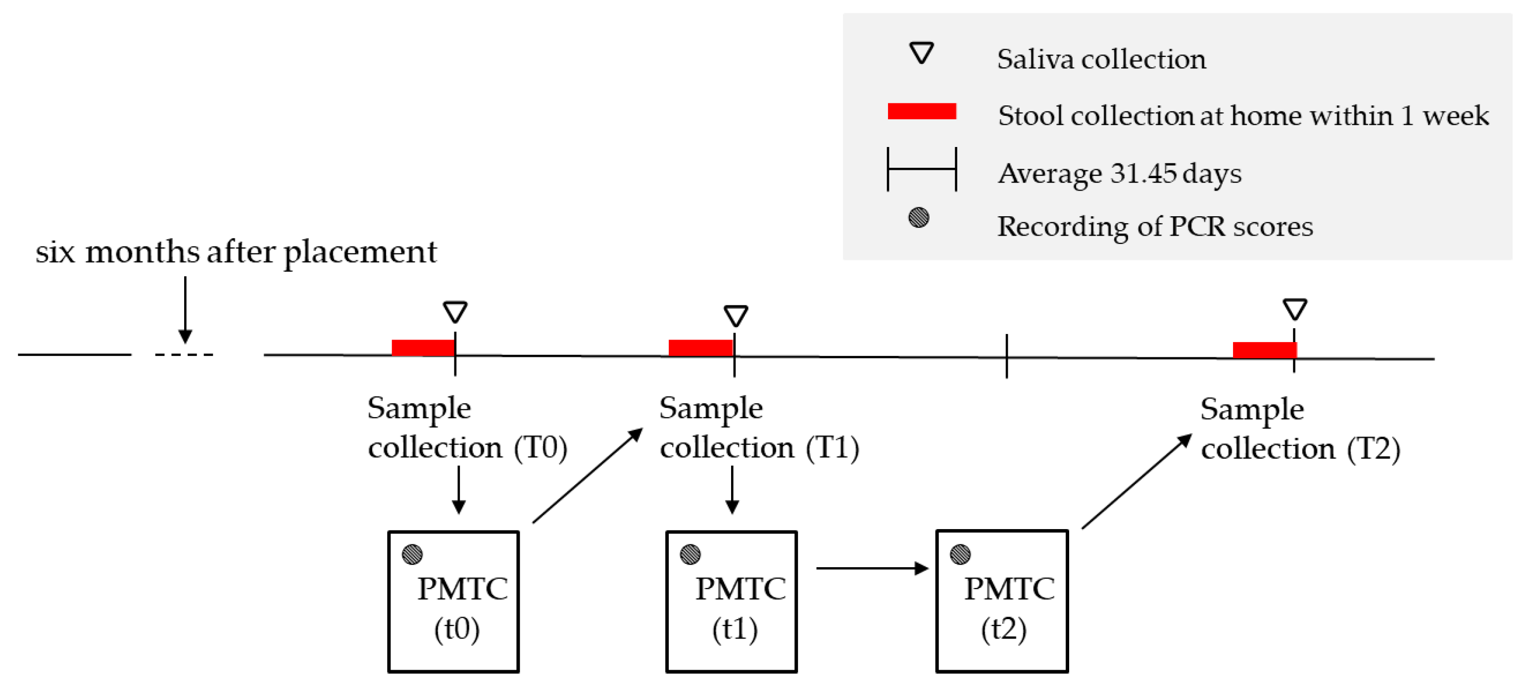
2.1.3. Sample Collection and Storage

While designing this study, the frequency of sample collection was determined from previous studies [8,9,16,21]. Collecting samples at three-time points was deemed necessary to capture dynamic changes during the study period. As multiple-time points are essential to assess temporal changes in bacterial flora, samples were collected at three different time points (T0, T1, and T2). Saliva and stool samples were collected three times each from every participant. The first collection (T0) of saliva and stool samples was performed before PMTC was initiated. The first PMTC (t0) was conducted after specimen collection. The second sampling (T1) was performed approximately one month after the first PMTC (t0). The second PMTC (t1) was performed after collecting the second specimen. One month after the second PMTC (t1), the third PMTC (t2) was performed. The final sample collection (T2) for the third time point was performed approximately one month after the third PMTC (t2). In total, 72 saliva and 72 stool samples were collected across these three-time points, thus providing a robust dataset for our analysis. A flowchart of this study is shown in Figure 1.
The unstimulated saliva was collected [8] from each patient at fixed time points within an average of 90 min. In individuals, the circadian rhythm of the salivary microflora is linked to the human body clock [22]. Considering the diurnal variation in the oral microbiota, sample collection was conducted in such a way that changes in the oral microbiota were not affected by differences in collection time.
Saliva was collected using an ORAL OM505 (DNA Genotek Inc., Kanata, ON, Canada) and stool was collected using FS-0014 (TechnoSuruga Laboratory Co., Ltd., Shizuoka, Japan). Participants were instructed not to eat or clean their mouths for at least 2 h before saliva collection [8,15]. Saliva and stool were frozen at −20 °C after collection [9,23].

2.1.4. PMTC

PMTC was performed once a month for 3 months by one dentist and two hygienists with sufficient experience in the oral cleaning of multi-bracket appliance wearers at the Kanagawa Dental University Hospital orthodontic clinic. The PMTC procedure was standardized for all patients and was followed consistently throughout all sessions, as follows: performed in regular medical treatment included TBI, and instructions were provided at t0, t1, and t2. Guidance was provided using visual aids that demonstrated oral hygiene methods, including toothbrushes (Ruscello; GC Co., Ltd., Tokyo, Japan), tufted brushes (Dent EX Onetuft; LION Dental Products Co., Ltd., Tokyo, Japan), interdental brushes (Prospec; GC Co., Ltd., Tokyo, Japan), and dental floss (Ci Floss; Ci Medical Co., Ltd., Ishikawa, Japan). With regard to TBI, the patients’ oral hygiene status was taken into consideration and the participants were instructed to practice self-cleaning (Figure 2).

2.1.5. PMTC Flow

  • Stain teeth with wires in place to visualize any plaque or debris (2 TONE; Kulzer Denta Ltd., Hanau, Germany).
  • Rinse their mouth with water three times.
  • Measure O’Leary Plaque Control Record (PCR) [24] using the 6-point method.
  • Perform oral cleaning instructions (TBI) using jaw models (TOMY INTERNATIONAL Inc., Tokyo, Japan), toothbrushes, tufted brushes, and interdental brushes.
  • Remove upper and lower wires.
  • Attach a scaler brush (Mini Taper; Ci Medical Co., Ltd., Ishikawa, Japan) to the tip of the air scaler (GC Co., Ltd., Tokyo, Japan) to clean periodontal pockets.
  • Remove tartar using an ultrasonic scaler (Solfy; Morita Co., Ltd., Tokyo, Japan).
  • Attach dental prophy brush (Ci Polishing Brush Flat Soft; Ci Medical Co., Ltd., Ishikawa, Japan) to the contra handpiece (GC Co., Ltd., Tokyo, Japan) and use dental paste (NEW PROCLEAR MEDIUM FINE; Ci Medical Co., Ltd., Ishikawa, Japan) to remove plaque and stain from the tooth surfaces.
  • Clean interdental spaces with floss.
  • Patients were provided instructions to follow the oral hygiene procedure at home.
PCR was calculated from the total number of plaque-attached tooth surfaces, and the number of surfaces was determined using the following formula [24]:
PCR = (total number of plaque-attached tooth surfaces/number of tooth surfaces examined) × 100.

2.2. Microbial DNA Extraction

Oral and gut microbial DNA was extracted from saliva and stool samples using previously described methods. Saliva and stool samples were mixed with a preservative solution and transferred to MORA bead tubes (AMR Inc., Gifu, Japan). The bacteria were crushed using zirconia beads. Following disruption, the supernatant was subjected to automated purification following the protocol of Genefind 2.0 (Beckman Coulter, Inc., Brea, CA, USA) and elution of the respective bacterial DNA in sterile water.

2.3. Microbial Community Analysis

Next-generation sequencing library conditioning and sequencing were performed according to the 16S metagenomic sequencing library conditioning protocol (15044223 Rev B) provided by Illumina (Illumina Inc., San Diego, CA, USA) [25]. The V3–V4 region of 16SrDNA, the target sequence, was amplified from bacterial DNA extracted from saliva and stool samples using the primers 341F (5′-TCGTCGGCAGCGTCAGATGTGTATAAGAGACAGCCTACGGGNGGCWGCAG-3′) and 806R (5′-GTCTCGTGGGCTCGGAGATGTGTATAAGAGACAGGACTACHVGGGTATCTAATCC-3′) (Takara Bio, Saga, Japan). The thermal cycling protocol consisted of 95 °C for 3 min, followed by 95 °C for 30 s, 55 °C for 30 s, 72 °C for 30 s, 28 times in saliva, 25 times in stool, and finally 72 °C for 5 min for amplification (first round of PCR). After confirming amplification, purification was performed using AmpureXP (Nippon Genetics Co., Ltd., Tokyo, Japan), followed by primer removal.
The PCR enzyme KAPA HiFi HotStart (Nippon Genetics Co., Ltd., Tokyo, Japan) was then used to perform eight cycles of PCR amplification (second round of PCR), similar to the thermal cycling conditions described above, to provide the adaptor and index sequences needed for sequencing. The PCR products were purified using AmpureXP and visualized by electrophoresis. Each sample was adjusted to the same concentration. After pooling equal amounts of each sample, next-generation sequencing analysis was performed using the Illumina MiSeq (Illumina Inc., San Diego, CA, USA) under sequencing conditions of 301 bp × 2 paired ends to obtain the nucleotide sequence. The sequences were analyzed using QIIME 2 software (version 2021.4) to estimate the bacterial species and flora [26]. Quality filtering and trimming were performed by using the DADA2 algorithm [27]. In this study, saliva samples were obtained from the V3–V4 region of the Human Oral Microbiome Database (eHOMD version 15.23; The Forsyth Institute, Cambridge, MA, USA), a reference database for oral microbiota [28]. Stool samples were obtained from the Silva database (version 138) (Max Planck Institute for Marine Microbiology, Bremen, Germany), a reference database for RNA gene sequences [29]. (https://qiime2.org/ accessed on 26 August 2022).

2.4. Biodiversity and Statistical Analysis

Alpha diversity, which denotes the abundance and diversity per sample, was analyzed using the Chao1 and Shannon indices. The indices were calculated using the QIIME 2 [25]. Friedman and Wilcoxon signed-rank tests were used to test the significance at T0, T1, and T2 (EZR [Easy R] version 1.54; Jichi Medical Uni-versity Saitama Medical Center, Saitama, Japan). The significance level was set at 5%.

2.5. Statistical Analysis

The relative abundance of each saliva (T0, T1, and T2) and stool (T0, T1, and T2) sample was analyzed using one-way repeated-measures analysis of variance (R version 4.2.1; R Development Core Team, R Foundation for Statistical Computing, Wien, Austria). Significant differences between the bacterial composition ratios at each sampling time point (T0, T1, and T2) were examined from the phylum to the species level. The significance level was set at 5%.
Friedman and Wilcoxon signed-rank tests were used to test the significance of changes in PCR values at t0, t1, and t2 (EZR [Easy R] version 1.54). The significance level was set at 1%.
The Pearson correlation coefficient was used to analyze the two quantitative relationships between treatment duration and microbiota, qualitative relationships between the vertical intermaxillary relationship and microbiota, and the relationship between mouth breathing and microbiota (R version 4.2.1). The significance level was set at 5%.

3. Results

3.1. Oral Microbiota

3.1.1. Bacterial Composition Ratio

Sequences were clustered into 13 phyla, 27 classes, 39 orders, 65 families, 135 genera, and 435 species. In all bacterial groups identified at each level, from the phylum to the species level, slight changes were observed at T0, T1, and T2. However, none of the bacteria exhibited a significant increase or decrease (p > 0.05). The six major phyla at the phylum level (Firmicutes, Bacteroidetes, Fusobacteria, Proteobacteria, Saccharibacteria, and Actinobacteria) accounted for 99.07% at T0, 98.78% at T1, and 98.72% at T2, and no significant changes were observed. The differences in T0, T1, and T2 between the major oral bacteria at the phylum and genus levels are shown in Table 2.

3.1.2. Alpha Diversity

The Shannon index was used to evaluate the evenness of the oral microbiota and the Chao1 index was used to evaluate the richness of the oral microbiota. The Shannon index increased over time at T0, T1, and T2; however, these differences were not significant (p > 0.05). The Chao1 index showed significant differences between T0 and T2, T1 and T2 (p < 0.05). The results are shown in Figure 3.

3.2. Gut Microbiota

3.2.1. Comparison of Bacterial Composition

The sequences were clustered into 19 phyla, 28 classes, 63 orders, 116 families, 299 genera, and 627 species. In all the bacterial groups identified at each level, from the phylum to the species level, slight changes were observed at T0, T1, and T2. However, none of the bacteria exhibited a significant increase or decrease (p > 0.05). The differences in T0, T1, and T2 between the major gut bacteria at the phylum and genus levels are shown in Table 3.

3.2.2. Duration and Gut Microbiota

We divided the different treatment durations from the beginning of orthodontic treatment into early- and long-term groups. Then, we investigated the changes in the relative abundance of each bacterium at different sampling times from T0 to T1, T0 to T2, and T1 to T2. As a result, for some bacteria, significant differences in the change in relative abundance of bacteria were observed for different treatment periods from the start of orthodontic treatment (p < 0.05). Table 4 lists the bacteria for which significant differences were detected.

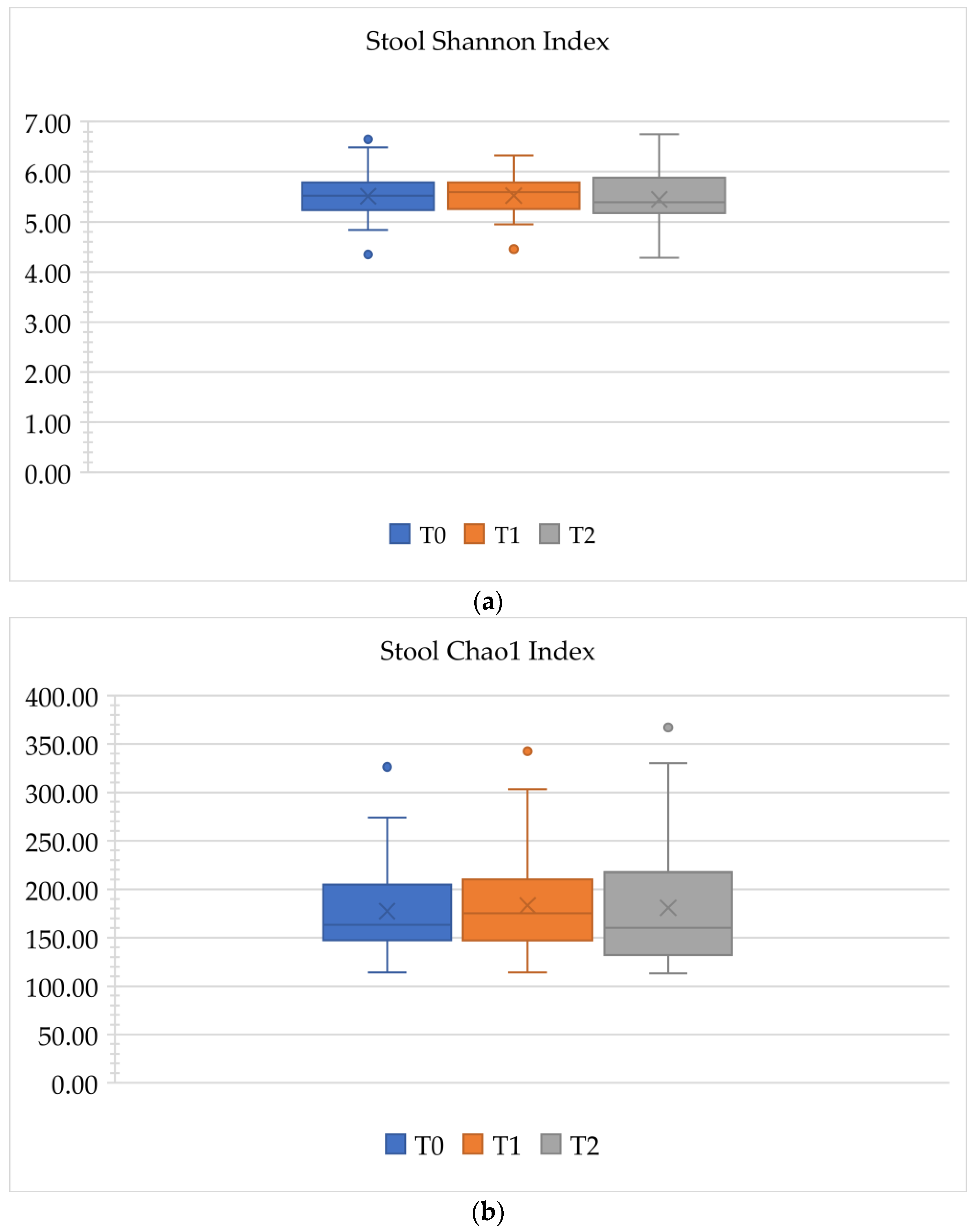
3.2.3. Alpha Diversity

The Shannon and Chao1 indices were used to evaluate the evenness and richness of the gut microbiota, respectively. No significant differences in the abundance and evenness of the bacterial microbiota were observed in the gut microbiota in any of the T0, T1, or T2 comparisons (p > 0.05). The results are shown in Figure 4.

3.3. PCR Score

A comparison of the first (T0), second (T1), and third (T2) PCR measurements revealed a p-value of 1.88 × 10⁻⁸, which decreased significantly over time. A statistical test to determine whether there was a difference between the groups showed a significant difference with p-values of 2.84 × 10⁻⁵ for T0 and T1, 5.83 × 10⁻³ for T1 and T2, and 1.19 × 10⁻⁶ for T0 and T2. The results are shown in Table 5.

4. Discussion

4.1. Summary

In this study, we investigated whether continuous PMTC, including TBI, could be used as a solution to reduce the risk of dental caries and periodontitis in orthodontic treatment with multi-bracket appliances; this is based on changes in the oral microbiota of patients more than 6 months after the start of treatment. We also evaluated the dynamics of the gut microbiota in multi-bracket appliance wearers and examined the relationship between orthodontic treatment and gut microbiota.
Our findings suggest that continuous PMTC administration may be effective in maintaining the oral environment and inhibiting the growth of pathogenic bacteria. The results indicate that continuous PMTC plays an important role in orthodontic treatment with multi-bracket appliances and should be considered as a solution to reduce the risk of caries and periodontitis. However, it is also clear that a decrease in PCR scores does not directly affect changes in oral microbiota. It is difficult to avoid the risks associated with the use of multi-bracket appliances [16]. The results provide a perspective on the effectiveness of manageable and sustainable risk avoidance methods. In this study, the participant selection was not based on their cleaning habits. If only patients with inferior plaque control among those wearing multi-bracket appliances were included in this study, the results might differ from those of the present study. Furthermore, a study on orthodontic treatment and gut microbiota suggested that changes in eating habits associated with different periods of wearing multi-bracket appliances may affect the gut microbiota. The gut microbiota is significantly affected by external factors such as changes in eating habits and lifestyle [30].

4.2. Multi-Bracket-PMTC-Microbiota

The oral microbiota is significantly more variable in individuals with high caries activity than in those with healthy oral environments [31]. The diversity of oral bacteria was significantly higher in individuals with a healthy oral environment, without caries or periodontal tissue disease, than in those with high caries activity [32]. This result indicates that an increased diversity of the oral microbiota is a critical factor for maintaining a healthy oral environment. In this study, no significant difference was observed in the change in the Shannon index with the continuation of PMTC, although it showed an increasing trend over time. The Chao1 index increased over time and showed statistical significance. These results suggest that the continuation of PMTC among multi-bracket wearers indicates an improving trend in the oral environment.
Plaque accumulation increases the risk of caries and periodontitis morbidity [33]. Wearing multi-bracket appliances allows for large amounts of plaque accumulation [34]. Furthermore, multi-bracket appliance wearers have an increased microbial load compared to individuals who do not wear any [35]. In this study, PCR scores decreased over time with continuous PMTC; thus, it indicates that continuous PMTC prevents dental caries and periodontal disease in multi-bracket appliances, and is an essential means of maintaining the oral environment. Enhancing patient motivational education regarding periodontal disease management is fundamental to the prevention of periodontal disease [36]. Ensuring PMTC and providing careful oral cleaning instructions based on an individual’s oral cleaning status will significantly reduce this risk among multi-bracket appliance wearers. Based on an investigation of the effectiveness of person-centered oral hygiene behavioral interventions in general dentistry and the reinforcement of instruction in behavior change [37], this study suggests the need to establish person-centered oral hygiene education programs targeting behavioral interventions in orthodontics.
PCR is a strict assessment of oral hygiene status. Smiech-Slomkowska et al. [38] showed that OHI (oral health education and oral hygiene instruction) significantly improved oral hygiene without affecting bacterial levels. This result is consistent with the lack of changes in the oral bacterial microbiota in the present study. A chemical approach is necessary to reduce bacterial levels [39]. A study on plaque index and oral hygiene instructions [40] showed significant short-term improvement in oral hygiene. However, 6 months after the education, oral hygiene levels declined. This suggests that continued long-term oral hygiene instruction during treatment may influence risk reduction and avoidance in multi-bracket appliance wearers.
In this study, no significant difference in the oral microbiota was observed with continuous PMTC at any stage from the phylum to the species level. Bacteroidetes were found in the oral cavity and intestines at the phylum level. Oral Bacteroidetes include periodontopathogenic bacteria, such as P. gingivalis and P. intermedia. Additionally, TM7 is associated with periodontitis, increases with age, and has been implicated in periodontitis [41,42]. Previous studies have shown an increasing trend in Bacteroidetes before and after wearing multi-bracket appliances [8]. A significant increase was also observed in TM7 [8]. In this study, TM7 showed no significant increase in relative abundance ratios at T0, T1, and T2, and a slightly decreasing trend at the Bacteroidetes group, suggesting that continuous PMTC during orthodontic treatment with multi-brackets may have inhibited the growth of periodontitis-associated bacteria, and may have influenced the stabilization of the oral environment.

4.3. Gut Microbiota

4.3.1. Treatment Duration and Gut Microbiota

Firmicutes, the largest phylum in the gut microbiota, is implicated in the pathogenesis of diabetes and obesity [43,44,45]. In particular, the relative abundance of Christensenellaceae, belonging to the phylum Firmicutes, was inversely correlated with host body mass index (BMI), according to a study by Waters et al. [46]. To date, the relationship between Christensenellaceae and BMI is the most robust and reproducible association between microbial ecology and metabolic diseases in the human gut. This suggests that it is also related to the health status of patients with many other diseases, including obesity and inflammatory bowel disease. Gnanasambandam et al. [47] found that BMI decreased during the first 3 months of orthodontic treatment and gradually recovered during the first year of treatment. Our study found significant differences in the Christensenellaceae abundance ratio across different treatment durations. Comparing the increase or decrease in the abundance ratio at T1 and T2, the shorter treatment duration group showed an increase in Christensenellaceae. In contrast, the longer treatment duration group exhibited decrease in Christensenellaceae. Although we did not observe changes in body weight over time in this study, the increase in Christensenellaceae in the short-treatment duration group may indicate that weight loss was initially observed. Therefore, weight loss may have improved in the long-term treatment group.
In the group with a shorter treatment duration, there may have been unfamiliar discomfort associated with wire adjustments compared with the group with a longer treatment duration, which could have led to greater restrictions on dietary intake and food choices. Conversely, in the group with a longer treatment duration, food intake restrictions due to pain relief and familiarity with orthodontic treatment were alleviated and eating habits were restored to pretreatment habits. Yaoita et al. [48] described how eating habits combined with appropriate masticatory activity and stimulation may play an important role in providing a favorable intestinal environment based on the interaction between the gut microbiota and the host immune system. The impact of orthodontic treatment-associated food choice restriction on masticatory activity and its associated effects on the gut microbiota are unclear. To clarify the relationship between orthodontic treatment and gut microbiota, it is important to examine changes in the gut microbiota associated with orthodontic treatment and masticatory activity in the future.

4.3.2. Oral and Gut Microbiota

Previous studies have also suggested that oral microbiota may significantly affect the development of intestinal diseases and colorectal cancer, with Fusobacterium nucleatum being of particular interest because of its association with colon cancer carcinogenesis and progression [10]. In S. mutans, a representative caries-pathogenic bacterium, its effects on diseases such as infective endocarditis and cerebral hemorrhage have been suggested [49]. P. gingivalis, a representative periodontopathogenic bacterium, has been suggested to cause atherosclerosis, rheumatism, and diabetes mellitus [50,51]. The saliva of patients with inflammatory bowel disease is rich in bacteria of the Prevotella genus and molecules involved in inflammation [52]. Disruption of oral microbiota significantly affects systemic diseases.
In this study, no significant changes in the gut microbiota were observed and there was no effect on systemic diseases associated with the use of a multi-bracket appliance. These results indicate that continuous PMTC for multi-bracket appliance wearers may stabilize the oral environment, with no growth of pathogenic bacteria in the mouth. However, further long-term studies are required to validate these findings.

4.4. Conditions for Participant Selection

4.4.1. Age

The prevalence of periodontopathogenic bacteria increases with age. For example, TM7 at the portal level of oral microbiota has been found to increase with age. It is involved in the initiation of periodontitis [40,41]; however, no marked differences in TM7 levels were observed in this study. The average age of the participants in this study was 25.7 years, and it is possible that the results of this study may change as the dynamics of bacterial composition may alter as the participants age. In the future, the aging society will advance further, and orthodontic treatment for the prosthetic management of missing teeth will be increasingly selected. Considering the possibility that the target age for orthodontic treatment will increase from the previous target age, it is necessary to compare the evaluation of continuous PMTC in the oral environment, according to age.

4.4.2. Number of Teeth

Two primary causes of tooth loss are caries and periodontal diseases. The participants in this study had 28 sound teeth or two or four intermediate teeth that were extracted to correct dental malocclusions. Those who had lost their teeth because of caries or periodontal disease were excluded. No previous studies have examined changes in the bacterial microbiota before and after treatment of cases with multi-bracket appliances for the prosthetic treatment of missing teeth. Because individuals with tooth loss due to dental caries or periodontal disease have a large variation in oral flora [30], changes in the number of teeth may also be observed if changes in flora during continuous PMTC in patients with missing teeth are verified.

4.4.3. Race

Comparisons of the oral microbiota before and after the use of multi-bracket appliances have been performed in various Western and Oriental countries. The participants in this study were Japanese, and the phylum-level predominance of Firmicutes, Proteobacteria, Actinobacteria, Bacteroidetes, and Fusobacteria in the oral microbiota was consistent with previous studies [53,54]. In particular, differences in the gut microbiota, which are greatly influenced by external factors, have been noted among racial groups owing to differences in food culture. Firmicutes and Bacteroidetes are the major intestinal bacterial factions [54,55], and the results of the present study are identical to those of previous studies on the predominant phyla in the gut microbiota. Racial differences in the predominant phyla may not be observed in patients with or without multi-bracket appliances.

4.4.4. Skeletal Pattern, Vertical Jaw Relationship, and Occlusal Relationship

In orthodontic treatment, skeletal pattern, vertical jaw relationship, and occlusal relationship are important in determining a treatment plan. Due to the large participant bias in this study, we did not evaluate the relationship between different skeletal patterns and bacterial microbiota. Regarding the occlusal relationship, more than 6 months had passed since the treatment course, and changes were already observed in the occlusal relationship before the start of treatment and at the time of sample collection. Therefore, we did not evaluate the relationship between the differences in occlusal relationships and bacterial microbiota. Regarding the vertical jaw relationship, we examined the differences in bacterial microbiota between the Dolico and Mesio facial patterns; however, we found no significant differences in the oral or gut microbiota. Given that patients with dental abnormalities present an oral environment that differs from normal, such as decreased masticatory efficiency, differences in salivary flow, and decreased occlusal pressure, it is highly possible that the differences in the oral and gut microbiota are due to differences in skeletal patterns, vertical jaw relationships, and occlusal relationships. This issue should be addressed in future studies.

4.5. Mouth Breathing

In most cases of dental abnormalities, patients experience functional problems with most dental abnormalities. Many Angle’s Class II division 1 cases, also known as occlusal relationship classifications, present with mouth breathing. Patients with mouth breathing often present with gingivitis symptoms such as gingival redness in the anterior teeth with a dry mouth. In this study, we examined whether there were differences in oral and gut microbiota between patients with and without mouth breathing. However, no statistically significant differences were observed between groups. A study on the oral microbiota of patients with xerostomia [56] showed changes in the major microbial phyla in the oral cavity between patients with xerostomia and healthy individuals; however, no statistically significant differences were found. However, they noted that the oral microbiota could be used as a diagnostic tool for xerostomia and that more samples are needed for this purpose.
A larger sample size may allow for the evaluation of the effects of mouth breathing and continuous PMTC on the oral microbiota of multi-bracket appliance wearers.

4.6. Method

Saliva and plaque are the two main sources used to assess the oral microbial communities. Saliva provides more information about the entire oral cavity than plaque [57], and its collection is noninvasive and easy to perform. Therefore, saliva was used in this study to investigate the oral microbiota. Caries and periodontal disease, two major oral diseases, are also major risks for multi-bracket appliance wearers. Their etiology and progression are better understood through a comprehensive understanding of the microbial community [58]. Therefore, in this study, 16SrRNA sequencing was performed.
In this study, we examined the changes in the oral and gut microbiota during 3 months of continuous PMTC; however, no significant differences were found. It is possible that a more extended observation period could yield different results than those obtained in this study. Additionally, new bioactive biomolecules, such as probiotics [59,60] and postbiotics [61], have been shown to influence the stabilization of the oral environment. These new bioactive biomolecules are effective as additional treatments in the prevention of dental caries and periodontal diseases. Research is also underway to investigate their impact on orthodontic dental treatments [62]. Therefore, we propose further studies on these products and emphasize the need to evaluate their effects on the oral and gut microbiota of orthodontic patients.

4.7. Limitations

There are several potential limitations of our study. First, the oral environment, such as the number of teeth, caries status, and number and type of prostheses worn, may influence the composition and diversity of the oral microbiota. However, caries and other aspects of the oral environment have not been investigated. Additionally, the presence or absence of bands or mini-screws and their numbers were not standardized for each patient.

5. Conclusions

The results of this study suggest that continuous PMTC during orthodontic treatment with multi-bracket appliances may inhibit the growth of pathogenic bacteria in the oral cavity and prevent dysbiosis of both the oral and gut microbiota by maintaining a clean oral environment. Considering that the stabilization of the oral environment may contribute to reducing the risk of systemic diseases, the achievement of continuous PMTC for multi-bracket appliances should be emphasized as a part of not only oral but also systemic management.
Significant changes in the gut microbiota with different treatment durations suggest that reduced dietary intake and food choices associated with different orthodontic treatment durations may affect the gut microbiota.
Fixed orthodontic appliances are not only used to treat systemically healthy individuals, but are also used to treat populations with systemic diseases. In light of this, research on changes in oral and gut microbiota as influenced by oral cleaning management in orthodontic appliance wearers needs to be conducted from a longer-term perspective.

Author Contributions

Conceptualization, S.O. and T.Y.; methodology, S.O. and T.Y.; formal analysis, S.O.; investigation, S.O. and T.Y.; resources, S.O.; data curation, S.O.; writing—original draft preparation, S.O. and T.Y.; writing—review and editing, K.H., A.T. and T.Y.; visualization, S.O.; supervision, T.Y.; project administration, T.Y.; funding acquisition, T.Y. All authors have read and agreed to the published version of the manuscript.

Funding

This research was funded by Kanagawa Dental University 2023 Project Research No. 6 Research Fund.

Institutional Review Board Statement

This study was conducted in accordance with the guidelines of the Declaration of Helsinki and approved by the Ethics Committee of Kanagawa Dental University (approval No. 795).

Informed Consent Statement

Informed consent was obtained from all the participants involved in this study.

Data Availability Statement

Data supporting the results of this study are available from the corresponding author upon reasonable request.

Acknowledgments

This study was supported by Kanagawa Dental University 2023 Project Research. We thank Makoto Taniguchi of the GenomeRead Corporation for the bacterial flora analysis and valuable advice.

Conflicts of Interest

The authors declare no conflict of interest.

Abbreviations

PMTCprofessional mechanical tooth cleaning
TBIincluding tooth-brushing instructions
S. mutansStreptococcus mutans
SDstandard deviation
PCRplaque control record
OHIoral health education and oral hygiene instruction
BMIbody mass index

References

  1. Lucchese, A.; Bondemark, L.; Marcolina, M.; Manuelli, M. Changes in oral microbiota due to orthodontic appliances: A systematic review. J. Oral. Microbiol. 2018, 10, 1476645. [Google Scholar] [CrossRef] [PubMed]
  2. Ristic, M.; Svabic, M.V.; Sasic, M.; Zelic, O. Effects of fixed orthodontic appliances on subgingival microflora. Int. J. Dent. Hyg. 2008, 6, 129–136. [Google Scholar] [CrossRef]
  3. Thornberg, M.J.; Riolo, C.S.; Bayirli, B.; Riolo, M.L.; Van Tubergen, E.A.; Kulbersh, R. Periodontal pathogen levels in adolescents before, during, and after fixed orthodontic appliance therapy. Am. J. Orthod. Dentofac. Orthop. 2009, 135, 95–98. [Google Scholar] [CrossRef] [PubMed]
  4. Julien, K.C.; Buschang, P.H.; Campbell, P.M. Prevalence of white spot lesion formation during orthodontic treatment. Angle Orthod. 2013, 83, 641–647. [Google Scholar] [CrossRef]
  5. Heymann, G.C.; Grauer, D. A contemporary review of white spot lesions in orthodontics. J. Esthet. Restor. Dent. 2013, 25, 85–95. [Google Scholar] [CrossRef] [PubMed]
  6. Reichardt, E.; Geraci, J.; Sachse, S.; Rödel, J.; Pfister, W.; Löffler, B.; Wagner, Y.; Eigenthaler, M.; Wolf, M. Qualitative and quantitative changes in the oral bacterial flora occur shortly after implementation of fixed orthodontic appliances. Am. J. Orthod. Dentofac. Orthop. 2019, 156, 735–744. [Google Scholar] [CrossRef]
  7. Mummolo, S.; Nota, A.; Albani, F.; Marchetti, E.; Gatto, R.; Marzo, G.; Quinzi, V.; Tecco, S. Salivary levels of Streptococcus mutans and Lactobacilli and other salivary indices in patients wearing clear aligners versus fixed orthodontic appliances: An observational study. PLoS ONE 2020, 15, e0228798. [Google Scholar] [CrossRef]
  8. Kado, I.; Hisatsune, J.; Tsuruda, K.; Tanimoto, K.; Sugai, M. The impact of fixed orthodontic appliances on oral microbiome dynamics in Japanese patients. Sci. Rep. 2020, 10, 21989. [Google Scholar] [CrossRef]
  9. Pan, S.; Liu, Y.; Zhang, L.; Li, S.; Zhang, Y.; Liu, J.; Wang, C.; Xiao, S. Profiling of subgingival plaque biofilm microbiota in adolescents after completion of orthodontic therapy. PLoS ONE 2017, 12, e0171550. [Google Scholar] [CrossRef]
  10. Uchino, Y.; Goto, Y.; Konishi, Y.; Tanabe, K.; Toda, H.; Wada, M.; Kita, Y.; Beppu, M.; Mori, S.; Hijioka, H.; et al. Colorectal Cancer patients have four specific bacterial species in oral and gut microbiota in common—A metagenomic comparison with healthy subjects. Cancers 2021, 13, 3332. [Google Scholar] [CrossRef]
  11. Chen, B.-Y.; Lin, W.-Z.; Li, Y.-L.; Bi, C.; Du, L.-J.; Liu, Y.; Zhou, L.-J.; Liu, T.; Xu, S.; Shi, C.-J.; et al. Roles of oral microbiota and oral-gut microbial transmission in hypertension. J. Adv. Res. 2023, 43, 147–161. [Google Scholar] [CrossRef] [PubMed]
  12. Olsen, I.; Yamazaki, K. Can oral bacteria affect the microbiome of the gut? J. Oral. Microbiol. 2019, 11, 1586422. [Google Scholar] [CrossRef]
  13. Hajishengallis, G.; Chavakis, T. Local and systemic mechanisms linking periodontal disease and inflammatory comorbidities. Nat. Rev. Immunol. 2021, 21, 426–440. [Google Scholar] [CrossRef]
  14. de Oliveira, C.; Watt, R.; Hamer, M. Toothbrushing, inflammation, and risk of cardiovascular disease: Results from Scottish Health Survey. BMJ 2010, 340, c2451. [Google Scholar] [CrossRef] [PubMed]
  15. Wang, Q.; Ma, J.-B.; Wang, B.; Zhang, X.; Yin, Y.-L.; Bai, H. Alterations of the oral microbiome in patients treated with the Invisalign system or with fixed appliances. Am. J. Orthod. Dentofac. Orthop. 2019, 156, 633–640. [Google Scholar] [CrossRef] [PubMed]
  16. Zhao, M.; Liu, M.; Chen, W.; Zhang, H.; Bai, Y.; Ren, W. Salivary microbial changes during the first 6 months of orthodontic treatment. PeerJ 2020, 8, e10446. [Google Scholar] [CrossRef] [PubMed]
  17. Boyd, R.L. Longitudinal evaluation of a system for self-monitoring plaque control effectiveness in orthodontic patients. J. Clin. Periodontol. 1983, 10, 380–388. [Google Scholar] [CrossRef]
  18. Awartani, F.; Atassi, F. Oral hygiene status among orthodontic patients. J. Contemp. Dent. Pract. 2010, 11, 25–32. [Google Scholar] [CrossRef]
  19. Zhao, R.; Huang, R.; Long, H.; Li, Y.; Gao, M.; Lai, W. The dynamics of the oral microbiome and oral health among patients receiving clear aligner orthodontic treatment. Oral. Dis. 2020, 26, 473–483. [Google Scholar] [CrossRef]
  20. Jing, D.; Hao, J.; Shen, Y.; Tang, G.; Lei, L.; Zhao, Z. Effect of fixed orthodontic treatment on oral microbiota and salivary proteins. Exp. Ther. Med. 2019, 17, 4237–4243. [Google Scholar] [CrossRef]
  21. Zivkovic-Sandic, M.; Popovic, B.; Carkic, J.; Nikolic, N.; Glisic, B. Changes in subgingival microflora after placement and removal of fixed orthodontic appliances. Srp. Arh. Za Celok. Lek. 2014, 142, 301–305. [Google Scholar] [CrossRef]
  22. Takayasu, L.; Suda, W.; Takanashi, K.; Iioka, E.; Kurokawa, R.; Shindo, C.; Hattori, Y.; Yamashita, N.; Nishijima, S.; Oshima, K.; et al. Circadian oscillations of microbial and functional composition in the human salivary microbiome. DNA Res. 2017, 24, 261–270. [Google Scholar] [CrossRef] [PubMed]
  23. Park, H.; Hosomichi, K.; Kim, Y.-I.; Hikita, Y.; Tajima, A.; Yamaguchi, T. Comprehensive Genetic Exploration of Fused Teeth by Whole Exome Sequencing. Appl. Sci. 2022, 12, 11899. [Google Scholar] [CrossRef]
  24. O’Leary, T.J.; Drake, R.B.; Naylor, J.E. The plaque control record. J. Periodontol. 1972, 43, 38. [Google Scholar] [CrossRef] [PubMed]
  25. Wang, D.; Nambu, T.; Tanimoto, H.; Iwata, N.; Yoshikawa, K.; Okinaga, T.; Yamamoto, K. Interdental Plaque Microbial Community Changes under In Vitro Violet LED Irradiation. Antibiotics 2021, 10, 1348. [Google Scholar] [CrossRef]
  26. Bolyen, E.; Rideout, J.R.; Dillon, M.R.; Bokulich, N.A.; Abnet, C.C.; Al-Ghalith, G.A.; Alexander, H.; Alm, E.J.; Arumugam, M.; Asnicar, F.; et al. Reproducible, interactive, scalable and extensible microbiome data science using QIIME 2. Nat. Biotechnol. 2019, 37, 852–857. [Google Scholar] [CrossRef] [PubMed]
  27. Callahan, B.J.; Mcmurdie, P.J.; Rosen, M.J.; Han, A.W.; Johnson, A.J.A.; Holmes, S.P. DADA2: High-resolution sample inference from Illumina amplicon data. Nat. Methods 2016, 13, 581–583. [Google Scholar] [CrossRef]
  28. Chen, T.; Yu, W.-H.; Izard, J.; Baranova, O.V.; Lakshmanan, A.; Dewhirst, F.E. The Human Oral Microbiome Database: A web accessible resource for investigating oral microbe taxonomic and genomic information. Database 2010, 6, baq013. [Google Scholar] [CrossRef]
  29. Quast, C.; Pruesse, E.; Yilmaz, P.; Gerken, J.; Schweer, T.; Yarza, P.; Peplies, J.; Glöckner, F.O. The SILVA ribosomal RNA gene database project: Improved data processing and web-based tools. Nucleic Acids Res. 2013, 41, D590–D596. [Google Scholar] [CrossRef]
  30. Arumugam, M.; Raes, J.; Pelletier, E.; Le Paslier, D.; Yamada, T.; Mende, D.R.; Fernandes, G.R.; Tap, J.; Bruls, T.; Batto, J.M.; et al. Enterotypes of the human gut microbiome. Nature 2011, 473, 174–180. [Google Scholar] [CrossRef]
  31. Yang, F.; Zeng, X.; Ning, K.; Liu, K.-L.; Lo, C.-C.; Wang, W.; Chen, J.; Wang, D.; Huang, R.; Chang, X.; et al. Saliva microbiomes distinguish caries-active from healthy human populations. ISME J. 2011, 6, 1–10. [Google Scholar] [CrossRef] [PubMed]
  32. Li, Y.; Ku, C.; Xu, J.; Saxena, D.; Caufield, P. Survey of oral microbial diversity using PCR-based denaturing gradient gel electrophoresis. J. Dent. Res. 2005, 84, 559–564. [Google Scholar] [CrossRef] [PubMed]
  33. Zaghrisson, B.U.; Zachrisson, S. Caries incidence and oral hygiene during orthodontic treatment. Scand. J. Dent. Res. 1971, 79, 394–401. [Google Scholar] [CrossRef] [PubMed]
  34. Jongsma, M.A.; Pelser, F.D.H.; van der Mei, H.C.; Atema-Smit, J.; van de Belt-Gritter, B.; Busscher, H.J.; Ren, Y. Biofilm formation on stainless steel and gold wires for bonded retainers in vitro and in vivo and their susceptibility to oral antimicrobials. Clin. Oral. Investig. 2013, 17, 1209–1218. [Google Scholar] [CrossRef]
  35. Hagg, U.; Kaveewatcharanont, P.; Samaranayake, Y.H.; Samaranayake, L.P. The effect of fixed orthodontic appliances on the oral carriage of Candida species and Enterobacteriaceae. Eur. J. Orthod. 2004, 26, 623–629. [Google Scholar] [CrossRef]
  36. Tonetti, M.S.; Chapple, I.L.C.; Jepsen, S.; Sanz, M. Primary and secondary prevention of periodontal and peri-implant diseases: Introduction to, and objectives of the 11th European Workshop on Periodontology consensus conference. J. Clin. Periodontol. 2015, 42 (Suppl. 16), S1–S4. [Google Scholar] [CrossRef]
  37. Dimenäs, S.L.; Jönsson, B.; Andersson, J.S.; Lundgren, J.; Petzold, M.; Abrahamsson, I.; Abrahamsson, K.H. A person-centred, theory-based, behavioural intervention programme for improved oral hygiene in adolescents: A randomized clinical field study. J. Clin. Periodontol. 2022, 49, 378–387. [Google Scholar] [CrossRef]
  38. Smiech-Slomkowska, G.; Jablonska-Zrobek, J. The effect of oral health education on dental plaque development and the level of caries-related Streptococcus mutans and Lactobacillus spp. Eur. J. Orthod. 2007, 29, 157–160. [Google Scholar] [CrossRef] [PubMed]
  39. Shimpo, Y.; Nomura, Y.; Sekiya, T.; Arai, C.; Okada, A.; Sogabe, K.; Hanada, N.; Tomonari, H. Effects of the dental caries preventive procedure on the white spot lesions during orthodontic treatment—An open label randomized controlled trial. J. Clin. Med. 2022, 11, 854. [Google Scholar] [CrossRef] [PubMed]
  40. Soldo, M.; Matijević, J.; Ivanišević, A.M.; Čuković-Bagić, I.; Marks, L.; Borić, D.N.; Krmek, S.J. Impact of oral hygiene instructions on plaque index in adolescents. Cent. Eur. J. Public. Health 2020, 28, 103–107. [Google Scholar] [CrossRef] [PubMed]
  41. Crielaard, W.; Zaura, E.; Schuller, A.A.; Huse, S.M.; Montijn, R.C.; Keijser, B.J. Exploring the oral microbiota of children at various developmental stages of their dentition in the relation to their oral health. BMC Med. Genom. 2011, 4, 22. [Google Scholar] [CrossRef]
  42. Duran-Pinedo, A.E.; Chen, T.; Teles, R.; Starr, J.R.; Wang, X.; Krishnan, K.; Frias-Lopez, J. Community-wide transcriptome of the oral microbiome in subjects with and without periodontitis. ISME J. 2014, 8, 1659–1672. [Google Scholar] [CrossRef]
  43. Rinninella, E.; Raoul, P.; Cintoni, M.; Franceschi, F.; Miggiano, G.A.D.; Gasbarrini, A.; Mele, M.C. What is the healthy gut microbiota composition? a changing ecosystem across age, environment, diet, and diseases. Microorganisms 2019, 7, 14. [Google Scholar] [CrossRef]
  44. Larsen, N.; Vogensen, F.K.; Van Den Berg, F.W.J.; Nielsen, D.S.; Andreasen, A.S.; Pedersen, B.K.; Al-Soud, W.A.; Sørensen, S.J.; Hansen, L.H.; Jakobsen, M. Gut microbiota in human adults with type 2 diabetes differs from non-diabetic adults. PLoS ONE 2010, 5, e9085. [Google Scholar] [CrossRef] [PubMed]
  45. Koliada, A.; Syzenko, G.; Moseiko, V.; Budovska, L.; Puchkov, K.; Perederiy, V.; Gavalko, Y.; Dorofeyev, A.; Romanenko, M.; Tkach, S.; et al. Association between body mass index and Firmicutes/Bacteroidetes ratio in an adult Ukrainian population. BMC Microbiol. 2017, 17, 120. [Google Scholar] [CrossRef] [PubMed]
  46. Waters, J.L.; Ley, R.E. The human gut bacteria Christensenellaceae are widespread, heritable, and associated with health. BMC Biol. 2019, 17, 83. [Google Scholar] [CrossRef]
  47. Gnanasambandam, V.; Gnaneswar, S.M. Effects of orthodontic treatment on body mass index, food habits and self-esteem of patients: A prospective single-arm cohort study. J. Taibah Univ. Med. Sci. 2022, 17, 818–825. [Google Scholar] [CrossRef] [PubMed]
  48. Yaoita, F.; Watanabe, K.; Kimura, I.; Miyazawa, M.; Tsuchiya, S.; Kanzaki, M.; Tsuchiya, M.; Tan-No, K. Impact of habitual chewing on gut motility via microbiota transition. Sci. Rep. 2022, 12, 13819. [Google Scholar] [CrossRef] [PubMed]
  49. Nakano, K.; Nomura, R.; Matsumoto, M.; Ooshima, T. Roles of oral bacteria in cardiovascular diseases—From molecular mechanisms to clinical cases: Cell-surface structures of novel serotype k Streptococcus mutans strains and their correlation to virulence. J. Pharmacol. Sci. 2010, 113, 120–125. [Google Scholar] [CrossRef]
  50. Arimatsu, K.; Yamada, H.; Miyazawa, H.; Minagawa, T.; Nakajima, M.; Ryder, M.I.; Gotoh, K.; Motooka, D.; Nakamura, S.; Iida, T.; et al. Oral pathobiont induces systemic inflammation and metabolic changes associated with alteration of gut microbiota. Sci. Rep. 2014, 4, 4828. [Google Scholar] [CrossRef]
  51. Nakajima, M.; Arimatsu, K.; Kato, T.; Matsuda, Y.; Minagawa, T.; Takahashi, N.; Ohno, H.; Yamazaki, K. Oral administration of P. gingivalis induces dysbiosis of gut microbiota and impaired barrier function leading to dissemination of enterobacteria to the liver. PLoS ONE 2015, 10, e0134234. [Google Scholar] [CrossRef]
  52. Ohkusa, T.; Yoshida, T.; Sato, N.; Watanabe, S.; Tajiri, H.; Okayasu, I. Commensal bacteria can enter colonic epithelial cells and induce proinflammatory cytokine secretion: A possible pathogenic mechanism of ulcerative colitis. J. Med. Microbiol. 2009, 58, 535–545. [Google Scholar] [CrossRef]
  53. Human Microbiome Project Consortium. Structure, function and diversity of the healthy human microbiome. Nature 2012, 486, 207–214. [Google Scholar] [CrossRef] [PubMed]
  54. Cueva, C.; Silva, M.; Pinillos, I.; Bartolomé, B.; Moreno-Arribas, M.V. Interplay between dietary polyphenols and oral and gut microbiota in the development of colorectal cancer. Nutrients 2020, 12, 625. [Google Scholar] [CrossRef]
  55. Kovatcheva-Datchary, P.; Nilsson, A.; Akrami, R.; Lee, Y.S.; De Vadder, F.; Arora, T.; Hallen, A.; Martens, E.; Björck, I.; Bäckhed, F. Dietary fiber-induced improvement in glucose metabolism is associated with increased abundance of Prevotella. Cell Metab. 2015, 22, 971–982. [Google Scholar] [CrossRef] [PubMed]
  56. Weng, C.-T.; Huang, S.-L.; Yang, H.-W.; Kao, C.-C.; Wei, C.-C.; Huang, Y.-F. Oral microbiota in xerostomia patients—A preliminary study. J. Dent. Sci. 2022, 17, 324–330. [Google Scholar] [CrossRef] [PubMed]
  57. Gao, X.; Jiang, S.; Koh, D.; Hsu, C.-Y.S. Salivary biomarkers for dental caries. Periodontol. 2000 2016, 70, 128–141. [Google Scholar] [CrossRef] [PubMed]
  58. Xu, X.; He, J.; Xue, J.; Wang, Y.; Li, K.; Zhang, K.; Guo, Q.; Liu, X.; Zhou, Y.; Cheng, L.; et al. Oral cavity contains distinct niches with dynamic microbial communities. Environ. Microbiol. 2015, 17, 699–710. [Google Scholar] [CrossRef] [PubMed]
  59. Vale, G.C.; Mayer, M.P.A. Effect of probiotic Lactobacillus rhamnosus by-products on gingival epithelial cells challenged with Porphyromonas gingivalis. Arch. Oral. Biol. 2021, 128, 105174. [Google Scholar] [CrossRef] [PubMed]
  60. Seminario-Amez, M.; Lopez-Lopez, J.; Estrugo-Devesa, A.; Ayuso-Montero, R.; Jane-Salas, E. Probiotics and oral health: A systematic review. Med. Oral. Patol. Oral. Cir. Bucal. 2017, 22, e282–e288. [Google Scholar] [CrossRef]
  61. Butera, A.; Pascadopoli, M.; Pellegrini, M.; Gallo, S.; Zampetti, P.; Cuggia, G.; Scribante, A. Domiciliary Use of Chlorhexidine vs. Postbiotic Gels in Patients with Peri-Implant Mucositis: A Split-Mouth Randomized Clinical Trial. Appl. Sci. 2022, 12, 2800. [Google Scholar] [CrossRef]
  62. Seidel, C.L.; Gerlach, R.G.; Weider, M.; Wölfel, T.; Schwarz, V.; Ströbel, A.; Schmetzer, H.; Bogdan, C.; Gölz, L. Influence of probiotics on the periodontium, the oral microbiota and the immune response during orthodontic treatment in adolescent and adult patients (ProMB Trial): Study protocol for a prospective, double-blind, controlled, randomized clinical trial. BMC Oral. Health 2022, 22, 148. [Google Scholar] [CrossRef] [PubMed]
Figure 1. Study flow diagram. PMTC, professional mechanical tooth cleaning; PCR, Plaque Control Record.
Applsci 13 10843 g001
Figure 2. Handouts provided describing the required oral cleaning methods.
Applsci 13 10843 g002
Figure 3. Alpha diversity of the oral microbiome. (a) The Shannon index and (b) the Chao1 index. The center line in the box and whisker diagram indicates the median value, and x indicates the mean value. (* Significant p < 0.05, Wilcoxon signed-rank tests). (a) (mean ± SD): T0 (6.08 ± 0.57), T1 (6.12 ± 0.56), and T3 (6.25 ± 0.54); (b) (mean ± SD): T0 (263.35 ± 77.11), T1 (271.37 ± 67.03), and T2 (295.69 ± 64.40).
Applsci 13 10843 g003
Figure 4. Alpha diversity of the gut microbiota. (a) The Shannon index and (b) the Chao1 index. The center line in the box and whisker diagram indicates the median value, and x indicates the mean value. (a) (mean ± SD): T0 (5.51 ± 0.49), T1 (5.53 ± 0.45), and T2 (5.45 ± 0.60); (b) (mean ± SD): T0 (177.40 ± 49.82), T1 (183.29 ± 55.87), and T2 (180.87 ± 68.01).
Applsci 13 10843 g004
Table 1. Clinical characteristics of the study participants.
Clinical CharacteristicsData
Age (year), mean ± SD25.7 ± 4.3
Sex, n (%)
         Male
         Female
1 (4.2)
23 (95.8)
Treatment duration
(year), mean ± SD
11.2 ± 5.0
SD, standard deviation.
Table 2. Differences in T0, T1, and T2 of major oral bacteria at the phylum and genus levels.
T0T1T2T0–T1–T2
ANOVA FDR p-Value
(a) Phylum level
Firmicutes29.86%29.93%29.01%0.70
Bacteroidetes27.49%27.34%27.04%0.95
Fusobacteria11.85%11.63%12.64%0.70
Proteobacteria19.64%19.39%19.65%0.99
Saccharibacteria
(TM7)
3.21%3.38%3.21%0.93
Actinobacteria7.01%7.10%7.18%0.98
(b) Genus level
Streptococcus13.51%12.98%13.18%0.83
Prevotella15.91%16.00%15.51%0.91
Fusobacterium6.83%6.85%6.58%0.89
Leptotrichia5.01%4.78%6.05%0.70
Haemophilus5.12%5.16%5.28%0.96
Veillonella5.54%6.12%5.49%0.70
Saccharibacteria
(TM7)[G-1]
2.08%2.40%2.18%0.78
Alloprevotella5.07%4.90%4.42%0.74
Neisseria8.06%8.28%7.22%0.70
Porphyromonas3.82%3.95%4.26%0.89
Rothia4.15%4.11%3.88%0.91
Gemella1.95%1.59%1.65%0.70
Granulicatella1.40%1.26%1.14%0.70
Serratia3.33%3.17%1.64%0.76
Campylobacter1.57%1.68%2.02%0.70
Oribacterium1.33%1.65%1.18%0.70
Capnocytophaga2.02%1.73%1.82%0.85
Table 3. Differences in T0, T1, and T2 of major gut bacteria at the phylum and genus levels.
T0T1T2T0–T1–T2
ANOVA
FDR
p-Value
(a) Phylum level
Firmicutes48.58%48.97%50.80%0.59
Bacteroidota39.01%36.91%37.75%0.61
Actinobacteriota6.36%7.62%5.99%0.59
Proteobacteria3.10%2.62%2.55%0.59
(b) Genus level
Blautia7.41%7.11%8.73%0.59
Bacteroides30.99%29.42%29.91%0.60
Faecalibacterium8.68%7.25%9.38%0.59
Bifidobacterium5.54%6.68%5.02%0.59
Parabacteroides3.18%3.19%3.36%1.00
Streptococcus2.06%2.40%2.16%0.80
Akkermansia1.42%2.73%1.03%0.59
Alistipes1.74%1.96%1.70%0.72
Table 4. Gut microbiota associated with treatment duration.
PhylumClassOrderFamilyGenusComparison
Period
Changes in the Short-Treatment Duration GroupChanges in the Long-Treatment Duration Groupp-Value
FirmicutesClostridiaChristensenellales T1–T20.08%−0.34%0.01 *
Christensenellaceae T1–T20.08%−0.34%0.01 *
Christensenellaceae_
R-7_group
T1–T20.09%−0.34%0.01 *
BacteroidotaBacteroidiaBacteroidalesPrevotellaceae T0–T10.00%−1.47%0.01 *
T1–T2−0.01%1.21%0.01 *
* Significant (p < 0.05).
Table 5. Average of plaque control record (PCR) scores for each session and statistical result.
Mean ± SDFriedman TestWilcoxson Test
T0T1T2T0–T1–T2T0–T1T1–T2T0–T2
PCR Score58.6 ± 16.738.0 ± 14.929.5 ± 16.91.88 × 10−8 *2.84 × 10−5 *5.83 × 10−3 *1.19 × 10−6 *
* Significant (p < 0.01.).
Disclaimer/Publisher’s Note: The statements, opinions and data contained in all publications are solely those of the individual author(s) and contributor(s) and not of MDPI and/or the editor(s). MDPI and/or the editor(s) disclaim responsibility for any injury to people or property resulting from any ideas, methods, instructions or products referred to in the content.

