Article

Thermal Stability of Oxidation-Resistant Ta-Zr-Si-B-N and Ta-Zr-Si-B-C Coatings under In Situ TEM Heating and Vacuum Annealing

by
Alina D. Sytchenko
*,
Pavel A. Loginov
,
Evgeny A. Levashov
and
Philipp V. Kiryukhantsev-Korneev
*
Laboratory “In Situ Diagnosis of Structural Transformations” of the Scientific—Educational Center of SHS, National University of Science and Technology MISIS, 119049 Moscow, Russia
*
Authors to whom correspondence should be addressed.
Appl. Sci. 2023, 13(18), 10440; https://doi.org/10.3390/app131810440
Submission received: 28 August 2023 / Revised: 14 September 2023 / Accepted: 17 September 2023 / Published: 18 September 2023

Abstract

:
Amorphous Ta-Zr-Si-B-C and Ta-Zr-Si-B-N coatings were deposited by magnetron sputtering using a ceramic target in Ar + C2H4 and Ar + N2 reaction media. Thermal stability under in situ TEM heating, vacuum annealing and nanoindentation, as well as the effect of heating on structural-phase transformations in coatings and their mechanical characteristics, have been studied. The results showed that the heating of amorphous Ta-Zr-Si-B-C and Ta-Zr-Si-B-N coatings in a TEM column leads to precipitation h-TaSi2 and c-TaC crystalline phases at a temperature of 600 °C and h-Ta5Si3 and c-TaN at a temperature of 1000 °C, respectively. Similar structural-phase transformations were revealed as a result of vacuum annealing. An increase in temperature from 20 to 1000 °C led to a decrease in the hardness of the carbon-containing coating from 21 to 16 GPa as a result of stress relaxation. For the Ta-Zr-Si-B-N coating at a temperature of 1000 °C, an increase in hardness was observed from 18 to 27 GPa, which is associated with the crystallization of the coating, with the formation of a nanocomposite structure with h-Ta5Si3 and c-TaN crystallites of 6–10 nm in size and an a-Si(Zr,B)N amorphous region.

1. Introduction

Coatings based on refractory metal silicides are one of the most promising materials for protecting critical parts exposed to high temperatures, aggressive gas environments and various types of wear. High working temperatures pose challenges, as they require materials that are resistant to oxidation and have good mechanical strength and high thermal and phase stability. Among silicide coatings, TaSi2 has a higher hardness and fracture toughness compared to other coatings such as NbSi2 and MoSi2 [1], as well as high corrosion resistance [2]. TaSi2 coatings have a high thermal stability and oxidation resistance up to 800 °C; however, above this temperature, the coating is intensively oxidized as a result of the formation of a loose TaxSiOy complex oxide [3]. The service properties of TaSi2 coatings can be improved using alloying by different elements, such as N, C, B, etc.
Our early studies have shown that the introduction of nitrogen into TaSi2 coatings by reactive magnetron sputtering makes it possible to increase the hardness from 10 to 27 GPa, the elastic recovery from 38 to 80%, the adhesion strength from 15 to 31 GPa and the oxidation resistance from 1000 to 1200 °C of the coatings by suppressing columnar grain growth, and it can lead to the formation of a nanocomposite structure with a high proportion of an amorphous a-Si3N4 phase [4,5]. It was shown in [6] that Ta-Si-N coatings with a high silicon content of 26 at.% were characterized by oxidation resistance up to 1300 °C due to the formation of a surface oxide layer consisting mainly of the orthorhombic o-Ta2O5 phase, which has high barrier properties and prevents the penetration of oxygen deep into the coating.
To increase tribological characteristics, carbon is introduced into the composition of TaSi2 coatings, which contributes to the formation of a solid lubricant during friction [7]. Ta-Si-C coatings are characterized by hardness at the level of 25–30 GPa, a low friction coefficient and high wear resistance at temperatures of 25–800 °C as well as a high thermal stability and oxidation resistance at a temperature of 800 °C [8].
The introduction of boron into Me-Si-C and Me-Si-N (Me = Ta, Ti) coatings increases oxidation resistance by reducing the viscosity of the a-SiOx oxide layer and increasing the self-healing effect [9,10,11]. The Ta-Si-B-C coating has a high oxidation resistance at a temperature of 1500 °C and exposure for 400 h due to the formation of a multilayer oxide film with an inner SiO2 layer and an outer Ta-Si-O [9].
Additionally, oxidation resistance can be increased by introducing zirconium, the oxidation of which forms a dense oxide film containing dispersed particles of ZrO2 and/or ZrSiO4, characterized by low oxygen permeability and high thermal stability [12,13]. Alloying Ta-Si-B-C coatings with zirconium leads to an increase in adhesive and cohesive strength [14,15]. The samples demonstrate good resistance to oxidation at a temperature of 1500 °C, which may be due to the formation of a protective oxide layer of ZrO2-SiO2.
The Ta-Zr-Si-B-C and Ta-Zr-Si-B-N coatings were developed based on the high oxidation resistance of Ta-Si-B-C coatings [9] and the positive effect of nitrogen and zirconium on the oxidation resistance and thermal stability of silicide coatings [16,17,18].
Previously, we have shown that the Ta-Zr-Si-B-C and Ta-Zr-Si-B-N coatings deposited by magnetron sputtering have a hardness of 30 GPa, a low friction coefficient of 0.2, high wear resistance and oxidation resistance at a temperature of 1200 °C [19,20,21]. Although the influence of the composition and structure on mechanical properties, tribological characteristics and oxidation resistance has been studied in detail, information on thermal stability, important from the point of view of the practical application of Ta-Zr-Si-B-C and Ta-Zr-Si-B-N coatings, is lacking.
To study the thermal stability of coatings, various research methods can be used, including: observation in a transmission electron microscope (TEM) under in situ heating [22,23,24] and vacuum annealing with subsequent study of the fine structure [25,26]. As of now, TEM is the only available equipment for observing atomic structures at the nanoscale, which can be exploited to characterize variations in the nanostructure during heating through direct observation [27]. Also, the nanoindentation method can be used to assess thermal stability, since it is sensitive to structural-phase transformations occurring in coatings during heating [28,29].
The purpose of this work is to study the thermal stability of Ta-Zr-Si-B-C-N coatings, including in situ studies during heating in a TEM column, vacuum annealing and nanoindentation, as well as studying the influence of the heating temperature on the structural-phase transformations and mechanical characteristics of coatings.

2. Materials and Methods

2.1. Coating Deposition

The coatings were deposited by magnetron sputtering in direct current mode using a TaSi2-Ta3B4-(Ta,Zr)B2 (70.8% Ta, 18.6%Si, 7.4% Zr, 2.9% B) ceramic target [30] with a diameter of 120 mm and a thickness of 6 mm. Coatings were manufactured using a UVN-2M vacuum installation [31]. The power supplied to the magnetron was maintained at 1 kW using a Pinnacle + 5 × 5 power supply manufactured by Advanced Energy (Denver, CO, USA). The residual pressure was 0.005 Pa. The substrate temperature was no higher than 250 °C. The coating deposition rate was 160–180 nm/min. The coatings were deposited at an operating pressure of 0.1–0.2 Pa in Ar + C2H4 and Ar + N2 gas mixtures based on Ar (99.9995%), C2H4 (99.95%) and N2 (99.999%) gases. The gas flow was controlled by a three-channel gas supply system: the flow rate of argon was 25 sccm, and the flow rate of the reaction gases (C2H4 or N2) was 5 sccm. The deposition time was 40 min. Plates of polycrystalline aluminum oxide (trademark VK-100-1) were used as substrates. The substrates were cleaned in isopropyl alcohol on an UZDN-2T unit at a frequency of 22 kHz for 5 min. Before deposition, the substrates were also cleaned in vacuum using an ion source (Ar+ ions, 2 keV) for 20 min.

2.2. Structural Characteristics

The structure was studied by scanning electron microscopy (SEM) using an S-3400 microscope (Hitachi, Tokyo, Japan) equipped with a Noran-7 Thermo attachment for energy-dispersive spectroscopy (EDS). The phase composition was studied by X-ray diffraction (XRD) on an Advance D8 Bruker diffractometer (Bruker, Billerica, MA, USA) using CuKα radiation. The fine structure of the coatings was examined by transmission electron microscopy (TEM) on a JEM-2100 (Jeol, Akishima, Japan) microscope. Lamellas for studying the fine structure were prepared using an FIB Quanta 200 3D scanning electron-ion microscope (FEI, Hillsboro, OR, USA). The TEM study produced bright-field (BF) images of the structure, and selected area electron diffraction (SAED) patterns of the coatings were obtained at an accelerating voltage of 200 kV. The processing of SAED and BF images, including the determination of interplanar distances and the crystallite size, was performed using ImageJ 2.5.1.0 and Olympus Radius 2.0 software. X-ray photoelectron spectroscopy (XPS) studies were carried out using a Kratos XPS spectrometer (Shimadzu, Kyoto, Japan) equipped with a monochromated Al Kα X-ray source (hν = 1486.6 eV). During the XPS measurements, pressure inside the chamber was maintained constant (~5 × 10⁻⁹ torr). The coatings were etched using an Ar gas cluster ion source (GCIS) at a voltage of 20 kV. An etched crater had a size of 1.9 mm × 1.0 mm; the time of each etching step was 300 s. The processing and calibration of the XPS spectra were carried out using Casa XPS 2.3.15 software.

2.3. Mechanical Characteristics

The hardness (H), Young’s modulus (E) and elastic recovery (W) were determined using a precision Nanohardness tester (CSM Instruments, Peuseux, Switzerland) at a load of 4 mN; the measurements were carried out according to the Oliver–Pharr method [32]. According to the nanoindentation data, the elastic strain to failure (H/E) and resistance to plastic deformation (H3/E2) were calculated. Internal stresses (σ) in the coating were evaluated by measuring the curvature of the substrate before and after the deposition of the coating using the Stoney formula [33]. Curvature measurements were performed using a WYKO-NT1100 (Veeco, New York, NY, USA) optical profilometer.

2.4. Thermal Stability

To determine the thermal stability, coatings were studied during heating in a column of a JEM-2100 TEM-microscope. The coating lamellas were heated at a rate of 100 °C/min in a Gatan 652 holder. BF imaging, SAED and fast Fourier transform (FFT) analysis were performed at each temperature. The structure was registered after isothermal holding for 15–30 min at temperatures of 20, 200, 400, 600, 800 and 1000 °C. For a better understanding of the processes of structural phase transformations in the coatings, SAED patterns were analyzed using the Olympus Radius program with the calculation of the full width at half maximum (FWHM).
Non-isothermal vacuum annealing of the coatings was carried out in a Thermionik T1 furnace at temperatures of 200–1000 °C with a step of 200 °C and a holding time of 15 min, which is similar to heating modes in a TEM column. The heating and cooling rate was 15 °C/min. After vacuum annealing, the coatings were studied by XRD, XPS and nanoindentation.

3. Results and Discussion

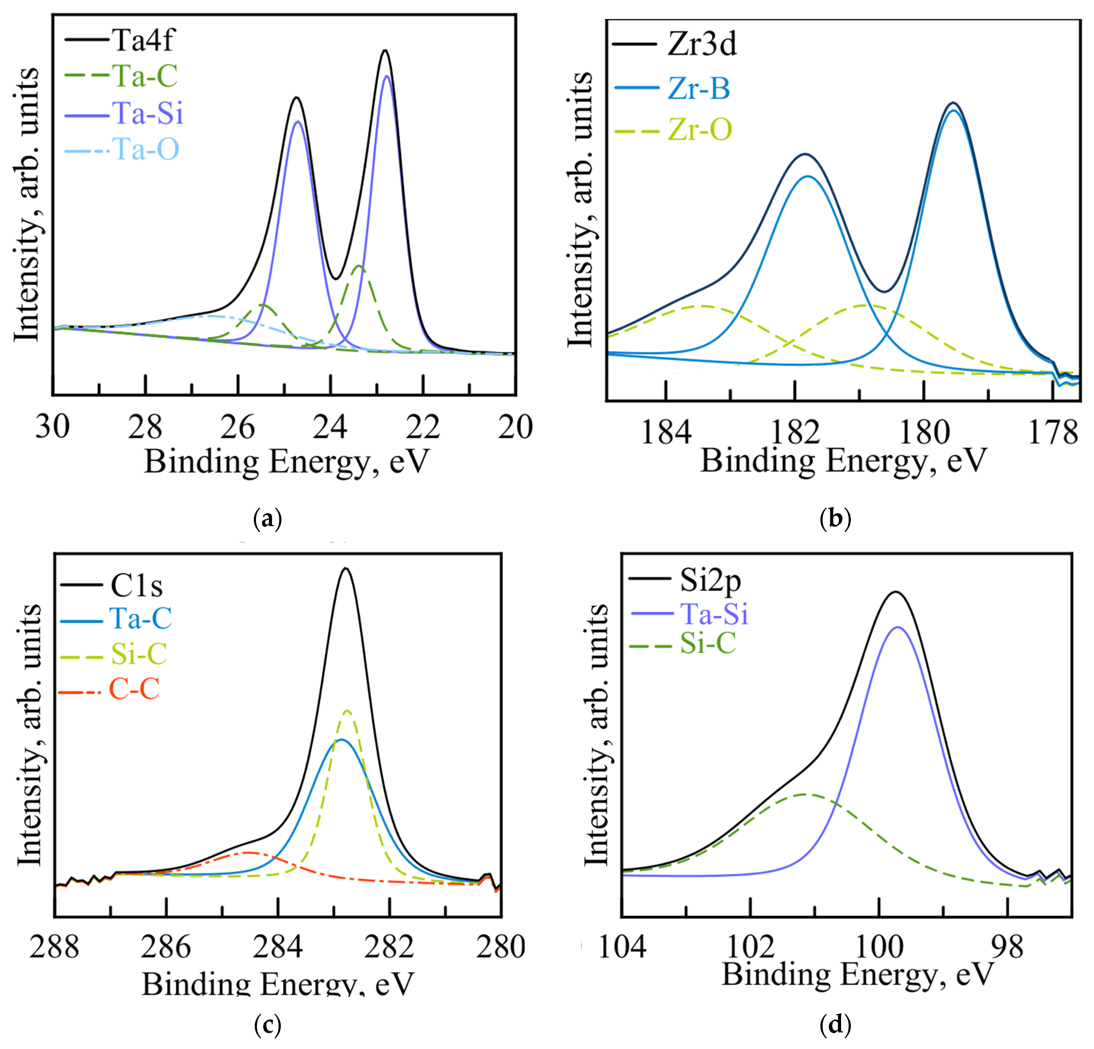
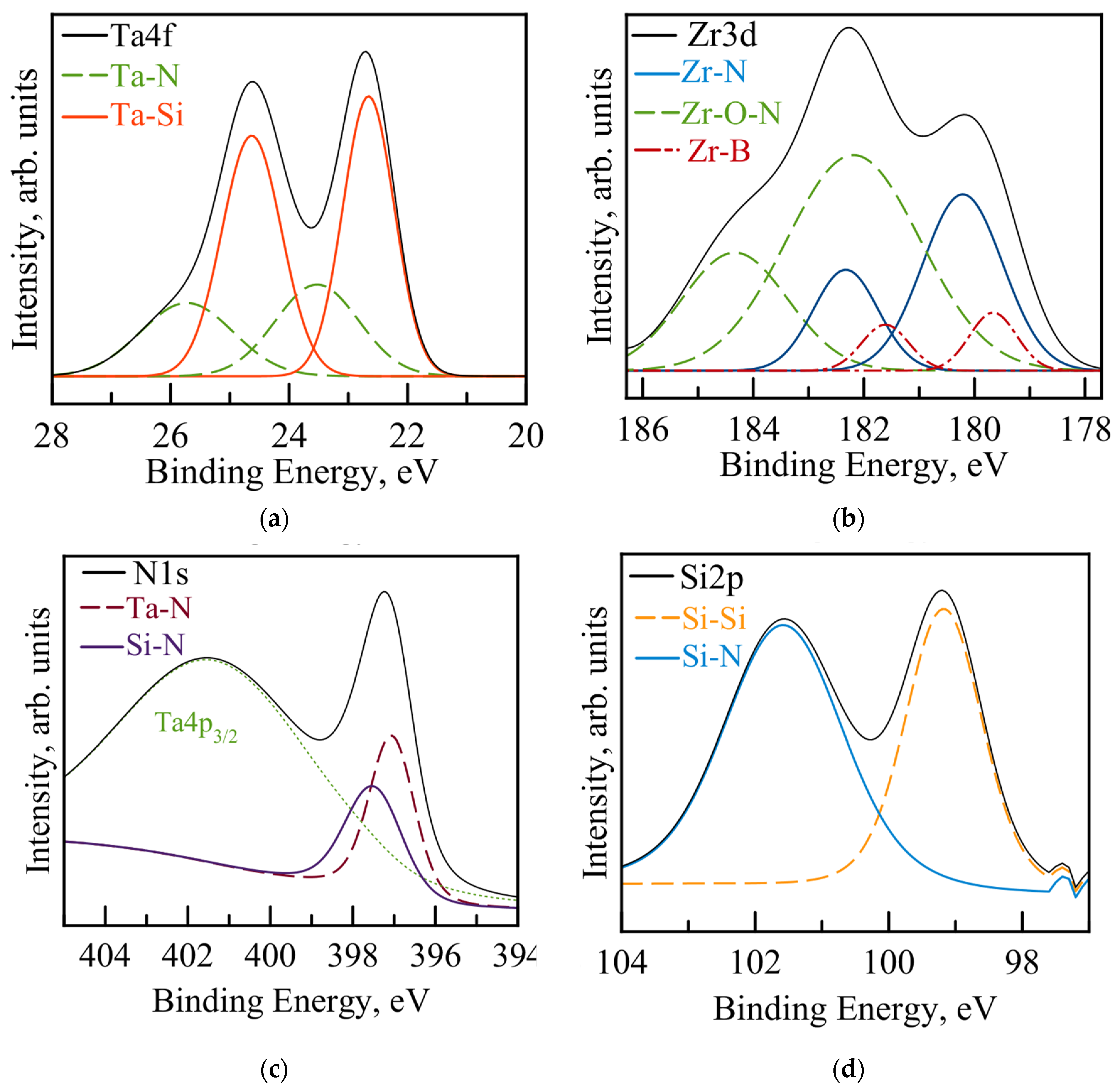
3.1. Structure and Mechanical Characteristics of As-Deposited Coatings

The coatings were characterized by a dense homogeneous structure without pronounced columnar grains. The Ta-Zr-Si-B-C and Ta-Zr-Si-B-N coatings, 7.2 and 6.4 µm thick, had the following composition at.%: Ta28Zr10Si26B24C12 and Ta27Zr8Si22B22N20. The roughness parameter (Ra) of the coatings was close and was in the range of 16–18 nm. A detailed description of the composition, structure and thickness of the as-deposited coatings is presented in [19,20,21].
According to the XRD data (Figure 1a), the coatings were X-ray amorphous. According to SAED patterns and BF TEM images (Figure 1b,c), the coatings are characterized by an amorphous structure.
On the SAED patterns of the Ta-Zr-Si-B-C and Ta-Zr-Si-B-N coatings, a broad reflection is observed, close to the position of the (110) and (111) lines of the h-TaSi2 crystalline phase (JCPDS 38-0483) with d/n = 0.239 and 0.224 nm, respectively.
In this work, the chemical and phase composition of the coatings was additionally studied using the XPS method. Figure 2 shows the Ta4f, Zr3d, C1s and Si2p spectra of the Ta-Zr-Si-B-C coating after etching.
In the Ta4f spectra, two doublets are observed at positions 22.7 and 24.7 eV and 23.4 and 25.5 eV, related to the Ta-Si and Ta-C bonds, respectively [34]. The low intensity peak at position 26.5 eV can be attributed to Ta-O bonds [34]. The high intensity of the Ta-Si peaks indicates the predominance of this bond in the coating. According to the Zr3d spectra, the doublet at binding energies of 179.5 and 181.9 eV indicates a Zr-B bond [35]. Peaks at 181.0 and 183.3 eV correspond to the bond of zirconium with oxygen [35]. Peaks in the C1s spectrum in the range of 282–284 eV indicate the presence of Ta–C and Si–C bonds; there is also a low-intensity C-C peak [34]. The Si2p spectrum confirms the presence of Si-C and Ta-Si bonds [36].
The spectra of Ta4f, Zr3d, N1s and Si2p for the Ta-Zr-Si-B-N coatings after etching are shown in Figure 3.
A doublet was found in the Ta4f spectrum at positions 22.7 and 24.7 eV, corresponding to the Ta-Si bond [34]. Low-intensity peaks at positions 23.7 and 25.7 eV correspond to the chemical state of Ta in the cubic or hexagonal Ta-N phase [37]. Also, the presence of Ta-Si-N bonds cannot be excluded. Three doublets were revealed in the spectrum of Zr3d, which can be attributed to the Zr-B, Zr-N and Zr-O-N bonds, respectively [38]. The spectra of N1s show an intense peak at 397.3 eV, which appeared as a result of the overlap of the Ta-N [39] and Si-N [40] peaks. It is also impossible to exclude the presence of B-N bonds at 396.7–397.5 eV [41]. Peaks at 99.3 and 101.6 eV in the Si2p spectrum correspond to Si-Si and Si-N bonds [40].
Thus, the XPS data showed that in the coatings, tantalum is predominantly bonded to silicon, and zirconium is bonded to boron. In addition, Ta-C and Si-C bonds were found for the carbon-containing coating, and Ta-N, Zr-N and Si-N bonds were found for the nitrogen-containing coating.
The Ta-Zr-Si-B-C coating had a hardness of 25 GPa, an elastic modulus of 259 GPa and an elastic recovery of 71%. For the Ta-Zr-Si-B-N coating: H = 23 GPa, E = 229 GPa and W = 74%. The high mechanical characteristics of the carbon-containing coating can be explained by the higher hardness of the TaC and SiC carbide phases compared to the TaN and Si3N4 phases [42,43,44].

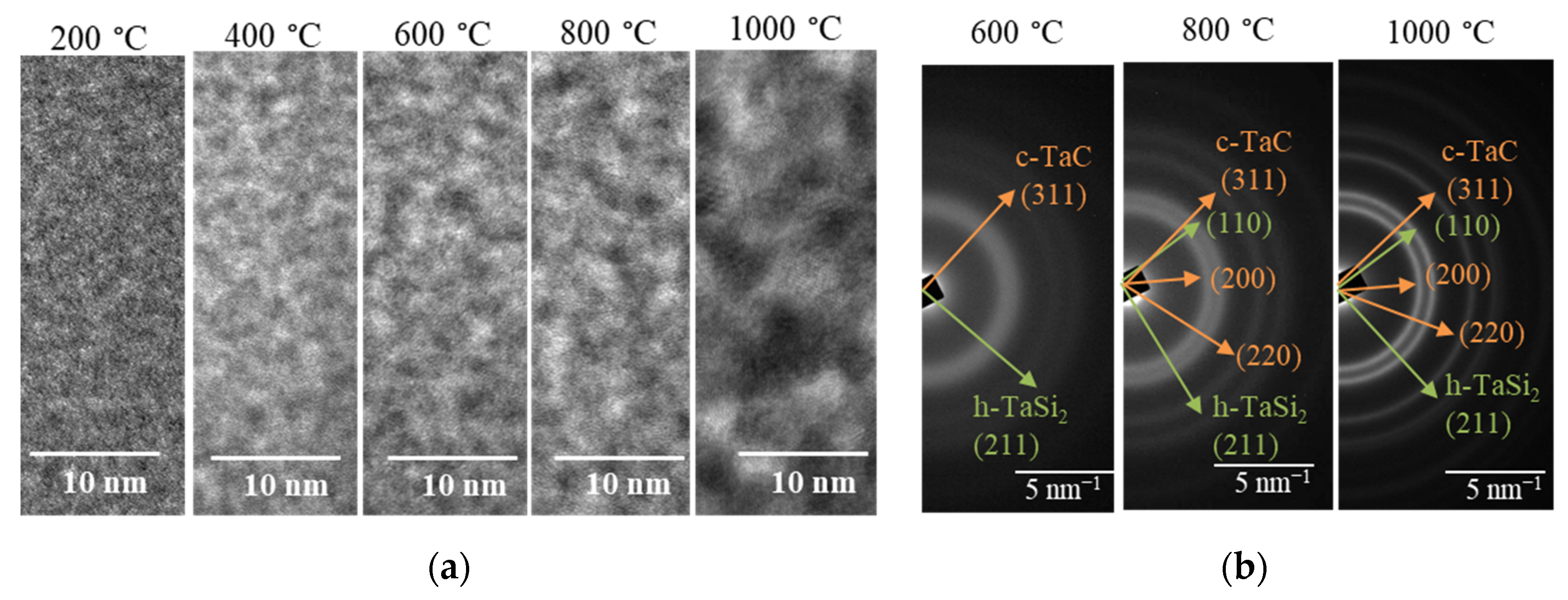
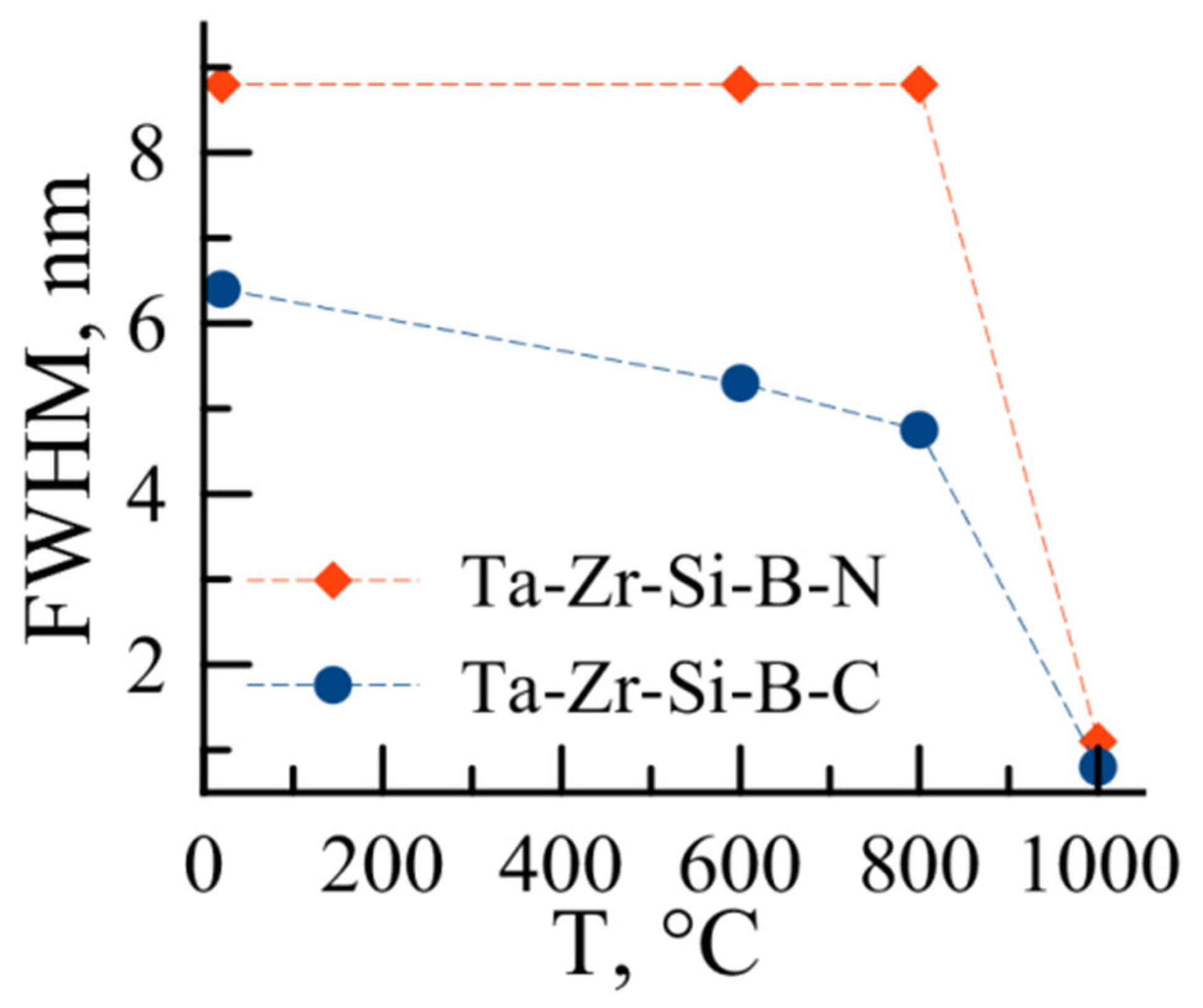
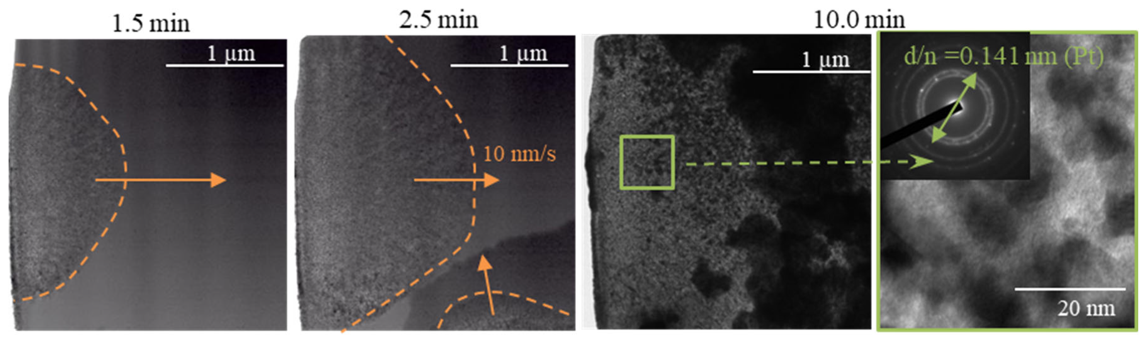
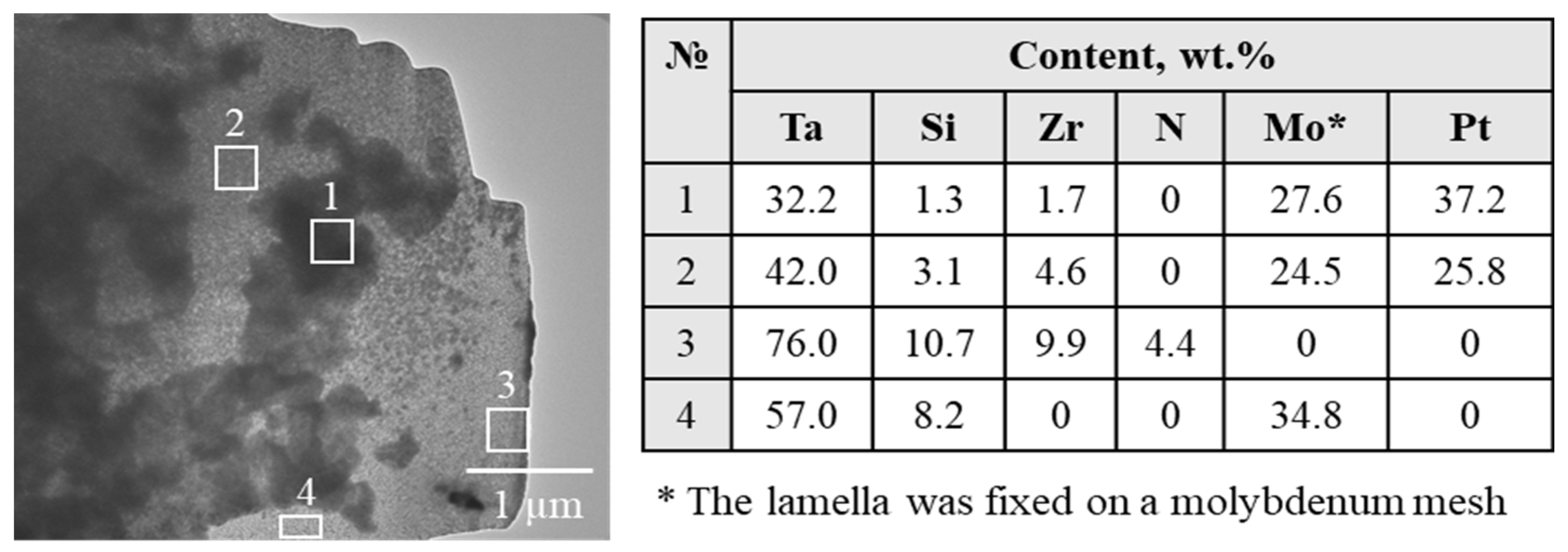
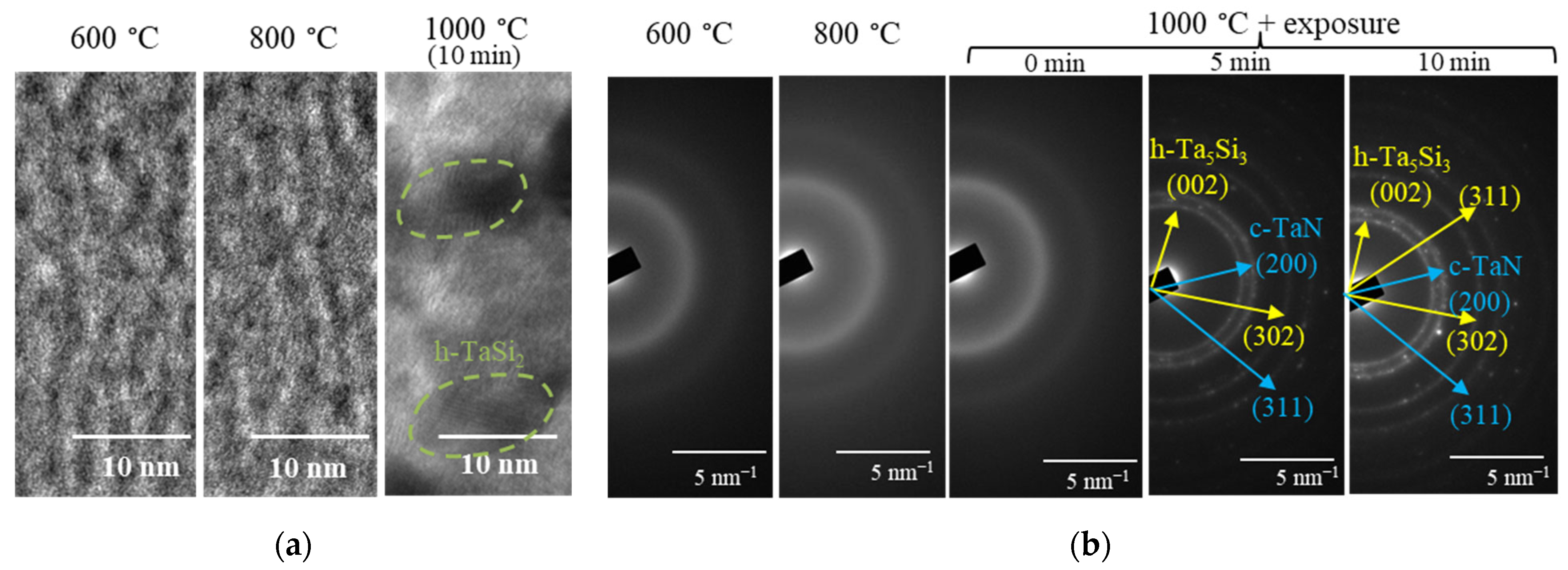
3.2. Thermal Stability under In Situ TEM Heating

Figure 4 shows BF TEM images and SAED patterns of Ta-Zr-Si-B-C coatings obtained during in situ TEM heating.
According to the BF images, heating at temperatures of 200–800 °C led to insignificant changes in the structure, while at a temperature of 1000 °C, after holding for 10 min, the coating crystallized (Figure 4a). It can be seen from the SAED patterns that the amorphous structure of the Ta-Zr-Si-B-C coating is retained up to a temperature of 400 °C (Figure 4b). At 600 °C, reflections from the (111) and (211) h-TaSi2 crystalline phases with d/n = 0.229 and 0.153 nm are observed on the SAED patterns. A reflection with d/n = 0.132 nm was also observed, provided by the cubic phase of c-TaC (311) (JCPDS 65-8145). Raising the temperature to 1000 °C led to a narrowing of the reflections and the formation of a more pronounced crystal structure.
For a more detailed study of the change in the structure of the Ta-Zr-Si-B-C coating during heating, the FWHM parameter was determined (Figure 5).
It can be seen that the FWHM parameter for the carbon-containing coating somewhat decreased in the range of 20–800 °C, which is associated with the narrowing of reflections as a result of structure ordering (Figure 4b). A pronounced decrease in temperature within 800–1000 °C indicates an accelerated crystallization of the coatings. The crystallite sizes corresponding to the FWHM parameter at temperatures of 800 and 1000 °C were ~3 and ~18 nm, respectively.
Figure 6 shows BF TEM images and SAED patterns of the Ta-Zr-Si-B-N coating obtained in the temperature range 600–1000 °C.
The results showed that heating in this temperature range did not lead to a change in the structural-phase state of the Ta-Zr-Si-B-N coating (Figure 6a). In all cases, the SAED patterns exhibited broad reflections (Figure 6b) typical of amorphous materials [45]. Crystallization of the coating was observed only during exposure at a temperature of 1000 °C. Shooting BF TEM images at low magnification made it possible to register the movement of the crystallization front at a speed of 10 nm/s (Figure 7). A video of the crystallization process is given in the Supplementary Material (Video S1).
Figure 8 shows the EDS data of Ta-Zr-Si-B-N coating lamellas after in situ TEM heating at 1000 °C.
At a maximum exposure of 10 min, the formation of local areas of grain accumulation (dark areas in TEM images) rich in platinum was observed (Figure 8, points 1, 2). According to EDS analysis, the studied zone at the lamella boundary (point 3) corresponded to the coating material and had the following composition: 76 wt.% Ta, 10.7 wt.% Si, 9.9 wt.% Zr and 4.4 wt.% N. Note that the determination of the boron concentration using the EDS method is difficult.
The process of crystallization and grain growth is well described by the SAED patterns taken during holding at 1000 °C (Figure 6b). It can be seen that at the initial moment of time (0–5 min), small crystallites are formed, giving reflections in the form of rings, the order of which corresponds to the FCC structure. Further, with an increase in exposure to 10 min, point reflections from individual larger grains appear. According to the SAED patterns taken at the maximum exposure time, lines with d/n = 0.248, 0.214, 0.166 and 0.156 nm were revealed, corresponding to reflections from the (002), (112), (302) and (113) planes of the hexagonal phase h-Ta5Si3 ( JCPDS 65-3598), and lines with d/n = 0.224 and 0.131 nm were revealed, which can be attributed to the reflection from the (200) and (311) planes of the c-TaN cubic phase (JCPDS 79-5712) (Figure 6). In addition to reflections from the h-Ta5Si3 and c-TaN phases, a line with d/n = 0.1406 nm, corresponding to platinum, was detected (Figure 7). It is important to note that Pt was used during the preparation of the lamella. The amorphous structure of the coating probably prevented the diffusion of platinum up to 1000 °C, which is due to the absence of grain boundaries serving as a channel for intense diffusion [46,47]. And at 1000 °C, due to the crystallization of the coating, the diffusion of platinum was activated.
Figure 5 shows the dependence of the FWHM parameter on temperature for the Ta-Zr-Si-B-N coating. In the range of 20–800 °C, no change in FWHM was observed; the structure remained amorphous up to 800 °C. At 1000 °C, a sharp decrease in the FWHM parameter indicates crystallization of the coating. The crystallite size was ~10 nm.
Thus, the Ta-Zr-Si-B-N coating began to crystallize during holding at a temperature of 1000 °C, which significantly exceeds the crystallization temperature of the Ta-Zr-Si-B-C coating, which is 600 °C.

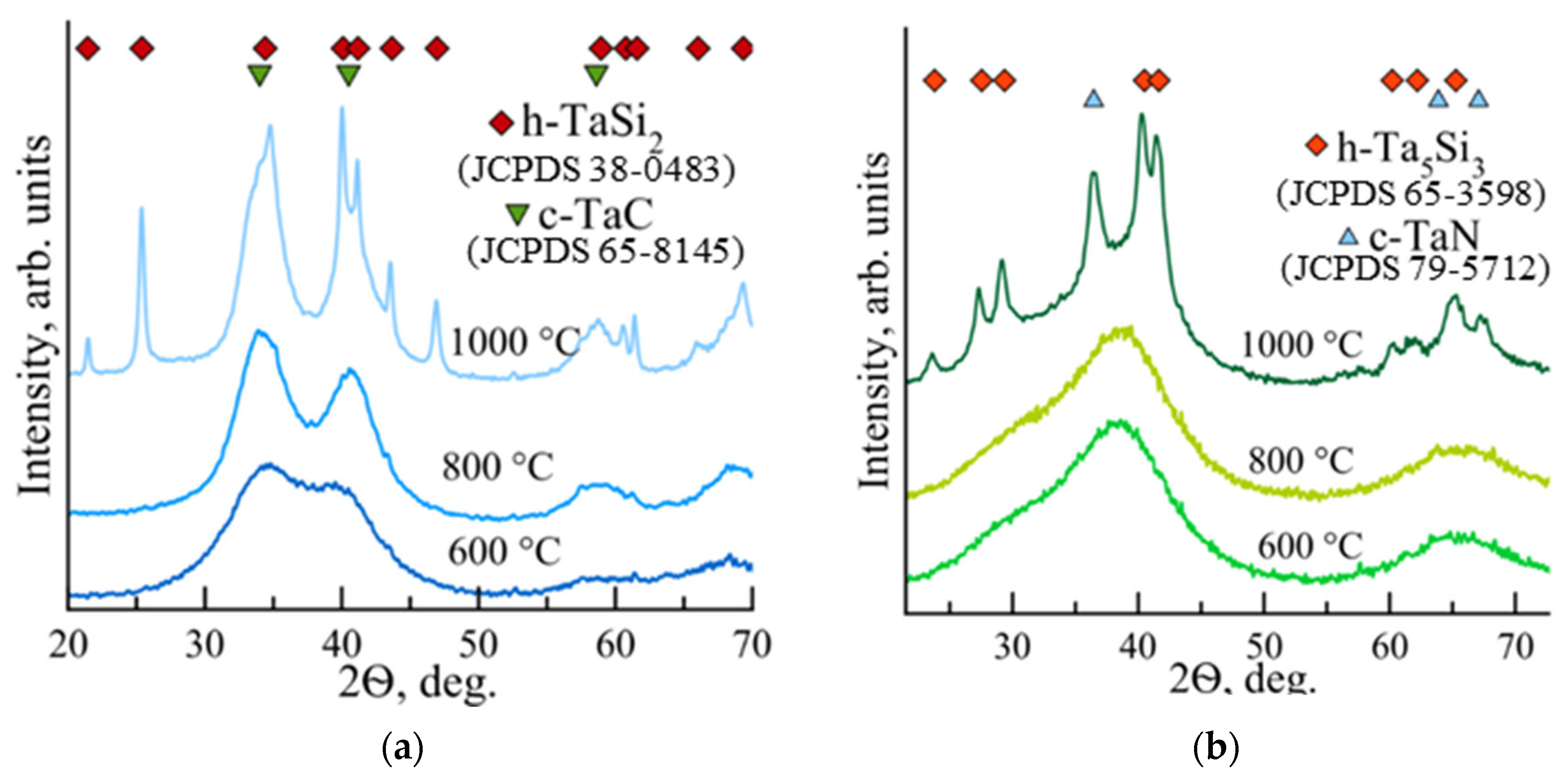
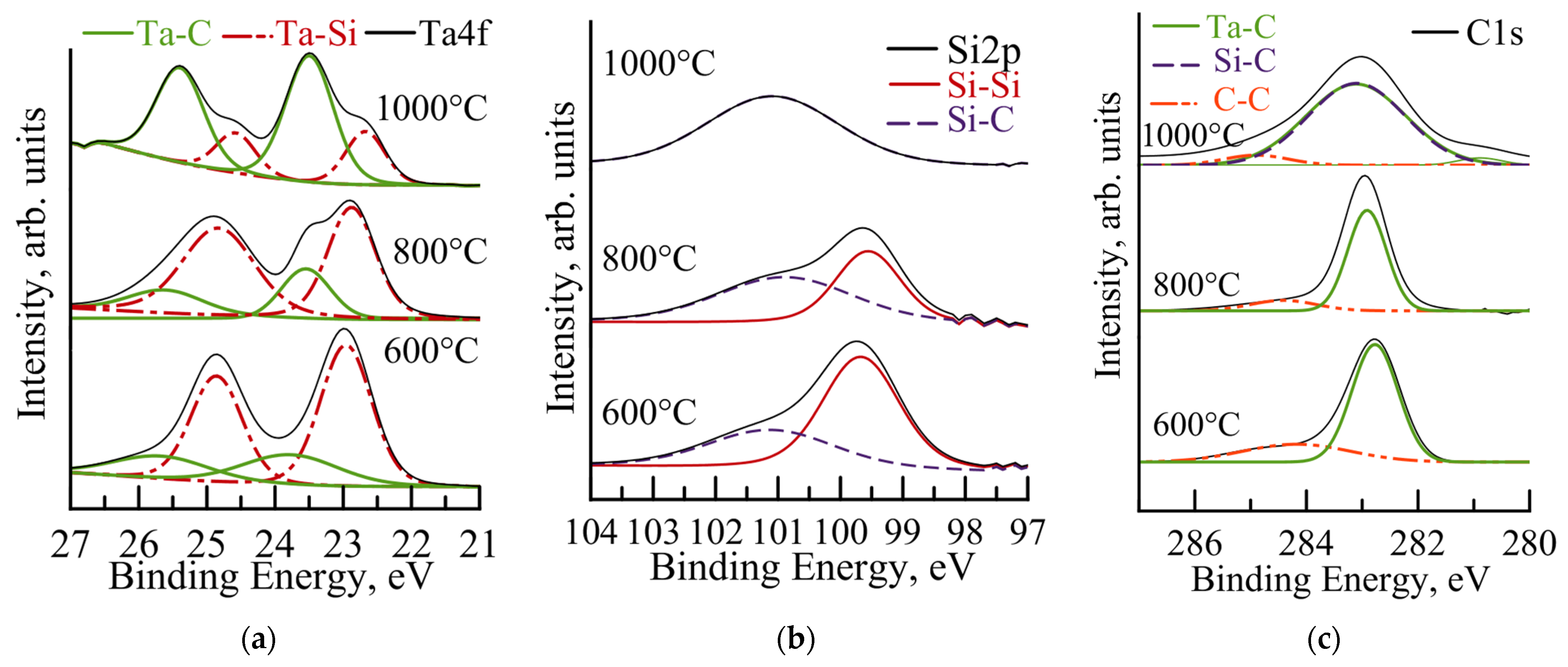
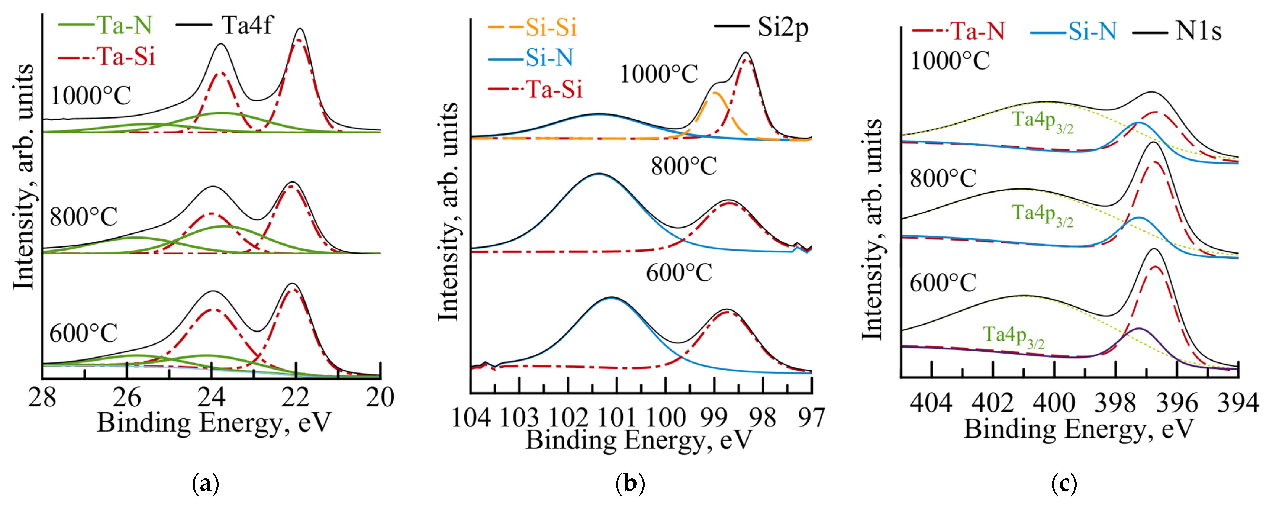
3.3. Thermal Stability under Vacuum Annealing

XRD patterns of coatings annealed in vacuum according to modes similar to TEM are shown in Figure 9.
For the Ta-Zr-Si-B-C coating (Figure 9b), an increase in temperature from 20 to 600 and 800 °C led to the separation of peaks in the 2Ꝋ = 20–50 and 55–75° and an increase in their intensity, which indicates phase segregation. At a temperature of 1000 °C, crisp peaks corresponding to reflections from the (100), (101), (102), (111), (112) and (114) planes of the h-TaSi2 phase and the (111), (200) and (220) planes of the c-TaC phase were detected on the XRD patterns. The size of the h-TaSi2 and c-TaC crystallites, determined by the most intense lines (111) using the Scherrer formula, was ~20 and ~10 nm, respectively.
According to XRD patterns, the amorphous structure of the Ta-Zr-Si-B-N coating was retained up to a temperature of 800 °C. At 1000 °C, crystallization of the Ta-Zr-Si-B-N coating was observed with the formation of h-Ta5Si3 and c-TaN phases. In this case, the size of h-Ta5Si3 and c-TaN crystallites, determined from the (211) and (111) lines, was ~10 and ~6 nm, respectively. In addition, the increase in the XRD intensity with the annealing temperature indicates that the number of crystallites increases and the coating becomes more crystalline [48].
The XPS spectra of Ta4f, Si2p and C1s for the Ta-Zr-Si-B-C coating after vacuum annealing at temperatures of 600, 800 and 1000 °C are shown in Figure 9. The positions of the Ta-Si and Ta-C lines in the Ta4f spectra (Figure 10a) remained almost unchanged from a temperature rise from 20 to 1000 °C.
The increase in the intensity of the Ta-C peaks is associated with an increase in the volume fraction of this bond in the coating. The XPS spectra of Si2p (Figure 10b) of the Ta-Zr-Si-B-C coating show that an increase in temperature from 20 to 800 °C does not lead to changes: Si-C and Si-Si bonds are observed in the coating in positions similar to the as-deposited coating. Heating to 1000 °C led to a change in the Si2p spectrum: a broad peak was observed at position 101.4 eV, corresponding to the Si-C bond. It can be assumed that with an increase in temperature from 800 to 1000 °C, all free silicon went to the formation of Si-C bonds, which is also evidenced by an increase in the intensity of the Si-C peak at an energy of 283.4 eV in the spectrum of C1s (Figure 10c).
The XPS spectra of Ta4f, Si2p and N1s for the Ta-Zr-Si-B-N coating after vacuum annealing are shown in Figure 11.
According to the Ta4f spectra (Figure 11a), the Ta-Zr-Si-B-N coating is heated from 20 to Si towards a lower binding energy, which is associated with an increase in the fraction of tantalum in Ta-Si bonds [49] and indicates the formation of the Ta5Si3 phase. The Si2p spectra (Figure 11b) show that an increase from 20 to 800 °C is accompanied by a decrease in the intensity of the Ta-Si peak. At 1000 °C, the division of the peak (97–100 eV) into two peaks at the positions 98.4 eV and 99.0 may be due to an increase in the fraction of tantalum in Ta-Si and the release of free silicon (Si-Si bond) due to the h-TaSi2 phase transition to h-Ta5Si3. At the same time, the intensity of the Si-N peak decreased. This effect is associated with the redistribution of silicon atoms between Si-N and Ta-Si with increasing temperature. The appearance of the N1s (Figure 11c) spectra practically did not change with increasing temperature.
It is important to note that the in situ TEM, FWHM, XRD and XPS data correlate well with each other. Crystallization of the Ta-Zr-Si-B-C coating was observed at a temperature of 600 °C, with the formation of a nanocomposite structure with h-TaSi2 and c-TaC crystallites ~10–20 nm in size and an amorphous interlayer based on a-SiC with dissolved zirconium and boron, whereas the Ta-Zr-Si-B-N coating crystallizes at a temperature of 1000 °C, with the formation of a nanocomposite structure with an h-Ta5Si3 and c-TaN crystallite ~6–10 nm in size, surrounded by an amorphous matrix a-Si(Zr,B)N. It can be concluded that the nitrogen-containing coating has a higher thermal stability compared to the carbon-containing sample.

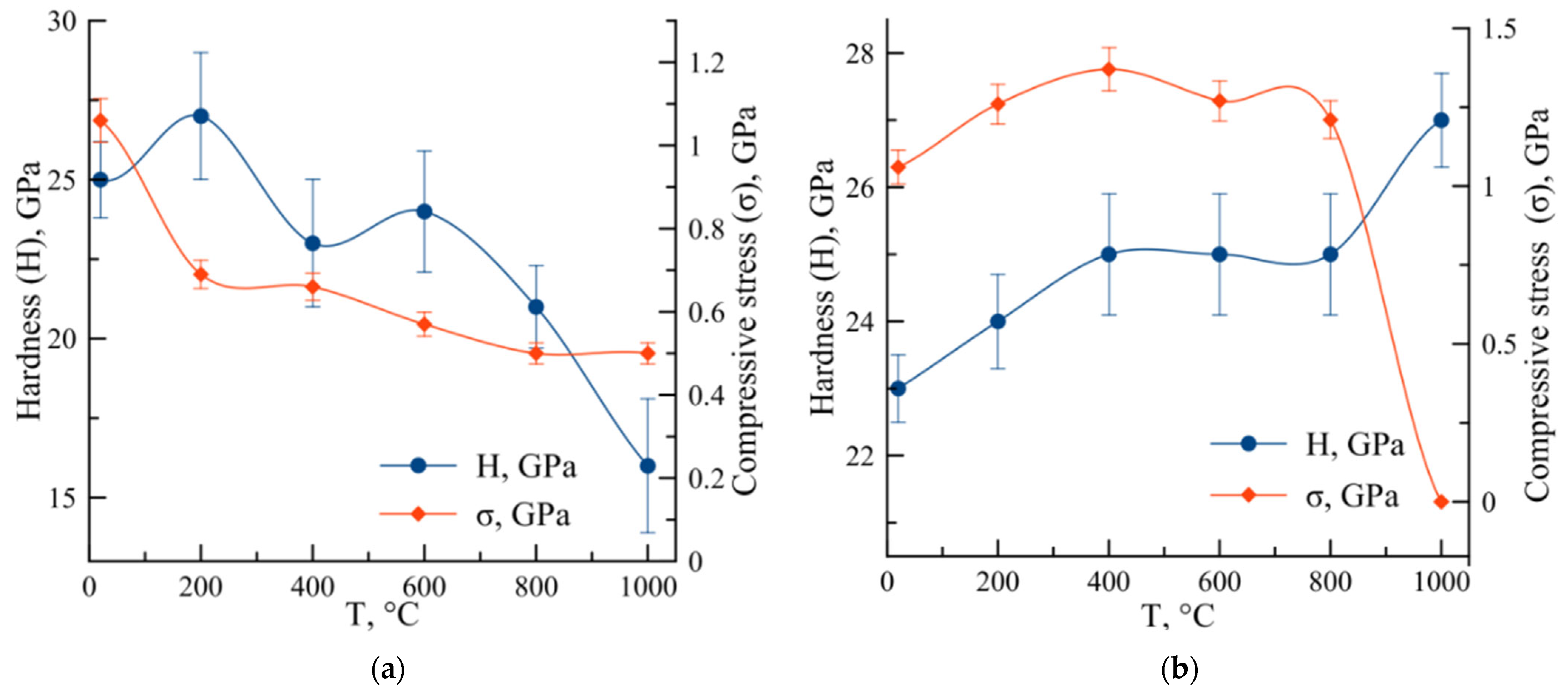
3.4. Thermal Stability under Nanoindentation

The temperature dependence of hardness and compressive stresses for Ta-Zr-Si-B-C and Ta-Zr-Si-B-N coatings is presented in Table 1.
In the case of Ta-Zr-Si-B-C coating, the temperature increase from 20 to 1000 °C led to a decrease in E by 8%, in W by 25% and in the parameters H/E and H3/E2 by 30 and 70%, respectively. For the Ta-Zr-Si-B-N coating, an increase in temperature from 20 to 1000 °C was accompanied by an increase in E from 229 to 275 GPa, in W from 74 to 77% and in H3/E2 from 0.232 to 0.260 GPa. The H/E parameter decreased slightly with an increase in temperature from 20 to 1000 °C.
For the Ta-Zr-Si-B-C coating, an increase in temperature from 20 to 1000 °C was accompanied by a gradual decrease in hardness from 25 to 16 GPa and in the level of internal stresses from −1.1 to 0 GPa (Figure 12a). A decrease in internal stresses during thermal exposure may be associated with the ordering of the coating structure and the crystallization process [50,51]. A decrease in the hardness of the Ta-Zr-Si-B-C coating by 36% may be associated with stress relaxation during thermal exposure, by analogy with [52].
When the Ta-Zr-Si-B-N coating was heated to 200 °C, the hardness increased slightly from 23 to 24 GPa; in the range of 200–800 °C, the values stabilized at ~25 GPa, which is explained by a minimal change in the level of internal stresses (Figure 12b). The absence of a change in σ is due to the absence of structural changes and phase transformations in the coating at temperatures of 20–800 °C. At 1000 °C, stress relaxation was observed, associated with the pronounced crystallization of the coatings. The abrupt rise in hardness to 27 GPa at 1000 °C cannot be explained by high internal stresses, since σ = 0 GPa at this temperature, and is most likely associated with the beginning of the crystallization process and the release of h-Ta5Si3 and c-TaN phases.
Thus, for the Ta-Zr-Si-B-C coating, a clear correlation was observed between the decrease in the level of residual stresses and the decrease in hardness with increasing temperature. The processes of structure ordering and crystallization occurring at temperatures of 600–1000 °C in the Ta-Zr-Si-B-C coating seem to have a negative effect on the level of mechanical properties. For the Ta-Zr-Si-B-N coating, the influence of crystallization prevails. A similar effect of crystallization on the mechanical properties was revealed for Ta-Si-N coatings [53]: the hardness of the coatings increased from 17 to 21 GPa with an increase in temperature to 750 °C, which is associated with the precipitation of hard TaxSiy binary or (Ta,Si)xNy ternary compounds.
The different effect of the crystallization process on the hardness of Ta-Zr-Si-B-N and Ta-Zr-Si-B-C coatings can be associated with their structure and the crystallite size of the main phases [54,55,56]. The nitrogen-containing coating, which is characterized by a maximum hardness, has a smaller crystallite size (~10 nm) compared to the carbon-containing coating (~20 nm), which is associated with the difference in crystallization temperatures and exposure times.
Note that simpler Ta-Si-N coatings have thermal stability up to 900 °C and a hardness of 15 GPa in the temperature range of 20–900 °C [57]. The complex doping of these coatings with Ta-Si-N Zr and B makes it possible to improve thermal stability up to 1000 °C and hardness up to 27 GPa.

4. Conclusions

The amorphous coatings Ta-Zr-Si-B-C and Ta-Zr-Si-B-N containing 12 at.% C and 20 at.% N were deposited by magnetron sputtering using a TaSi2-Ta3B4-(Ta,Zr)B2 ceramic target in Ar + C2H4 and Ar + N2 reaction media, respectively. According to XPS data, Ta is predominantly bonded to Si, and Zr is predominantly bonded to B. In addition, Ta-C and Si-C bonds were detected for the carbon-containing coating, and Ta-N, Zr-N and Si-N bonds were detected for the nitrogen-containing coating.
The Ta-Zr-Si-B-C coating was characterized by higher values of hardness of 25 GPa and an elastic modulus of 259 GPa compared to 25 GPa and 229 GPa for the Ta-Zr-Si-B-N coating. The high mechanical properties of the carbon-containing coating are ensured by the presence of Ta-C and Si-C carbide bonds.
In situ TEM heating led to the crystallization of the amorphous Ta-Zr-Si-B-C coating at a temperature of 600 °C with the precipitation of the h-TaSi2 and c-TaC phases. The Ta-Zr-Si-B-N coating crystallized during holding at 1000 °C with the precipitation of the h-Ta5Si3 and c-TaN phases.
The XRD and XPS data of vacuum-annealed coatings correlate well with the TEM data. Crystallization of the Ta-Zr-Si-B-C coating was observed at a temperature of 600 °C, with the formation of a nanocomposite structure with h-TaSi2 and c-TaC crystallites ~10–20 nm in size and an amorphous interlayer based on a-Si(Zr,B)C, whereas the Ta-Zr-Si-B-N coating crystallized at 1000 °C, with the formation of h-Ta5Si3 and c-TaN crystallites ~6–10 nm in size, surrounded by an a-Si(Zr,B)N amorphous matrix.
An increase in temperature led to a decrease in the hardness of the carbon-containing coating from 25 to 16 GPa at 1000 °C as a result of stress relaxation. For the Ta-Zr-Si-B-N sample at a temperature of 1000 °C, an increase in hardness from 23 to 27 GPa was observed, which is associated with the crystallization of the coating and the formation of a nanocomposite structure with an optimal crystallite size of about 6–10 nm.
The combination of high mechanical and tribological characteristics with high oxidation resistance and good thermal stability makes Ta-Zr-Si-B-C coatings promising as protective coatings for parts of heavily loaded friction units. High-oxidation-resistance coatings are ensured due to the formation of SiO2+ B2O3 and Ta2O5 oxides. High wear resistance is achieved due to free carbon, which acts as a solid lubricant, and due to the formation of TaSiOx particles, which reduce friction.
Ta-Zr-Si-B-N coatings, due to their high oxidation resistance, their high thermal stability and the absence of phase transformations, can be used as coatings for high-temperature microelectronic resistors. The hardening effect of coatings can also be used to improve the performance of high-performance dry machining tools.
A logical continuation of this work is the study of higher concentrations of nitrogen and carbon regarding the thermal stability of coatings, as well as the study of the thermodynamics of phase transitions and the study of crystallization kinetics.

Supplementary Materials

The following supporting information can be downloaded at: https://www.mdpi.com/article/10.3390/app131810440/s1, Video S1: Movement of the crystallization front in the Ta-Zr-Si-B-N coating at 1000 °C.

Author Contributions

Conceptualization, A.D.S. and P.V.K.-K.; formal analysis, A.D.S.; investigation, A.D.S., P.A.L. and P.V.K.-K.; resources, E.A.L.; writing—original draft preparation, A.D.S.; writing—review and editing, A.D.S. and P.V.K.-K. All authors have read and agreed to the published version of the manuscript.

Funding

This work was supported in the framework of the “Priority 2030″ (NUST MISIS, project K7-2023-009, “Promising powder materials and additive technologies for the aerospace and nuclear industries”). The TEM studies and nanoindentation experiments were supported by the Ministry of Science and Higher Education of the Russian Federation (State Assignment project No. 0718-2020-0034).

Institutional Review Board Statement

Not applicable.

Data Availability Statement

Not applicable.

Acknowledgments

The authors are grateful to A.V. Bondarev for the help with the XPS study.

Conflicts of Interest

The authors declare no conflict of interest.

References

  1. Bahr, A.; Richter, S.; Hahn, R.; Wojcik, T.; Podsednik, M.; Limbeck, A.; Ramm, J.; Hunold, O.; Kolozsvári, S.; Riedl, H. Oxidation Behaviour and Mechanical Properties of Sputter-Deposited TMSi2 Coatings (TM = Mo, Ta, Nb). J. Alloys Compd. 2023, 931, 167532. [Google Scholar] [CrossRef]
  2. Shon, I.-J.; Ko, I.-Y.; Chae, S.-M.; Na, K. Rapid consolidation of nanostructured TaSi2 from mechanochemically synthesized powder by high frequency induction heated sintering. Ceram. Int. 2011, 37, 679–682. [Google Scholar] [CrossRef]
  3. Niu, Y.; Huang, L.; Zhai, C.; Zeng, Y.; Zheng, X.; Ding, C. Microstructure and Thermal Stability of TaSi2 Coating Fabricated by Vacuum Plasma Spray. Surf. Coat. Technol. 2015, 279, 1–8. [Google Scholar] [CrossRef]
  4. Sytchenko, A.D.; Kozlova, N.S.; Zabelina, E.V.; Loginov, P.A.; Levashov, E.A.; Kiryukhantsev-Korneev, P.V. The Effect of the Ar/N2 Gas Ratio on the Structure and Properties of Ta-Si-N Coatings Produced by Magnetron Sputtering of TaSi2 Target. Surf. Interfaces 2023, 37, 102654. [Google Scholar] [CrossRef]
  5. Sytchenko, A.D.; Levashov, E.A.; Kiryukhantsev-Korneev, P.V. Structure and Properties of Ta–Si–N Coatings Produced by Pulsed Magnetron Sputtering. Russ. J. Non-Ferrous Met. 2021, 62, 611–617. [Google Scholar] [CrossRef]
  6. Zeman, P.; Musil, J.; Daniel, R. High-Temperature Oxidation Resistance of Ta–Si–N Films with a High Si Content. Surf. Coat. Technol. 2006, 200, 4091–4096. [Google Scholar] [CrossRef]
  7. Du, S.; Zhang, K.; Wen, M.; Ren, P.; Meng, Q.; Hu, C.; Zheng, W. Tribochemistry Dependent Tribological Behavior of Superhard TaC/SiC Multilayer Films. Surf. Coat. Technol. 2018, 337, 492–500. [Google Scholar] [CrossRef]
  8. Bondarev, A.V.; Vorotilo, S.; Shchetinin, I.V.; Levashov, E.A.; Shtansky, D.V. Fabrication of Ta-Si-C Targets and Their Utilization for Deposition of Low Friction Wear Resistant Nanocomposite Si-Ta-C-(N) Coatings Intended for Wide Temperature Range Tribological Applications. Surf. Coat. Technol. 2019, 359, 342–353. [Google Scholar] [CrossRef]
  9. Ren, X.; Li, H.; Fu, Q.; Li, K. Ultra-High Temperature Ceramic TaB2–TaC–SiC Coating for Oxidation Protection of SiC-Coated Carbon/Carbon Composites. Ceram. Int. 2014, 40, 9419–9425. [Google Scholar] [CrossRef]
  10. Korotaev, A.D.; Borisov, D.P.; Moshkov, V.Y.; Ovchinnikov, S.V.; Oskomov, K.V.; Pinzhin, Y.P.; Savostikov, V.M.; Tymentsev, A.N. Nanocomposite and Nanostructured Superhard Ti-Si-B-N Coatings. Russ. Phys. J. 2007, 50, 969–979. [Google Scholar] [CrossRef]
  11. Kiryukhantsev-Korneev, P.V.; Kozlova, N.S.; Levashov, E.A. Optical Characteristics of NUV-VIS-NIR Transparent Zr-Si-B-N Hard Films for Protection of Optical and Laser Devices. J. Phys. Conf. Ser. 2020, 1692, 012022. [Google Scholar] [CrossRef]
  12. Wang, Z.; Niu, Y.; Hu, C.; Li, H.; Zeng, Y.; Zheng, X.; Ren, M.; Sun, J. High Temperature Oxidation Resistance of Metal Silicide Incorporated ZrB2 Composite Coatings Prepared by Vacuum Plasma Spray. Ceram. Int. 2015, 41, 14868–14875. [Google Scholar] [CrossRef]
  13. Zhang, Y.; Zhou, X.; Cheng, H.; Geng, Z.; Li, W. Fabrication and Oxidation Resistance of a Novel MoSi2-ZrB2-Based Coating on Mo-Based Alloy. Materials 2023, 16, 5634. [Google Scholar] [CrossRef] [PubMed]
  14. Ren, Y.; Qian, Y.; Xu, J.; Jiang, Y.; Zuo, J.; Li, M. Oxidation and Cracking/Spallation Resistance of ZrB2–SiC–TaSi2–Si Coating on Siliconized Graphite at 1500 °C in Air. Ceram. Int. 2020, 46, 6254–6261. [Google Scholar] [CrossRef]
  15. Wang, R.-q.; Wang, N.; Zhu, S.-z.; Liu, Y.-b.; Ma, Z. Study on the Mechanism of Ultra-High Temperature Ablation of ZrB2–SiC–TaSi2 Coatings by Low-Pressure Plasma Spraying on the C/C Composites. Ceram. Int. 2022, 49, 11344–11354. [Google Scholar] [CrossRef]
  16. Farhadizadeh, A.; Vlček, J.; Houška, J.; Haviar, S.; Čerstvý, R.; Červená, M.; Zeman, P.; Matas, M. Effect of Nitrogen Content on High-Temperature Stability of Hard and Optically Transparent Amorphous Hf-Y-Si-B-C-N Coatings. Ceram. Int. 2023, 49, 6086–6093. [Google Scholar] [CrossRef]
  17. Houska, J.; Mares, P.; Simova, V.; Zuzjakova, S.; Cerstvy, R.; Vlcek, J. Dependence of Characteristics of MSiBCN (M = Ti, Zr, Hf) on the Choice of Metal Element: Experimental and Ab-Initio Study. Thin Solid Films 2016, 616, 359–365. [Google Scholar] [CrossRef]
  18. Kiryukhantsev-Korneev, P.; Sytchenko, A.; Pogozhev, Y.; Vorotilo, S.; Orekhov, A.; Loginov, P.; Levashov, E. Structure and Properties of Zr-Mo-Si-B-(N) Hard Coatings Obtained by D.C. Magnetron Sputtering of ZrB2-MoSi2 Target. Materials 2021, 14, 1932. [Google Scholar] [CrossRef] [PubMed]
  19. Kiryukhantsev-Korneev, P.V.; Sytchenko, A.D.; Vorotilo, S.A.; Klechkovskaya, V.V.; Lopatin, V.Y.; Levashov, E.A. Structure, Oxidation Resistance, Mechanical, and Tribological Properties of N- and C-Doped Ta-Zr-Si-B Hard Protective Coatings Obtained by Reactive D.C. Magnetron Sputtering of TaZrSiB Ceramic Cathode. Coatings 2020, 10, 946. [Google Scholar] [CrossRef]
  20. Kiryukhantsev-Korneev, P.V.; Sytchenko, A.D.; Levashov, E.A.; Lobova, T.A. Mechanical Properties and Heat Resistance of Ta–Zr–Si–B–C–N Coatings Obtained upon the Magnetron Sputtering of the TaZrSiB Target in Ar, N2, and C2H4 Atmosphere. Russ. J. Non-Ferr. Met. 2020, 61, 732–738. [Google Scholar] [CrossRef]
  21. Sytchenko, A.D.; Vakhrushev, R.A.; Kiryukhantsev-Korneev, P.V. Investigation of the Tribological Characteristics of Ta–Zr–Si–B–C–N Coatings. Powder Metall. Funct. Coat. 2023, 17, 62–70. [Google Scholar] [CrossRef]
  22. Morgiel, J.; Poliarus, O.; Pomorska, M.; Maj, Ł.; Szlezynger, M. Thermal Stability of Plasma-Sprayed NiAl/CrB2 Composite Coatings Investigated through in-Situ TEM Heating Experiment. Mater. Charact. 2020, 159, 110068. [Google Scholar] [CrossRef]
  23. Falqui, A.; Loche, D.; Casu, A. In Situ TEM Crystallization of Amorphous Iron Particles. Crystals 2020, 10, 41. [Google Scholar] [CrossRef]
  24. Jin, H.W.; Park, C.G.; Kim, M.C. In Situ TEM Heating Studies on the Phase Transformation of Metastable Phases in Fe–Cr–B Alloy Spray Coatings. Mater. Sci. Eng. A 2001, 304–306, 321–326. [Google Scholar] [CrossRef]
  25. Pogrebnjak, A.D.; Shpak, A.P.; Beresnev, V.M.; Kolesnikov, D.A.; Kunitskii, Y.A.; Sobol, O.V.; Uglov, V.V.; Komarov, F.F.; Shypylenko, A.P.; Makhmudov, N.A.; et al. Effect of Thermal Annealing in Vacuum and in Air on Nanograin Sizes in Hard and Superhard Coatings Zr-Ti-Si-N. J. Nanosci. Nanotechnol. 2012, 12, 9213–9219. [Google Scholar] [CrossRef] [PubMed]
  26. Koller, C.M.; Hollerweger, R.; Sabitzer, C.; Rachbauer, R.; Kolozsvári, S.; Paulitsch, J.; Mayrhofer, P.H. Thermal Stability and Oxidation Resistance of Arc Evaporated TiAlN, TaAlN, TiAlTaN, and TiAlN/TaAlN Coatings. Surf. Coat. Technol. 2014, 259, 599–607. [Google Scholar] [CrossRef]
  27. Rao, J.C.; Diao, H.Y.; Ocelík, V.; Vainchtein, D.; Zhang, C.; Kuo, C.; Tang, Z.; Guo, W.; Poplawsky, J.D.; Zhou, Y.; et al. Secondary Phases in AlxCoCrFeNi High-Entropy Alloys: An in-Situ TEM Heating Study and Thermodynamic Appraisal. Acta Mater. 2017, 131, 206–220. [Google Scholar] [CrossRef]
  28. Zhu, Q.; Liu, Y.; Cong, W.; Chang, D.; Fan, Q.; Cao, F.; Wang, T.G. The Effect of Vacuum Annealing Temperature on the Properties of AlCrTiSiN Coating Prepared by Arc Ion Plating. Coatings 2022, 12, 316, Retracted. [Google Scholar] [CrossRef]
  29. Penkov, O.V.; Kopylets, I.A.; Khadem, M.; Kondratenko, V.V.; Malykhin, S.V.; Surovitskiy, S.V.; Fedchenko, A.V. Evaluation of Structure and Mechanical Properties of TiZrNi Coatings under Annealing. Thin Solid Films 2022, 748, 139149. [Google Scholar] [CrossRef]
  30. Vorotilo, S.; Levashov, E.A.; Kurbatkina, V.V.; Patsera, E.I.; Loginov, P.A.; Lopatin, V.Y.; Orekhov, A.S. Theoretical and Experimental Study of Combustion Synthesis of Microgradient ULTRA High-Temperature Ceramics in Zr-Ta-Si-B System. J. Eur. Ceram. Soc. 2021, 41, 4728–4746. [Google Scholar] [CrossRef]
  31. Kiryukhantsev-Korneev, P.V.; Sytchenko, A.D.; Sviridova, T.A.; Sidorenko, D.A.; Andreev, N.V.; Klechkovskaya, V.V.; Polčak, J.; Levashov, E.A. Effects of Doping with Zr and Hf on the Structure and Properties of Mo-Si-B Coatings Obtained by Magnetron Sputtering of Composite Targets. Surf. Coat. Technol. 2022, 442, 128141. [Google Scholar] [CrossRef]
  32. Oliver, W.C.; Pharr, G.M. Measurement of Hardness and Elastic Modulus by Instrumented Indentation: Advances in Understanding and Refinements to Methodology. J. Mater. Res. 2004, 19, 3–20. [Google Scholar] [CrossRef]
  33. Chen, P.Y.; Wang, W.C.; Wu, Y.T. Experimental Investigation of Thin Film Stress by Stoney’s Formula. Measurement 2019, 143, 39–50. [Google Scholar] [CrossRef]
  34. Du, S.; Wen, M.; Yang, L.; Ren, P.; Meng, Q.; Zhang, K.; Zheng, W. Structural, Hardness and Toughness Evolution in Si-Incorporated TaC Films. Ceram. Int. 2018, 44, 9318–9325. [Google Scholar] [CrossRef]
  35. Zhang, M.; Li, M.; Yan, Z.; Zhang, L.; Yin, J.; Ma, X.; Li, W.; Deng, L. Multifunctional Ag-ZrB2 Composite Film with Low Infrared Emissivity, Low Visible Light Reflectance and Hydrophobicity. Appl. Surf. Sci. 2022, 604, 154626. [Google Scholar] [CrossRef]
  36. Kaur, A.; Chahal, P.; Hogan, T. Selective Fabrication of SiC/Si Diodes by Excimer Laser under Ambient Conditions. IEEE Electron Device Lett. 2016, 37, 142–145. [Google Scholar] [CrossRef]
  37. Liu, X.; Ma, G.J.; Sun, G.; Duan, Y.P.; Liu, S.H. Effect of Deposition and Annealing Temperature on Mechanical Properties of TaN Film. Appl. Surf. Sci. 2011, 258, 1033–1037. [Google Scholar] [CrossRef]
  38. Muneshwar, T.; Cadien, K. Comparing XPS on Bare and Capped ZrN Films Grown by Plasma Enhanced ALD: Effect of Ambient Oxidation. Appl. Surf. Sci. 2018, 435, 367–376. [Google Scholar] [CrossRef]
  39. Zaman, A.; Meletis, E.I. Microstructure and Mechanical Properties of TaN Thin Films Prepared by Reactive Magnetron Sputtering. Coatings 2017, 7, 209. [Google Scholar] [CrossRef]
  40. Yamamoto, K.; Koga, Y.; Fujiwara, S. XPS Studies of Amorphous SiCN Thin Films Prepared by Nitrogen Ion-Assisted Pulsed-Laser Deposition of SiC Target. Diam. Relat. Mater. 2001, 10, 1921–1926. [Google Scholar] [CrossRef]
  41. Prakash, A.; Sundaram, K.B. Optical and XPS Studies of BCN Thin Films by Co-Sputtering of B4C and BN Targets. Appl. Surf. Sci. 2017, 396, 484–491. [Google Scholar] [CrossRef]
  42. Hu, J.; Li, H.; Li, J.; Huang, J.; Kong, J.; Zhu, H.; Xiong, D. Structure, Mechanical and Tribological Properties of TaCx Composite Films with Different Graphite Powers. J. Alloys Compd. 2020, 832, 153769. [Google Scholar] [CrossRef]
  43. Koller, C.M.; Marihart, H.; Bolvardi, H.; Kolozsvári, S.; Mayrhofer, P.H. Structure, Phase Evolution, and Mechanical Properties of DC, Pulsed DC, and High Power Impulse Magnetron Sputtered Ta–N Films. Surf. Coat. Technol. 2018, 347, 304–312. [Google Scholar] [CrossRef]
  44. Li, X.; Yin, X.; Zhang, L.; Pan, T. Comparison in Microstructure and Mechanical Properties of Porous Si3N4 Ceramics with SiC and Si3N4 Coatings. Mater. Sci. Eng. A 2009, 527, 103–109. [Google Scholar] [CrossRef]
  45. Hirata, A. Local Structure Analysis of Amorphous Materials by Angstrom-Beam Electron Diffraction. Microscopy 2021, 70, 171. [Google Scholar] [CrossRef] [PubMed]
  46. Cao, Z.H.; Hu, K.; Meng, X.K. Diffusion Barrier Properties of Amorphous and Nanocrystalline Ta Films for Cu Interconnects. J. Appl. Phys. 2009, 106, 113513. [Google Scholar] [CrossRef]
  47. Kuo, Y.L.; Lee, C.; Lin, J.C.; Yen, Y.W.; Lee, W.H. Evaluation of the Thermal Stability of Reactively Sputtered (Ti, Zr)Nx Nano-Thin Films as Diffusion Barriers between Cu and Silicon. Thin Solid Films 2005, 484, 265–271. [Google Scholar] [CrossRef]
  48. Lin, C.; Al-Muhtaseb, S.A.; Ritter, J.A. Thermal Treatment of Sol-Gel Derived Nickel Oxide Xerogels. J. Sol-Gel Sci. Technol. 2003, 28, 133–141. [Google Scholar] [CrossRef]
  49. Zier, M.; Oswald, S.; Reiche, R.; Wetzig, K. XPS Investigations of Thin Tantalum Films on a Silicon Surface. Anal. Bioanal. Chem. 2003, 375, 902–905. [Google Scholar] [CrossRef] [PubMed]
  50. Solov’ev, A.A.; Sochugov, N.S.; Oskomov, K.V. Effect of Residual Internal Stresses in TiN Coatings On Specific Losses in Anisotropic Electrical Steel. Phys. Met. Metallogr. 2010, 109, 111–119. [Google Scholar] [CrossRef]
  51. Shugurov, A.R.; Panin, A.V. Mechanisms of Stress Generation in Thin Films and Coatings. Tech. Phys. 2020, 65, 1881–1904. [Google Scholar] [CrossRef]
  52. Chen, Y.I.; Cheng, Y.R.; Chang, L.C.; Lu, T.S. Chemical Inertness of Ta-Si-N Coatings in Glass Molding. Thin Solid Films 2015, 584, 66–71. [Google Scholar] [CrossRef]
  53. Chung, C.K.; Chen, T.S.; Chang, N.W.; Chang, S.C.; Liao, M.W. Oxidation Resistance and Mechanical Property of Cosputtered Quasi-Amorphous Ta–Si–N Films under Vacuum Rapid Thermal Annealing. Surf. Coat. Technol. 2010, 205, 1268–1272. [Google Scholar] [CrossRef]
  54. Mayrhofer, P.H.; Hörling, A.; Karlsson, L.; Sjölén, J.; Larsson, T.; Mitterer, C.; Hultman, L. Self-Organized Nanostructures in the Ti–Al–N System. Appl. Phys. Lett. 2003, 83, 2049–2051. [Google Scholar] [CrossRef]
  55. Wang, C.; Shi, K.; Gross, C.; Pureza, J.M.; de Mesquita Lacerda, M.; Chung, Y.W. Toughness Enhancement of Nanostructured Hard Coatings: Design Strategies and Toughness Measurement Techniques. Surf. Coat. Technol. 2014, 257, 206–212. [Google Scholar] [CrossRef]
  56. Pogrebnjak, A.D.; Beresnev, V.M.; Pogrebnjak, A.D.; Beresnev, V.M. Hard Nanocomposite Coatings, Their Structure and Properties. In Nanocomposites-New Trends Development; InTech: Houston, TX, USA, 2012. [Google Scholar] [CrossRef]
  57. Chung, C.K.; Chen, T.S.; Peng, C.C.; Wu, B.H. Thermal Stability of Ta–Si–N Nanocomposite Thin Films at Different Nitrogen Flow Ratios. Surf. Coat. Technol. 2006, 201, 3947–3952. [Google Scholar] [CrossRef]
Figure 1. XRD patterns (a), SAED patterns and BF TEM images of the structure (b) of Ta-Zr-Si-B-N and Ta-Zr-Si-B-C as-deposited coatings.
Applsci 13 10440 g001
Figure 2. XPS spectra of Ta4f (a), Zr3d (b), C1s (c) and Si2p (d) for Ta-Zr-Si-B-C as-deposited coatings after etching for 5 min.
Applsci 13 10440 g002
Figure 3. XPS spectra of Ta4f (a), Zr3d (b), N1s (c) and Si2p (d) for Ta-Zr-Si-B-N as-deposited coatings after etching for 5 min.
Applsci 13 10440 g003
Figure 4. BF TEM images at temperatures of 200–1000 °C (a) and SAED patterns (b) at temperatures of 600–1000 °C for the Ta-Zr-Si-B-C coating.
Applsci 13 10440 g004
Figure 5. FWHM parameter for Ta-Zr-Si-B-C and Ta-Zr-Si-B-N coatings depending on temperature.
Applsci 13 10440 g005
Figure 6. BF TEM images (a) and SAED patterns (b) at temperatures of 600, 800 and 1000 °C, recorded during exposure (b), for the Ta-Zr-Si-B-N coating.
Applsci 13 10440 g006
Figure 7. BF TEM images obtained for the Ta-Zr-Si-B-N coating at 1000 °C at various exposition times (Supplementary Material, Video S1).
Applsci 13 10440 g007
Figure 8. EDS data of Ta-Zr-Si-B-N coating lamellas after in situ TEM heating at 1000 °C.
Applsci 13 10440 g008
Figure 9. XRD patterns of Ta-Zr-Si-B-C (a) and Ta-Zr-Si-B-N (b) coatings after vacuum annealing at temperatures of 600, 800 and 1000 °C.
Applsci 13 10440 g009
Figure 10. XPS spectra of Ta4f (a), Si2p (b) and C1s (c) for Ta-Zr-Si-B-C coatings after vacuum annealing at temperatures of 600, 800 and 1000 °C.
Applsci 13 10440 g010
Figure 11. XPS spectra of Ta4f (a), Si2p (b) and N1s (c) for Ta-Zr-Si-B-N coatings after vacuum annealing at temperatures of 600, 800 and 1000 °C.
Applsci 13 10440 g011
Figure 12. Temperature dependence of hardness and compressive stresses for Ta-Zr-Si-B-C (a) and Ta-Zr-Si-B-N (b) coatings.
Applsci 13 10440 g012
Table 1. Mechanical characteristics of coatings depending on temperature.
T, °CCoating Ta-Zr-Si-B-CCoating Ta-Zr-Si-B-N
H, GPaE, GPaW, %σ, GPaH/EH3/E2, GPaH, GPaE, GPaW, %σ, GPaH/EH3/E2, GPa
202525971−1.10.0960.2332322974−1.10.1000.232
2002727780−0.70.0970.2562424175−1.30.0990.238
4002325070−0.60.0920.1952524578−1.40.1020.260
6002428172−0.60.0850.1752525274−1.30.0990.246
800212736700.0770.1242525972−1.20.0960.233
1000162395300.0670.072272757700.0980.260
Disclaimer/Publisher’s Note: The statements, opinions and data contained in all publications are solely those of the individual author(s) and contributor(s) and not of MDPI and/or the editor(s). MDPI and/or the editor(s) disclaim responsibility for any injury to people or property resulting from any ideas, methods, instructions or products referred to in the content.

