Article

Physiological Effects of Hydrolyzed Skim Milk and Probiotics on Osteoporosis Models

1
Department of Animal Biotechnology, College of Life Science, Sangji University, Wonju 26339, Republic of Korea
2
Department of Livestock, Korea National University of Agriculture and Fisheries, Jeonju 54874, Republic of Korea
3
Animal Products Research and Development Division, National Institute of Animal Science, Rural Development Administration, Wanju 55365, Republic of Korea
*
Author to whom correspondence should be addressed.
These authors contributed equally to this work.
Appl. Sci. 2023, 13(18), 10424; https://doi.org/10.3390/app131810424
Submission received: 14 August 2023 / Revised: 16 September 2023 / Accepted: 17 September 2023 / Published: 18 September 2023
(This article belongs to the Section Food Science and Technology)

Abstract

:
Osteoporosis, a skeletal metabolic disease characterized by low bone mineral density and deterioration of bone microarchitecture, frequently occurs in postmenopausal women older than 50 years. Milk and dairy products are essential calcium sources recommended for bone health. In this study, we analyzed the effects of skim milk and probiotics in an ovariectomized osteoporosis model. Body weight significantly increased, whereas the consumption of skim milk and probiotics significantly decreased (approximately 20%) in the ovariectomized models. In addition, the concentration of calcium was significantly 0.5 mg/dL higher in the skim-milk-with-probiotic group than in the ovariectomized group. The bone volume/tissue volume ratio, trabecular thickness, trabecular number, and trabecular separation were higher in the skim-milk and skim-milk-with-probiotic groups than in the ovariectomized group. Histological analysis of the small intestine revealed that the consumption of skim milk alone or in combination with probiotics increased the lengths of the villus, crypt, and serosa. These results verify the beneficial effects of milk products in osteoporosis models, which may enable higher milk and milk product consumption by older women.

1. Introduction

Osteoporosis is a metabolic bone disorder characterized by low bone mass and microarchitectural deterioration of the bone tissue. On average, up to 50% of older women (>50 years) are at risk of fractures due to osteoporosis, which affects women twice as often as men [1,2]. Osteoporosis has evolved to include compromised bone strength and skeletal fragility caused by several factors, including defects in the microarchitecture of the trabeculae, defective intrinsic material properties of the bone tissue, defective repair of microdamage from normal daily activities, and an excessive bone remodeling rate. These factors are associated with age-related bone loss [3]. Osteoporosis can be caused by various diseases, including endocrine disorders, celiac disease, and inflammatory bowel disease [4]. As the average life expectancy of humans has increased globally, people’s interest in bone health has also increased. Many studies have suggested the benefits of vitamin D and calcium supplementation for bone formation, preservation, and prevention of osteoporosis [5]; this is because peak bone mass is influenced by genetic and environmental factors, including diet [6]. Low levels of vitamin D in the body decrease the intestinal absorption of calcium, eventually leading to osteoporosis [7].
Milk and dairy products are among the most important sources of calcium. For example, 100 g of cow’s milk contains 112 mg of calcium, which is over three times higher than that in the same amount of human milk [8]. Other minerals found in milk, such as phosphorus, iodine, potassium, vitamins, proteins, and other nutrients, may be beneficial for skeletal growth and bone strength [9]. When the effects of dairy product consumption on human health were assessed, the bone mineral content (BMC) was found to be lower (approximately 5.6%) in adult women who had consumed less than one portion of milk weekly during childhood than in those who had consumed more than one portion [10].
Prior studies examined the effects of milk or whey extracts on animals and surrogate markers in humans, such as bone remodeling markers or bone mineral density. Several observational studies using large cohorts also revealed the positive effects of milk on bone health or the risk of hip fracture [9].
Skim-milk powder (SMP) is defined as whole milk in which the fat is removed. SMP contains 80% casein and 20% whey protein. Fried et al. reported that SMP enhanced trabecular bone (TB) architecture compared to casein or whey in diet-induced obese rats [11]. As people age, their basal metabolic rate decreases, and if older people consume excessive milk for bone health, obesity can develop owing to milk fat.
Probiotics and prebiotics are essential components of milk and dairy products that modulate bone turnover [12]. A recent study suggested the relevance of intestinal microbiota and bone health. Further, several studies have demonstrated that probiotics can prevent primary and secondary osteoporotic bone loss [12,13]. In addition, probiotic-based compounds have been introduced in many fields of medicine such as dermatology, dentistry, and gastroenterology [14,15,16].
Ovariectomized (OVX) animals are typical experimental models used to investigate postmenopausal osteoporosis caused by estrogen deficiency. OVX mouse and rat models have been widely used to evaluate and develop novel drugs for the treatment of postmenopausal osteoporosis [17]. Fermented soy skim milk attenuates bone loss in OVX mice and reduces the risk of osteoporosis [18]. Two probiotic strains were found to inhibit osteoporosis in an OVX mouse model, with L. paracasei GKS6 outperforming L. plantarum GKM3 [19].
In this study, we determined the synergistic effects of skim milk and probiotics in an osteoporosis model. In particular, changes in body weight (BW), calcium concentration, lipid profiles, bone mineral density, bone mineral content, and length of the small intestine components were determined.

2. Materials and Methods

2.1. Materials

SMP was purchased from Seoul Milk Co-op (Seoul, Republic of Korea). The milk proteins of this powder were hydrolyzed using proteases (FoodPro, Bision Biochem, Kyunggido, Korea). SMP was mixed with distilled water at a ratio of 1:10. The enzyme (protease)/substrate (SMP) ratio was kept constant (1:5000). The mixtures were heated to 40 °C for 5 h and shaken at 150 rpm. Thereafter, the suspension samples were harvested from hydrolyzed SMP via centrifugation (12,000 rpm, 20 min, 4 °C). The collected suspension samples were dried at 65 °C for 3 h and stored at −20 °C. The probiotic combinations were prepared using several bacterial strains, including Lactobacillus acidophilus (Culture System Inc., IN, USA), L. delbrueckii ssp. bulgaricus (Chr Hansen, Hoersholm, Denmark), L. lactis (Sacco srl, CO, Italy), Bifidobacterium animalis ssp. lactis (Sacco srl, Italy), and Streptococcus thermophiles (Chr. Hansen, Hoersholm, Denmark), with a ratio of 55:10:5:5:25 (total number: 1.0 × 10¹⁰ CFU/mL). Mice were administered 1.0 × 10⁸ CFU/100 μL of phosphate-buffered saline. To employ hydrolyzed SMP as the skim-milk group, Pro was used as the probiotic mixture.

2.2. Animal Study

All animal experimental protocols were approved by the Animal Care Committee of Sangji University (Registration No. 2021-26). Six-week-old female CD1 mice (Dae Han Bio Link Co., Chungbuk, Republic of Korea) were housed in an environmentally controlled room (temperature: 23 ± 2 °C, relative humidity: 50 ± 10%, programmed ventilation, and 12:12 h light:dark cycle). After five days of acclimatization to a standard diet and water, mice were ovariectomized or non-ovariectomized (control). After the hairs of anesthetized mice were shaved and the skin was cleaned with alcohol, 0.5 cm of the skin layer was cut to expose the tissue. The ovary and oviduct were excised, the remaining tissue was inserted into the peritoneal cavity, and the skin was sutured. One week after surgery, mice were divided into four groups for milk protein treatment: (1) Ctrl, (2) OVX, (3) OVX mice administered hydrolyzed skim milk (1.5 g/kg/day), and (4) OVX mice administered hydrolyzed skim milk (1.5 g/kg/day) and probiotics (1.0 × 10⁸ CFU/100 μL in PBS). Oral injections were administered every other day for eight weeks, and BW was measured once every two weeks. Mice were killed at eight weeks, and femur, blood, and intestinal samples were collected. Blood samples were collected via cardiac puncture and analyzed at a central laboratory (Samkwang Medical Laboratories, Seoul, Republic of Korea) for accuracy and consistency.

2.3. Micro-CT Analysis

The right femurs of five mice from each group were analyzed using a micro-CT imaging system. The distal metaphysical right femur was scanned using a high-resolution micro-CT imaging system (Quantum GX Macro CT; PerkinElmer, Waltham, MA, USA) and then using a high-resolution (8 µm) micro-CT scanner (GE eXplore Locus SP, GH Health Care Ltd., London, UK) to evaluate bone mass, geometry, and trabecular microarchitecture. The parameters computed from the data were bone volume/tissue volume (BV/TV), trabecular thickness (Tb.Th), trabecular number (Tb.N), and trabecular separation (Tb.Sp).

2.4. Histological Preparation and Immunostaining

The intestine samples were fixed in 4% paraformaldehyde at 4 °C for 24 h and gradually dehydrated in 30–100% ethanol for 60 min. Thereafter, the tissues were cleaned with xylene for 60 min, embedded in paraffin blocks, sectioned into 5 µm thick slices using a microtome (Leica, Nussloch, Germany), and stained with hematoxylin and eosin (Sigma-Aldrich, Waltham, MA, USA), as previously described [20]. Images were captured using a microscope (Olympus IX73; Tokyo, Japan), and a minimum of six areas of the intestine for each biological replicate were evaluated and scored by an analyst blinded to the experimental conditions. The villi, crypt, and serosa lengths in the intestine of each sample were measured using the Motic Images Advanced 3.2 Software (Kowloon, Hong Kong). Measurements of lengths were determined for whole well-orientated villi, crypt, and serosa per small intestinal tissue section per mouse, and the values were averaged (five difference slides from each intestine sample). Immunostaining was performed as previously described [21]. Briefly, the sectioned samples were deparaffinized, and antigen retrieval was performed by boiling in 10 mM sodium citrate buffer for 10 min. After incubating the samples in blocking solution (0.02% Triton X-100, 1% bovine serum albumin) and washing three times with PBD, they were incubated overnight at 4 °C with the anti-Mucin2 antibody (sc-59859, Santa Cruz Biotechnology, Santa Cruz, CA, USA) as the primary antibody. The samples were then washed three times with PBS and incubated with the secondary antibody (Alexa 488 secondary antibody, #A32723, Invitrogen, Carlsbad, CA, USA). DAPI was used for nuclear staining, and the stained images were observed under a microscope (Olympus IX73, Tokyo, Japan).

2.5. Quantitative Real-Time PCR

Total intestinal RNA was retrieved from each sample using a Qiagen RNeasy Mini Kit (Qiagen, Germantown, MD, USA) with DNase treatment (Qiagen, Germantown, MD, USA), according to the manufacturer’s instructions. cDNA was synthesized from RNA using the RevertAid cDNA synthesis kit (Thermo Scientific, Rockford, IL, USA), and real-time PCR was performed as previously described [9] using the QuantStudio 1 system (Applied Biosystems, Foster City, CA, USA). The qPCR cycling conditions were as follows: 94 °C for 1 min, followed by 40 cycles at 94 °C for 10 s, 57 °C for 10 s, and 72 °C for 20 s. The data were analyzed using the comparative Ct method [18]. The results were normalized to that of GAPDH. The relative quantification (RQ) means and SEM were plotted as folds. Table 1 shows the primer sequences used for qPCR.

2.6. Western Blotting

Total protein was isolated from mouse intestines using RIPA buffer (Thermo Fisher Scientific, Waltham, MA, USA) with a protease inhibitor (Roche, Mannheim, Germany). Thereafter, Western blot analysis was performed as previously described [21]. Briefly, 40 µg of total protein from each sample was separated via 4–20% gradient acrylamide gel (Bio-Rad Laboratories, Hercules, CA, USA) electrophoresis and transferred onto PVDF membranes. The membranes were blocked with 5% nonfat milk for 1 h at room temperature (RT) and incubated overnight at 4 °C with the following primary antibodies: occludin (Thermo Fisher Scientific, Waltham, MA, USA; 1:1000 dilution) and β-actin (SC-517582; Santa Cruz Biotechnology, Santa Cruz, CA, USA). After three washes with Tris-buffered saline-Tween, the membranes were incubated with the appropriate secondary antibodies for 2 h at RT. Protein expression was confirmed using enhanced chemiluminescence (32106; Thermo Fisher Scientific, Waltham, MA, USA). The signal images were visualized using the iBrightTM imaging system (Thermo Fisher Scientific Inc., Waltham, MA, USA). Three technical replicates were used for Western blot analysis. The protein levels were normalized to that of β-actin. The relative protein expression is displayed in the graph.

2.7. Statistical Analysis

The data are expressed as the mean ± standard error of the mean (SEM), while mean data are expressed as mean ± standard deviation (SD) of at least three independent experiments. The data were evaluated using one-way analysis of variance (ANOVA) (Tukey’s test as a post hoc test). Statistical significance was determined using t-test with the SPSS statistical package (version 21.0; SPSS Inc., Chicago, IL, USA). The values of * p < 0.05 and ** p < 0.01 were considered to indicate statistical significance.

3. Results and Discussion

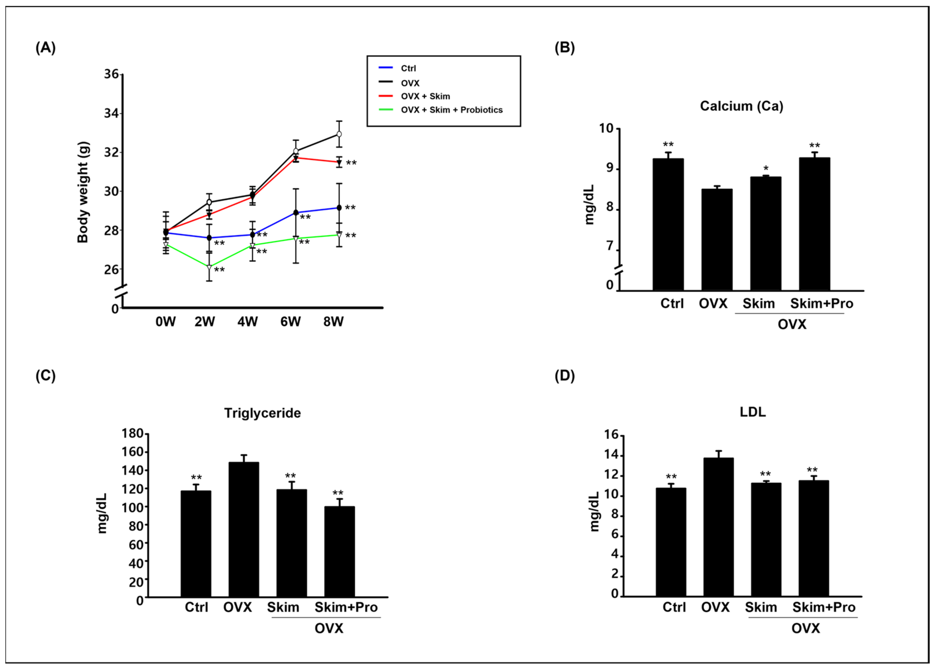
3.1. Physiological Effect of Skim Milk and Probiotics on Osteoporosis Models

BW was measured to investigate the physiological changes in the control, OVX, skim-milk, and skim-milk-with-probiotic groups. From weeks 2 to 8, the BW of the OVX group increased significantly, while that of the skim-milk-with-probiotic group decreased significantly. At week 8, the BW of the skim-milk group decreased compared with that of the OVX group; however, a statistically significant difference was not observed until week 6 (Figure 1A). These results suggest that skim milk and probiotics can prevent the OVX-induced increase in BW.
The serum calcium concentration significantly decreased (approximately 10%) in the OVX models but recovered following the consumption of skim milk and skim milk with probiotics (Figure 1B). In particular, when the OVX model was administered skim milk and probiotics, their calcium levels were similar to those of the control, which was not an OVX model.
The concentrations of triglyceride and low-density lipoprotein cholesterol (LDL-C) significantly increased in the OVX models but were reduced by the consumption of skim milk and probiotics (Figure 1C,D), as expected. A previous report revealed that ovariectomy increased BW, serum tartrate-resistant acid phosphatase concentration, and cathepsin K expression in bone; decreased serum estradiol concentration; and induced significant bone loss manifested as a decrease in BV/TV, connectivity density, trabecular number, and trabecular thickness, with increased trabecular separation and structural model index [22]. Another study revealed that the WW strain of L. plantarum is a potential probiotic in functional foods that can alter lipid metabolism and reduce cholesterol levels [23]. Previously, the BW and levels of total cholesterol (TC), triglycerides (TG), and LDL-C were found to increase in OVX rats [24]. Our results are consistent with those of prior studies and suggest that the levels of calcium and triglycerides have a positive effect on the body when skim milk and probiotics are consumed together, rather than alone.

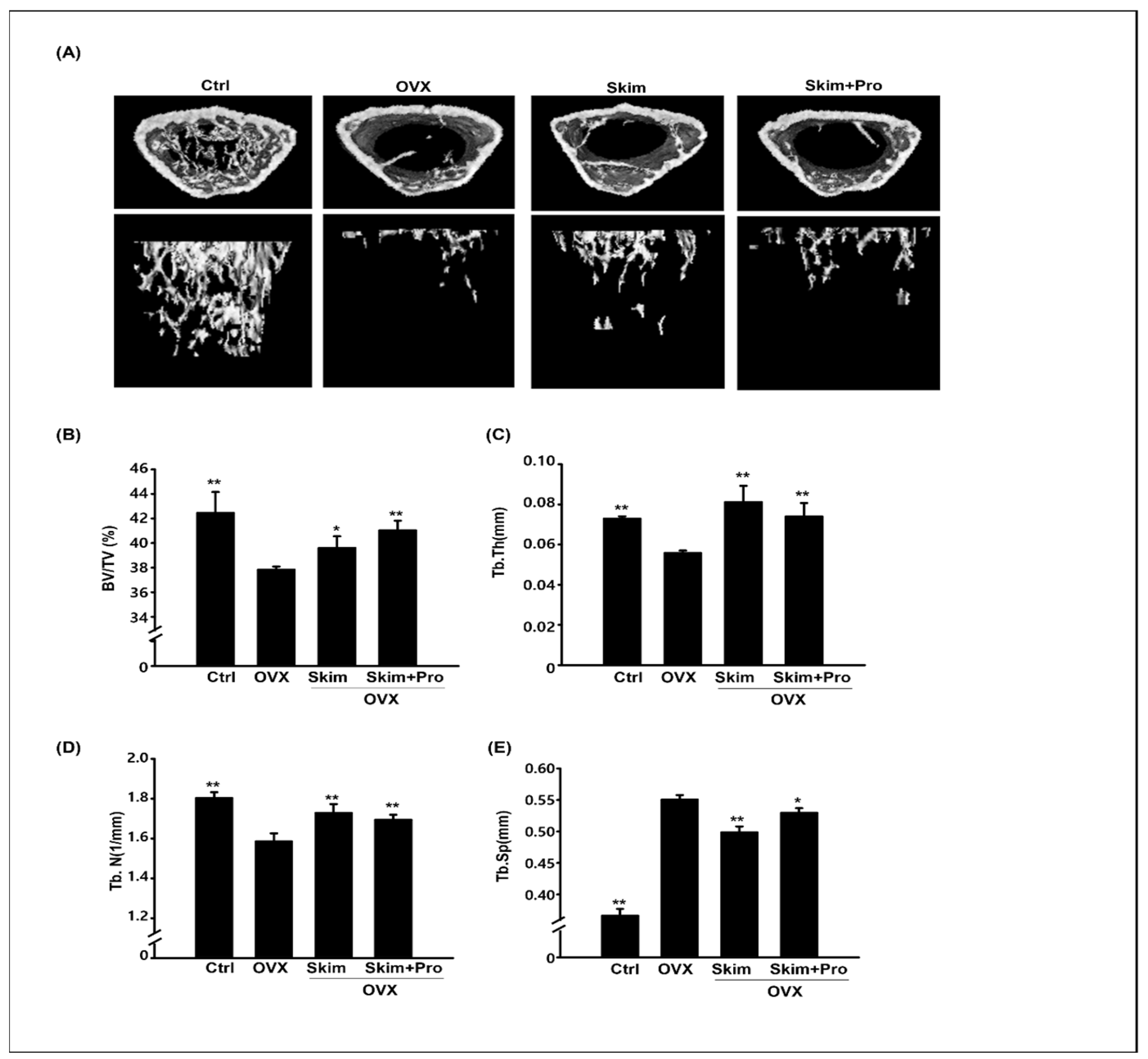
3.2. Attenuation of the OVX-Driven Effects of Skim Milk and Probiotics

Micro-CT imaging of the distal metaphysis of femurs was performed to analyze the effects of skim milk and probiotics on OVX-induced osteoporosis models. The number of reconstructed femurs decreased in the OVX group and was restored by the consumption of skim milk and probiotics (Figure 2A). Compared to the control group, the percentages of BV/TV, Tb.Th, and Tb.N significantly decreased (Figure 2B–D), while that of Tb.Sp significantly increased in the OVX group (Figure 2E). For the OVX groups administered skim milk and skim milk with probiotics, the levels of BV/TV, Tb.Th, and Tb.N were significantly increased by the consumption of skim milk and skim milk with probiotics (Figure 2B–D). The percentage of Tb.Sp significantly decreased when skim milk was consumed alone or in combination with probiotics (Figure 2E). Milk and dairy products contain proteins, minerals, and vitamins that are beneficial for bone health [25]. Skim or nonfat milk is milk in which the milk fat has been removed. As a result, this milk has fewer calories and a higher percentage of calcium by weight. According to a prior study, the calcium concentration in skim milk is 244 mg/200 mL, which is higher than that in whole milk (236 mg/200 mL) [12]. Skim milk contains higher amounts of vitamins than whole milk owing to fortification. In recent years, several studies have reported the potential benefits of probiotic supplementation on bone health under both healthy and pathological conditions. L. casei and L. acidophilus, either alone or in combination, decreased bone damage and effectively restored the antioxidant status of the liver and kidneys [26]. L. reuteri treatment significantly protects OVX mice from bone loss. The levels of osteoclast bone resorption markers and activators (Trap5 and RANKL) and osteoclastogenesis were significantly downregulated in L. reuteri-treated mice [27]. In this study, we used the probiotic combination of Lactobacillus, L. delbrueckii ssp. bulgaricus Lc. Lactis, Bif. animalis ssp. lactis, and Streptococcus thermophiles, which are abundant in yogurt starter cultures. Based on the results, the consumption of skim milk with probiotics was similar to yogurt consumption, and skim milk had a positive effect on bone density when supplemented with probiotics. Fermented milk consists of yogurt, cultured cream, and buttermilk. However, various processes and equipment are required for milk fermentation. Here, we suggest that a mixture of skim milk and probiotics without fermentation is effective at reducing weight gain and preventing osteoporosis in elderly individuals. This mixture is also economical because it excludes the fermentation process, despite evidence that various products, such as acids, produced through the fermentation process, positively affect human health [28].

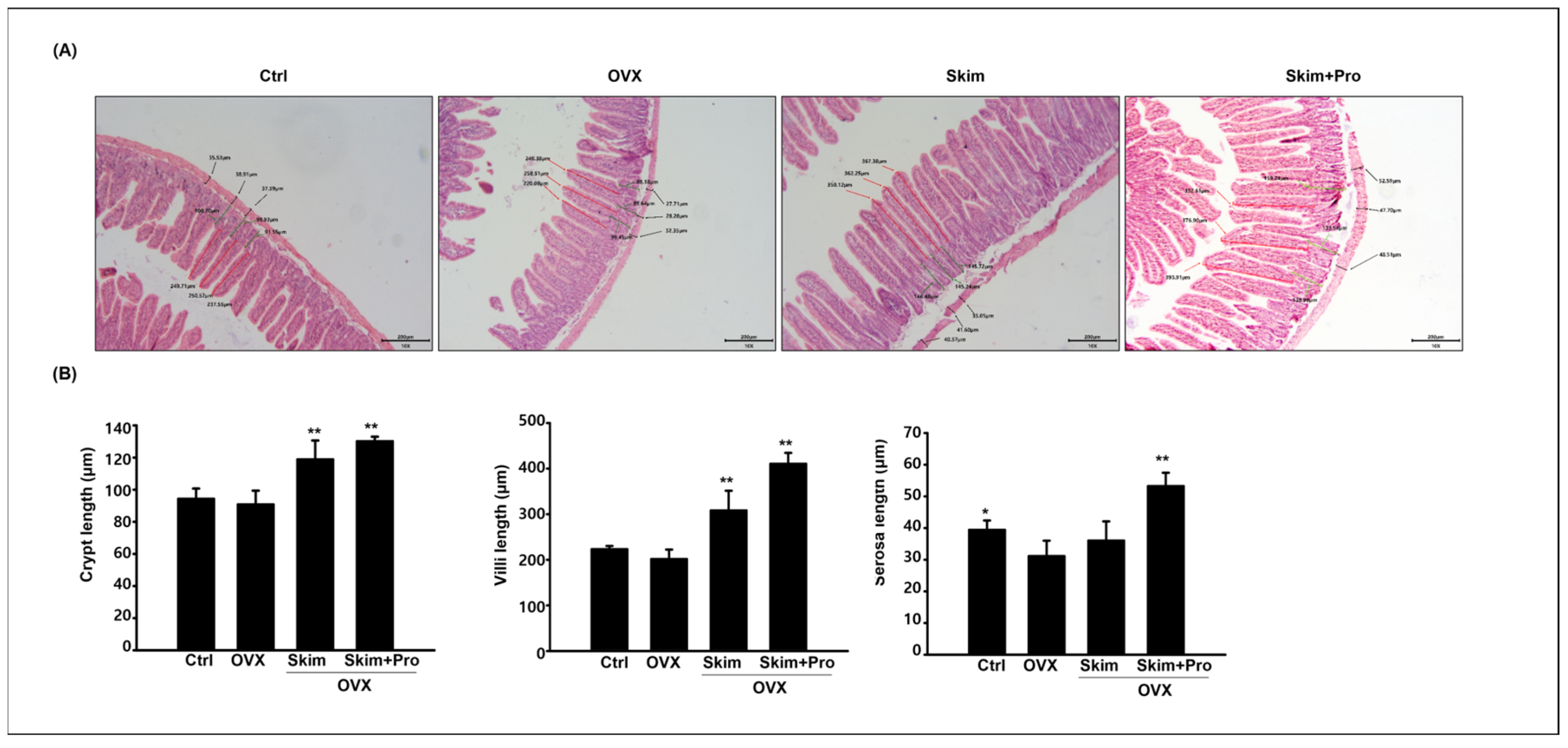
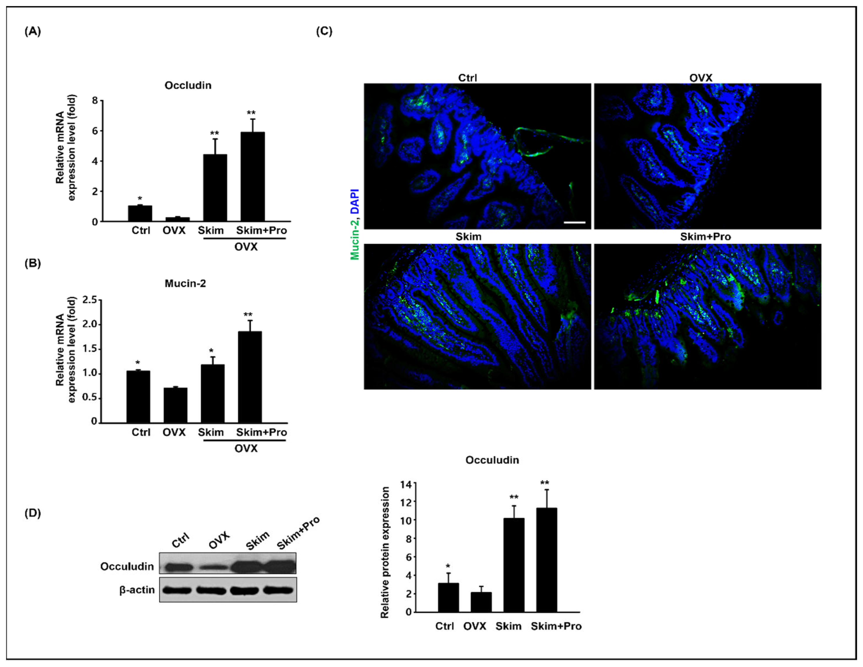
3.3. Effect of Skim Milk and Probiotics on the Small Intestine

According to our results, the consumption of both skim milk and probiotics had positive effects on weight loss and the prevention of osteoporosis. Several studies have reported that probiotics positively affect gut health [29]. Therefore, we analyzed the histological changes and lengths of the villi, crypts, and serosa. The lengths of the villi and crypts were significantly increased in the skim-milk and skim-milk-with-probiotic groups compared to those in the OVX and control groups (Figure 3A). In addition, the length of the serosa significantly increased in the skim-milk-with-probiotic group compared with that in the other groups (Figure 3B). A previous study revealed a significant decrease in intestinal villus height, density, and the villus height:crypt ratio in OVX rats compared with those in sham-operated rats, suggesting a compromised gut barrier [30]. However, our results did not reveal a significant difference in villus or crypt lengths between the control and OVX groups. Long villi are associated with an increased absorptive surface area and absorption capacity of the intestine [31].
In piglets, feeding with probiotics enhanced intestinal integrity, lengthened intestinal villi in the jejunum, and positively impacted the intestinal microbiota [32]. The addition of probiotics or synbiotics increased the villus-height-to-crypt-depth ratio and villus height in both the duodenum and ileum of broiler chickens [33]. The increase in villus height and villus-height-to-crypt-depth ratio was associated with an improvement in the growth performance of both synbiotics and probiotics. Our study revealed the positive effect of milk production and probiotics on OVX-induced osteoporosis in mice, indicating their beneficial effects in older women; this will have a different effect compared to the growth time window.
We analyzed the gene expression of occludin and mucin 2 in the intestinal samples. The expression levels of occludin and mucin 2 significantly increased in the skim-milk and skim-milk-with-probiotic groups compared with those in the OVX groups (Figure 4A,B). Consistently, the immunostaining result showed that the mucin 2 positive area increased in the skim-milk and skim-milk-with-probiotic groups compared with that in the OVX group (Figure 4C). Quantification of the occludin protein also revealed a statistically significant increase in its level in both the skim-milk and skim-milk-with-probiotic groups compared with that in the OVX groups (Figure 4D).
The intestinal epithelial tight junction (TJ) barrier controls the paracellular permeation of contents from the intestinal lumen into the intestinal tissue and systemic circulation [30]. Commensal bacteria and probiotics have been demonstrated to promote intestinal barrier integrity in vitro and in vivo. Probiotics were found to preserve the intestinal barrier in mouse models of colitis [34]. Mucin is a major component of the intestinal mucosal layer, plays an essential role in maintaining intestinal mucosal homeostasis, and has been detected in various organs, tissues, and body fluids.
Mucin 2, the most abundant mucin family member in the small intestine and colon [35], is a key player during infection in the body. For instance, Bergstrom et al. reported that Muc2-/-mice had higher susceptibility to attaching and effacing one of the mucosal pathogens, Citrobacter rodentium [36]; these mice also showed clinical signs of colitis. Muc2-/-mice exhibited mucosal thickening, superficial erosion, and hyperproliferation [37]. Prior studies strongly suggest that mucin 2 is critical for gut health. Furthermore, our results clearly show that the intake of skim milk and skim milk with probiotics can improve intestinal health by increasing mucin 2 production in the intestine. In particular, the expression of the Mucin2 gene was more pronounced in the co-administration group, which involved the administration of skim milk and probiotics, compared with that in the skim-milk-only group.
A previous study revealed that human-milk-oligosaccharide-treated cells exhibit increased mucin 2 expression, decreased bacterial attachment, and increased dextran permeability when challenged with enteric pathogens [25]. In addition, several types of Lactobacillus reportedly increase the expression of mucin protein in HT29 cells, a human intestinal cell line [38]. An extract of L. acidophilus A 4 cell increased mucin 2 expression in HT29 cells [39]. These studies highlight the positive synergistic effect of simultaneous intake on the increase in Mucin 2 in our study.
The intestine is a very important organ that integrates immunity, nutrient absorption, and digestion. TJs, which form in the intestinal epithelia, play a critical role in the physical intestinal barrier and modulate the transfer of various substances. Among TJs, occludin is abundantly expressed at cell-to-cell contact sites. Occludin-/-mice exhibited increased inflammation, growth retardation, and hyperplasia, despite normal TJ structure in the intestine [40]. Accordingly, increased occludin levels improve the TJ barrier function and prevent TJ damage [41,42]. Based on our results, the protein levels of occludin were markedly increased in the skim-milk and skim-milk-and-probiotic groups compared to those in the OVX groups, indicating that the intake of skim milk and probiotics has positive effects on intestinal TJ function. In addition, comparative analysis of the various effectiveness mentioned above with fermented skim milk using probiotics will be necessary as further study.

4. Conclusions

Our results revealed the positive effect of hydrolyzed skim milk and milk with probiotics on weight loss, calcium increase, triglyceride and LDL decrease, osteoporosis prevention, and gut health in OVX mouse models. In conclusion, milk products and probiotic composite materials have positive effects on osteoporosis, may prevent weight gain due to menopause in older women, and improve intestinal health by controlling the length of intestinal villi and expression of TJs and mucus. In addition, several indicators displayed a greater effect in the group administered skim milk with probiotics than in the group administered skim milk alone. The findings of this study are relevant to the dairy industry and the production of various functional health products that combine hydrolyzed skim milk and probiotics.

Author Contributions

Conceptualization: H.-J.P. and W.-Y.L.; Investigation: H.-J.P., W.-Y.L. and H.-W.S.; Writing—original draft: H.-J.P. and W.-Y.L.; Validation: H.W.K., J.-k.P. and K.C.; Methodology: H.-J.P., J.M.Y., K.C. and J.-k.P. All authors have read and agreed to the published version of the manuscript.

Funding

This study was supported by the National Research Foundation of Korea (NRF) grant funded by the Korean government (MIST) (No. 2021R1G1A1091690).

Institutional Review Board Statement

All experiments in this study were performed following the guidelines of the Institutional Animal Care and Use Committee of Sangji University (approval number # 2021–20).

Data Availability Statement

The author confirm that the data supporting the finding of this study are available within the article.

Conflicts of Interest

The authors declare no conflict of interest.

References

  1. Eastell, R.; O’Neill, T.W.; Hofbauer, L.C.; Langdahl, B.; Reid, I.R.; Gold, D.T.; Cummings, S.R. Postmenopausal osteoporosis. Nat. Rev. Dis. Primers 2016, 2, 16069. [Google Scholar] [CrossRef] [PubMed]
  2. Rosen, C.J. The Epidemiology and Pathogenesis of Osteoporosis; MDText.com, Inc.: South Dartmouth, MA, USA, 2020. [Google Scholar]
  3. Armas, L.A.; Recker, R.R. Pathophysiology of osteoporosis: New mechanistic insights. Endocrinol. Metab. Clin. N. Am. 2012, 41, 475–486. [Google Scholar] [CrossRef]
  4. Janiszewska, M.; Kulik, T.; Dziedzic, M.; Żołnierczuk-Kieliszek, D.; Barańska, A. Osteoporosis as a social problem- pathogenesis, symptoms and risk factors of postmenopausal osteoporosis. Probl. Hig. Epidemiol. 2015, 96, 106–114. [Google Scholar]
  5. Cymet, T.C.; Wood, B.; Orbach, N. Osteoporosis. J. Am. Osteopath. Assoc. 2000, 100, S9–S15. [Google Scholar]
  6. Fiona, E.A.M.; Liam, M.; Alison, G.; George, D.; Neville, C.E.; Rob, V.H.; Boreham, C.; Ralston, S.H. Genetic and environmental determinants of peak bone mass in young men and women. J. Bone Miner. Res. 2002, 17, 1273–1279. [Google Scholar]
  7. Ankit, D.; Sameer, A.; Prasoon, K.; Vishal, K.; Deba, P.D.; Sanjay, K.B. High prevalence of vitamin D deficiency and osteoporosis in patients with fragility fractures of hip: A pilot study. J. Clin. Orthop. Trauma. 2019, 10, 1097–1100. [Google Scholar]
  8. Verduci, E.; D’Elios, S.; Cerrato, L.; Comberiati, P.; Calvani, M.; Palazzo, S.; Martelli, A.; Landi, M.; Trikamjee, T.; Peroni, D.G. Cow’s Milk Substitutes for Children: Nutritional Aspects of Milk from Different Mammalian Species, Special Formula and Plnat-Based Beverages. Nutrients 2019, 11, 1739. [Google Scholar] [CrossRef]
  9. Fardellone, P. The effect of milk consumption on bone and fracture incidence, an update. Aging Clin. Exp. Res. 2019, 31, 759–764. [Google Scholar] [CrossRef]
  10. Heidi, J.K.; Jane, C.K.; Bruce, P.L. Milk intake during childhood and adolescence, adult bone density, and osteoporotic fractures in US women. Am. J. Clin. Nutr. 2003, 77, 257–265. [Google Scholar]
  11. Aviv, F.; Sarah, L.M.; Lindsay, K.E.; Caeley, L.; Raylene, A.R.; Ronald, F.Z. Skim milk powder enhances trabecular bone architecture compared with casein or whey in diet-induced obese rats. Nutrition 2012, 28, 331–335. [Google Scholar]
  12. Ratajczak, A.E.; Zawada, A.; Rychter, A.M.; Dobrowolska, A.; Krela-Kaźmierczak, I. Milk and dairy products: Good or bad for human bone? Practical dietary recommendations for the prevention and management of osteoporosis. Nutrients 2021, 13, 1329. [Google Scholar] [CrossRef] [PubMed]
  13. Jing, Z.; Katherine, J.M.; Regina, I.; Ormond, A.M.; Robert, A.B.; Laura, R.M. Loss of bone and Wnt10b expression in male type 1 diabetic mice is blocked by the probiotic Lactobacillus reuteri. Endocrinology 2015, 153, 3169–3182. [Google Scholar]
  14. Xu, B.; Ling, S.; Xu, X.; Liu, X.; Wang, A.; Zhou, Y.; Luo, Y.; Li, W.; Yao, X. A New Formulation of Probiotics Attenuates Calcipotriol-Induced Dermatitis by Inducing Regulatory Dendritic Cells. Front. Immunol. 2021, 12, 775018. [Google Scholar] [CrossRef] [PubMed]
  15. Butera, A.; Pascadopoli, M.; Pellegrini, M.; Gallo, S.; Zampetti, P.; Guggia, G.; Scribante, A. Domiciliary Use of Chlorhexidine vs. Postbiotic Gels in Patients with Peri-Implant Mucositis: A Split-Mouth Randomized Clinical Trial. Appl. Sci. 2022, 12, 2800. [Google Scholar] [CrossRef]
  16. Zhou, B.; Jin, G.; Pang, X.; Mo, Q.; Bao, J.; Liu, T.; Wu, J.; Xie, R.; Liu, X.; Liu, J.; et al. Lactobacillus rhamnosus GG colonization in early life regulates gut-brain axis and relieves anxiety-like behavior in adulthood. Pharmacol. Res. 2022, 177, 106090. [Google Scholar] [CrossRef] [PubMed]
  17. Inada, M.; Matsumoto, C.; Miyaura, C. Animal models for bone and joint disease. Ovariectomized and orchidectomized animals. Clin. Calcium 2011, 21, 164–170. [Google Scholar] [PubMed]
  18. Chiang, S.S.; Pan, T.M. Antiosteoporotic effects of Lactobacillus-fermented soy skim milk on bone mineral density and the microstructure of femoral bone in ovariectomized mice. J. Agric. Food Chem. 2011, 59, 7734–7742. [Google Scholar] [CrossRef]
  19. Yang, L.C.; Lin, S.W.; Li, I.C.; Chen, Y.P.; Tzu, S.Y.; Chou, W.; Chen, C.C.; Lin, W.C.; Chen, Y.L.; Lin, W.H. Lactobacillus plantarum GKM3 and Lactobacillus paracasei GKS6 supplementation ameliorates bone loss in ovariectomized mice by promoting osteoblast differentiation and inhibiting osteoclast formation. Nutrients 2020, 12, 1914. [Google Scholar] [CrossRef]
  20. Park, H.J.; Lee, W.; Zhang, M.; Hong, K.; Park, C.; Kim, J.; Song, H. Evaluation of resmethrin toxicity to neonatal testes in organ culture. Toxicol. Sci. 2020, 173, 53–64. [Google Scholar] [CrossRef]
  21. Lee, W.Y.; Park, H.J. Toxicology of cerium oxide nanoparticles on neonatal testicular development in mouse organ culture. Reprod. Toxicol. 2022, 111, 120–128. [Google Scholar] [CrossRef]
  22. Cao, J.J.; Gregoire, B.R. A high-fat diet increases body weight and circulating estradiol concentration but does not improve bone structural properties in ovariectomized mice. Nutr. Res. 2016, 36, 320–327. [Google Scholar] [CrossRef]
  23. Li, J.Y.; Chassaing, B.; Tyagi, A.M.; Vaccaro, C.; Luo, T.; Adams, J.; Darby, T.M.; Weitzmann, M.N.; Mulle, J.G.; Gewirtz, A.T.; et al. Sex steroid deficiency-associated bone loss is microbiota dependent and prevented by probiotics. J. Clin. Investig. 2016, 126, 2049–2063. [Google Scholar] [CrossRef]
  24. Park, I.S.; Lee, H.W.; Ryuk, J.A.; Ko, B.S. Effects of an aqueous extract of dangguijagyagsan on serum lipid levels and blood flow improvement in ovariectomized rats. Evid. Based Complement. Alternat Med. 2014, 2014, 497836. [Google Scholar] [CrossRef]
  25. Tunick, M.H.; Van Hekken, D.L. Dairy products and health: Recent insights. J. Agric. Food Chem. 2015, 63, 9381–9388. [Google Scholar] [CrossRef]
  26. Amdekar, S.; Kumar, A.; Sharma, P.; Singh, R.; Singh, V. Lactobacillus protected bone damage and maintained the antioxidant status of liver and kidney homogenates in female Wistar rats. Mol. Cell Biochem. 2012, 368, 155–165. [Google Scholar] [CrossRef]
  27. Britton, R.A.; Irwin, R.; Quach, D.; Schaefer, L.; Zhang, J.; Lee, T.; Parameswaran, N.; McCabe, L.R. Probiotic L. reuteri treatment prevents bone loss in a menopausal ovariectomized mouse model. J. Cell Physiol. 2014, 229, 1822–1830. [Google Scholar] [CrossRef] [PubMed]
  28. Kok, C.R.; Hutkins, R. Yogurt and other fermented foods as sources of health-promoting bacteria. Rev. Nutr. Rev. 2018, 76, 1–15. [Google Scholar] [CrossRef]
  29. Butel, M. Probiotics, gut microbiota and health. Med. Mal. Infect. 2013, 44, 1–8. [Google Scholar] [CrossRef] [PubMed]
  30. Jia, L.; Tu, Y.; Jia, X.; Du, Q.; Zheng, X.; Yuan, Q.; Zheng, L.; Zhou, X.; Xu, X. Probiotics ameliorate alveolar bone loss by regulating gut microbiota. Cell Prolif. 2021, 54, e13075. [Google Scholar] [CrossRef] [PubMed]
  31. Izadi, H.; Arshami, J.; Golian, A.; Raji, M.R. Effects of chicory root powder on growth performance and histomorphometry of jejunum in broiler chicks. Vet. Res. Forum 2013, 4, 169–174. [Google Scholar]
  32. Pupa, P.; Apiwatsiri, P.; Sirichokchatchawan, W.; Pirarat, N.; Maison, T.; Koontanatechanon, A.; Prapasarakul, N. Use of Latobacillus plantarum (strains 22F and 25F) and Pediococcus acidilactici (strain 72N) as replacements for antibiotic-growth promotants in pigs. Sci. Rep. 2021, 11, 12028. [Google Scholar] [CrossRef]
  33. Awad, W.A.; Ghareeb, K.; Abdel-Raheem, S.; Böhm, J. Effects of dietary inclusion of probiotic and synbiotic on growth performance, organ weights, and intestinal histomorphology of broiler chickens. Poult. Sci. 2009, 88, 49–56. [Google Scholar] [CrossRef] [PubMed]
  34. Kaminsky, L.W.; Al-Sadi, R.; Ma, T.Y. IL-1β and the intestinal epithelial tight junction barrier. Front. Immunol. 2021, 12, 767456. [Google Scholar] [CrossRef]
  35. Madsen, K.; Cornish, A.; Soper, P.; McKaigney, C.; Jijon, H.; Yachimec, C.; Doyle, J.; Jewell, L.; De Simone, C. Probiotic bacteria enhance murine and human intestinal epithelial barrier function. Gastroenterology 2001, 121, 580–591. [Google Scholar] [CrossRef] [PubMed]
  36. Johansson, M.E.V.; Thomsson, K.A.; Hansson, G.C. Proteomic analysis of the two mucus layers of the colon barrier reveal that their main component, the Muc2 mucin, is strongly bound to the Fcgbp protein. J. Proteome Res. 2009, 8, 3549–3557. [Google Scholar] [CrossRef] [PubMed]
  37. Brett, F.; Kris, C.; Bruce, A.V. Muc2 protects against lethal infectious colitis by disassociating pathogenic and commensal bacteria from the colonic mucosa. PLoS Pathog. 2010, 6, e1000902. [Google Scholar]
  38. Maria, V.S.; Barbara, A.; Adrianus, C.; Anna, V.; Jules, P.P.M.; Johannes, B.V.G.; Hans, A.B.; Jan, D.; Isabelle, V.S.; Ingrid, B.R.; et al. Muc2-deficient mice spontaneously develop colitis, indicating that MUC2 is critical for colonic protection. Gastroenterology 2006, 131, 117–129. [Google Scholar]
  39. Mack, D.R.; Ahrne, S.; Hyde, K.; Wei, S.; Hollingsworth, M.A. Extracellular MUC3 mucin secretion follows adherence of Lactobacillus strains to intestinal epithelial cells in vitro. Gut 2003, 52, 827–833. [Google Scholar] [CrossRef]
  40. Kim, Y.; Kim, S.; Whang, K.; Kim, Y.; Oh, S. Inhibition of Escherichia coli O157:H7 attachment by interactions between lactic acid bacteria and intestinal epithelial cells. J. Microbiol. Biotechnol. 2008, 18, 1278–1285. [Google Scholar]
  41. Saitou, M.; Furuse, M.; Sasaki, H.; Schulzke, J.D.; Fromm, M.; Takano, H.; Noda, T.; Tsukita, S. Complex phenotype of mice lacking occludin, a component of tight junction strands. Mol. Biol. Cell 2000, 11, 4131–4142. [Google Scholar] [CrossRef]
  42. AI-Sadi, R.; Khatib, K.; Guo, S.; Ye, D.; Youssef, M.; Ma, T. Occludin regulates macromolecule flux across the intestinal epithelial tight junction barrier. Am. J. Physiol.-Gastrointest. Liver Physiol. 2011, 300, G1054–G1064. [Google Scholar] [CrossRef] [PubMed]
Figure 1. Changes in body weights and serum lipid profiles due to ovariectomy (OVX) and skim-milk and probiotic treatment. Changes in body weight from weeks 0 to 8 (A). Concentrations of calcium (B), triglycerides (C), and low-density lipoprotein cholesterol (LDL-C) in the serum of each experiment group (D). Data from the experimental group were compared with data from the OVX group. Data are presented as mean ± SD (n = 5). The values of * p < 0.05 and ** p < 0.01 were considered to indicate statistical significance.
Applsci 13 10424 g001
Figure 2. Effects of ovariectomy (OVX) and the consumption of skim milk and probiotics on bone mineral density and bone mineral content. (A) Longitudinal and transverse cross-sections of representative micro-CT reconstructed femurs after a four-week treatment with skim milk and probiotics. Skim-milk- and probiotic-driven suppression of OVX-induced loss of femoral bone volume/tissue volume ratio (BV/TV) (B), trabecular number (Tb.N) (C), trabecular thickness (Tb.Th) (D), and trabecular spacing (Tb.Sp) (E). The results are presented as mean ± SD. * p < 0.05 and ** p < 0.01 were considered to indicate statistical significance. Data from the experimental group were compared with data from the OVX group.
Applsci 13 10424 g002
Figure 3. Analysis of the villi, crypt, and serosa lengths in the ovariectomy (OVX) and skim-milk-and-probiotic groups. Hematoxylin and eosin staining analysis of mice intestine from each experimental group (A) and the lengths of villi, crypt, and serosa. Lengths are presented as μm (B). Data from the experimental group were compared with data from the OVX group. The results are expressed as mean ± SD. * p < 0.05 and ** p < 0.01 were considered to indicate statistical significance. Data from the experimental group were compared with data from the OVX group.
Applsci 13 10424 g003
Figure 4. Expression of occludin and mucin 2 in the small intestines of the control, ovariectomy (OVX), and skim-milk-and-probiotic groups. The mRNA expression levels of occludin (A) and mucin 2 (B) in the intestine of each experimental group. Immunohistochemical analysis of the mucin 2 protei. Scale bar = 50 μm (C). Western blotting analysis of the occludin protein; the graph shows the results of densitometric analysis of the band normalized to that of β-actin (D). The results are presented as mean ± SD. * p < 0.05 and ** p < 0.01 were considered to indicate statistical significance. Data from the experimental group were compared with data from the OVX group.
Applsci 13 10424 g004
Table 1. Mice primer sequences used for real-time quantitative PCR.
GeneForward PrimerReverse Primer
Occludin5′-ATGTCCGGCCGATGCTCTC-3′5′-TTTGGCTGCTCTTGGGTCTGTAT-3′
Mucin 25′-GCCTGTTTG ATAGCTGCTATGTGCC-35′-GTTCCGCCAGTCAATGCAGACAC-3
GAPDH5′-GTCGGTGTGAACGGATTTG-35′-CTTGCCGTGGGTAGAGTCAT-3′
Disclaimer/Publisher’s Note: The statements, opinions and data contained in all publications are solely those of the individual author(s) and contributor(s) and not of MDPI and/or the editor(s). MDPI and/or the editor(s) disclaim responsibility for any injury to people or property resulting from any ideas, methods, instructions or products referred to in the content.

