Article

The Investigation of the Effect of a-Tomatine as a Novel Matrix Metalloproteinase Inhibitor on the Bond Strength of Sound and Eroded Dentine through In Vitro and In Silico Methods

1
Department of Restorative Dentistry, Faculty of Dentistry, Altinbas University, Istanbul 34147, Turkey
2
Department of Chemistry, Faculty of Science, Gazi University, Ankara 06590, Turkey
3
Department of Biochemistry, Faculty of Dentistry, Istanbul University, Istanbul 34320, Turkey
4
Department of Restorative Dentistry, Faculty of Dentistry, Istanbul University, Istanbul 34320, Turkey
*
Author to whom correspondence should be addressed.
Appl. Sci. 2023, 13(18), 10322; https://doi.org/10.3390/app131810322
Submission received: 22 August 2023 / Revised: 7 September 2023 / Accepted: 11 September 2023 / Published: 14 September 2023
(This article belongs to the Collection Dental Composites and Adhesives in Dentistry)

Abstract

:
This study aims to examine the effect of a-tomatine, a new matrix metalloproteinase inhibitor for dentistry, as a surface pretreatment on the bonding strength of different types of dentine via in vitro and in silico methods. The binding efficacy of both a-tomatine and chlorhexidine to MMP-2, 8, and 9 was evaluated through molecular docking and dynamics analyses. For microtensile testing (µTBS), specimens (n = 84) were categorized into two groups based on the type of dentin: sound (SD) and eroded (ED) (n = 42). Each group was further divided into three subgroups according to the utilization of surface pretreatment agents (1.5 µM of tomatine, 2% chlorhexidine (CHX), and the control). Composite buildups were gradually created via a three-step etch-and-rinse technique. The specimens were sectioned into sticks and subsequently subjected to µTBS after aging for either 24 h (n = 7) or 6 months (n = 7). The data were subjected to analysis using two-way ANOVA with a Bonferroni correction post hoc test. The significance level was evaluated at a minimum of p < 0.05. According to molecular docking and dynamic simulation analyses, a-tomatine exhibits a higher affinity for MMP-2, -8, and -9 enzymes compared to chlorhexidine. Lower µTBS values were observed in all ED groups compared to the SD groups. Following 24-h aging, the CHX application in both the SD and ED groups achieved lower µTBS values compared to the control group (p < 0.01 and p > 0.05, respectively). The most favorable results were consistently achieved across all the subgroups subjected to a-tomatine applications (p < 0.05). a-tomatine is a more effective MMP inhibitor than chlorhexidine in terms of preserving bond strength values over time and its capacity to bind to MMP-2,8, and 9 for inhibition.

1. Introduction

Erosion, described as a non-caries lesion resulting from the dissolution of hard tissues via acids that are not bacterial by products, is characterized as a progressive condition [1]. Initially, the weakening of the enamel tissue occurs due to wear and attrition, while prolonged exposure to extrinsic or intrinsic acids disrupts the remineralization–demineralization balance over time, potentially affecting the underlying dentin tissue [2]. In dentin erosion, due to the acidic pH, dentin demineralization occurs, exposing collagen fibrils, and the endogenous collagenolytic activity (MMP) present in dentin and saliva begins to function [1]. The suppression of this MMP, which contributes to the degradation of the hybrid layer formed by adhesive restorations, holds significance for the longevity of restorations achieved via the use of inhibitory agents [3]. Specific MMP inhibitors, such as chlorhexidine (CHX) or galardin, can potentially slow down the degradation in the hybrid layer by suppressing collagenolytic and gelatinolytic activity [4,5]. Several analyses have revealed the significant role of endogenous enzymes such as MMP-2, -8, and -9 in dental caries or dental erosion, demonstrating their involvement in the process of degradation [6,7,8]. Recent research has indicated that a-tomatine, a glycoalkaloid found in tomatoes, can inhibit MMP-2 and MMP-9 activation in certain cancer cells, displaying anti-metastatic effects [9,10]. The MMP activity in dentin can be detected through various and costly techniques, such as in situ zymography [11], SDS-PAGE [5], or the mass loss measurement of collagen degradation [12].
Molecular docking is a computational chemistry method used to predict the binding affinities of ligands to protein structures such as enzymes and receptors [13]. In this method, each of the ligands is rotated around the rotatable bonds (i.e., their various conformations are changed) and approximated to the actual protein–ligand complex via a complete, systematic search for its orientation and position [14,15]. The results obtained are important in terms of providing guidance for in vitro and in vivo results. The molecular docking method has been accepted by most scientific authorities since the early 2000s to shed light on experimental results [13,16,17,18]. The movements of atoms, both interactions with each other and with the solvent in the physiological environment, are included in the molecular dynamics calculation method [19]. This makes computational chemistry a set of methods whose accuracy continues to improve as technology is developed [15]. According to the literature, there is no existing study within the dental literature regarding the potential utilization of a-tomatine as an MMP inhibitor, and furthermore, an in silico approach (molecular or dynamic docking) to assess the effectiveness of a-tomatine on MMP enzymes has not been applied.
In light of this information, the present study aimed to scrutinize the application of a-tomatine and chlorhexidine (Figure 1) as MMP inhibitors in both sound and eroded dentin tissues, investigating their immediate and long-term (6-month) effects on adhesive bond strength. Furthermore, the intention behind this was to substantiate, through both molecular and dynamic docking methods, the enzymatic relationship of these two agents with MMP-2, -8, and -9. The formulated hypotheses were as follows: (1) The bond strength values in sound dentin are higher compared to those of eroded dentin. (2) a-tomatine is equally efficacious as chlorhexidine in terms of MMP inhibition.

2. Materials and Methods

2.1. Experimental Design and Sample Size Calculation

The research was conducted utilizing dentin samples procured from extracted human teeth at the Research Laboratory of the Istanbul University Faculty of Dentistry. The study was carried out with the approval of the Clinical Research Ethics Committee of the Istanbul University Faculty of Dentistry (File no: 2017/66). G. Power 3.1.7 software was employed for power analysis to determine the appropriate subgroup sample size (n) in the study. The study power was denoted as 1-ß (where ß represents the probability of a type II error). After the data from the study by Carvalho et al. [20] were taken into account, the effect size (d) was initially determined to be 1.7105250. Subsequently, using this value and aiming for 80% power at the α: 0.05 level, it was calculated that a minimum of 7 teeth should be used in each subgroup for the sample size (n = 84). These 84 molars were halved to form eroded and sound dentin groups. Each of these groups was further subdivided into 3 subgroups based on the intended MMP inhibitor application and then 2 subgroups based on aging time, resulting in a total of 12 subgroups for the study. After extraction, the teeth were cleansed of debris and stored in a thymol solution for 1 week. Subsequently, they were affixed to acrylic resin blocks and sectioned using a low-speed microtome (Isomet 1000 Buehler Precision Saw, Düsseldorf, Germany) positioned 3 mm below the occlusal surface. Following the sectioning, the dentin surfaces were meticulously prepared utilizing 600-grit silicon carbide to achieve a flat surface. The absence of any enamel residue was verified using a stereomicroscope at a magnification of ×30.

2.2. Formation of Erosion on the Dentine Surface

Half of the experimental teeth (n = 42) were immersed in a 1% citric acid (pH: 3.5) solution for 5 min and then soaked in artificial saliva, which was prepared as suggested by Zimmerli et al., for 3.5 h [21]. According to Zimmerli et al., this process is applied to dentine surfaces 6 times a day for 8 days. The content of the remineralizing agent was composed of 0.002 g of ascorbic acid, 0.58 g of NaCl, 0.17 g of CaCl2, 0.16 g of NH4Cl, 1.27 g of KCl, 0.16 g of NaSCN, 0.33 g of KH2PO4, and 0.34 g of Na2HPO4 for 1 L. It was prepared through dissolution in demineralized water. Before the solution volume was completed, the pH level was adjusted to 6.4 with 1 N HCl.

2.3. Preparation of a-tomatine

The powdered tomatine material (phyproof© PhytoLab, Vestenbergsgreuth, Germany) was dissolved in a dimethyl sulfoxide (DMSO) liquid using a precise balance. A preliminary investigation was conducted on 6 sound dentine surfaces to ascertain the appropriate concentration and exposure time of a-tomatine for this study. Initially, 9.31 mg of a-tomatine was weighed with precision and dissolved in DMSO, yielding a total volume of 3 mL. Subsequently, a 3 mM a-tomatine stock solution was diluted by a factor of 1/1000 using distilled water, resulting in a 3 µM a-tomatine stock solution. Consequently, solutions of 1.5 µM, 1 µM, and 0.75 µM of a-tomatine were formulated, which are recognized for their inhibitory effects against MMP enzymes. These solutions were created via further dilution with distilled water at ratios of 1/2000, 1/3000, and 1/4000, respectively [16,19]. In accordance with the outcomes of the preliminary investigation, the decision was made to administer 1.5 µM of tomatine to the dentine surfaces for a duration of 20 s (Table 1).

2.4. Restoration Stage and Bond Strength Measurement

The application was conducted on both the sound (SD) and eroded (ED) flat dentin surfaces using 37.5% orthophosphoric acid for a duration of 15 s, followed by a 15-s rinse under 2 bar air pressure from a 10 mm distance and subsequent drying. In the experimental groups, MMP inhibitors of 2% CHX (Cavity Cleanser, Bisco Inc., Schaumburg, IL, USA) and 1.5 µM of tomatine were administered for 20 s. Conversely, only Optibond FL (Kerr, Orange, CA, USA) was employed in the control groups, adhering to the recommendations of the manufacturing company. Following the elimination of any excess inhibitory agent using an absorbent pellet, the Optibond FL application in the experimental groups was finalized. A microhybrid-based composite material was polymerized onto the flat dentin surface with the assistance of a halogen light (a minimum intensity of 400 mW/cm2 and 40 s). The experimental samples belonging to individual subgroups were stored in an incubator at 37 °C in distilled water until the testing phase. After the aging process, dentin sticks were affixed at both extremities utilizing a cyanoacrylate adhesive (Pattex, Henkel, Düsseldorf, Germany) to a testing device (Microtensile Tester, Bisco, Inc., USA) to assess the microtensile bond strength. This involved the application of a tensile force of 500 N at a speed of 1 mm/min within a universal testing apparatus until the point of failure, with the resulting µTBS values measured in MPa. The failures that occurred during the microtensile bond strength analysis in the dentin sticks were scrutinized under ×30 magnification using a stereomicroscope (Olympus SZ 6.1, Munster, Germany). The identified failure modes were categorized into three distinct types: adhesive/mixed, cohesive in dentin, and cohesive in composite. Detailed information regarding the utilized materials and their compositions is provided in Table 2.

2.5. Computational Method

All molecular docking and molecular dynamics calculations were performed using the Maestro Interface and sub-software available in Schrödinger Release 2023-2 [23].

2.5.1. Molecular Docking

The molecular docking method was used to calculate the affinity of a-tomatine and chlorhexidine molecules for MMP-2, MMP-8, and MMP-9 enzymes.

Ligand Preparation

Three-dimensional structures of a-tomatine and chlorhexidine molecules were downloaded from PubChem. The downloaded three-dimensional structures were imported into Maestro [24]. All ligands were prepared for molecular docking at a pH of 6.9 [25] using the LigPrep [26] application in Maestro, giving a standard deviation of 0.5 using the embedded EPIK [27,28] software, taking into account the ionization state and tautomerization. The main reason for setting the pH at 6.9 was to determine the ionization state of the molecule in the mouth and to accurately predict the conformational state in a realistic way. Thirty-two steroisomers were generated for each of the alta-tomatin and chlorhexidine molecules, and the OPLS3e [29] force field was used.

Protein Preparation

Proteins to be used for molecular docking and molecular dynamics calculations were imported from the Protein Data Bank to the Maestro interface using Protein Preparation Wizard [30]. The protein constructs included three of the MMP enzymes. The PDB ID for MMP-2 protein structure was 1HOV [31], while the PDB ID for the MMP-8 protein structure was 1ZP5 [32], and the PDB ID for the MMP-9 protein structure was 2OVX [33]. Since the protein structures would also be used in molecular dynamics simulations, no crystal water was deleted. The pH value was set to 6.9 [25] to take into account the ionization state of the protein structures in the mouth, the standard deviation was set to 0.7, and a preprocess was applied using the other settings as a default. The hydrogen bonds were then optimized using PROPKA [34] for a pH of 6.9. Finally, the protein structures were minimized using the OPLS3e force field. A Receptor Grid Generation file was prepared to determine the docking sites of the prepared ligands in the protein structures. The cognate ligand, a-tomatine, and chlorhexidine compounds in the protein structures were used to determine the active sites in the protein structures.

Ligand Docking

The prepared ligands were individually docked to the protein structures with the Standard Sensitivity (SP) setting using Maestro’s Glide [35] module. Different conformations were included in the docking process via requesting 10 poses per ligand. The partial load cut-off value was kept at 0.15 and the scaling factor at 0.80.

2.5.2. Molecular Dynamics Study

Molecular dynamics simulations were prepared separately for the MMP-2, MMP-8, and MMP-9 enzymes in a solvent alone (apo form) and when a-tomatine and chlorhexidine molecules were bound to the protein structures (halo form).

Molecular Dynamics Simulation System Setup

The preparation of the protein systems for molecular dynamics simulations was done via immersing each protein and protein–ligand complex in a solvent box with dimensions of 10 Å × 10 Å × 10 Å. The SPC [36,37] setting was chosen for the water molecules in the solvent box. The SPC setting was preferred to ensure more realistic results. In the SPC setting, the bonds in the water molecules are fixed at 1 Å, and the angle between hydrogens is fixed at 109.5 degrees. The solvent box required the addition of inorganic ions to create an exact model of the physiological environment in the mouth. An NaCl ion at a concentration of 0.15 M was added to the solvent box using the Monte Carlo method. A system setup was performed using the OPLS3e force field.

Molecular Dynamic Simulation Protocols

The next step was the initiation of molecular dynamics simulations, which involved the study of the motion of atoms in the solvent environment, following rigorous molecular docking calculations. The protein–solvent systems prepared for this purpose were subjected to molecular dynamics simulations separately in halo form and apo form. Molecular dynamics simulations were performed using the Desmond [38] module in Maestro. The simulations were performed as 100 nanosecond (ns) simulations. A relaxation protocol of 2 ns was used. Pressure, temperature, and the particle number were kept constant. The temperature was kept constant at 310 K on a Nosé–Hoover thermostat [39], and the pressure was kept constant at a 1.01325 bar on a Martyna–Tobias–Klein [40] barostat. These precise and detailed adjustments ensured complete control over the system and produced reliable and accurate results. A total of nine molecular dynamics simulations of the MMP-2, MMP-8, and MMP-9 enzymes, an apo form for each protein, a tomatine–protein complex, and a chlorhexidine–protein complex were run at the same settings for accurate comparison.

2.6. Statistical Analysis

The statistical analysis of the data was conducted using the NCSS (Number Cruncher Statistical System) software (Kaysville, UT, USA). Descriptive statistical methods, including measures such as means, standard deviations, medians, frequency, ratios, minimums, and maximums, were employed for data analysis. The normal distribution adequacy of quantitative data was assessed using a Kolmogorov–Smirnov test (due to the sample size (n) > 50), skewness–kurtosis values, and Q-Q Plots graphical evaluations. For the evaluation of the bond strength measurements of quantitative data exhibiting a normal distribution, a two-way ANOVA test was applied, based on the applied agent and dentin type. For assessments based on the applied agents, a one-way ANOVA test was employed, and a Bonferroni test was used for post hoc evaluations. The bond strength measurements conducted for the two groups were assessed using Student’s t-test. The significance level was evaluated at a minimum of p < 0.05.

3. Results

3.1. Microtensile Bond Strength

After 24 h, the effects of the MMP inhibitor, the dentin type, and the interaction between the MMP inhibitor and the dentin type were found to be statistically significant (p < 0.05). However, after 6 months, the effects of the MMP inhibitor and dentin type were statistically significant (p < 0.05), while the interaction between the applied agent and dentin type was not statistically significant (p > 0.05) (Table 3). All subgroups using a-tomatine exhibited higher bond strength values compared to the chlorhexidine and control groups, and this difference was statistically significant (p < 0.05). When comparing the 24-h and 6-month aging periods in sound dentin for the chlorhexidine-treated groups, no statistically significant difference was observed (p > 0.05). Similarly, there was no significant difference between the chlorhexidine-treated group and the control group in eroded dentin after 24-h aging (p > 0.05). After this aging, chlorhexidine exhibited inferior values in both sound and eroded dentin compared to the control group (Table 4).
The adhesive/mixed failure type observed across all subgroups was adhesive/mixed failure, with the highest occurrence rate of 93.8%. Conversely, cohesive failure was the least frequent in dentin, accounting for 1.9% of cases. Following the 24-h aging period, the most prevalent adhesive/mixed failure was identified in the a-tomatine group for erosive dentin. Upon 6-month aging, the most prevalent adhesive/mixed failure was found in the a-tomatine group for sound dentin, while the chlorhexidine group exhibited the highest rate for erosive dentin (Table 5).

3.2. Molecular Docking

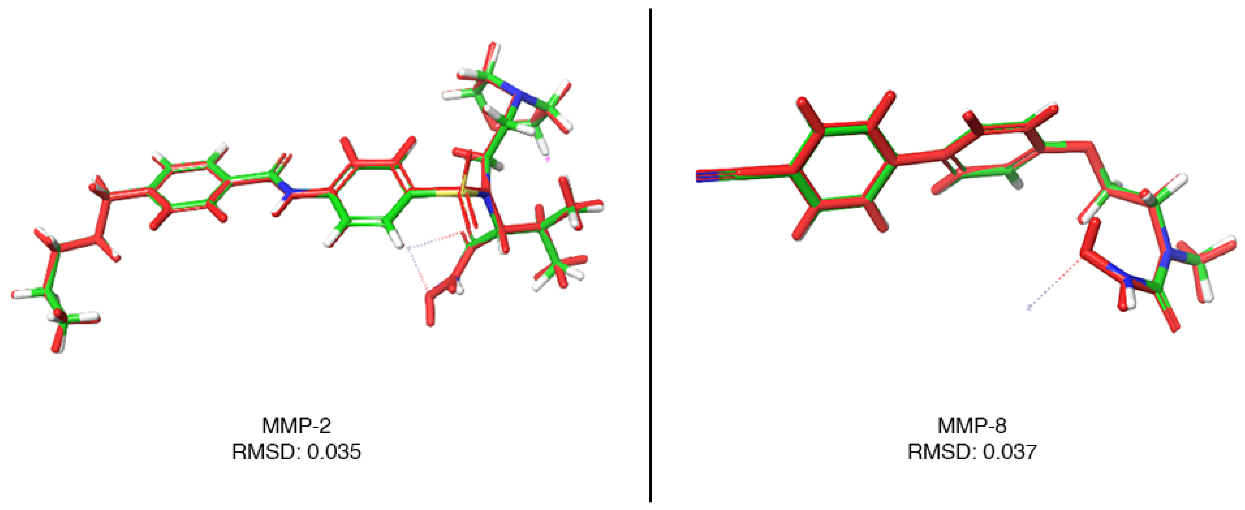
Molecular docking was successfully applied to determine the affinity of a-tomatine and chlorhexidine compounds for MMP-2, MMP-8, and MMP-9 enzymes. The MMP-2 and MMP-8 enzymes were imported together with the cognate ligand, i.e., the ligand present in the protein structure in the Protein Data Bank. The cognate ligand was removed from the protein structure, and the accuracy of the molecular docking process was checked via docking again [41,42]. The results obtained from this docking were in direct agreement with the position of the incoming ligand in the crystal structure and showed extremely low deviation. The low deviation values indicated docking extremely close to the crystal data, i.e., the experimental result. This indicated that docking using these settings would yield results extremely close to reality for a-tomatine and chlorhexidine. The validation image of the MMP-2 and MMP-8 enzymes and the RMSD value, which gives the mean value of the deviation, are given in Figure 1. Since MMP-9 does not contain a cognate ligand, validation was not performed (Figure 2).
Settings whose reliability was proven through validation studies were used to dock the a-tomatine and chlorhexidine molecules to the MMP-2, MMP-8, and MMP-9 enzymes. The docking scores of the ligands for which molecular docking calculations were successfully performed are shown in kcal/mol in Table 6. In the molecular docking calculations, a-tomatine showed a higher affinity for all enzymes than chlorhexidine. The a-tomatine compound obtained scores of −9.358, −9.663, and −7.996 kcal/mol for the MMP-2, MMP-8, and MMP-9 enzymes, respectively. The chlorhexidine compound obtained scores of −7.673, −7.132, and −5.102 kcal/mol for the MMP-2, MMP-8, and MMP-9 enzymes, respectively.

3.3. Molecular Dynamics

In the molecular docking study, 100 ns molecular dynamics studies were successfully performed to analyze whether the interactions in the complexes formed by the a-tomatine and chlorhexidine compounds with the MMP-2, MMP-8, and MMP-9 enzymes continued in the solvent medium and the changes in interactions. Molecular dynamics simulations were performed to see whether the interactions persisted during 100 ns and to determine the potential compatibility with in vitro results. The results of the molecular dynamics simulations and the first step in the analysis involved monitoring and visually analyzing the simulation from start to finish.

3.4. Root-Mean-Square Deviation (RMSD) Analysis

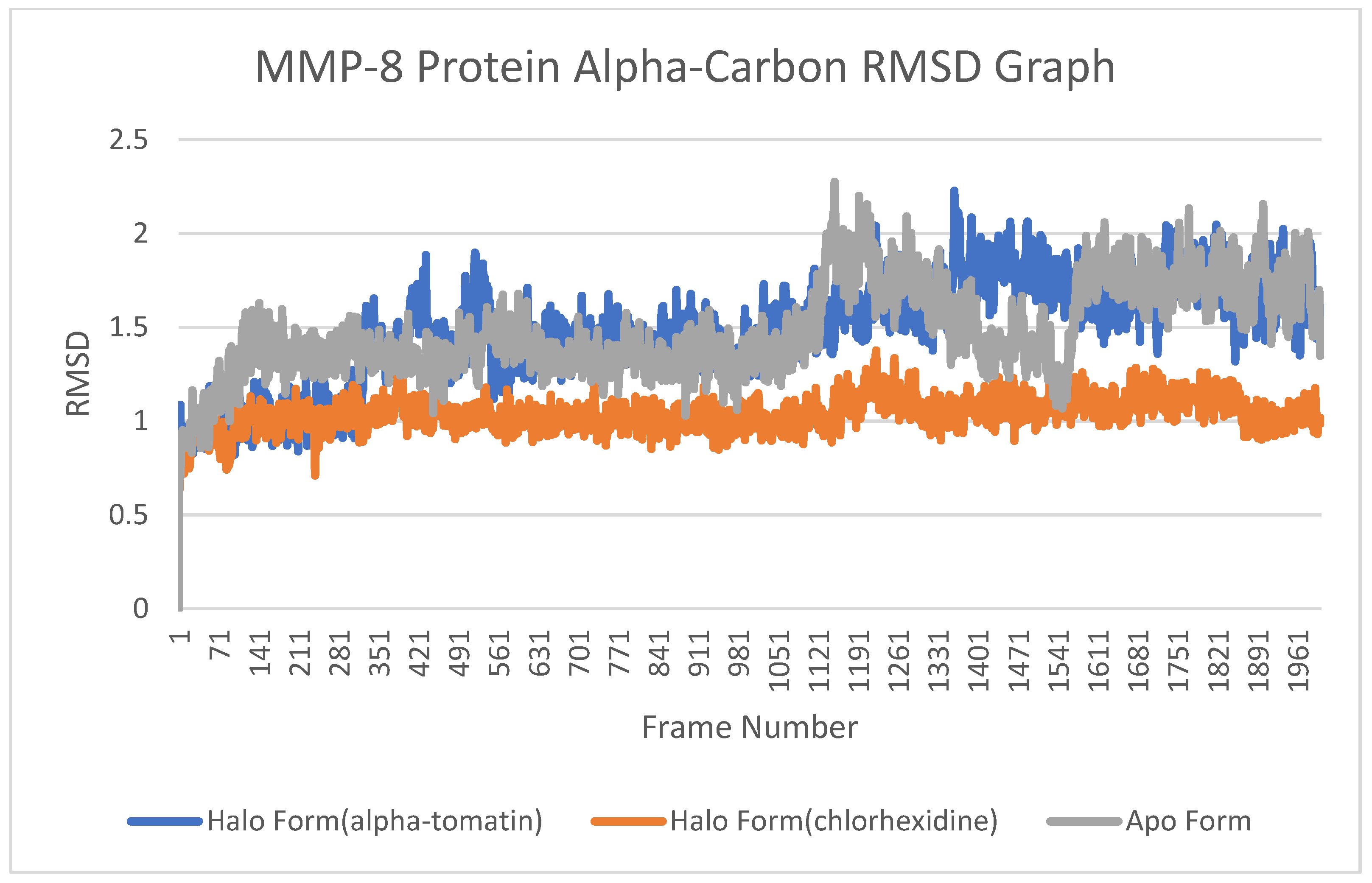
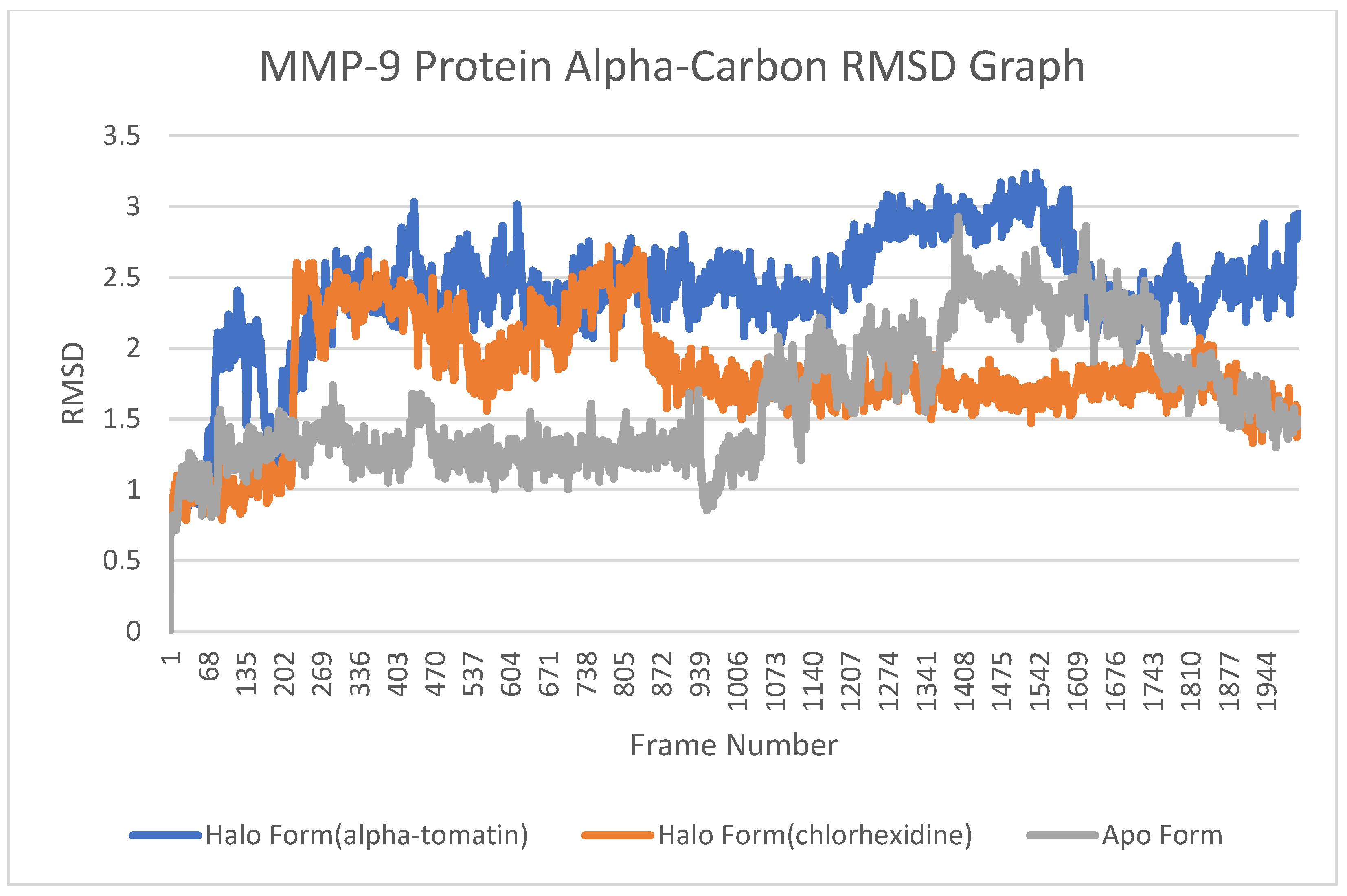
The root-mean-square deviation (RMSD) plot is a method that allows us to understand how frames change relative to an aligned reference frame (the first frame) throughout a molecular dynamics simulation. That is, the RMSD value allows us to understand the extent to which the average position of the protein structure changes relative to the first frame throughout the molecular dynamics simulation. This is an effective method for monitoring conformational changes in protein structure. The MMP-2, MMP-8, and MMP-9 protein structures carry out their natural movements in solvent media, i.e., physiological media. The fact that the ligands bound to these enzymes limit the conformational change that the enzyme performs while doing its work shows the inhibition potential of the ligands. For this purpose, alpha-carbon RMSD plots of both the apo form (without any ligand) and the halo form (protein–ligand complexes containing a-tomatine and chlorhexidine compounds separately) were combined under the same graph to observe how ligands changed the conformation of the protein structure.

3.5. Root-Mean-Square Fluctuation (RMSF) Analysis

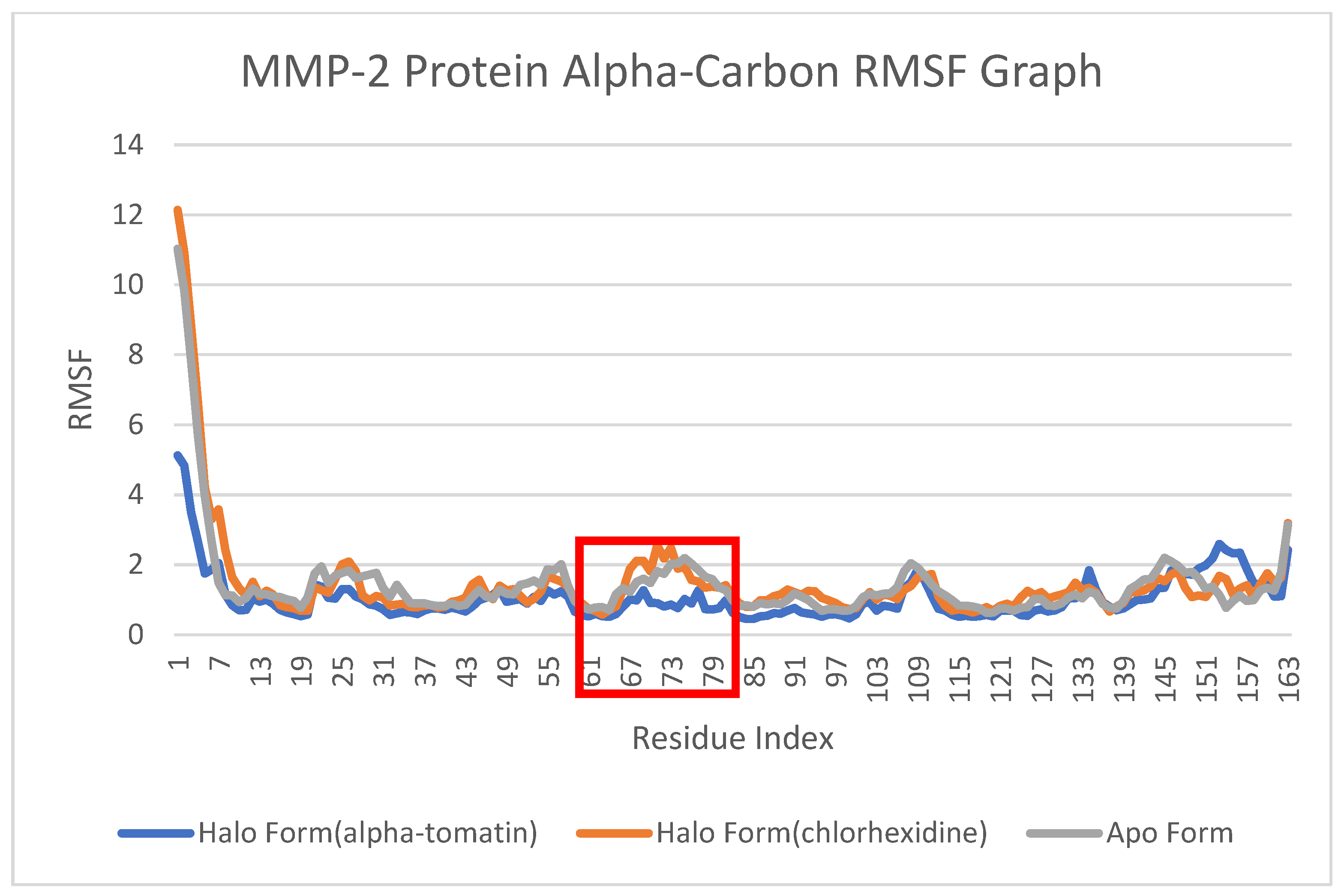
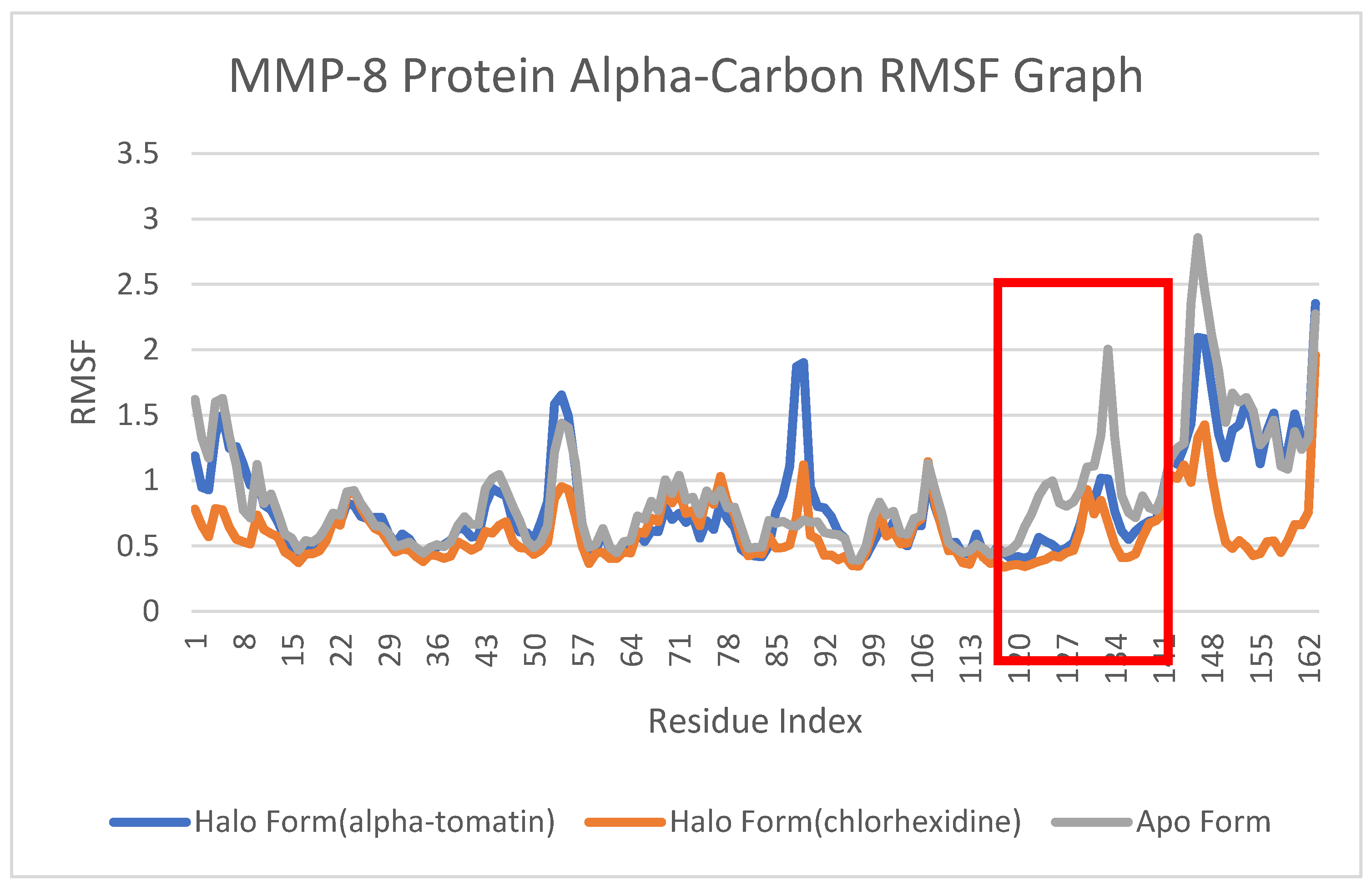
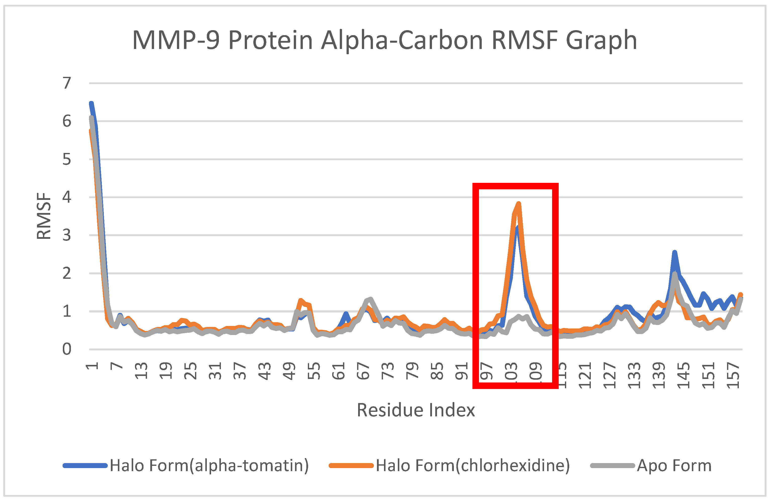
A root-mean-square fluctuation (RMSF) plot was successfully used to detect local fluctuations in protein structure. Graphs containing RMSF values were superimposed to compare each halo-form complex and apo-form structure with each other. Thus, it was possible to detect all the effects of the ligand binding on the protein structure in terms of both the modification and enhancement of the protein structure. As expected, in all RMSF graphs, the ends of the proteins fluctuated a lot, while the ribbon structures fluctuated less. This situation was used to determine the fluctuation of the active center, which is the part that does the work in enzymes, and the effect of ligand contact on the fluctuations in the protein was examined.

4. Discussion

The confirmation of the first hypothesis occurred with the determination of higher binding values in sound dentin compared to eroded dentin. The second hypothesis was formulated based on the assumption that a-tomatine would be as effective as chlorhexidine in terms of bond strength. The obtained µTBS values and the results of docking analyses confirmed this hypothesis and demonstrated that a-tomatine is more effective than chlorhexidine.
Owing to various types of acids that are cost-effective, user-friendly, and can be repeatedly employed, it becomes plausible to artificially induce erosive zones in dental hard tissues [21,43,44,45]. In scientific investigations, hydrochloric acid is utilized to replicate internal dentinal erosion, while citric acid predominantly emulates external erosion processes [46]. Diverse parameters encompassing acid solution concentration, pH, solution composition, and the duration of acid activity hold the potential to influence the corrosive capacity of acids, consequently yielding histological distinctions in dentin [45,47,48]. Despite the utilization of carbonated beverages such as cola to induce erosive conditions in recent times, these tissues exhibit a restricted erosive impact due to the presence of calcium, phosphate, and fluoride components [45,49,50]. Furthermore, citric acid emerges as another surrogate that effectively imitates the acidic nature of fruits and a vegetarian diet, serving to fabricate simulated erosive zones on non-reparative dentin surfaces. Prolonged processes of demineralization and remineralization lead to the accumulation of more denatured irregular fibrils and foster profound demineralization. However, the adhesive resin’s inability to adequately permeate dentin leads to compromised bond strength. In a meticulous inquiry appraising the efficacy of carbonated beverages and citric acid in tissue via the infiltration–demineralization interface, heightened nanoleakage was observed subsequent to a citric acid application compared to carbonated beverages. This underscores the potential for compromised bond strength due to insufficient adhesive resin penetration, as further evidenced by a higher nanoleakage incidence in the study employing citric acid vis-à-vis carbonated beverages [51].
To forestall the deterioration of the hybrid layer established in restorations crafted from sound, eroded [8,21,52,53,54,55,56], or remineralized dentin tissue [57], as per the existing literature, investigators proposed an extended primer application [56], the utilization of crosslinkers [52], or MMP enzyme suppressive agents such as chlorhexidine [21,53,54,55,56], along with preparatory techniques including NaOCl [8,56], diamond bur [21,56], air abrasion, pumice stone, and silicone polishing rubber [21] prior to acid application. It has been suggested that these approaches can mitigate the time-dependent reduction in bond strength. Chlorhexidine, which can be employed at even extremely low concentrations such as 0.002%, 0.02%, 0.2% [58,59], or 0.004% [55], may potentially be restrained by calcium ions. An elevated level of chlorhexidine within the tissue could influence the bonding strength to dentin [60,61]. Research has revealed that, with an escalation of chlorhexidine concentration from 2% to 4%, detrimental effects on microtensile bond strength emerge [58,62]. Since the literature’s reports indicate that a 2% chlorhexidine application curbs degradation in the hybrid layer and yields more favorable long-term bond strength outcomes than other percentages [4,54,58,63], the utilization of chlorhexidine to enhance bond strength remains a contentious topic. While it has been reported that the immediate bond strength of the etch-and-rinse adhesive material post chlorhexidine application remains unaffected [64,65,66], a study by Soares et al. [67] highlighted that the combined application of 2% chlorhexidine and etch-and-rinse adhesive material leads to statistically significant decreases in bond strength values in the short term. Subsequent to a 37% phosphoric acid application, attempts were made to remove chlorhexidine from the surface of chlorhexidine-treated dentin tissue using an absorbent cotton pellet, yet the hydroxyapatite within the dentin tissue continued to harbor chlorhexidine remnants. Through X-ray photoelectron spectroscopy, a study by Sodhi et al. [68] determined that chlorhexidine residues persisted on acid-etched hydroxyapatite surfaces, possibly undermining the subsequent primer’s binding capacity. Moreover, Perdigao et al. [69] demonstrated through SEM analysis that the detrimental effects of chlorhexidine applied after acid treatment, such as in plug formation and debris accumulation in the dentin canals, impeded the formation of resin extensions in the desired dimensions. In present study, it was considered that the low bond strength values observed in the short term in the groups where chlorhexidine was applied may be attributed to these reasons.
Research has demonstrated that a-tomatine, a representative glycoalkaloid present in the roots, stems, and leaves of tomatoes, plays a crucial role in countering potential pathogens and is universally found across tomato genotypes [70]. It comprises a tomatidine (aglycone moiety), ß-licotetraose (tetrasaccharide moiety) containing two glucose molecules, a xylose, and a galactose [71,72]. a-tomatine has been found to exhibit the suppression of certain cancer cell lines [22,73,74]. Notably, it significantly inhibits MMP-2 and -9 activation compared to untreated positive controls [9,75]. Additionally, a-tomatine, known for its antiproliferative activity, can restrain the growth of cancer cells, creating cytotoxicity and suppressing lymphoma and lung cancer cell proliferation [76,77]. When combined with anti-cancer drugs, tomatine has been reported to be able to hinder cancer cells’ spread via curbing MMP activation [9]. Some studies have also indicated its antibiotic activity against diverse microorganisms [78,79,80]. Although, in the medical field, a-tomatine has demonstrated the capacity to inhibit MMP-2 and -9 release [9,76,77], it has yet to be utilized in dentistry. This study marks the inaugural application of a-tomatine as an MMP inhibitor in dentin tissue. A pilot study was required to determine the concentration and duration of application of a-tomatine on the dentin surfaces before starting the study. Regarding the application duration, when examining studies related to CHX in the literature, it was known that the CHX application time is a crucial factor for bond strength. Therefore, durations of 20 and 30 s, based on the application time of CHX, which are among two of the most commonly used values, were selected for a-tomatine [81]. A preliminary investigation involving the dentin surfaces of six sound teeth was conducted to ascertain the appropriate molarity and application duration for a-tomatine. 0.75-1 and 1.5 µM of tomatine solutions, previously reported as effective against MMP enzymes, were prepared following suitable conditions and formulations [9,10,82]. Subsequent to applying the a-tomatine solutions at three distinct µM levels for durations of 20 and 30 s, microtensile bond strength values were assessed and subjected to statistical analysis. The findings led to the selection of 1.5 µM of a-tomatine, with a determined application time of 20 s to dentin surfaces.
a-Tomatine impedes the transport and intracellular signaling of specific DNA cues facilitating MMP-2 and -9 enzyme production [76], and it hinders the formation of these enzymes by obstructing mRNA structures [10]. Consequently, we speculate that a-tomatine could exhibit efficacy via curtailing the attributes of MMP enzymes pivotal in hybrid layer degradation, encompassing tissue invasion, motility, and adhesion [9,10]. Cell studies and gelatin zymography approaches have indicated that a-tomatine intervenes in MMP-2 and -9 enzyme transcription and curbs their release via enzymatic degradation [9,10,22,71,76]. Furthermore, a-tomatine averts cell membrane polarization by interacting with the sterols in the membrane’s structure, instigating cellular content leakage [83,84]. All of this information bolsters the findings of our study, showcasing a-tomatine’s superior MMP inhibition compared to chlorhexidine in sound dentin and its more favorable impact on microtensile bond strength values than chlorhexidine. DMSO, a non-polar and versatile solvent adept at dissolving both polar and non-polar substances for a-tomatine preparation [85,86], facilitates the dissolution of cross-linked collagens into sparse fibrils within the dentin matrix it is applied to [86,87], thereby enhancing resin penetration on the biological surface [86,87,88]. Additionally, we posit that DMSO’s reinforcing effect, through inducing structural biomodifications within the substrate [89,90], contributes to superior outcomes in microtensile bond strength values. However, a study contrary to the aforementioned evidence posits that DMSO has no influence on bonding strength [91]. The omission of DMSO as a distinct group could be regarded as a limitation of our study. A comprehensive examination of this scenario could involve future studies delving into distinct dentin surfaces (such as caries-affected) where DMSO is considered a separate entity. These studies could encompass molecular docking and dynamic analyses of DMSO or investigate the performance of a-tomatine in an alternative solvent.

4.1. Molecular Docking

a-Tomatine enveloped all three enzymes, MMP-2, MMP-8, and MMP-9, with its bulky structure. In addition, the hydroxyl moieties in the glucoside group in this compound coordinated with the zinc atom in the enzymes. This is one of the main reasons for the affinity of a-tomatine for enzymes.
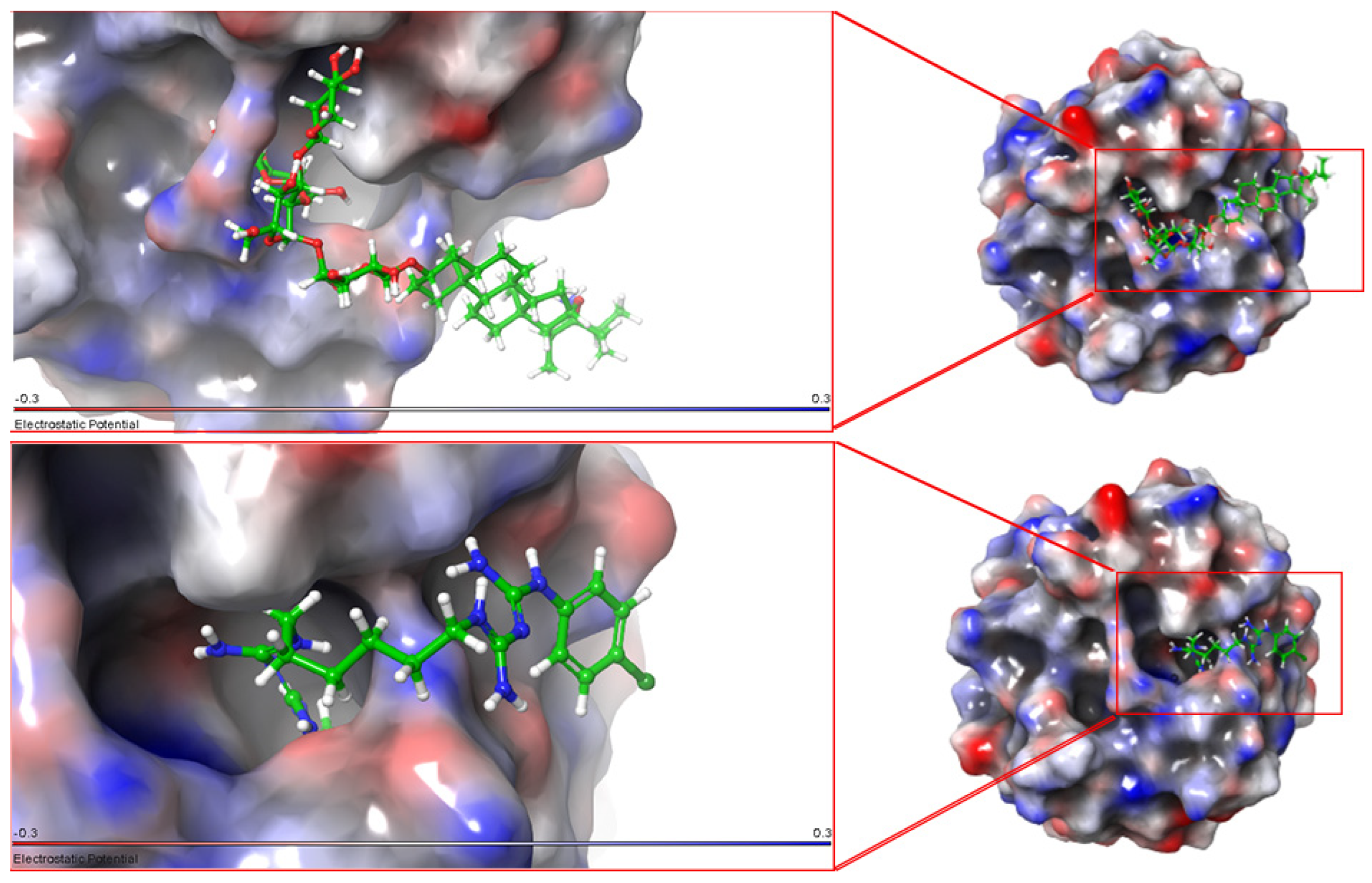
MMP-2: a-Tomatine and chlorhexidine molecules are docked to the recesses and protrusions in the protein structure of the MMP-2 enzyme. The rotation of the rotatable bonds in the a-tomatine compound allows the molecule to reach the optimal conformation of the protein structure through electronegative and electropositive effects. The interaction of chlorhexidine and a-tomatine with the protein structure of the MMP-2 enzyme is shown in Figure 3. The a-tomatine compound was well adhered to the surface of the protein structure, thanks to its hydroxyl groups, whereas the chlorhexidine compound acted mostly by embedding itself in the protein structure. The spreading of the MMP-2 positive charge density on the surface enabled the a-tomatine compound to make non-covalent interactions.
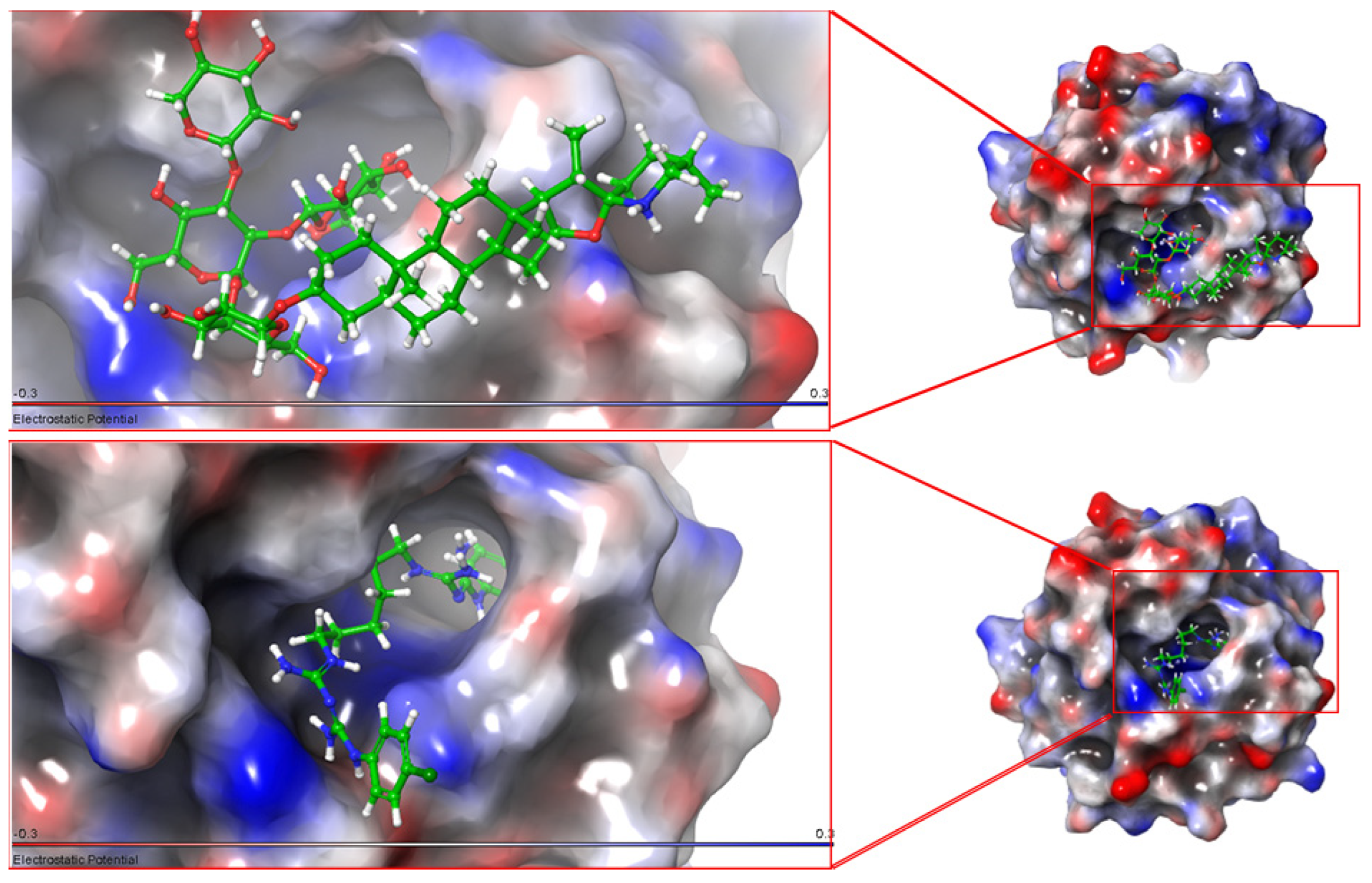
MMP-8: The a-tomatine compound interacts strongly with the MMP-8 enzyme through hydroxyl groups from glycoside groups. These interactions are not involved in the other end of the compound, and this part is released into the solvent medium without any interaction with the protein structure. This means that a-tomatine interacts only at the site of the glycoside groups. One end of the chlorhexidine compound is embedded in the MMP-8 protein structure and interacts with the protein surface from the other end. Figure 4 shows the interactions of a-tomatine and chlorhexidine with the MMP-8 protein. These interactions are noncovalent interactions. Nitrogen atoms in the chlorhexidine compound strongly interact with electronegative sites on the protein structure.
MMP-9: The enzyme for which the a-tomatine and chlorhexidine compounds potentially show the least affinity is the MMP-9 protein structure. All groups of the a-tomatine compound formed strong interactions with the surface amino acids of the MMP-9 protein structure. The glycoside groups tended to penetrate deeper into the protein structure, while the other parts interacted less. One end of the chlorhexidine compound is buried in the protein structure while the other part interacts with the surface of the protein structure. Figure 5 shows that the surface interactions are electropositive. These interactions are the driving force of the part of the chlorhexidine compound embedded in the protein structure. This is mainly due to the fact that the nitrogen atoms in the chlorhexidine compound on the outside are electropositive. The repulsion of two positive effects is one of the effects that reduce the docking score.
The movement of the enzymes in the solvent media was decisive for the enzyme activity. The movement of each enzyme was different when the ligand was bound and when the ligand was not bound. Simulations were successfully performed to investigate and compare the effect of a-tomatine and chlorhexidine molecules on the MMP-2, MMP-8, and MMP-9 enzymes. After calculating the affinity of a-tomatine and chlorhexidine molecules to the MMP-2, MMP-8, and MMP-9 enzymes via a molecular docking study, molecular dynamics simulations were performed to model the states of the formed ligand–protein complexes in a physiological environment. For this purpose, the complexes were modeled in a solvent medium.

4.2. Molecular Dynamics

MMP-2: The a-tomatine compound started the molecular dynamics simulation by coordinating with the zinc atom in the binding pocket of the MMP-2 enzyme. This interaction was mediated by two hydroxyl moieties in the glycoside group of a-tomatine. a-Tomatine interacted with the zinc atom in two places at the same time. This interaction with the zinc atom continued throughout the molecular dynamics simulation and significantly inhibited enzyme functioning. While the steroid part of the a-tomatine compound oscillated in the solvent environment, the part containing the glycoside groups was extremely stable and rigid. The change in conformation was caused by the rotation of the glycoside groups around the single bond where the steroidal part was attached. Throughout the molecular dynamics simulation, the a-tomatine compound did not show any significant conformational change and remained in its initial binding position and conformation. The primary reason for the absence of a significant change was the strong coordination with the zinc atom and the rigidity of the structure of the compound. All these properties indicate that a-tomatine is a potential potent inhibitor of the MMP-2 enzyme.
The chlorhexidine compound started the molecular dynamics simulation by forming a coordination with the zinc atom from the nitrogen atoms. Unlike a-tomatine, the chlorhexidine compound contained a part embedded in the protein structure of the MMP-2 enzyme. In this embedded part, Thr142 and Thr144 amino acid residues acted as a lid, and the conformational changes of these amino acids were very important for the chlorhexidine compound to provide continuity in the inhibition of the MMP-2 enzyme. At 21 nanoseconds of the molecular dynamics simulation, the part of the chlorhexidine compound embedded in the protein structure was directed further inwards, and three-quarters of the compound was embedded in the protein structure. Although the chlorhexidine compound was a compound with a large number of flexible and rotatable bonds, it completed the molecular dynamics simulation in the first binding pocket, thanks to the noncovalent interactions performed by the nitrogen atoms and coordination with the zinc atom, and it exhibited potential strong inhibitor properties.
MMP-8: The a-tomatine compound started the molecular dynamics simulation by coordinating with the zinc atom and two hydroxyl moieties from the glycoside groups in interactions with the MMP-8 enzyme. Coordination with the zinc atom continued throughout the molecular dynamics simulation. The a-tomatine compound exhibited a flat structure and conformation, and this conformation continued throughout the molecular dynamics simulation. The compound remained in a rigid structure. The compound showed potential to be a good inhibitor when evaluated as a whole through the overall molecular dynamics simulation.
The chlorhexidine compound, unlike the a-tomatine compound, started the molecular dynamics simulation slightly away from the zinc atom and without coordination with the zinc atom. The compound was in a “U” shaped conformation, and this conformation changed as the simulation time progressed. The simulation began with some of the part embedded in the protein structure. Although the embedded part remained inside the protein structure from the first moments of the simulation, the other end of the compound began to be released into the solvent environment. The molecular dynamics simulation ended with this repetitive motion. The chlorhexidine compound always remained in the binding pocket throughout the simulation. However, the lack of coordination with the zinc atom indicated a weak interaction. Therefore, it was observed that the chlorhexidine compound had less MMP-8 inhibition potential than a-tomatine.
MMP-9: The interactions of the a-tomatine compound with the MMP-9 enzyme were much less than with the other enzymes. The compound started the molecular dynamics simulation by forming a coordination with the zinc atom, and this effect continued until the end of the simulation. One of the glycoside groups retained the interaction with the zinc atom, while the other parts of the compound were released from the protein structure after 20 ns and released Into the solvent environment. This effect indicates the potential partial inhibition of the protein structure. It is a negative situation that the other parts of the a-tomatine compound do not participate in the interactions. Therefore, the a-tomatine compound partially inhibits the MMP-9 protein structure.
The chlorhexidine compound started the molecular dynamics simulation without coordination at a position just above the zinc atom. Part of the chlorhexidine compound, which has a flexible structure, was embedded in the protein structure, and the other part was released into the solvent environment as soon as the molecular dynamics simulation started. After 60 nanoseconds, the part of the chlorhexidine compound released into the solvent environment was directed towards the surface of the protein structure and interacted with the surface. However, at 80 nanoseconds of the simulation, the part of the compound embedded in the protein structure came out of the protein. The interactions with the surface were insufficient for the compound to remain bound to the protein structure, and the compound somersaulted and moved completely away from the protein structure. From this moment on, the chlorhexidine compound started to circulate in the solvent environment, that is, it had no relation with the protein structure. This shows that the chlorhexidine compound cannot inhibit the MMP-9 protein structure.

4.3. Root-Mean-Square Deviation (RMSD) Analysis

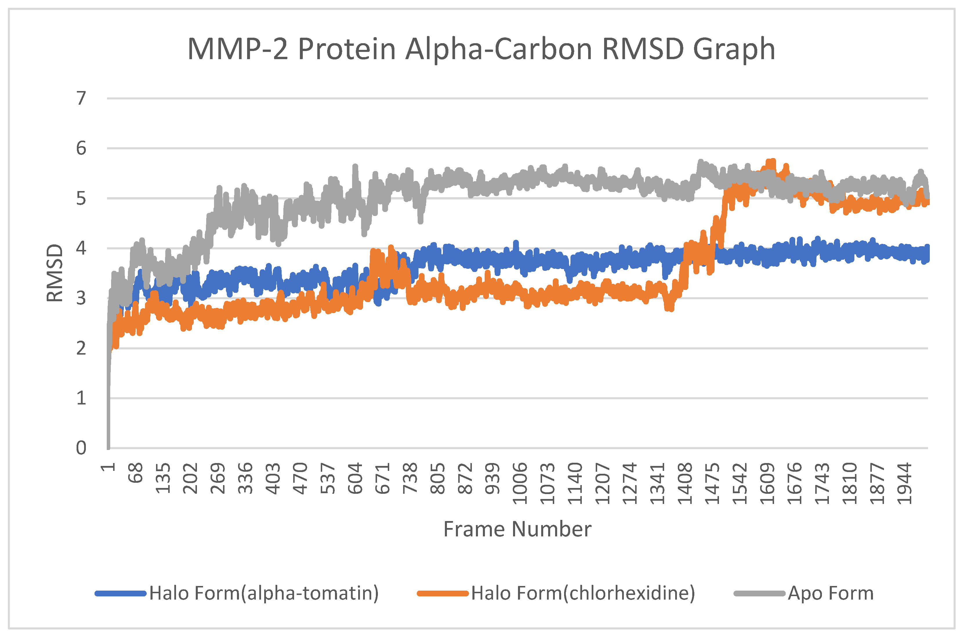
MMP-2: The apo form’s RMSD values increased sharply since the beginning of the simulation. This rise then reached a plateau level. A comparison of the apo-form MMP-2 graphics and the halo-form MMP-2 graphics is given in Figure 6. It is shown that the RMSD value of the complex formed by the a-tomatine compound with MMP-2 remained under the apo form graphic throughout all frames. This means that, when a-tomatine binds to the MMP-2 protein, it changes conformation, that is, prevents the enzyme from performing its normal function. This effect was also seen in the chlorhexidine compound, but the MMP-2 protein structure to which the chlorhexidine compound was attached overlapped with the apo form graph after the simulation, that is, when it came to the final frames. This situation shows that a-tomatine has more inhibitory potential when the capacity of the a-tomatine and chlorhexidine compounds to inhibit MMP-2 enzyme is compared.
MMP-8: In the MMP-8 protein structure RMSD plot, the apo form was above both the halo form lines. a-Tomatine mostly intersected with the apo form. a-Tomatine is, therefore, less restricted than the chlorhexidine compound. This means that the movement of the MMP-8 protein structure in the solvent medium was more inhibited by the chlorhexidine compound. The halo form formed by the chlorhexidine compound remained below all lines, meaning that the MMP-8 protein structure changed conformation less. The halo form formed by the a-tomatine compound was, on average, lower than the apo form. This is an indication that the MMP-8 protein is inhibited by a-tomatine. The conformational change caused by the a-tomatine and chlorhexidine compounds in the MMP-8 protein structure is shown as an RMSD graph in Figure 7.
MMP-9: The conformation changes caused by a-tomatine and chlorhexidine compounds in the MMP-9 protein structure are shown in Figure 7 as both halo forms and apo form graphs. The graph gives us mixed signals. This is mainly due to the fact that the chlorhexidine compound could not remain embedded and bound to the MMP-9 protein structure in the molecular dynamics simulation. The chlorhexidine compound was not effective in the long-term activation of biological systems because the compound left the protein structure in a short time. The a-tomatine compound, unlike the chlorhexidine compound, remained bound throughout the molecular dynamics simulation by coordinating with the zinc atom with the MMP-9 protein structure. It was seen that the a-tomatine halo form graph, which was always above the apo form line in the RMSD graph, did not limit the conformation of the protein structure. Therefore, the potential for partial inhibition via a-tomatine may be in question when the simulation is combined with three-dimensional image analysis. However, the chlorhexidine compound had no effect on the MMP-9 protein (Figure 8).

4.4. Root-Mean-Square Fluctuation (RMSF) Analysis

MMP-2: During the comparison of the halo-form complexes and apo-form complexes of the MMP-2 protein structure with each other, the fluctuation of the residues of the active center, which is the center that does the work in the protein structure, was examined in detail. Large changes occurred in the fluctuation of the area between residue indexes 66 and 81, the region where a-tomatine and chlorhexidine compounds bind to the protein structure. The fluctuation of the apo form was higher than all the other fluctuations, as shown in Figure 9. This fluctuation shows that the protein structure was functional and active. However, with a-tomatine binding, great damping of the fluctuation in this region occurred. This means that a-tomatine binding to MMP-2 inactivated the center that did the work in the enzyme. Upon the binding of the chlorhexidine compound to the protein structure, the fluctuation of this region increased to apo-form levels but did not show a clear increase in fluctuation. The lack of an increase above the apo form graph indicates that the chlorhexidine compound achieves partial inhibition in the MMP-2 protein structure.
MMP-8: The fluctuating parts of the MMP-8 protein structure are distributed over a large area. The fluctuating areas are enclosed in the red quadrilateral in Figure 9. These parts contain residue indexes, especially between 121 and 141. The residue indexes between 121 and 141 contain the residues of the active center, the part of the MMP-8 enzyme that does the work. The quenching of the active center residues indicates that the enzyme had become inoperable due to the effect of the a-tomatine and chlorhexidine compounds. The fluctuations in the MMP-8 protein structure are shown in Figure 10, with a-tomatine binding in the blue line and chlorhexidine binding in the orange line. It was seen that this was well below the apo form line. This indicates that both compounds have a significant dampening effect on the fluctuations of the active center residues. The other fluctuating regions of the MMP-8 protein structure are distant from the active center and, therefore, have no direct effect on enzyme functioning.
MMP-9: The MMP-9 enzyme is a globular protein and, therefore, fluctuates very little in all regions. The active center of the MMP-9 enzyme protein consists of the region containing residue indexes 96-116, and the fluctuation in this region directly affects the enzyme functioning. A lower fluctuation in this region than the apo form indicates inhibition, while a higher fluctuation than the apo form indicates that there is no significant change in enzyme activity. In the region enclosed in the red quadrilateral in Figure 11, it was seen that the a-tomatine and chlorhexidine compounds were located well above the apo form graph, that is, the fluctuation increased with the binding of the compounds to the protein structure. In the molecular dynamics simulation of the chlorhexidine compound, it was seen that it was completely separated from the protein structure, and it was already seen that it had no significant effect on the MMP-9 protein structure. It is also clear from the RMSF graph that the chlorhexidine compound could not cause the enzyme activity to stop by causing high fluctuation. It is possible to say the same thing for a-tomatine in Figure 11. In the molecular dynamics simulation of the a-tomatine compound, only the zinc atom interacted, not the other sites. The interaction with the zinc atom alone could not prevent the fluctuation of residues around the active center in the enzyme activity. This means that a-tomatine would achieve partial inhibition or no inhibition at all. Formun Üstü.
One limitation of this study is the use of a single adhesive system (three-step etch-and-rinse). The effectiveness of the material should be assessed with different types of adhesive agents. Additionally, the discussion of how a-tomatine interacts with adhesive systems is a subject of interest. It should be addressed through chromatographic and electrochemical determination methods to investigate whether it forms bonds with the carboxylic or phosphate groups of adhesive materials. Moreover, further research is needed involving the application of a-tomatine in different solvents, possibly in a solvent other than DMSO. Molecular docking or dynamic analyses of DMSO could be conducted. Furthermore, various methods not included in this study, such as in situ zymography and SDS Page, can be employed to detect MMP inhibition activity with samples obtained from dentin tissue for a-tomatine. Prolonged aging procedures could be applied, and studies on different types of dentin surfaces (e.g., caries-affected) could be conducted. Lastly, the efficacy of a-tomatine should be evaluated in vivo or in cell studies to assess its impact on living tissues such as odontoblasts.

5. Conclusions

According to the results obtained, a-tomatine and chlorhexidine compounds were found to achieve potentially strong inhibition of the MMP-2 enzyme. For the MMP-8 enzyme, it was determined that a-tomatine achieves a potential strong inhibition and the chlorhexidine compound achieves a potential inhibition or partial inhibition. For the MMP-9 enzyme, it was determined that the chlorhexidine compound caused no inhibition and had no potential to have any effect on enzyme functioning. The a-tomatine compound was found to achieve either potentially no inhibition or potentially partial inhibition of the MMP-9 protein structure. According to molecular dynamic analysis regarding the affinity for MMP enzymes, it is anticipated that the system is in equilibrium, and the existing affinities will be preserved over time. The finding that a-tomatine exhibited higher bond strength values in all subgroups compared to chlorhexidine and the control group (regardless of dentine type) in the in vitro aspect of this study corroborates the findings of the in silico method. In line with the findings of this study, it can be said that the tomatine material that was found to increase bond strength showed better results than chlorhexidine.

Author Contributions

M.K.U.: conceptualization, methodology, data curation, writing—original draft, and reviewing and editing. A.B.O.: formal analysis, methodology, data curation, writing—original draft, and reviewing and editing. S.B.: data curation, methodology, and resources. E.Y.: methodology, validation, visualization, supervision, project administration, and reviewing and editing. All authors have read and agreed to the published version of the manuscript.

Funding

The present work was supported by the Research Fund of Istanbul University, Project No. 32045.

Institutional Review Board Statement

This article does not contain any studies with human participants or animals performed by any of the authors. All procedures performed involving human tissues were in accordance with the ethical standards of the institutional and/or national research committee and with the Declaration of Helsinki and its later amendments or comparable ethical standards. The study was approved by the Ethics Committee of the Istanbul University Faculty of Dentistry Clinical Research (File no: 2017/66).

Informed Consent Statement

Written informed consent has been obtained from patients to publish this paper.

Data Availability Statement

Not applicable.

Conflicts of Interest

The authors do not have any financial interest in the companies whose materials are included in this article.

References

  1. Buzalaf, M.A.R.; Kato, M.T.; Hannas, A.R. The Role of Matrix Metalloproteinases in Dental Erosion. Adv. Dent. Res. 2012, 24, 72–76. [Google Scholar] [CrossRef] [PubMed]
  2. Viana, Í.E.L.; Alania, Y.; Feitosa, S.; Borges, A.B.; Braga, R.R.; Scaramucci, T. Bioactive Materials Subjected to Erosion/Abrasion and Their Influence on Dental Tissues. Oper. Dent. 2020, 45, E114–E123. [Google Scholar] [CrossRef] [PubMed]
  3. Tezvergil-Mutluay, A.; Pashley, D.; Mutluay, M.M. Long-Term Durability of Dental Adhesives. Curr. Oral. Health Rep. 2015, 2, 174–181. [Google Scholar] [CrossRef]
  4. Carrilho, M.R.O.; Carvalho, R.M.; de Goes, M.F.; di Hipólito, V.; Geraldeli, S.; Tay, F.R.; Pashley, D.H.; Tjäderhane, L. Chlorhexidine Preserves Dentin Bond In Vitro. J. Dent. Res. 2007, 86, 90–94. [Google Scholar] [CrossRef] [PubMed]
  5. Breschi, L.; Martin, P.; Mazzoni, A.; Nato, F.; Carrilho, M.; Tjäderhane, L.; Visintini, E.; Cadenaro, M.; Tay, F.R.; Dorigo, E.D.S. Use of a Specific MMP-Inhibitor (Galardin) for Preservation of Hybrid Layer. Dent. Mater. 2010, 26, 571–578. [Google Scholar] [CrossRef]
  6. Do Amaral, S.F.; Scaffa, P.M.C.; Rodrigues, R.D.S.; Nesadal, D.; Marques, M.M.; Nogueira, F.N.; Sobral, M.A.P. Dynamic Influence of PH on Metalloproteinase Activity in Human Coronal and Radicular Dentin. Caries Res. 2018, 52, 113–118. [Google Scholar] [CrossRef]
  7. Gendron, R.; Grenier, D.; Sorsa, T.; Mayrand, D. Inhibition of the Activities of Matrix Metalloproteinases 2, 8, and 9 by Chlorhexidine. Clin. Diagn. Lab. Immunol. 1999, 6, 437–439. [Google Scholar] [CrossRef]
  8. Siqueira, F.S.F.; Cardenas, A.M.; Ocampo, J.B.; Hass, V.; Bandeca, M.C.; Gomes, J.C.; Reis, A.; Loguercio, A.D. Bonding Performance of Universal Adhesives to Eroded Dentin. J. Adhes. Dent. 2018, 20, 121–132. [Google Scholar] [CrossRef]
  9. Yelken, B.Ö.; Balcı, T.; Süslüer, S.Y.; Kayabaşı, Ç.; Avcı, Ç.B.; Kırmızıbayrak, P.B.; Gündüz, C. The Effect of Tomatine on Metastasis Related Matrix Metalloproteinase (MMP) Activities in Breast Cancer Cell Model. Gene 2017, 627, 408–411. [Google Scholar] [CrossRef]
  10. Shi, M.-D.; Shih, Y.-W.; Lee, Y.-S.; Cheng, Y.-F.; Tsai, L.-Y. Suppression of 12-O-Tetradecanoylphorbol-13-Acetate-Induced MCF-7 Breast Adenocarcinoma Cells Invasion/Migration by α-Tomatine through Activating PKCα/ERK/NF-ΚB-Dependent MMP-2/MMP-9 Expressions. Cell Biochem. Biophys. 2013, 66, 161–174. [Google Scholar] [CrossRef]
  11. Maravic, T.; Breschi, L.; Paganelli, F.; Bonetti, G.A.; Martina, S.; Di Giorgio, G.; Bossù, M.; Polimeni, A.; Checchi, V.; Generali, L. Endogenous Enzymatic Activity of Primary and Permanent Dentine. Materials 2021, 14, 4043. [Google Scholar] [CrossRef]
  12. Ahmet, B.S.O.; Seseogullari-Dirihan, R.; Tezvergil-Mutluay, A. Activation of Matrix-Bound Endogenous Proteases by Self-Etch Adhesives. Dent. Mater. J. 2020, 39, 1044–1049. [Google Scholar] [CrossRef] [PubMed]
  13. Morris, G.M.; Lim-Wilby, M. Molecular Docking. Methods Mol. Biol. 2008, 443, 365–382. [Google Scholar]
  14. Pagadala, N.S.; Syed, K.; Tuszynski, J. Software for Molecular Docking: A Review. Biophys. Rev. 2017, 9, 91–102. [Google Scholar] [CrossRef] [PubMed]
  15. Guedes, I.A.; de Magalhães, C.S.; Dardenne, L.E. Receptor-Ligand Molecular Docking. Biophys. Rev. 2014, 6, 75–87. [Google Scholar] [CrossRef]
  16. Bakir Boga, O.; Bugra Ortaakarsu, A.; Kadırustaoglu, B.; Basaran Kurbanoglu, E. Phytochemical Profiling, in Vitro Biological Activities and in Silico Molecular Docking Studies of the Crude Extract of Crambe Orientalis, an Endemic Plant in Turkey. Chem. Biodivers. 2023, 20, e202201142. [Google Scholar] [CrossRef] [PubMed]
  17. Tunc, T.; Ortaakarsu, A.B.; Hatipoglu, S.M.; Kazancı, U.; Karabocek, S.; Karabocek, N.; Dege, N.; Karacan, N. New Schiff Bases with a 2,6-Bis(2-Aminophenylthio)Pyridine Moiety Acting as Glutathione Reductase Activator and Inhibitors: Synthesis and Molecular Docking Studies. J. Mol. Struct. 2022, 1254, 132299. [Google Scholar] [CrossRef]
  18. Shoichet, B.K.; McGovern, S.L.; Wei, B.; Irwin, J.J. Lead Discovery Using Molecular Docking. Curr. Opin. Chem. Biol. 2002, 6, 439–446. [Google Scholar] [CrossRef]
  19. Hollingsworth, S.A.; Dror, R.O. Molecular Dynamics Simulation for All. Neuron 2018, 99, 1129–1143. [Google Scholar] [CrossRef]
  20. Carvalho, C.; Fernandes, F.P.; da Freitas, V.P.; França, F.M.G.; Basting, R.T.; Turssi, C.P.; do Amaral, F.L.B. Effect of Green Tea Extract on Bonding Durability of an Etch-and-Rinse Adhesive System to Caries-Affected Dentin. J. Appl. Oral. Sci. 2016, 24, 211–217. [Google Scholar] [CrossRef]
  21. Zimmerli, B.; De Munck, J.; Lussi, A.; Lambrechts, P.; Van Meerbeek, B. Long-Term Bonding to Eroded Dentin Requires Superficial Bur Preparation. Clin. Oral. Investig. 2012, 16, 1451–1461. [Google Scholar] [CrossRef]
  22. Lee, S.T.; Wong, P.F.; Cheah, S.C.; Mustafa, M.R. Alpha-Tomatine Induces Apoptosis and Inhibits Nuclear Factor-Kappa B Activation on Human Prostatic Adenocarcinoma PC-3 Cells. PLoS ONE 2011, 6, e18915. [Google Scholar] [CrossRef]
  23. Schrödinger Release 2023-2; Maestro, Schrödinger, LLC: New York, NY, USA, 2023.
  24. Schrödinger Schrödinger Release 2023-2. In Maestro-Desmond Interoperability Tools, Desmond Molecular Dynamics System; Maestro, Schrödinger, LLC: New York, NY, USA, 2023.
  25. Baliga, S.; Muglikar, S.; Kale, R. Salivary PH: A Diagnostic Biomarker. J. Indian. Soc. Periodontol. 2013, 17, 461–465. [Google Scholar] [CrossRef]
  26. Schrödinger LigPrep. Schrödinger Release 2023-2; Schrödinger, LLC: New York, NY, USA, 2023. [Google Scholar]
  27. Schrödinger Epik. Schrödinger Release 2023-2; Schrödinger, LLC: New York, NY, USA, 2023. [Google Scholar]
  28. Greenwood, J.R.; Calkins, D.; Sullivan, A.P.; Shelley, J.C. Towards the Comprehensive, Rapid, and Accurate Prediction of the Favorable Tautomeric States of Drug-like Molecules in Aqueous Solution. J. Comput. Aided Mol. Des. 2010, 24, 591–604. [Google Scholar] [CrossRef]
  29. Roos, K.; Wu, C.; Damm, W.; Reboul, M.; Stevenson, J.M.; Lu, C.; Dahlgren, M.K.; Mondal, S.; Chen, W.; Wang, L.; et al. OPLS3e: Extending Force Field Coverage for Drug-Like Small Molecules. J. Chem. Theory Comput. 2019, 15, 1863–1874. [Google Scholar] [CrossRef] [PubMed]
  30. Schrödinger, L.L.C. Schrödinger, L.L.C. Schrödinger Release 2023-2: Protein Preparation Wizard. In Epic Impact Prime Schrödinger; Schrödinger, LLC: New York, NY, USA, 2023. [Google Scholar]
  31. Feng, Y.; Likos, J.J.; Zhu, L.; Woodward, H.; Munie, G.; Mcdonald, J.J.; Stevens, A.M.; Howard, C.P.; De Crescenzo, G.A.; Welsch, D.; et al. Solution Structure and Backbone Dynamics of the Catalytic Domain of Matrix Metalloproteinase-2 Complexed with a Hydroxamic Acid Inhibitor. Biochim. Biophys. Acta BBA Proteins Proteom. 2002, 1598, 10–23. [Google Scholar] [CrossRef] [PubMed]
  32. Campestre, C.; Agamennone, M.; Tortorella, P.; Preziuso, S.; Biasone, A.; Gavuzzo, E.; Pochetti, G.; Mazza, F.; Hiller, O.; Tschesche, H.; et al. N-Hydroxyurea as Zinc Binding Group in Matrix Metalloproteinase Inhibition: Mode of Binding in a Complex with MMP-8. Bioorg. Med. Chem. Lett. 2006, 16, 20–24. [Google Scholar] [CrossRef] [PubMed]
  33. Tochowicz, A.; Maskos, K.; Huber, R.; Oltenfreiter, R.; Dive, V.; Yiotakis, A.; Zanda, M.; Bode, W.; Goettig, P. Crystal Structures of MMP-9 Complexes with Five Inhibitors: Contribution of the Flexible Arg424 Side-Chain to Selectivity. J. Mol. Biol. 2007, 371, 989–1006. [Google Scholar] [CrossRef]
  34. Olsson, M.H.M.; SØndergaard, C.R.; Rostkowski, M.; Jensen, J.H. PROPKA3: Consistent Treatment of Internal and Surface Residues in Empirical p K a Predictions. J. Chem. Theory Comput. 2011, 7, 525–537. [Google Scholar] [CrossRef]
  35. Users Manual. Schrödinger Release 2023-2: Glide, Schrödinger, LLC: New York, NY, 2023. In Schrödinger Release 2023-2: LigPrep; Schrödinger, LLC: New York, NY, USA, 2023.
  36. Gopal, S.M.; Kuhn, A.B.; Schäfer, L.V. Systematic Evaluation of Bundled SPC Water for Biomolecular Simulations. Phys. Chem. Chem. Phys. 2015, 17, 8393–8406. [Google Scholar] [CrossRef]
  37. Mark, P.; Nilsson, L. Structure and Dynamics of the TIP3P, SPC, and SPC/E Water Models at 298 K. J. Phys. Chem. A 2001, 105, 9954–9960. [Google Scholar] [CrossRef]
  38. Desmond Molecular Dynamics System, D.E. Shaw Research, New York, NY, 2023; Maestro-Desmond Interoperability Tools, Schrödinger: New York, NY, USA, 2023.
  39. Evans, D.J.; Holian, B.L. The Nose-Hoover Thermostat. J. Chem. Phys. 1985, 83, 4069–4074. [Google Scholar] [CrossRef]
  40. Martyna, G.J.; Tobias, D.J.; Klein, M.L. Constant Pressure Molecular Dynamics Algorithms. J. Chem. Phys. 1994, 101, 4177–4189. [Google Scholar] [CrossRef]
  41. Friesner, R.A.; Murphy, R.B.; Repasky, M.P.; Frye, L.L.; Greenwood, J.R.; Halgren, T.A.; Sanschagrin, P.C.; Mainz, D.T. Extra Precision Glide: Docking and Scoring Incorporating a Model of Hydrophobic Enclosure for Protein-Ligand Complexes. J. Med. Chem. 2006, 49, 6177–6196. [Google Scholar] [CrossRef]
  42. Hevener, K.E.; Zhao, W.; Ball, D.M.; Babaoglu, K.; Qi, J.; White, S.W.; Lee, R.E. Validation of Molecular Docking Programs for Virtual Screening against Dihydropteroate Synthase. J. Chem. Inf. Model. 2009, 49, 444–460. [Google Scholar] [CrossRef] [PubMed]
  43. Cruz, J.B.; Bonini, G.; Lenzi, T.L.; Imparato, J.C.P.; Raggio, D.P. Bonding Stability of Adhesive Systems to Eroded Dentin. Braz. Oral. Res. 2015, 29, S1806-83242015000100290. [Google Scholar] [CrossRef] [PubMed]
  44. Forgerini, T.V.; Ribeiro, J.F.; Rocha, R.O.; Soares, F.Z.M.; Lenzi, T.L. Role of Etching Mode on Bonding Longevity of a Universal Adhesive to Eroded Dentin. J. Adhes. Dent. 2017, 19, 69–75. [Google Scholar] [CrossRef]
  45. Shellis, R.P.; Ganss, C.; Ren, Y.; Zero, D.T.; Lussi, A. Methodology and Models in Erosion Research: Discussion and Conclusions. Caries Res. 2011, 45, 69–77. [Google Scholar] [CrossRef]
  46. Zero, D.T. Etiology of Dental Erosion-Extrinsic Factors. Eur. J. Oral. Sci. 1996, 104, 162–177. [Google Scholar] [CrossRef]
  47. Murrell, S.; Marshall, T.A.; Moynihan, P.J.; Qian, F.; Wefel, J.S. Comparison of in Vitro Erosion Potentials between Beverages Available in the United Kingdom and the United States. J. Dent. 2010, 38, 284–289. [Google Scholar] [CrossRef]
  48. Hannig, C.; Hamkens, A.; Becker, K.; Attin, R.; Attin, T. Erosive Effects of Different Acids on Bovine Enamel: Release of Calcium and Phosphate in Vitro. Arch. Oral. Biol. 2005, 50, 541–552. [Google Scholar] [CrossRef] [PubMed]
  49. Lussi, A. Erosive Tooth Wear—A Multifactorial Condition of Growing Concern and Increasing Knowledge. Dental Erosion: From Diagnosis to Therapy. Monogr. Oral. Sci. 2006, 20, 1–8. [Google Scholar]
  50. Young, A.; Tenuta, L.M.A. Initial Erosion Models. Caries Res. 2011, 45, 33–42. [Google Scholar] [CrossRef] [PubMed]
  51. de Siqueira, F.S.F.; Hilgemberg, B.; Araujo, L.C.R.; Hass, V.; Bandeca, M.C.; Reis, A.; Gomes, J.C.; Cardenas, A.F.M.; Loguercio, A.D. Effect of Phosphoric Acid Containing MMP-Inactivator on the Properties of Resin Bonding to Eroded Dentin. J. Adhes. Dent. 2019, 21, 149–158. [Google Scholar] [CrossRef]
  52. de Siqueira, F.S.F.; Hilgemberg, B.; Araujo, L.C.R.; Hass, V.; Bandeca, M.C.; Gomes, J.C.; Reis, A.; Loguercio, A.D.; Cardenas, A.F.M. Improving Bonding to Eroded Dentin by Using Collagen Cross-Linking Agents: 2 Years of Water Storage. Clin. Oral. Investig. 2020, 24, 809–822. [Google Scholar] [CrossRef] [PubMed]
  53. Machado, C.M.; Zamuner, A.C.; Modena, K.C.D.S.; Ishikiriama, S.K.; Wang, L. How Erosive Drinks and Enzyme Inhibitors Impact Bond Strength to Dentin. Braz. Oral. Res. 2015, 29, S1806-83242015000100300. [Google Scholar] [CrossRef]
  54. Francisconi-Dos-Rios, L.F.; Casas-Apayco, L.C.; Calabria, M.P.; Francisconi, P.A.; Borges, A.F.; Wang, L. Role of Chlorhexidine in Bond Strength to Artificially Eroded Dentin over Time. J. Adhes. Dent. 2015, 17, 133–139. [Google Scholar]
  55. Francisconi-Dos-Rios, L.F.; Calabria, M.P.; Casas-Apayco, L.C.; Honório, H.M.; De Oliveira Carrilho, M.R.; Pereira, J.C.; Wang, L. Chlorhexidine Does Not Improve but Preserves Bond Strength to Eroded Dentin. Am. J. Dent. 2015, 28, 28–32. [Google Scholar]
  56. Deari, S.; Wegehaupt, F.J.; Tauböck, T.T.; Attin, T. Influence of Different Pretreatments on the Microtensile Bond Strength to Eroded Dentin. J. Adhes. Dent. 2017, 19, 147–155. [Google Scholar]
  57. Krithi, B.; Vidhya, S.; Mahalaxmi, S. Microshear Bond Strength of Composite Resin to Demineralized Dentin after Remineralization with Sodium Fluoride, CPP-ACP and NovaMin Containing Dentifrices. J. Oral. Biol. Craniofac. Res. 2020, 10, 122–127. [Google Scholar] [CrossRef]
  58. Loguercio, A.D.; Stanislawczuk, R.; Polli, L.G.; Costa, J.A.; Michel, M.D.; Reis, A. Influence of Chlorhexidine Digluconate Concentration and Application Time on Resin-Dentin Bond Strength Durability. Eur. J. Oral. Sci. 2009, 117, 587–596. [Google Scholar] [CrossRef]
  59. Ozan, G.; Sar Sancakli, H.; Yucel, T. Effect of Black Tea and Matrix Metalloproteinase Inhibitors on Eroded Dentin in Situ. Microsc. Res. Technol. 2020, 83, 834–842. [Google Scholar] [CrossRef]
  60. Zhou, J.; Tan, J.; Chen, L.; Li, D.; Tan, Y. The Incorporation of Chlorhexidine in a Two-Step Self-Etching Adhesive Preserves Dentin Bond in Vitro. J. Dent. 2009, 37, 807–812. [Google Scholar] [CrossRef] [PubMed]
  61. Ozsoy, A.; Erdemir, U.; Yucel, T.; Yildiz, E. Effects of Cavity Disinfectants on Bond Strength of an Etch-and-Rinse Adhesive to Water- or Ethanol-Saturated Sound and Caries-Affected Dentin. J. Adhes. Sci. Technol. 2015, 29, 2551–2564. [Google Scholar] [CrossRef]
  62. de Menezes, L.R.; da Silva, E.O.; Maurat da Rocha, L.V.; Ferreira Barbosa, I.; Rodrigues Tavares, M. The Use of Clays for Chlorhexidine Controlled Release as a New Perspective for Longer Durability of Dentin Adhesion. J. Mater. Sci. Mater. Med. 2019, 30, 132. [Google Scholar] [CrossRef] [PubMed]
  63. Hamdan-Nassar, T.; Bellot-Arcís, C.; Paredes-Gallardo, V.; García-Sanz, V.; Pascual-Moscardó, A.; Almerich-Silla, J.M.; Montiel-Company, J.M. Effect of 2% Chlorhexidine Following Acid Etching on Microtensile Bond Strength of Resin Restorations: A Meta-Analysis. Medicine 2019, 55, 769. [Google Scholar] [CrossRef]
  64. de Castro, F.L.A.; de Andrade, M.F.; Duarte Júnior, S.L.L.; Vaz, L.G.; Ahid, F.J.M. Effect of 2% Chlorhexidine on Microtensile Bond Strength of Composite to Dentin. J. Adhes. Dent. 2003, 5, 129–138. [Google Scholar]
  65. Dalkilic, E.E.; Arisu, H.D.; Kivanc, B.H.; Uctasli, M.B.; Omurlu, H. Effect of Different Disinfectant Methods on the Initial Microtensile Bond Strength of a Self-Etch Adhesive to Dentin. Lasers Med. Sci. 2012, 27, 819–825. [Google Scholar] [CrossRef] [PubMed]
  66. Lenzi, T.L.; Soares, F.Z.M.S.; de Rocha, R.O. Degradation of Resin-Dentin Bonds of Etch-and-Rinse Adhesive System to Primary and Permanent Teeth. Braz. Oral. Res. 2012, 26, 511–515. [Google Scholar] [CrossRef]
  67. Soares, C.J.; Pereira, C.A.; Pereira, J.C.; Santana, F.R.; Do Prado, C.J. Effect of Chlorhexidine Application on Microtensile Bond Strength to Dentin. Oper. Dent. 2008, 33, 183–188. [Google Scholar] [CrossRef]
  68. Sodhi, R.N.S.; Grad, H.A.; Smith, D.C. Examination by x-Ray Photoelectron Spectroscopy of the Adsorption of Chlorhexidine on Hydroxyapatite. J. Dent. Res. 1992, 71, 1493–1497. [Google Scholar] [CrossRef] [PubMed]
  69. Perdigao, J.; Swift, E.J.; Denehy, G.E.; Wefel, J.S.; Donly, K.J. In Vitro Bond Strengths and SEM Evaluation of Dentin Bonding Systems to Different Dentin Substrates. J. Dent. Res. 1994, 73, 44–55. [Google Scholar] [CrossRef] [PubMed]
  70. Davies, J.N.; Hobson, G.E. The Constituents of Tomato Fruit—The Influence of Environment, Nutrition, and Genotype. C. R. C. Crit. Rev. Food Sci. Nutr. 1981, 15, 205–280. [Google Scholar] [CrossRef] [PubMed]
  71. Shieh, J.-M.; Cheng, T.-H.; Shi, M.-D.; Wu, P.-F.; Chen, Y.; Ko, S.-C.; Shih, Y.-W. α-Tomatine Suppresses Invasion and Migration of Human Non-Small Cell Lung Cancer NCI-H460 Cells Through Inactivating FAK/PI3K/Akt Signaling Pathway and Reducing Binding Activity of NF-ΚB. Cell Biochem. Biophys. 2011, 60, 297–310. [Google Scholar] [CrossRef] [PubMed]
  72. Huang, H. Matrix Metalloproteinase-9 (MMP-9) as a Cancer Biomarker and MMP-9 Biosensors: Recent Advances. Sensors 2018, 18, 3249. [Google Scholar] [CrossRef]
  73. Lee, K.R.; Kozukue, N.; Han, J.S.; Park, J.H.; Chang, E.Y.; Baek, E.J.; Chang, J.S.; Friedman, M. Glycoalkaloids and Metabolites Inhibit the Growth of Human Colon (HT29) and Liver (HepG2) Cancer Cells. J. Agric. Food Chem. 2004, 52, 2832–2839. [Google Scholar] [CrossRef] [PubMed]
  74. Sucha, L.; Hroch, M.; Rezacova, M.; Rudolf, E.; Havelek, R.; Sispera, L.; Cmielova, J.; Kohlerova, R.; Bezrouk, A.; Tomsik, P. The Cytotoxic Effect of α-Tomatine in MCF-7 Human Adenocarcinoma Breast Cancer Cells Depends on Its Interaction with Cholesterol in Incubation Media and Does Not Involve Apoptosis Induction. Oncol. Rep. 2013, 30, 2593–2602. [Google Scholar] [CrossRef]
  75. Benson, C.S.; Babu, S.D.; Radhakrishna, S.; Selvamurugan, N.; Sankar, B.R. Expression of Matrix Metalloproteinases in Human Breast Cancer Tissues. Dis. Mrk. 2013, 34, 395–405. [Google Scholar] [CrossRef]
  76. Shih, Y.W.; Shieh, J.M.; Wu, P.F.; Lee, Y.C.; Chen, Y.Z.; Chiang, T.A. α-Tomatine Inactivates PI3K/Akt and ERK Signaling Pathways in Human Lung Adenocarcinoma A549 Cells: Effect on Metastasis. Food Chem. Toxicol. 2009, 47, 1985–1995. [Google Scholar] [CrossRef]
  77. Chao, M.W.; Chen, C.H.; Chang, Y.L.; Teng, C.M.; Pan, S.L. α-Tomatine-Mediated Anti-Cancer Activity In Vitro and In Vivo through Cell Cycle- and Caspase-Independent Pathways. PLoS ONE 2012, 7, e44093. [Google Scholar] [CrossRef]
  78. Thorne, H.V.; Clarke, G.F.; Skuce, R. The Inactivation of Herpes Simplex Virus by Some Solanaceae Glycoalkaloids. Antivir. Res. 1985, 5, 335–343. [Google Scholar] [CrossRef] [PubMed]
  79. Friedman, M. Tomato Glycoalkaloids: Role in the Plant and in the Diet. J. Agric. Food Chem. 2002, 50, 5751–5780. [Google Scholar] [CrossRef] [PubMed]
  80. Simons, V.; Morrissey, J.P.; Latijnhouwers, M.; Csukai, M.; Cleaver, A.; Yarrow, C.; Osbourn, A. Dual Effects of Plant Steroidal Alkaloids on Saccharomyces Cerevisiae. Antimicrob. Agents Chemother. 2006, 50, 2732–2740. [Google Scholar] [CrossRef]
  81. Collares, F.M.; Rodrigues, S.B.; Leitune, V.C.; Celeste, R.K.; Borba de Araújo, F.; Samuel, S.M.; Borba de Araujo, F.; Samuel, S.M. Chlorhexidine Application in Adhesive Procedures: A Meta-Regression Analysis. J. Adhes. Dent. 2013, 15, 11–18. [Google Scholar] [CrossRef]
  82. Zhao, B.; Zhou, B.; Bao, L.; Yang, Y.; Guo, K. Alpha-Tomatine Exhibits Anti-Inflammatory Activity in Lipopolysaccharide-Activated Macrophages. Inflammation 2015, 38, 1769–1776. [Google Scholar] [CrossRef] [PubMed]
  83. Milner, S.E.; Brunton, N.P.; Jones, P.W.; O’Brien, N.M.; Collins, S.G.; Maguire, A.R. Bioactivities of Glycoalkaloids and Their Aglycones from Solanum Species. J. Agric. Food Chem. 2011, 59, 3454–3484. [Google Scholar] [CrossRef] [PubMed]
  84. Chowański, S.; Adamski, Z.; Marciniak, P.; Rosiński, G.; Büyükgüzel, E.; Büyükgüzel, K.; Falabella, P.; Scrano, L.; Ventrella, E.; Lelario, F.; et al. A Review of Bioinsecticidal Activity of Solanaceae Alkaloids. Toxins 2016, 8, 60. [Google Scholar] [CrossRef]
  85. Zimmerley, M.; McClure, R.A.; Choi, B.; Potma, E.O. Following Dimethyl Sulfoxide Skin Optical Clearing Dynamics with Quantitative Nonlinear Multimodal Microscopy. Appl. Opt. 2009, 48, D79–D87. [Google Scholar] [CrossRef]
  86. Tjäderhane, L.; Mehtälä, P.; Scaffa, P.; Vidal, C.; Pääkkönen, V.; Breschi, L.; Hebling, J.; Tay, F.R.; Nascimento, F.D.; Pashley, D.H.; et al. The Effect of Dimethyl Sulfoxide (DMSO) on Dentin Bonding and Nanoleakage of Etch-and-Rinse Adhesives. Dent. Mater. 2013, 29, 1055–1062. [Google Scholar] [CrossRef]
  87. Stape, T.H.S.; Tjäderhane, L.; Marques, M.R.; Aguiar, F.H.B.; Martins, L.R.M. Effect of Dimethyl Sulfoxide Wet-Bonding Technique on Hybrid Layer Quality and Dentin Bond Strength. Dent. Mater. 2015, 31, 676–683. [Google Scholar] [CrossRef]
  88. Marren, K. Dimethyl Sulfoxide: An Effective Penetration Enhancer for Topical Administration of NSAIDs. Phys. Sport. 2011, 39, 75–82. [Google Scholar] [CrossRef] [PubMed]
  89. Stape, T.H.S.; Tjäderhane, L.; Tezvergil-Mutluay, A.; Yanikian, C.R.F.; Szesz, A.L.; Loguercio, A.D.; Martins, L.R.M. Dentin Bond Optimization Using the Dimethyl Sulfoxide-Wet Bonding Strategy: A 2-Year in Vitro Study. Dent. Mater. 2016, 32, 1472–1481. [Google Scholar] [CrossRef] [PubMed]
  90. Tekbas Atay, M.; Seseogullari-Dirihan, R.; Mutluay, M.M.; Tezvergil-Mutluay, A. Long-Term Effect of Curcuminoid Treatment on Resin-to-Dentin Bond Strength. Eur. J. Oral. Sci. 2022, 130, e12837. [Google Scholar] [CrossRef] [PubMed]
  91. de Mello, R.M.M.; Alcântara, B.A.R.; França, F.M.G.; do Amaral, F.L.B.; Basting, R.T. Dimethyl Sulfoxide Dentin Pretreatments Do Not Improve Bonding of a Universal Adhesive in Etch-and-Rinse or Self-Etch Modes. J. Adhes. Dent. 2022, 24, 49–56. [Google Scholar] [CrossRef]
Figure 1. The chemical structure of a-tomatine and chlorhexidine.
Applsci 13 10322 g001
Figure 2. Validation image and RMSD values of MMP-2 and MMP-8 enzymes. The red-colored ligand is the molecular docking ligand. The green-colored ligand is the position in the crystal structure.
Applsci 13 10322 g002
Figure 3. Interactions of a-tomatine and chlorhexidine with the protein structure of MMP-2 enzyme. a-tomatine on top and chlorhexidine on the bottom.
Applsci 13 10322 g003
Figure 4. Interactions of a-tomatine and chlorhexidine with the protein structure of MMP--8 enzyme. a-tomatine on top and chlorhexidine on the bottom.
Applsci 13 10322 g004
Figure 5. Interactions of a-tomatine and chlorhexidine with the protein structure of MMP-9 enzyme. a-tomatine on top and chlorhexidine on the bottom.
Applsci 13 10322 g005
Figure 6. Alpha-carbon RMSD plots of the halo form and apo form of the MMP-2 protein.
Applsci 13 10322 g006
Figure 7. Alpha-carbon RMSD plots of the halo forms and apo form of the MMP-8 protein.
Applsci 13 10322 g007
Figure 8. Alpha-carbon RMSD plots of the halo form and apo form of the MMP-9 protein.
Applsci 13 10322 g008
Figure 9. Alpha-carbon RMSF plots of the halo form and apo form of the MMP-2 protein.
Applsci 13 10322 g009
Figure 10. Alpha-carbon RMSF plots of the halo form and apo form of the MMP-8 protein.
Applsci 13 10322 g010
Figure 11. Alpha-carbon RMSF plots of the halo form and apo form of the MMP-9 protein.
Applsci 13 10322 g011
Table 1. Evaluation of pilot study to calculate the amount and application time of tomatine.
Dentine TypeDifferent Amounts of a-Tomatine *Bond Strength (24-h)
20 s30 s
Mean ± SdMean ± Sd
Sound dentine
(n = 1)
0.75 µM14.07 ± 4.3114.12 ± 6.01
1 µM25.43 ± 8.9625.38 ± 8.48
1.5 µM43.17 ± 7.4042.55 ± 16.86
p0.001 **0.001 **
* According to Shi and Lee et al. [10,22], selecting for a-tomatine’s µM.
Table 2. Content of the materials.
MaterialCompositionManufacturer and Batch Numbers
Citric acidCitric acid monohydrate
C6H8O7H2O
Merck, KGaA, Darmstadt, Germany
(5949-29-1)
Dimethyl sulfoxide
extra pure
Dimethyl sulfoxide
C2H6OS
Merck, Sigma Aldrich, Germany
Saliva0.002 g of ascorbic acid, 0.58 g of NaCl, 0.17 g of CaCl2, 0.16 g of NH4Cl, 1.27 g of KCl, 0.16 g of NaSCN, 0.33 g of KH2PO4, and 0.34 g of Na2HPO4 for 1 LProduced in the lab as artificial
Etching dental gel37.5% phosphoric acid gelKerr, Gel Etchant, Orange, USA
(5887888)
Cavity cleanser2% chlorhexidine digluconateBisco, Inc., Schaumburg, IL, USA
(1900000744)
Optibond FLPrimer: HEMA, PAMM, GPDM, water, ethanol, photoinitiator Adhesive: TEGDMA, DMA, GPDM, HEMA, BIS-GMA, filler, photoinitiatorKerr, Orange, CA, USA
(26684)
FiltekTM Z250Organic matrix: BIS-GMA, UDMA, BIS-EMA
Inorganic matrix: zirconia/silica as a filler, the loading of the inorganic filler (without treatment with silane) was 60% by volume with a particle size in the range of 0.01–3.5 μm
3M ESPE, St Paul, MN, USA
(6020A2)
Tomatinea-tomatine (powdered)PhyProof, PhytoLab GmbH
Dutendorfer, Germany
(89905)
Table 3. The two-way ANOVA results for dentin type and applied agents (tests of between-subjects effects).
Aging TimeSourceFp
24 hMMP inhibitor10.2200.049 *
Type of dentin536.7270.002 **
MMP inhibitor * type of dentin3.6070.037 *
6 monthsMMP inhibitor131.4220.008 **
Type of dentin4236.3520.000 **
MMP inhibitor * type of dentin0.2950.746
* p < 0.05, ** p < 0.01.
Table 4. Distribution of bond strengths based on dentin type, applied agents, and application time.
Dentin SurfaceMMP
Inhibitor Agent
24 h6 Months
Min–Max (Median)Mean ± SDMin–Max (Median)Mean ± Sdp
Sound dentin1 Control28.8–53.6 (35.8)36.30 ± 5.07 a18.2–36.2 (28.2)27.94 ± 3.93 ba > b **
2 Chlorhexidine22.3–44.6 (32.9)33.01 ± 5.02 a22.2–44.1 (30.4)31.43 ± 4.46 a>0.05
3 a-Tomatine23.6–55.2 (38.1)39.03 ± 6.97 a18.7–49.2 (33.3)34.00 ± 6.24 ba > b **
p0.001 **0.001 **
Post hoc test1 > 2 **
3 > 1,2 **
1 < 2,3 **
2 < 3 *
Eroded dentin1 Control10.4–26.2 (16.3)16.65 ± 3.89 a3.4–18.2 (9.8)9.89 ± 3.64 ba > b **
2 Chlorhexidine6.6–27.1 (14.6)15.27 ± 4.71 a4.2–22.1 (11.9)13.13 ± 3.92 ba > b **
3 a-Tomatine7.5–32.2 (17.8)18.50 ± 5.19 a5.8–24.5 (15.6)15.01 ± 3.84 ba > b **
p0.001 **0.001 **
Post hoc test3 > 1 *
3 > 2 **
1 < 2 *
1 < 3 **
2 < 3 *
One-way ANOVA test and post hoc Bonferroni test (1,2,3 numbers were plotted to compare the MMP inhibitor agents within the same column). Student’s t-test (a,b lettering was used to compare between two different aging durations within the same row). * p < 0.05, ** p < 0.01.
Table 5. Number of specimens according to the fracture pattern mode of each subgroup *.
SoundEroded
24 h6 Months24 h6 Months
a-Tomatine65/3/269/1/067/2/167/3/0
Chlorhexidine64/5/167/2/164/4/269/1/0
Control63/5/264/3/362/5/367/2/1
* The numbers represent the number of sticks that showed adhesive-mixed/cohesive-in-composite/cohesive-in-dentine failures for all subgroups.
Table 6. Molecular docking scores of a-tomatin and chlorhexidine molecules with MMP-2, MMP-8, and MMP-9 enzymes in kcal/mol.
MMP-2MMP-8MMP-9
a-Tomatine−9.358−9.663−7.996
Chlorhexidine−7.673−7.132−5.102
Disclaimer/Publisher’s Note: The statements, opinions and data contained in all publications are solely those of the individual author(s) and contributor(s) and not of MDPI and/or the editor(s). MDPI and/or the editor(s) disclaim responsibility for any injury to people or property resulting from any ideas, methods, instructions or products referred to in the content.