Communication

New Operative Protocol for Immediate Post-Extraction Implant in Lower-First-Molar Region with Rex-Blade Implants: A Case Series with 18 Months of Follow-Up

1
Department of Clinical Sciences and Stomatology, Polytechnic University of Marche, 60121 Ancona, Italy
2
Independent Researcher, 16149 Genova, Italy
3
Independent Researcher, 41121 Reggio Emilia, Italy
4
Department of Life, Health and Environmental Sciences, Università degli Studi dell’Aquila, 67100 L’Aquila, Italy
*
Author to whom correspondence should be addressed.
Appl. Sci. 2023, 13(18), 10226; https://doi.org/10.3390/app131810226
Submission received: 28 July 2023 / Revised: 6 September 2023 / Accepted: 8 September 2023 / Published: 12 September 2023
(This article belongs to the Special Issue Current Advances in Dentistry)

Abstract

:
In this manuscript, the authors propose a new technique for inserting implants immediately into the sockets corresponding to the lower first molars and, in any case, in the sockets in which the alveolar septum is still present. Immediate post-extraction implants are a widely discussed topic in the literature. Most authors currently consider the insertion of implants immediately after extraction less useful in terms of the procedure’s questionable benefits in maintaining the height of the alveolar bone and more useful in terms of lessening patients’ discomfort. Due to the anatomy of the post-extraction socket and its traditionally cylindrical geometry, this procedure is not always possible.: Next-generation REX-type blade implants were used via their insertion into the septum accompanied by a cortical lamina for periosteal inhibition without filling any of the alveoli.: In the 20 patients treated, the REX implant proved to be stable and surrounded by newly formed bone at the 18-month follow-up. This simple, easily employable technique allows an implant to be inserted immediately after extraction and in the same surgical session, with good patient compliance and good preservation of the alveolus due to the facilitation of periosteal inhibition. The excellent clinical results obtained with the use of a blade implant in the posterior sectors suggest that it is possible to reduce surgical sessions even in conditions of post-extraction sockets whose septum alone can ensure the primary stability essential for osseointegration. The use of a larger number of patients will also provide us with significant statistical results in support of this preliminary clinical work. New clinical studies are needed to understand the true potential of this method for application in daily clinical practice.

1. Introduction

Immediate post-extraction implants are a widely debated topic in the literature. In previous studies, this method was recommended by some authors [1,2,3,4,5]. The reason for this type of decision regarding immediate post-extraction implants essentially lay in the fact that it was believed that the bone remodeling carried out subsequently could be reduced, in order to avoid the occurrence of mucositis and, subsequently, peri-implantitis uncovering the implant threads. However, uncertainty regarding bone remodeling in the case of an immediate post-extraction implant is particularly significant in cases in which an immediate post-extraction implant is inserted in the upper anterior areas. In such cases, potential bone resorption can lead to very serious aesthetic problems. For this reason, many authors advise against this surgical practice [6,7,8]. However, the bone resorption that occurs in the months following the extraction of a dental element has been the subject of numerous studies [6]. There is a difference between aesthetic and non-aesthetic zones in terms of clinical recommendations. More attention has been paid to the aesthetic zone in relation to bone resorption around the implant [9]. However, other authors have described the correlation between bone resorption. the surgical and prosthetic techniques employed [10,11], and the timing of the insertion of the implant [12,13,14]. Many authors have focused to a greater extent on how to fill the socket and which biomaterial to use to avoid bone resorption after extraction and to obtain bone in the socket with good gloveless properties and good quality [15,16,17,18,19,20,21]. The authors of these papers [22] studied many types of biomaterials, but no studies seem to report a material that is better than the others, with the exception of autologous bone or homologous dentine taken from a patient intraorally; other authors have argued that biomaterials may be capable of supporting or even promoting the formation of alveolar bone, effectively avoiding the possibility of resorbed sockets and sockets unsuitable for the insertion of implants. However, in the case of biomaterials that determine the formation of new bone, the true quality, vascularization, and, in general, cytoarchitecture of the bone are unclear [23]. The possibility of inserting an implant in compact but poorly vascularized bone does not in itself imply certainty on long-term follow-up compared to sockets with regenerated and well-revascularized bone. Therefore, obtaining regenerated bone with a cytoarchitecture similar or identical to the native type is, in fact, a guarantee of the integration of the implant and its duration over time. Some authors have highlighted the possibility of obtaining regenerated bone in the alveolus by simply inhibiting the osteoclastic action exerted by the periosteum by inserting a PTFE membrane between the periosteum and the cortex [24,25,26,27,28,29]: in this technique, a non-resorbable Gore-Tex PTFE membrane placed between the bone and the periosteum in the vestibular area of the alveolus subjected to tooth extraction can inhibit reabsorption via the inhibition of the osteoclasts contained in the periosteum. For this reason, subsequently, another author described a technique that simply involves the insertion of collagen inside the alveolus and a PTFE membrane; the latter is then removed, leading to the acquisition of bone volumes of excellent quality. However, the removal of the PTFE membrane and its cost are important limitations in terms of clinical use. Other authors have modified this technique, effectively replacing the PTFE membrane with a porcine or bovine cortex membrane glued to the cortex with the human fibrin glue Tisseel [27,28,29,30,31,32] or with the application of a biodegradable magnesium membrane [33]. The use of collagen inside the alveolus together with this type of technique led to the authors’ excellent clinical results [34]. Regarding the insertion of immediate post-extraction implants, Degidi published a study [35] in which he highlights how the same abutment screwed into the implant in one step can, with its flared geometry, enable the creation of a chamber for a blood clot, defined by the same author as the “coagulum chamber.” In this way, the clot, in addition to promoting the formation of new tissue, actually revascularizes the more coronal cortex, which is the type most susceptible to reabsorption, which is also due to mere post-extraction vascular deficits. Bambini et al. [36] have shown how it is possible to avoid post-extraction bone resorption by interpreting the same resorption as an apical vertical reorganization of the biological width described by Berglundh in 1996 [37]. In particular, it is possible to correlate the thickness of the soft tissues and the position of the implant with respect to the bone crest using a mathematical method. In relation to thin biotypes, it is convenient to move the implant to a sub-crestal position in proportion with the thickness of the soft tissues, and this movement must always be in accordance with the 3 mm of biological width described by Bergludh. In the posterior sectors and, in particular, both the upper and lower 6 s, it is possible that the extraction of the element may leave a completely empty alveolus or an alveolus with only the residual presence of a thin inter-radicular bone septum; however, this bone septum may not be able to support a traditional cylindrical implant. In order to render this bone septum usable, some drills for implant preparation have been described in the literature that do not alter the geometry of the septum and allow the insertion of a cylindrical implant. These burs, called Densah® burs [38], have a particular design that allows them to maintain the geometry of the septum through a technique that is clearly specified in the dedicated user manual and results in “osseo densification”. Unlike traditional dental drilling techniques, osteodensification does not entail the excavation of bone tissue. Rather, the bone is simultaneously compacted and self-grafted in outwardly expanding directions from the osteotomy. When a Densah® bur is rotated at high speed in a reverse, non-cutting direction, with constant external irrigation, a strong, dense layer of bone tissue forms along the walls and at the base of the osteotomy [39]. Dense and compact bone tissue offers a greater hold of the dental implant and can accelerate healing. However, there is a limit to the thickness of the septum, which, even with this type of drill, prevents the immediate post-extraction insertion of the implant; this limit is around 2 mm, which, in fact, constitutes the limit that clinicians most frequently encounter after extractions. This surgical problem, which many clinicians have faced, led the author to use a new type of blade implant, called a Rex implant. These implants are inserted with a dedicated surgical kit described later, in the Section 2.
The purpose of this work is to demonstrate a new technique for inserting Rex post-extraction implants into sockets with a reduced residual inter-radicular septum and less than 2 mm thick. This technique can be used routinely and in the hands of any clinician due to its very low grade of difficulty.

2. Materials and Methods

2.1. Study Population

The present multi-center, retrospective case series study was performed in two different private clinics, in compliance with the principles of the Declaration of Helsinki on medical protocol and ethics in 1964 and its later amendments, in agreement with post-market clinical follow-up (PMCF) UE 2017/745. Twenty patients who met the inclusion criteria were selected for the study (age range: 31–77 years old; 12 males and 8 females) [30,31]. The participants were healthy patients requiring single-tooth six inferior extraction with the vestibular bone intact at a total of thirty sites.
Inclusion criteria:
  • Age > 18 years old;
  • General good health (ASA I–II);
  • Adequate oral hygiene (full mouth plaque score ≤ 20%, full mouth bleeding score ≤ 20%);
  • Presence of one hopeless tooth requiring extraction.
Exclusion Criteria:
  • Pregnant or within lactating period;
  • Untreated periodontitis;
  • Osteometabolic disease;
  • Intravenous bisphosphonates therapy;
  • History of chemotherapy or radiation therapy applied to the neck–head area;
  • Frequent smoking (>15 cigarettes/per day);
  • Absence of buccal bone plate.
Written informed consent was signed by all patients for the clinical procedure and the present study. Pre-operative CBCTs were performed. Patients were administered antibiotic prophylaxis starting on the day before extraction; this regimen consisted of the administration of 2 g per day of amoxicillin and clavulanic acid for 6 days.

2.2. Surgical Technique

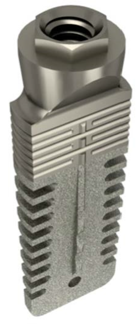
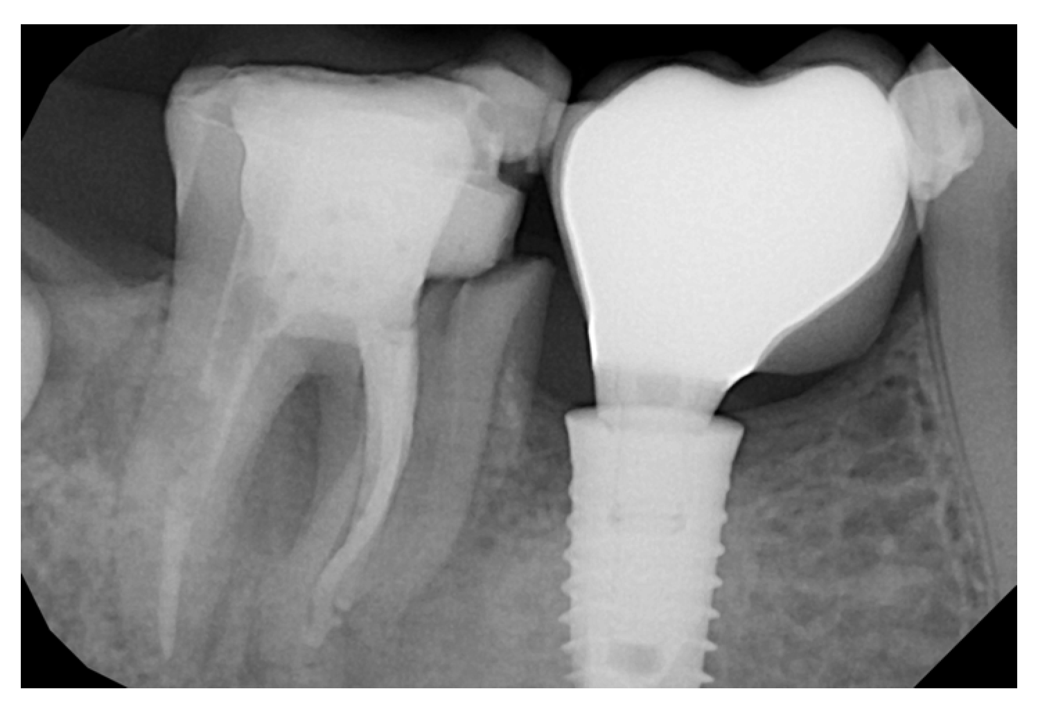
Rex implant: Implants 9 mm and 11 mm in length with a transgingival neck and an external hexagon were used. This type of implant is produced by Mectron (Figure 1, Figure 2 and Figure 3) (Mectron Electronics SPA, Genova, Italy) and consists of a body with a width of 6 mm, a thickness of 1.8 mm, and a height varying from 9 to 13 mm. For this study, implants with a height of 9 mm or 11 mm were used. The technique used for the study was as follows: The surgical extraction of 6 with rhizotomy and selective rhizotomy was performed with the aid of piezo-surgery (Figure 4). The residual septum was prepared using a dedicated drill included in the Rex implant surgical kit; the cutter was flat, and it is described in Figure 5. The depth of the implant was decided on the basis of the formula described by Bambini et al. [37], according to which Y = X − 3, where X represents the thickness of the soft tissues measured using Rinn’s centering device and an intraoral X-ray. The bur for the sagittal cut of the septum was used by mounting it on the piezo-surgery handpiece, with the “implant” setting elected, and using the classic “come and go” movement.
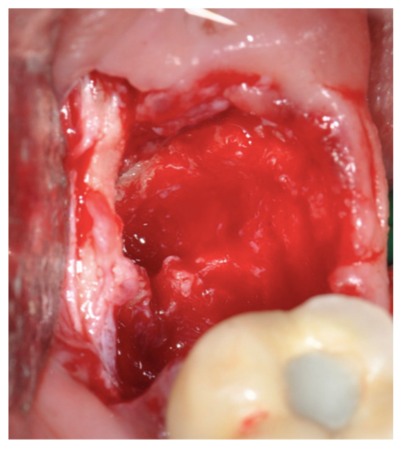
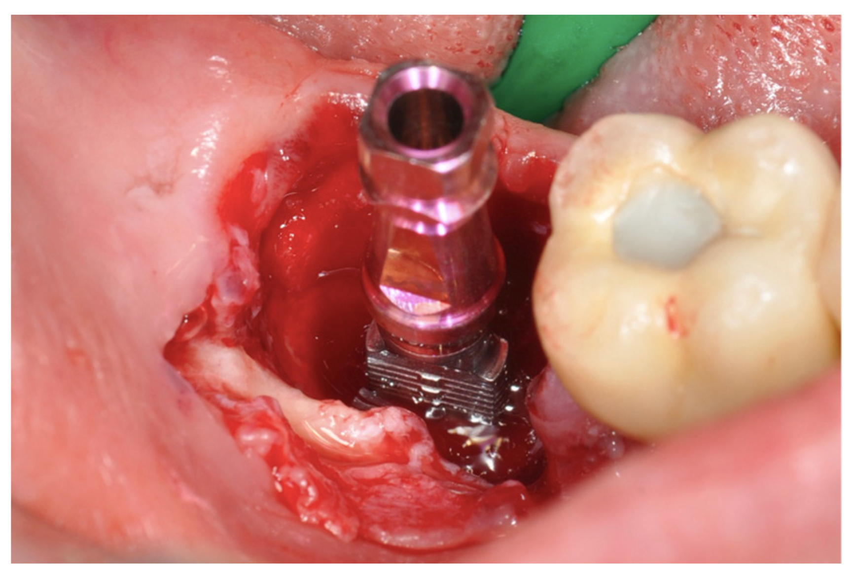
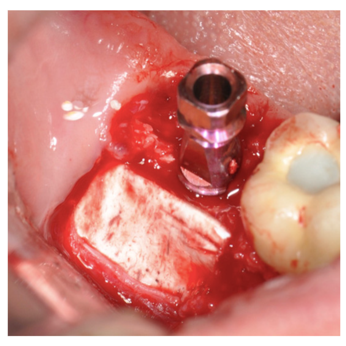
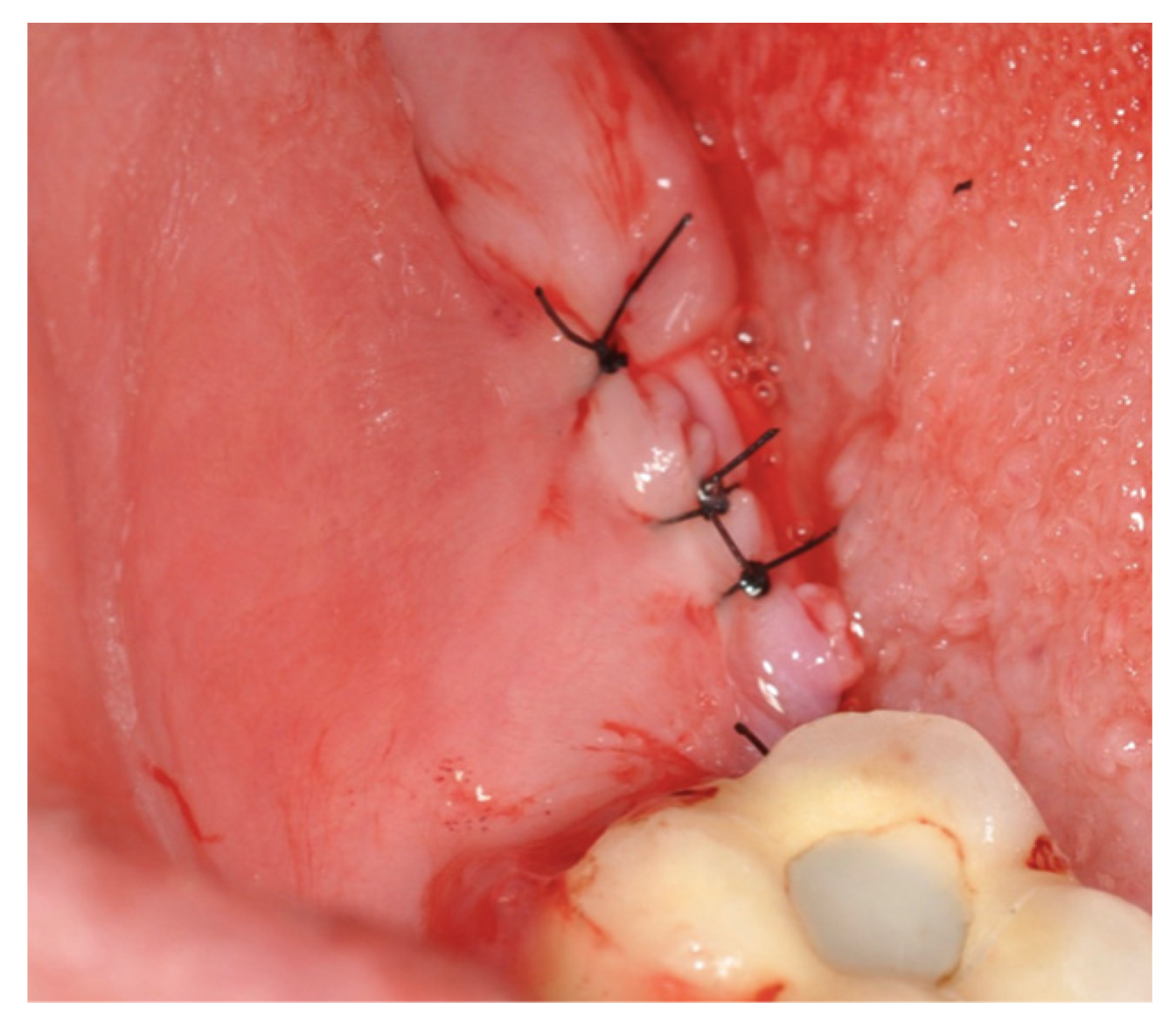
Once the canal was exposed on the inter-root septum (Figure 5), it was possible to position the Rex implant and lower it, in accordance with the protocol, with a magnetic mallet (Figure 6). Once the desired position was achieved, it was possible to insert the cover screw into the implant. A full-thickness flap was incised buccally, the periosteum was sectioned at 1 cm from the enamel–cement junction, and two oblique relief lines were incised to obtain an elastic flap that could be moved buccally up to the suture with the lingual part. Before performing the apical repositioning of the flap and applying the 5.0 suture to the synthetic poly filament coated in PTFE, a 3-millimeter cortical flex membrane fig was blocked with fibrin glue in order to exploit the periosteal inhibition that this membrane provokes (Figure 7). The alveolus was filled using a collagen sponge. The suture was inserted around the implant’s neck up to the lingual part to completely close the flap (Figure 8). Antibiotic therapy with amoxicillin and clavulanic acid was administered, consisting of 2 g per day for 5 days and 1100 mg of NSAIDs for 3 days. The sutures were removed routinely after 2 weeks. Radiographic control trials were performed after the surgery (Figure 9) and at 30, 60, and 90 days and after loading at 4 months after surgery.

3. Results

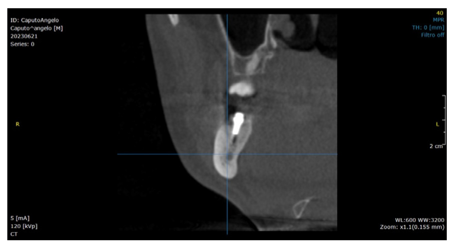
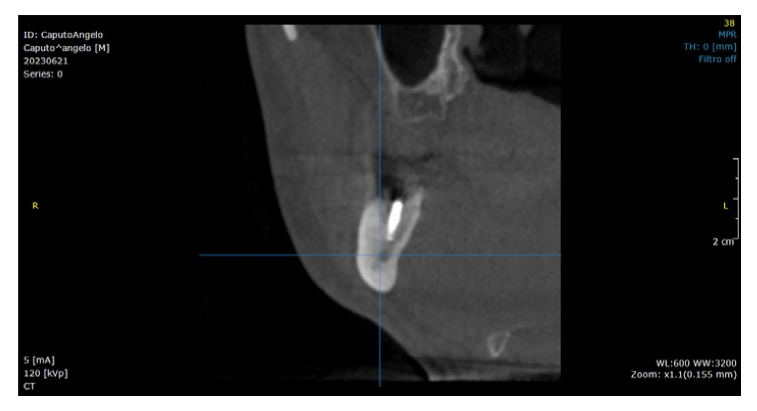
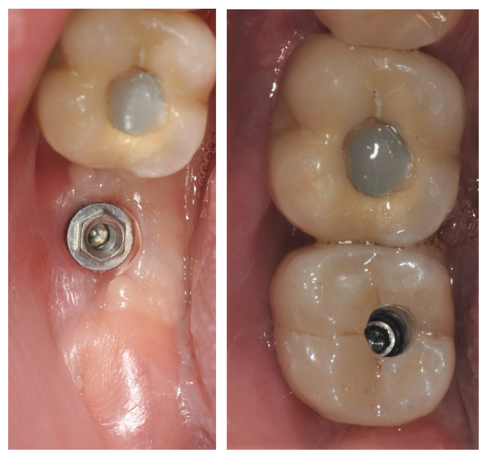
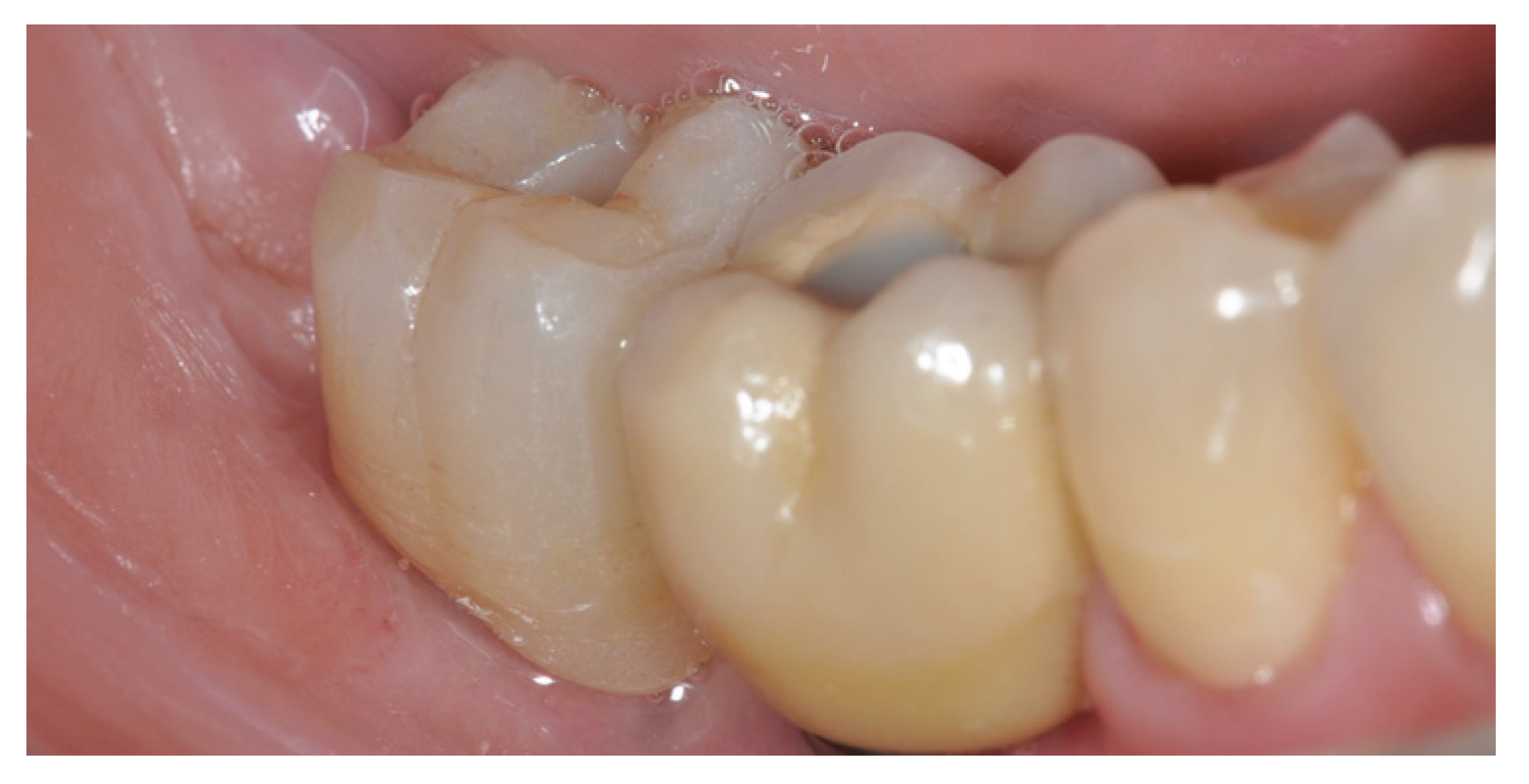
The 20 patients who underwent surgery with the Rex implants showed an adequate degree of healing of the soft tissues, in accordance with our original intention. For all the patients, the sutures were removed on the fourteenth day. Furthermore, healing of the hard tissues was observed at 4 months, revealing complete filling of the post-extraction alveoli, as evidenced by the CBCT images (Figure 10). The prosthesis was implanted such that monolithic zirconium crowns could be screwed into the implants and an abutment could be glued inside them so that they could be screwed into the portion of the Rex implant with an external hexagon as its anti-rotational geometry. The clamping screws tightened, according to Mectron’s indications, at 25 n/cm did not loosen over the following 18 months. The emergence profiles of the prosthetic crowns corresponded with the biological width already present when choosing the depth of the Rex implant. At a standard checkup after 18 months, no bone resorption or initial signs of bone damage were observed in the portion of the implant neck, and the state of the soft tissues indicated the absence of inflammatory phenomena, which serve as a prelude to mucositis or peri-implantitis (Figure 11, Figure 12 and Figure 13). Confirmation of the absence of a requirement for surgical re-entry for submerged implants was not necessary as Rex implants are non-submerged implants. The biological seal obtained during the 4 months of healing was not altered by any potential loosening or unscrewing of the healing screw, as described by Abrahamsson et al. in 1998 [40]. The achievement of this biological seal, which was also described in the cytoarchitecture by Kawahara and Hashimoto in 1998 [41], was confirmed by the absence of inflammatory phenomena around the implant neck, bleeding, and a pink coloration of the tissues. This clinical sign of the absence of inflammatory processes was verified by the presence of hemidesmosomal bonds between the titanium of the Rex neck and the epithelium, as described by Abrahamsson [42]. The control CBCTs revealed, in correspondence with the lingual and buccal areas with respect to the central position of the blade, the conspicuous formation of bone completely filling the “tub” that was previously filled exclusively with collagen (CondressR) to support the clot. The excellent adaptation of the soft tissue to the Rex implant’s neck and the pink coloration of the soft tissue proves the absence of bacteria and inflammation around the implant neck.

4. Discussion

In this study, we used, for the first time, a Rex implant in the inferior molar region. This geometric inspirations for this implant are the blade implants produced and used in the past, namely, those designed by Prof. Leonard Linkow [43]. These implants have been the subject of numerous criticisms, as they were thought to be susceptible to incorporation by fibrous tissue, which, in the past, was also called “fiber integration” [44]. In reality, this phenomenon was due to implant failure; the reasons for this early failure often lay in the way in which the blade housing was prepared, wherein cutters were mounted on a turbine, and worked at very high speeds. This led to the overheating of the bone and surrounding necrosis of the hard tissues, with a consequent fibrous reaction to the blade. Another type of late failure consisted of the fracture of the neck of the blade that supported the prosthetic element. This failure was the result of the incongruous and unsuitable geometry used to support the prosthetic load. Later, for the reasons described above, this type of implant procedure was abandoned. Subsequently, Tommaso Vercellotti [45] proposed an implant and, using current metallurgical, biological, and prosthetic knowledge, re-upgraded the Linkow blade implant with a new blade implant, called the Rex implant. This implant is now supported by important scientific documentation, which validates its use in bone crests of volumes that normally do not allow the insertion of cylindrical implants. This type of implant consists of an implant body that is 6 mm wide and has a height varying from 9 to 13 mm; this geometry exposes a transmucosal part that supports an external hexagonal system for the connection of a prosthetic abutment. This system appears particularly robust and suitable for functional loading, avoiding the fracturing of the prosthetic neck caused by the previous Linkow blade implants. The insertion of an immediate post-extraction implant, which has been discussed and strongly approved in the literature, represents a clinical practice that allows a patient to undergo, in a single operation, the extraction of a compromised dental element and the insertion of an implant on which the prosthetic element is screwed. Using this technique, it is also possible to inhibit the activity of the periosteum by simply inserting a small cortical membrane, thereby guaranteeing the maximum quantity and quality of bone possible according to the current state of scientific knowledge. There are no biological or economic expenses regarding the use of biomaterials inserted in the sockets beyond those associated with the use of collagen to suppress blood clots. This additional advantage guarantees the quality of the regenerated bone, which is the result of the avoidance of the formation of bloods clot due to the support provided by the collagen. The chemotactic function performed by the alpha granules of platelets, particularly by the growth factors contained therein (TGF beta and PDGF), and neo-angiogenic action determine the course of the differentiation of the still-undifferentiated osteoblasts and neo-angiogenesis; these effects serve as the basis of the formation of new bone, whose quality and quantity depend on the outcomes of clots and the platelets they contain. A particularly important role is played by the surgical planning of implant positioning; many authors [46,47,48] have demonstrated how perfect knowledge of the bone anatomy and the consequent guided surgical planning can improve the optimal selection of the implant, reduce surgical times, obtain optimal prosthetic results, and, in what is probably the most important aspect, improve the attitudes of patients to implant surgery. In the future, it will be possible to make complete libraries relating to the Rex implant available to clinicians. In order to be able to insert implants into post-extraction sites of the lower seta, where we face large, empty volumes, only the particular innovative geometry of this Rex implant can ensure the insertion of immediate post-extraction implants in the posterior sectors without causing any structural alterations to the inter-radicular septum, and this process is achieved by exploiting the implant’s simple anatomical shape. This very simple and economical clinical process allows this technique to be placed in the hands of every clinician and facilitates the performance of the extraction, guided bone regeneration, modified periosteal inhibition, and, obviously, the insertion of the implant.

Author Contributions

Conceptualization, F.B. and L.M.; methodology, R.R.; validation, A.G. and L.M. formal analysis, S.G.; investigation, S.G.; writing—original draft preparation, F.B. and L.M.; writing—review and editing, L.M.; visualization, S.M. and A.G.; supervision, F.B and S.M. All authors have read and agreed to the published version of the manuscript.

Funding

This research received no external funding.

Institutional Review Board Statement

The study was conducted in accordance with the Declaration of Helsinki.

Informed Consent Statement

Informed consent was obtained from all subjects involved in the study.

Conflicts of Interest

The authors declare no conflict of interest.

References

  1. Quaranta, A.; Perrotti, V.; Putignano, A.; Malchiodi, L.; Vozza, I.; CalvoGuirado, J.L. Anatomical Remodeling of Buccal Bone Plate in 35 Premaxillary Post-Extraction Immediately Restored Single TPS Implants: 10-Year Radiographic Investigation. Implant. Dent. 2016, 25, 186–192. [Google Scholar] [CrossRef] [PubMed]
  2. Roberto, C.; Paolo, T.; Giovanni, C.; Ugo, C.; Bruno, B.; Giovanni-Battista, M.F. Bone remodeling around implants placed after socket preservation: A 10-year retrospective radiological study. Int. J. Implant. Dent. 2021, 7, 74. [Google Scholar] [CrossRef]
  3. Menchini-Fabris, G.B.; Cosola, S.; Toti, P.; Hwan Hwang, M.; Crespi, R.; Covani, U. Immediate Implant and Customized Healing Abutment for a Periodontally Compromised Socket: 1-Year Follow-Up Retrospective Evaluation. J. Clin. Med. 2023, 12, 2783. [Google Scholar] [CrossRef]
  4. Muñoz-Cámara, D.; Gilbel-Del Águila, O.; Pardo-Zamora, G.; Camacho-Alonso, F. Immediate post-extraction implants placed in acute periapical infected sites with immediate prosthetic provisionalization: A 1-year prospective cohort study. Med. Oral Patol. Oral Cir. Bucal 2020, 25, e720–e727. [Google Scholar] [CrossRef] [PubMed]
  5. Wakankar, J.; Mangalekar, S.B.; Kamble, P.; Gorwade, N.; Vijapure, S.; Vhanmane, P. Comparative Evaluation of the Crestal Bone Level Around Pre- and Post-loaded Immediate Endoosseous Implants Using Cone-Beam Computed Tomography: A Clinico-Radiographic Study. Cureus 2023, 15, e34674. [Google Scholar] [CrossRef]
  6. Carosi, P.; Lorenzi, C.; Di Gianfilippo, R.; Papi, P.; Laureti, A.; Wang, H.L.; Arcuri, C. Immediate vs. Delayed Placement of Immediately Provisionalized Self-Tapping Implants: A Non-Randomized Controlled Clinical Trial with 1 Year of Follow-Up. J. Clin. Med. 2023, 12, 489. [Google Scholar] [CrossRef]
  7. Juodzbalys, G.; Daugela, P.; Duruel, O.; Fernandes, M.H.; de Sousa Gomes, P.; Goyushov, S.; Mariano, L.; Poskevicius, L.; Stumbras, A.; Tözüm, T.F. The 2nd Baltic Osseointegration Academy and Lithuanian University of Health Sciences Consensus Conference 2019. Summary and Consensus Statements: Group I—Biological Aspects of Tooth Extraction, Socket Healing and Indications for Socket Preservation. J. Oral Maxillofac. Res. 2019, 10, e4. [Google Scholar] [CrossRef]
  8. Gjelvold, B.; Kisch, J.; Chrcanovic, B.R.; Albrektsson, T.; Wennerberg, A. Clinical and radiographic outcome following immediate loading and delayed loading of single-tooth implants: Randomized clinical trial. Clin. Implant. Dent. Relat. Res. 2017, 19, 549–558. [Google Scholar] [CrossRef]
  9. Javaid, M.A.; Khurshid, Z.; Zafar, M.S.; Najeeb, S. Immediate Implants: Clinical Guidelines for Esthetic Outcomes. Dent. J. 2016, 4, 21. [Google Scholar] [CrossRef]
  10. Crespi, R.; Fabris, G.B.M.; Crespi, G.; Toti, P.; Marconcini, S.; Covani, U. Effects of different loading protocols on the bone remodeling volume of immediate maxillary single implants: A 2- to 3-year follow-up. Int. J. Oral Maxillofac. Implant. 2019, 34, 953–962. [Google Scholar] [CrossRef]
  11. Ionescu, A.; Dodi, A.; Petcu, L.C.; Nicolescu, M.I. Open Healing: A Minimally Invasive Protocol with Flapless Ridge Preservation in Implant Patients. Biology 2022, 11, 142. [Google Scholar] [CrossRef]
  12. Testori, T.; Weinstein, T.; Scutellà, F.; Wang, H.L.; Zucchelli, G. Implant placement in the esthetic area: Criteria for positioning single and multiple implants. Periodontol. 2000 2018, 77, 176–196. [Google Scholar] [CrossRef] [PubMed]
  13. Berberi, A.N.; Tehini, G.E.; Noujeim, Z.F.; Khairallah, A.A.; Abousehlib, M.N.; Salameh, Z.A. Influence of surgical and prosthetic techniques on marginal bone loss around titanium implants. Part I: Immediate loading in fresh extraction sockets. J. Prosthodont. 2014, 23, 521–527. [Google Scholar] [CrossRef]
  14. Menchini-Fabris, G.B.; Toti, P.; Crespi, G.; Covani, U.; Furlotti, L.; Crespi, R. Effect of Different Timings of Implant Insertion on the Bone Remodeling Volume around Patients’ Maxillary Single Implants: A 2–3 Years Follow-Up. Int. J. Environ. Res. Public Health 2020, 17, 6790. [Google Scholar] [CrossRef]
  15. Araújo, M.G.; Lindhe, J. Ridge preservation with the use of Bio-Oss collagen: A 6-month study in the dog. Clin. Oral Implant. Res. 2009, 20, 433–440. [Google Scholar] [CrossRef]
  16. Araújo, M.; Linder, E.; Wennström, J.; Lindhe, J. The influence of Bio-Oss Collagen on healing of an extraction socket: An experimental study in the dog. Int. J. Periodontics Restor. Dent. 2008, 28, 123–135. [Google Scholar]
  17. Santarelli, A.; Mascitti, M.; Orsini, G.; Memè, L.; Rocchetti, R.; Tiriduzzi, P.; Sampalmieri, F.; Putignano, A.; Procaccini, M.; Lo Muzio, L.; et al. Osteopontin, osteocalcin and OB-cadherin expression in synthetic nanohydroxyapatite vs bovine hydroxyapatite cultured osteoblastic-like cells. J. Biol. Regul. Homeost. Agents 2014, 28, 523–529. [Google Scholar] [PubMed]
  18. Bernardi, S.; Mummolo, S.; Varvara, G.; Marchetti, E.; Continenza, M.A.; Marzo, G.; Macchiarelli, G. Bio-morphological evaluation of periodontal ligament fibroblasts on mineralized dentin graft: An in vitro study. J. Biol. Regul. Homeost. Agents 2019, 33, 275–280. [Google Scholar]
  19. Baskaran, P.; Prakash, P.S.G.; Appukuttan, D.; Mugri, M.H.; Sayed, M.; Subramanian, S.; Al Wadei, M.H.D.; Ahmed, Z.H.; Dewan, H.; Porwal, A.; et al. Clinical and Radiological Outcomes for Guided Implant Placement in Sites Preserved with Bioactive Glass Bone Graft after Tooth Extraction: A Controlled Clinical Trial. Biomimetics 2022, 7, 43. [Google Scholar] [CrossRef]
  20. Mummolo, S.; Mancini, L.; Quinzi, V.; D’Aquino, R.; Marzo, G.; Marchetti, E. Ri genera® autologous micrografts in oral regeneration: Clinical, histological, and radiographical evaluations. Appl. Sci. 2020, 10, 5084. [Google Scholar] [CrossRef]
  21. Memè, L.; Santarelli, A.; Marzo, G.; Emanuelli, M.; Nocini, P.F.; Bertossi, D.; Putignano, A.; Dioguardi, M.; Lo Muzio, L.; Bambini, F. Novel hydroxyapatite biomaterial covalently linked to raloxifene. Int. J. Immunopathol. Pharmacol. 2014, 27, 437–444. [Google Scholar] [CrossRef] [PubMed]
  22. Sultan, N.; Jayash, S.N. Evaluation of osteogenic potential of demineralized dentin matrix hydrogel for bone formation. BMC Oral Health 2023, 23, 247. [Google Scholar] [CrossRef]
  23. Zhao, R.; Yang, R.; Cooper, P.R.; Khurshid, Z.; Shavandi, A.; Ratnayake, J. Bone Grafts and Substitutes in Dentistry: A Review of Current Trends and Developments. Molecules 2021, 26, 3007. [Google Scholar] [CrossRef] [PubMed]
  24. Nguyen, V.; Von Krockow, N.; Pouchet, J.; Weigl, P.M. Periosteal Inhibition Technique for Alveolar Ridge Preservation as It Applies to Implant Therapy. Int. J. Periodontics Restor. Dent. 2019, 39, 737–744. [Google Scholar] [CrossRef]
  25. Marconcini, S.; Denaro, M.; Cosola, S.; Gabriele, M.; Toti, P.; Mijiritsky, E.; Proietti, A.; Basolo, F.; Giammarinaro, E.; Covani, U. Myofibroblast gene expression profile after tooth extraction in the rabbit. Materials 2019, 9, 3697. [Google Scholar] [CrossRef] [PubMed]
  26. Tomaseck, J.J.; Gabbiani, G.; Hinz, B.; Chaponnier, C.; Brown, R.A. Myofibroblast and mechanoregulation of connective tissue remodeling. Nat. Rev. Mol. Cell Biol. 2022, 3, 349–363. [Google Scholar] [CrossRef]
  27. Grassi, A.; Bernardello, F.; Cavani, F.; Palumbo, C.; Spinato, S. Lindhe Three-Punch Alveolar Ridge Reconstruction Technique: A Novel Flapless Approach in Eight Consecutive Cases. Int. J. Periodontics Restor. Dent. 2021, 41, 875–884. [Google Scholar] [CrossRef] [PubMed]
  28. Wongpairojpanich, J.; Kijartorn, P.; Suwanprateeb, J.; Buranawat, B. Effectiveness of bilayer porous polyethylene membrane for alveolar ridge preservation: A randomized controlled trial. Clin. Implant. Dent. Relat. Res. 2021, 23, 73–85. [Google Scholar] [CrossRef]
  29. Lutz, R.; Sendlbeck, C.; Wahabzada, H.; Tudor, C.; Prechtl, C.; Schlegel, K.A. Periosteal Elevation induces supracortical peri-implant bone formation. J. Craniomaxillofac. Surg. 2017, 45, 1170–1178. [Google Scholar] [CrossRef] [PubMed]
  30. Grassi, A.; Memè, L.; Strappa, E.M.; Martini, E.; Bambini, F. Modified Periosteal Inhibition (MPI) Technique for Extraction Sockets: A Case Series Report. Appl. Sci. 2022, 12, 12292. [Google Scholar] [CrossRef]
  31. Rossi, R.; Modoni, M.; Monterubbianesi, R.; Dallari, G.; Memè, L. The ‘Guided Tissue Regeneration (GTR) Effect’ of Guided Bone Regeneration (GBR) with the Use of Bone Lamina: A Report of Three Cases with More than 36 Months of Follow-Up. Appl. Sci. 2022, 12, 11247. [Google Scholar] [CrossRef]
  32. Schuh, P.L.; Wachtel, H.; Beuer, F.; Goker, F.; Del Fabbro, M.; Francetti, L.; Testori, T. Multi-Layer Technique (MLT) with Porcine Collagenated Cortical Bone Lamina for Bone Regeneration Procedures and Immediate Post-Extraction Implantation in the Esthetic Area: A Retrospective Case Series with a Mean Follow-Up of 5 Years. Materials 2021, 14, 5180. [Google Scholar] [CrossRef] [PubMed]
  33. Elad, A.; Rider, P.; Rogge, S.; Witte, F.; Tadić, D.; Kačarević, Ž.P.; Steigmann, L. Application of Biodegradable Magnesium Membrane Shield Technique for Immediate Dentoalveolar Bone Regeneration. Biomedicines 2023, 11, 744. [Google Scholar] [CrossRef] [PubMed]
  34. Rossi, R.; Memè, L.; Strappa, E.M.; Bambini, F. Restoration of Severe Bone and Soft Tissue Atrophy by Means of a Xenogenic Bone Sheet (Flex Cortical Sheet): A Case Report. Appl. Sci. 2023, 13, 692. [Google Scholar] [CrossRef]
  35. Degidi, M.; Daprile, G.; Nardi, D.; Piattelli, A. Immediate provisionalization of implants placed in fresh extraction sockets using a definitive abutment: The chamber concept. Int. J. Periodontics Restor. Dent. 2013, 33, 559–565. [Google Scholar] [CrossRef]
  36. Bambini, F.; Orilisi, G.; Quaranta, A.; Memè, L. Biological oriented immediate loading: A new mathematical implant vertical insertion protocol, five-year follow-up study. Materials 2021, 14, 387. [Google Scholar] [CrossRef]
  37. Berglundh, T. Dimension of the periimplant mucosa Biological width revisited. J. Clin. Periodontol. 1996, 23, 971–973. [Google Scholar] [CrossRef]
  38. Mullings, O.; Tovar, N.; Abreu de Bortoli, J.P.; Parra, M.; Torroni, A.; Coelho, P.G.; Witek, L. Osseodensification Versus Subtractive Drilling Techniques in Bone Healing and Implant Osseointegration: Ex Vivo Histomorphologic/Histomorphometric Analysis in a Low-Density Bone Ovine Model. Int. J. Oral Maxillofac. Implant. 2021, 36, 903–909. [Google Scholar] [CrossRef] [PubMed]
  39. Seo, D.-J.; Moon, S.-Y.; You, J.-S.; Lee, W.-P.; Oh, J.-S. The Effect of Under-Drilling and Osseodensification Drilling on Low-Density Bone: A Comparative Ex Vivo Study. Appl. Sci. 2022, 12, 1163. [Google Scholar] [CrossRef]
  40. Abrahamsson, I. The mucosal barrier following abutment dis/reconnection: An experimental study in dogs. J. Clin. Periodontol. 1997, 24, 568–572. [Google Scholar] [CrossRef]
  41. Kawahara, H.; Kawahara, D.; Mimura, Y.; Takashima, Y.; Ong, J.L. Morphologic Studies on the Biologic Seal of Titanium Dental Implants. Report II. In Vivo Study on the Defending Mechanism of Epithelial Adhesion/Attachment Against Invasive Factors. Int. J. Oral Maxillofac. Implant. 1998, 13, 465–473. [Google Scholar]
  42. Abrahamsson, I.; Berglundh, T.; Glantz, P.-O.; Lindhe, J. The mucosal attachment at different abutments: An experimental study in dogs. J. Clin. Periodontol. 1998, 25, 721–727. [Google Scholar] [CrossRef] [PubMed]
  43. Linkow, L.I. The blade vent—A new dimension in endosseous implantology. Dent. Concepts 1968, 11, 3–12. [Google Scholar]
  44. Participants of CSP No., 86; Kapur, K.K. Veterans administration cooperative dental impant study—Comparisons between fixed partial dentures supported by blade-vent implants and removable partial dentures. Part II: Comparison of success rates and periodontal health between two treatment modalities. J. Prosthet. Dent. 1989, 62, 685–702. [Google Scholar] [CrossRef]
  45. Vercellotti, T.; Troiano, G.; Oreglia, F.; Lombardi, T.; Gregorig, G.; Morella, E.; Rapani, A.; Stacchi, C. Wedge-shaped implants for minimally invasive treatment of narrow ridges: A multicenter prospective cohort study. J. Clin. Med. 2020, 9, 3301. [Google Scholar] [CrossRef]
  46. Shetty, S.R.; Arya, S.; Kamath, V.; Al-Bayatti, S.; Marei, H.; Abdelmagyd, H.; El-Kishawi, M.; Al Shehadat, S.; Al Kawas, S.; Shetty, R. Application of a Cone-Beam Computed Tomography-Based Index for Evaluating Surgical Sites Prior to Sinus Lift Procedures—A Pilot Study. BioMed Res. Int. 2021, 2021, 9601968. [Google Scholar] [CrossRef]
  47. Shetty, S.R.; Murray, C.A.; Al Kawas, S.; Jaser, S.; Al-Rawi, N.; Talaat, W.; Narasimhan, S.; Shetty, S.; Adtani, P.; Hegde, S. Impact of fully guided implant planning software training on the knowledge acquisition and satisfaction of dental undergraduate students. Med. Educ. Online 2023, 28, 2239453. [Google Scholar] [CrossRef]
  48. Shetty, S.R.; Murray, C.; Kawas, S.A.; Jaser, S.; Talaat, W.; Madi, M.; Kamath, V.; Manila, N.; Shetty, R.; Ajila, V. Acceptability of fully guided virtual implant planning software among dental undergraduate students. BMC Oral Health 2023, 23, 336. [Google Scholar] [CrossRef]
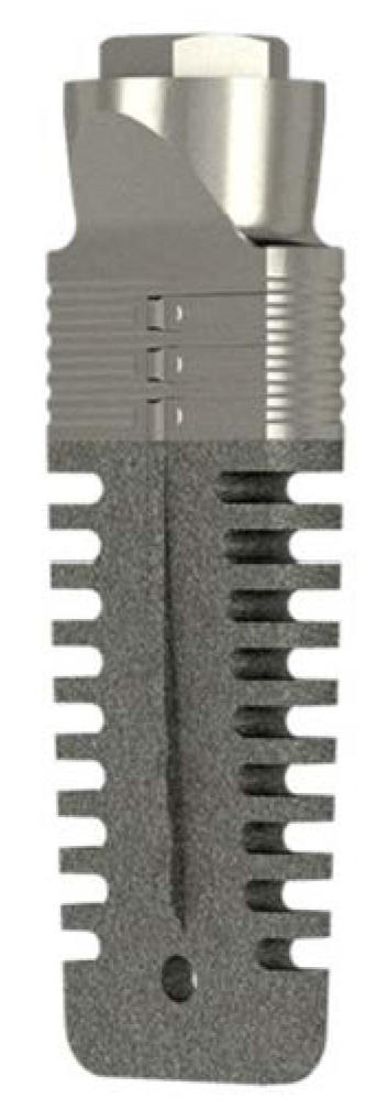
Figure 1. Rex implant.
Applsci 13 10226 g001
Figure 2. Rex implant.
Applsci 13 10226 g002
Figure 3. Kit for Rex implants’ positioning.
Applsci 13 10226 g003
Figure 4. Pre-operative Rx.
Applsci 13 10226 g004
Figure 5. Image taken after extraction.
Applsci 13 10226 g005
Figure 6. Rex inside socket after extraction.
Applsci 13 10226 g006
Figure 7. MPI technique.
Applsci 13 10226 g007
Figure 8. Suture after surgery.
Applsci 13 10226 g008
Figure 9. Post-operative Rx.
Applsci 13 10226 g009
Figure 10. CBCT control at 4 months.
Applsci 13 10226 g010
Figure 11. Soft tissue around neck of Rex.
Applsci 13 10226 g011
Figure 12. Screw-retained zirconia crown on Rex.
Applsci 13 10226 g012
Figure 13. Screw-retained zirconia crown on Rex.
Applsci 13 10226 g013
Disclaimer/Publisher’s Note: The statements, opinions and data contained in all publications are solely those of the individual author(s) and contributor(s) and not of MDPI and/or the editor(s). MDPI and/or the editor(s) disclaim responsibility for any injury to people or property resulting from any ideas, methods, instructions or products referred to in the content.