1. Introduction
Modern society is entering an era of digitalization with the Fourth Industrial Revolution, and the significance of display technology in visually representing various types of information has increased. Recently, OLED displays have garnered attention as the next generation display technology. However, the OLED market faces competitive challenges due to the low cost display strategies employed by global companies. Nevertheless, the OLED market is growing through continuous investments and technological developments by domestic companies [
1]. If this growth continues steadily, the domestic OLED industry is expected to secure a competitive edge and evolve into a key player in the future market, leading to the development of next generation display technologies [
2]. Therefore, this study aims to explore the growth potential of the domestic OLED industry and proposes strategies for its development. This study also focuses on improving the yield by applying an optimal deep learning algorithm to detect defects in OLED cells.
Detecting defects within OLED cells is crucial for several reasons. Firstly, it aims to prevent the production of faulty items during the manufacturing process. Defective products can compromise quality and performance, potentially leading to unreliable products for consumers. Therefore, accurately identifying and addressing defects within OLED cells is of paramount importance. Furthermore, reducing the occurrence of product disposal due to defects is economically significant within the OLED industry. Identifying defects early and taking corrective actions can prevent the need for product disposal or rework, thereby resulting in cost savings. According to the study by Li Wei et al., utilizing artificial intelligence technology to automatically detect defects has been explored [
3]. They developed and validated a network that could automatically identify defects, such as scratches, on the surface of products. This research emphasizes the potential for enhancing product quality and reducing the number of defective items through effective defect detection methods. Efficiently identifying and addressing cell defects can increase production yield. This can contribute to the advancement of OLED display manufacturing technology and have positive effects on the OLED display market.
The yield of OLEDs is determined throughout the production process, from manufacturing to encapsulation. The careful detection and identification of defects or issues are crucial during the OLED panel manufacturing process. Generally, OLED cell defects arise owing to the use of faulty materials, improper process conditions, or impacts during transportation in the manufacturing process [
4]. The encapsulation process, which occurs after the preceding manufacturing steps, involves covering the OLED panel with encapsulation glass to protect it from external influences and ensure long term usage without interference. The encapsulation process is essential because the organic materials and electrodes in OLEDs are highly sensitive to oxygen and moisture, which can lead to the loss of luminous characteristics. This step directly preserves or enhances the lifespan of the OLED panels [
5]. However, cell defects occur even with the encapsulation process used to improve the OLED yield. This is because defects can be introduced during the manufacturing process through impurity infiltration or the introduction of moisture and oxygen into organic materials. Despite thorough processing during manufacturing and encapsulation, complete detection and discrimination between defects during the inspection is challenging [
6]. During the inspection process, visual inspection by human operators may result in misjudgments, where good OLED panels may be incorrectly classified as defective or vice versa. Defects in OLEDs can be caused by degradation during panel production, which can be attributed to physical factors such as temperature, humidity, and pressure. In addition, defects can arise during the encapsulation process owing to the presence of impurities. This complexity makes it difficult to perfectly detect and discriminate defects, resulting in dark spots within the panels. Dark spots are formed because of cell discoloration within the panel when defects occur. This study compared and analyzed deep learning algorithms to identify and measure the shape and size of dark spots formed within cells. These dark spots were used as criteria for determining cell defects. We conclude that improving the yield of OLEDs and reducing the quantity of defective products during the manufacturing process is feasible by using deep learning algorithms to enhance defect detection and classification.
Therefore, this study explores an optimal deep learning algorithm for detecting defects in OLED cells and discriminating between them to improve the yield. The VGG-16 deep learning algorithm was used to classify the dark spot images. A model was developed to distinguish between acceptable and defective cells by considering the shapes and sizes of the dark spots.
2. Related Research
2.1. Research on the Causes of Defects in OLED Cells
In the field of OLED displays, continuous research is being performed to extend the panel lifespan and improve yield. The presence of defects in OLED cells directly influences the panel lifespan, which is determined by the operational conditions and production process of the OLED panels. When investigating the formation process of dark spots, defects can arise because the organic materials are not stable. These materials can break down gradually over time, causing degradation that results in the appearance of dark spots within the cells. In addition, particles or impurities might get trapped in the display while it is being manufactured, leading to the creation of defects that could show up as dark spots. OLED panels are susceptible to moisture and oxygen; dark spots emerge if these substances penetrate the display surface. Dark spots are defects that gradually emerge while OLED panels are being used, usually as a result of electric current flowing through the panel over time. The review article by Azrain et al., discusses the relationship between dark spots and current in OLED cells, as well as the impact of current on OLED cells [
7]. The environmental instability of OLED components and resultant dark spot formation are significant concerns, influenced by factors like external impurities, pinholes, and high current density. These elements trigger gas formation within OLED cells, alongside processes like electrical migration and crystallization, shortening OLED lifespan and performance. Another study by Phatak et al. highlights the significance of employing rigorous encapsulation technology in OLED manufacturing [
8]. This is because exposure to oxygen or humidity results in the formation of dark spots, which has a detrimental impact on OLED performance. Therefore, applying rigorous encapsulation technology during manufacturing is crucial for preventing the infiltration of various impurities from the surrounding environment. In addition, the formation of dark spots can be minimized, and growth can be inhibited by heating the substrate to high temperatures during cathode deposition in the OLED organic layer. However, this approach has certain limitations.
Based on these studies, it is evident that dark spots are formed in OLED panels owing to various factors, and the impact of these dark spots on OLED displays is understood. Focusing on dark spots as one of the primary defects in OLEDs, this study uses artificial intelligence to differentiate OLED cell defects by analyzing the size and distribution of dark spots that develop within cells. Through this approach, the number of defective products can be reduced, thereby improving the yield and suggesting potential cost savings in production.
2.2. Study on Defect Detection in OLED Using Deep Learning
Recent technological advancements have led to the use of deep learning in OLED displays for detecting minor defects. OLED panels contain microscopic cells, which limit the effectiveness of visual inspection. Although recent approaches use camera-based image analysis for defect detection, accurately classifying defect images without controlling for all environmental factors remains challenging. This trend is shifting towards utilizing artificial-intelligence-based image analysis techniques rather than camera-based image discrimination. In a previously reported study, we classified and studied the defects that occur during OLED production [
9]. Our focus was on general and disjointed cell defects. A CNN-based model was developed to discriminate between cell defects. This involved collecting an OLED defect image dataset to train the deep learning model and evaluating its performance using test data. The research findings indicate that the developed model accurately detects defects in flexible OLEDs, leading to improved accuracy in defect detection and the reduced time and cost of defect detection through deep learning. In another study, Singh, R. B. presented a defect detection technique for the manufacturing process using deep learning [
10]. This study used deep learning, specifically Convolutional Neural Networks (CNNs) such as Res-Net and Dense-Net, to identify different types of defects in X-ray images. The Dense-Net model demonstrated the highest performance, and data augmentation techniques such as rotation, translation, and flipping were used to increase the dataset size and improve the model performance. The proposed system achieved an F1-score of 80%, indicating its potential as a valuable technology for defect detection in metal manufacturing processes. A study was conducted on anomaly detection in LCD/OLED display panel manufacturing using Generative Adversarial Networks [
11]. The detection of defects in display panels requires the identification and categorization of anomalies based on specific criteria. The proposed method employs GAN based anomaly detection for real time and automatic data labeling to construct an efficient training dataset. Anomalies were detected in real time, and the resulting anomalous data were automatically labeled to create an effective training dataset. S. Ye et al. used deep learning techniques for defect detection in patterned OLED panels with fine scale defects [
12]. The proposed method uses defect inpainting and a multiscale Siamese neural network to detect minor defects in patterned OLED panels. This approach aims to detect defects using limited training data by leveraging few shot learning, which enables the detection of fine scale defects.
Alireza Saberironaghi et al. conducted research from the perspective of image defect detection, utilizing deep learning techniques for detecting defects in industrial products. He explored surface defect detection based on deep learning technology, identifying imbalanced samples and detecting defects in real time with limited sample sizes [
13].
Van Huan et al. suggested that automatic defect detection and classification in OLED panels are essential for quality management [
14]. Therefore, this study designed an efficient and high performance system for defect classification by combining machine learning algorithms such as Support Vector Machines (SVMs), Random Forest, and K-Nearest Neighbors (K-NN). The approach involved using principal component analysis (PCA) to design potential features and automatically select the most effective features using Random Forest. A hierarchical structure for classifiers was proposed to achieve the efficient adjustment of the ratio between good and defective items in defect classification. The designed system achieved an accuracy of 94% for the binary classification. Chen et al. employed deep convolutional neural networks (DCNNs) to segment small targets in LCD defects accurately [
15]. These small targets refer to defects that can occur in LCDs, such as pixel damage, spots, panel degradation, and dust accumulation. The deep learning technique resulted in a 91% accuracy rate for defect identification. In another study, the same group used the VGG-16 deep learning algorithm to differentiate between papillary thyroid carcinomas and normal thyroid cells in cytological images [
16]. The VGG-16 model was used with either pretrained weights or retrained weights to analyze the images. The experiment results showed an accuracy of 0.976 for fragmented images and 0.95 for patient targeted images, indicating successful differentiation between thyroid carcinoma and normal thyroid cells. In the medical field, a study by Geng et al. focused on automatically detecting and classifying lung regions in CT images to aid medical diagnosis [
17]. The experiment used 137 images, and the primary performance metric, the Dice Similarity Coefficient (DSC), yielded an accuracy of 0.986 for lung segmentation.
Based on the aforementioned studies, we compared and analyzed deep learning algorithms, including CNN, ResNet-50, and VGG-16, to detect defects in OLED cells. After comparing the performance metrics of the algorithms, we selected the optimal algorithm for image processing and defect discrimination in OLED cells. The aim was to enhance the efficiency of defect detection using this process.
2.3. Research on Artificial-Intelligence-Based Virtual Image Dataset Generation
According to a report by You et al., dark spots are defects caused by degradation and various factors in the production process of OLED panels [
18]. These dark spots can be formed by multiple factors and are particularly influenced by parameters such as the thickness (L) of the organic light emitting layer, temperature (T), humidity (C0), pressure (P), and degradation time (t). The researchers conducted performance experiments under various process conditions and used Monte Carlo simulations and deep learning to generate virtual sample data. In another study on OLED cell simulation and detection phases based on the A
2G algorithm for artificial intelligence applications, virtual dark spot data were generated because of the challenge of obtaining real datasets. Data were generated based on the defined conditions for dark spot growth in relation to humidity. The researchers established criteria for identifying display defects after the minimum requirement of 10,000 h of OLED lifespan. They analyzed the growth of dark spots over time using an FEM solver program and investigated the factors influencing the distribution and size of dark spots. Virtual dark spot data were generated using AI technology and the consideration of physical phenomena [
19,
20]. Furthermore, in another study, the growth of dark spots in OLEDs and factors that accelerate the growth were investigated [
21]. OLEDs, as light emitting diodes, consist of organic semiconductors and emitting layers. However, the prolonged usage or application of high voltages in OLEDs form dark spots, which are defects that occur within the emitting layer. These dark spots degrade the performance of the OLED and reduce its lifespan. Therefore, the growth and acceleration factors of dark spots were investigated using simulations. The group also evaluated the growth rate and acceleration factors of dark spots.
Lee et al. investigated the thermal evaporation process that affects the performance and quality of OLEDs, and they concluded through the simulation of thin-film thickness distribution in the OLED thermal evaporation process that the distribution of thin-film thickness can impact the performance and quality of OLED devices [
22].
According to the report by Shin et al., having an adequate amount of both normal and defective data is necessary to ensure quality and reliability in the directed energy deposition process [
23]. However, collecting a large amount of data is challenging owing to the lengthy processing time and high costs of the metal powder associated with the directed energy deposition process. Therefore, this paper proposes a virtual process data generation algorithm for the deposition process. The generated virtual process data were validated using data collected from an actual deposition process monitoring system. The findings of the study suggest that the generated virtual data can be used. In another study by Jung et al., the researchers used AI to generate artificial image data to address the problem through large scale data mining techniques [
24]. In the medical field, securing large-scale data is challenging because of constraints such as privacy protection. From the findings of this study, it is concluded that if virtual data can be generated to resemble real data, they can be used as training data for developing data mining models. Based on the aforementioned studies, this study defines and applies virtual dataset generation methods using AI technology and the FEM to create virtual dark spot image data.
3. Research Methodology
3.1. Generating Virtual Dark Spot Image Dataset
This study aimed to identify OLED dark spot images. However, obtaining real OLED dark spot image data is challenging owing to the confidentiality of the OLED industry. In addition, direct data generation methods are time consuming and costly. Therefore, inspired by this research report, we used a virtual data generation method to create a dataset of OLED dark spots. The data for the virtual dark spots were generated by considering the humidity exposure conditions, and the criteria for defects were established after 10,000 h of the OLED lifetime. The FEM was employed to analyze the growth of dark spots over time and to identify the factors influencing their distribution and size. The analysis results generated virtual dark spot data using the FEM Solver program [
19].
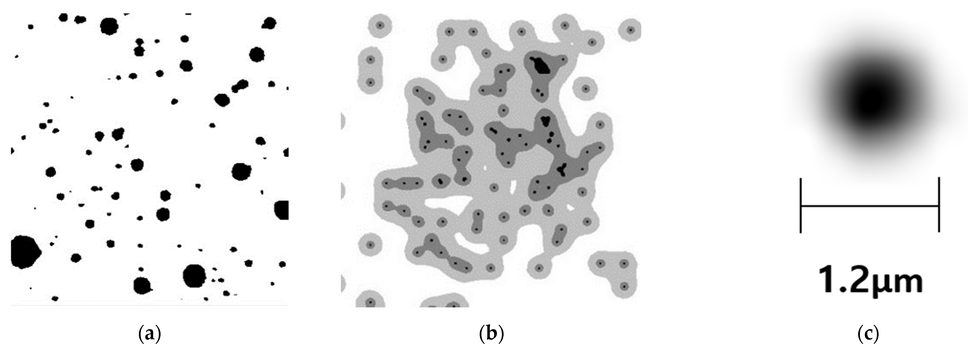
Figure 1 depicts real and dark spot images generated using the FEM Solver.
Figure 1a shows the dark spots formed inside the OLED cell.
Figure 1b shows the virtual dark spot images generated using the FEM Solver program (V6.0). The figure shows the actual dark spot images and virtual dark spot images generated using the FEM Solver.
Figure 1c shows the initial dark spot that has been magnified 100 times. The size of the dark spot is 1.2 μm. The generated dark spot dataset consisted of 2000 images, classified into pass and non-pass groups based on their initial state and the state after 10,000 h. The pixel values and color intensities, represented as bit values, were adjusted for the classified dark spot images. According to a study, optical inspection systems are highly noise-sensitive [
25]. Therefore, image filtering must be performed using the adjusted pixel values for defect detection. This approach improves the efficiency of filtering algorithms and reduces error rates when filtering algorithms are applied to images. Therefore, the attribute values were transformed to utilize the virtually generated image data for model training and validation. The original image data had a color depth of 24 bits and a resolution of 760 × 609 pixels, and the file format was set to PNG. Finally, the modified image data were transformed into gray-scale images.
A preprocessing procedure was carried out on the 1300 training dataset images utilized during the training process. This step aimed to enhance the model’s performance by making adjustments to the image data. The original image dimensions were resized from 760 × 609 to 300 × 300, and the bit value representing the color format was set to 24. Additionally, to promote diversity and improve the generalization capability of the data, image augmentation was performed. This involved applying various transformations to the images to enhance the model’s performance. To facilitate this, the image dimensions of the training dataset were set to 224 × 224, and a batch size of 32 was chosen to perform data augmentation using the generated image data.
Following the same procedure, the training dataset images were also processed for the validation dataset. In this process, the image dimensions of the validation dataset were resized from 760 × 609 to 300 × 300, and the color format bit value was set to 24. Similarly, data augmentation was applied to the validation dataset to improve the model’s performance and generalization capability. The image dimensions were adjusted to 224 × 224, and a batch size of 32 was employed for data augmentation. Consistent preprocessing steps were thus applied to the validation dataset images as well. After converting the image data format, a comparative analysis was conducted between the actual OLED dark spot dataset and the virtually generated dark spot dataset.
Table 1 summarizes the virtually generated dark spot image data. A total of 2000 data samples were created and used in the training and validation processes. Looking at the image dataset, the data in the “pass” category represent images of cells in good condition, while the data in the “non-pass” category represent images of cells with defects. Among the dataset categories, the “Initial Image” category comprises 950 images representing the initial dark spots formed in the OLED cells. The “10,000 (H) Image” category consists of 1050 images depicting dark spots that formed after 10,000 h.
To determine the optimal deep learning algorithm for dark spot detection, various algorithms were selected and utilized as the foundation for developing a defect detection model. For this purpose, 1300 images from the “Training Dataset” were used for model training, and 600 images from the “Validation Dataset” were utilized to perform model validation. The remaining 50 images from the “Test Dataset” were reserved for evaluating the performance of the defect detection model in identifying actual dark spots.
3.2. Comparison and Selection of Deep Learning Algorithms
Deep learning is a subfield of machine learning that relies on the multilayered structure of mathematically modeled artificial neural networks. It can achieve exceptional performance by learning from vast amounts of data. Deep learning technology is used in the medical field for diagnoses using images [
26]. Various deep learning algorithms for image processing have been employed in different domains. Representative convolutional neural networks (CNNs), ResNet-50, and VGG-16 have been compared and analyzed. Seo et al. developed an automated identification and classification system using a CNN algorithm for switch defect detection [
27]. CNNs have demonstrated exceptional performance in image and pattern recognition tasks. Simonyan et al. introduced the innovative deep learning architecture called VGG-Net in 2014 [
28]. This architecture has shown remarkable performance in image recognition and classification tasks. VGG-16 is the most popular version of VGG-Net and consists of 16 layers, 13 of which are convolutional. The architecture of VGG-16 consists of convolutional, pooling, and fully connected layers. VGG-16 extensively used small-sized filters in its pooling layers to extract diverse features from data. In addition, it has a regular structure comprising convolutional and pooling layers. The convolution layers use 3 × 3 filters with padding, while the pooling layers use 2 × 2 filters. The fully connected layers consist of three layers. After convolution and pooling, the feature maps are flattened into one-dimensional vectors for image classification. Unlike convolutional layers, where neurons are only connected to a local input region, fully connected layers connect every neuron to all the neurons in the previous layer. This allows each neuron to learn its own set of weights independent of the inputs. The learned weights are then used to combine the features of the image and make a final prediction. The output of the last fully connected layer is passed through the soft-max function to convert it into probability values that determine the class to which the input image belongs. This structure is easy to implement and extend, enabling consistent processing for various image sizes.
VGG-16 was pretrained on the ImageNet dataset and achieved high performance in image classification tasks. The VGG-16 algorithm was selected for our study because it outperformed ResNet-50.
Table 2 compares and analyzes the performance of three commonly used algorithms in image analysis: the CNN (Convolutional Neural Network), VGG-16, and ResNet-50. The performance metrics of the CNN algorithm exhibited an accuracy of 0.875, loss value of 0.384, specificity of 0.586, and recall of 0.671. When comparing VGG-16 and ResNet-50, most model performance metrics were relatively similar. However, the performance of the VGG-16 algorithm was slightly better than that of ResNet-50. Finally, the comparative results indicated that the VGG-16 algorithm outperformed the CNN and ResNet-50 regarding relative performance. VGG-16, pretrained on the ImageNet dataset, demonstrated excellent performance in image classification tasks; therefore, the VGG-16 algorithm was chosen for application in this study.
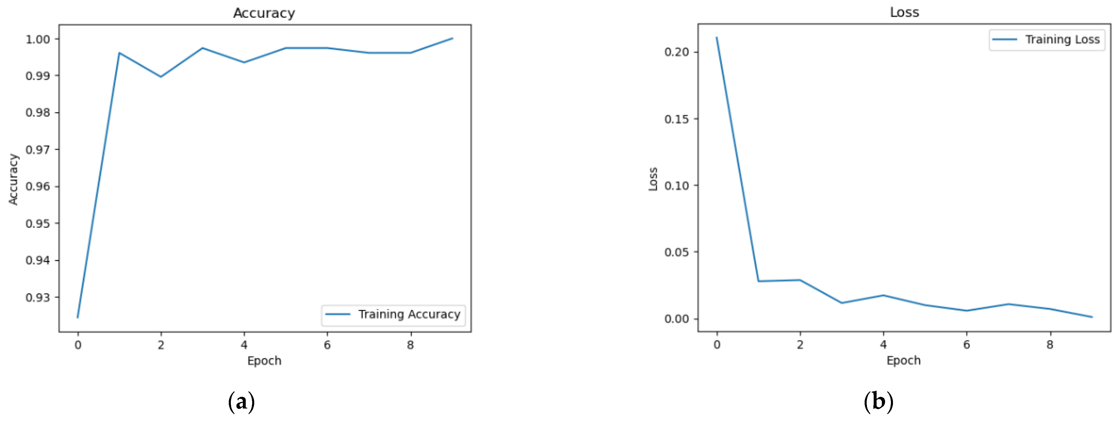
A model for discriminating cell defects was developed based on the VGG-16. The accuracy and loss values were analyzed to validate the generated model. The fitness of the model was determined based on the performance metrics. The results of the performance metrics, accuracy, and loss are shown in the graphs below.
As shown in
Figure 2, 600 image data points were used to validate the discrimination model generated using the VGG-16 algorithm. The
x-axis represents the epoch, which indicates the number of times the entire training dataset was used for learning. The
y-axis represents the measured values.
Figure 2a illustrates the accuracy of the discrimination model. The model progressively learned from the training dataset through epochs and improved its performance. Setting an epoch value to 10 or higher can affect the training time and performance, potentially leading to overfitting. Therefore, the epoch values were appropriately adjusted. The average accuracy values were 0.988, with the highest recorded value of 0.998.
Figure 2b presents the loss values for the VGG-16 algorithm. For validation, 600 images were used, and the epochs were plotted on the
x-axis, whereas the
y-axis shows the measured loss values for each epoch. Comparing epoch values up to 10, as higher epochs can impact the training time and performance, the average loss value was 0.026, with the lowest recorded value being 0.017.
Based on these performance metrics, the VGG-16 algorithm was the optimal choice; therefore, it was applied to discriminate the OLED cell defects to ensure precise defect classification.
3.3. Development of an OLED Defect Detection Model Using VGG-16 Algorithm
After comparing and analyzing various deep learning algorithms, the optimal choice, VGG-16, was selected to construct the OLED defect detection model. To construct the model using the VGG-16 algorithm, the required libraries, including TensorFlow, Matplotlib, and OpenCV, were used. We imported the required libraries, such as TensorFlow, Matplotlib, and OpenCV. The VGG-16 algorithm was implemented using the default implementation of VGG-16 provided by TensorFlow. VGG-16 is a binary classification algorithm designed to train images for specific defect identification tasks. Pretrained weights can be utilized, and layers can be frozen to prevent them from being trained during the process. In addition, a flattened layer was added to convert the 4D tensor output of VGG-16 into a 2D tensor. The rectified linear unit ReLu activation function was applied, and a dropout layer was incorporated to mitigate overfitting. The final dense layer used a sigmoid activation function for binary classification to generate the ultimate output. The model was compiled using a binary cross-entropy loss function and the RMSprop optimizer. The learning rate was set to 10
−4, and an accuracy metric was specified for the evaluation. The pseudocode for implementing the OLED defect detection using the VGG-16 algorithm is presented in Algorithm 1.
| Algorithm 1. VGG-16 for OLED Cell Defect Detection |
# Library
Import TensorFlow, Matplotlib, OpenCV
# Application of VGG16 Algorithm
x = base model VGG16
input shape = (224, 224, 3)
weights = image net’
Flatten = base model.output
Dense = 256, Activation = relu
Dropout = 0.5,
Dense = 1, Activation = sigmoid
# Model Compile
model.compile
loss = binary crossentropy,
optimizer = RMSprop 10−4
metrics = accuracy
# Image Upload and Preprocessing
Read image in directory
For image in directory:
Target size = (224, 224)
VGG16 preprocessed Input images
# Displaying an image
x = image to array
x = np.expand dims(axis = 0)
# If the result is 0.95 or higher, output ‘pass’; otherwise, output ‘nonpass’
val = model images
IF val ≥ 0.95
Print “pass”
ELSE:
Print “nonpass”
END |
In Algorithm 1, the image data were resized to 224 × 224 for defect detection. This resizing was performed to optimize the image data for the defect detection model. Cell defect detection involves classifying the defects as either “pass” or “non-pass” based on a threshold value represented as ‘val’. The selection of the threshold value ‘val’ was based on the performance metrics of the model, such as accuracy, recall, and specificity. Setting the threshold value to 0.95 was motivated by achieving high performance in detecting cell defects. Therefore, the defects in OLED cells can be effectively detected by adjusting the image size and defining a threshold value. This automation of the cell defect detection process is expected to enhance efficiency, making it more effective and reliable.
4. Results
In this study, a set of 100 virtually generated defect images was employed to assess the defect detection model. The defect detection model successfully recognized 100 images with defects. Among these, 25 initial defect images were classified as “pass” and 25 defect images after 10,000 h of testing. Among the 50 tested images, the model accurately classified 47 images as “pass,” while the remaining 3 images were classified as “non-pass”. In order to illustrate the formed dark spots in detail, they have been marked with red circles, indicating the presence of dark spots in OLED cells.
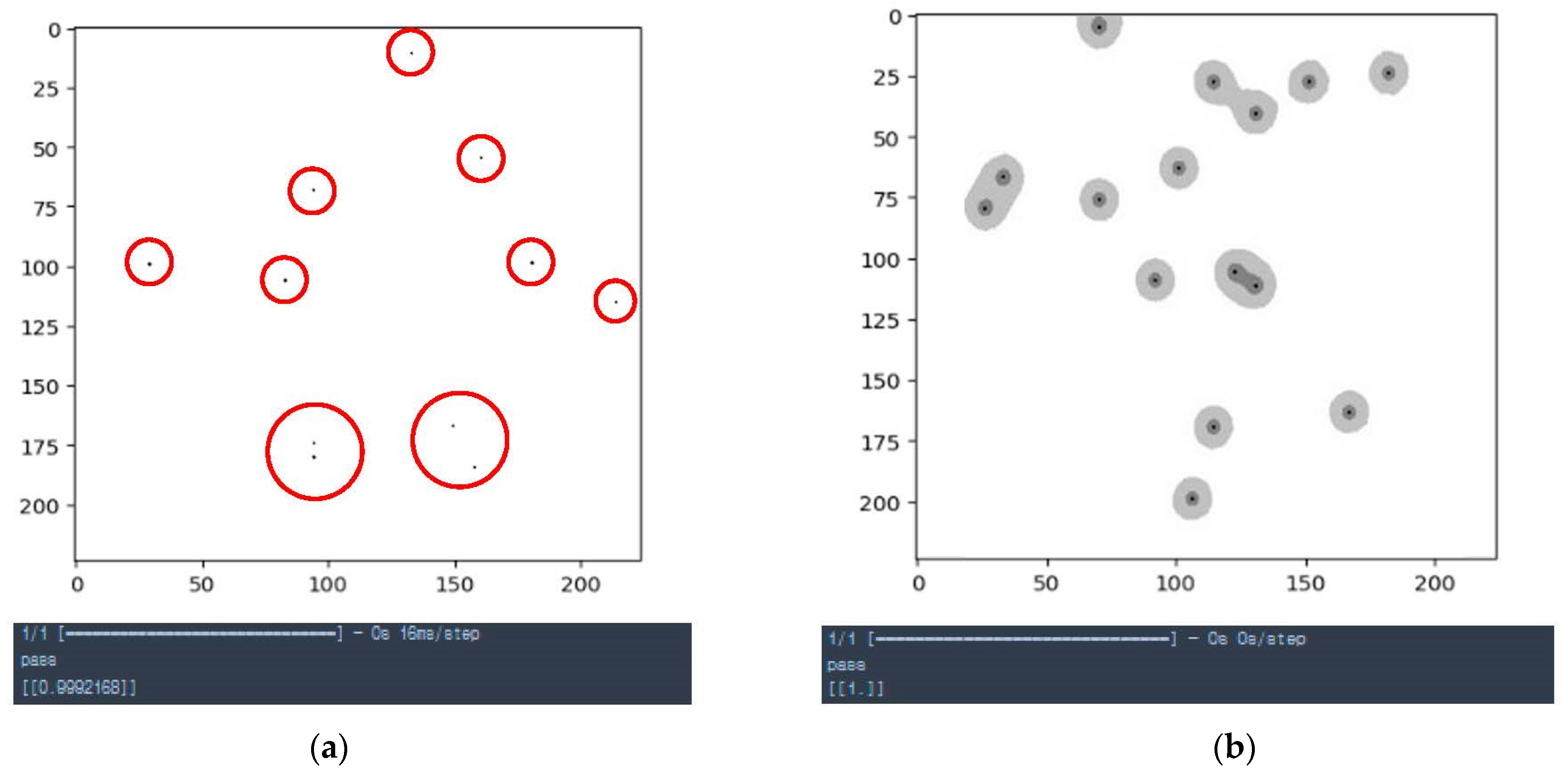
Figure 3 shows the results of applying the virtually generated defect images to the OLED cell-defect detection model. The model analyzes the size and distribution of defects inside the OLED cell to classify them as either pass or non-pass. The generated defect image in
Figure 3a is the initial defect image. The defect detection model classified it as “pass” with a confidence score of 0.999, indicating that the defect image
Figure 3a was classified as a defect-free sample. In contrast,
Figure 3b represents a defective image after 10,000 h. After 10,000 h, dark spots have formed due to humidity, causing the shapes of the dark spots to superposition. As a result, a dispersed pattern emerges around the center of the dark spots.
Analysis of the morphology of this defect revealed that, over time, the defects overlapped and darkened in appearance. In the case of the superposition defect image
Figure 3b, the model confidently classified it as “pass” with a score of 1, indicating that it was also classified as a defect-free sample.
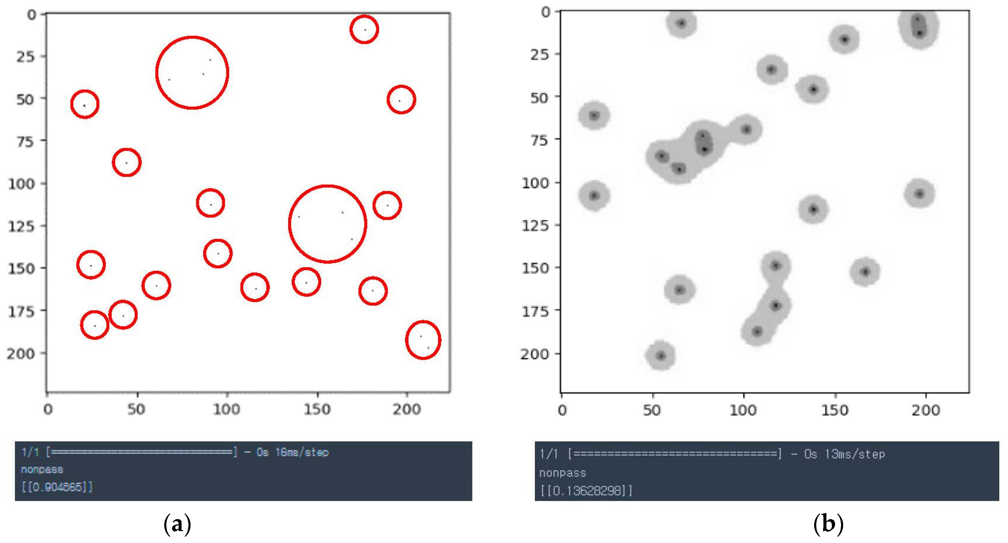
Figure 4 shows the results of applying the virtually generated defect images to the defect detection model. A total of 50 defect images were used, including 25 initial defect images classified as non-pass and 25 defect images after 10,000 h. The model correctly classified 42 out of 50 images as non-pass and the remaining seven images as passes. The criteria for defect detection are based on the distribution area, size, and number of defects in the image data. Notably, the initial defect image
Figure 4a was classified as a non-pass with a confidence score of 0.904, whereas the defect image after 10,000 h
Figure 4b showed overlapping defect patterns and was classified as a non-pass with a confidence score of 0.136.
The defect detection model used a classification threshold of 0.95. Scores equal to or higher than 0.95 were considered passes, while scores below 0.95 were considered non-pass. The overall accuracy for classifying 100 defect images, including initial and 10,000 h images, was 0.89. When applying real image data to the model for validation, it was possible to achieve an accuracy level of around 90%. These results demonstrate the effectiveness of the OLED cell defect detection model in accurately identifying and classifying defects based on the specified criteria.
5. Conclusions
In this study, deep learning algorithms were applied to detect defects in OLED cells. After comparing and analyzing various deep learning algorithms, VGG-16 was selected as the optimal algorithm. To facilitate defect identification, image data were necessary; however, due to the unique characteristics of the OLED industry, obtaining real image data was challenging. Therefore, a total of 2000 virtual dark spot image data were generated using the FEM Solver program. To select the optimal algorithm, a comparative analysis of the CNN, ResNet-50, and VGG-16 algorithms was conducted. Among them, VGG-16 demonstrated the best results, achieving an accuracy of 0.982, loss value of 0.053, specificity of 0.981, and recall of 0.985. VGG-16 was selected to create an OLED cell defect discrimination model based on these performance metrics. The model was trained using 1300 defect images and validated using 600 images, resulting in an accuracy of 0.988 and a loss value of 0.026. The performance of the model indicated its potential applicability in research. In the testing phase, the model achieved an accuracy of 0.89 with 100 defect images. Based on the study, it was possible to identify cell defects by implementing a cell defect classification model using the VGG-16 algorithm. While the study did not utilize actual images of dark spots, it successfully simulated the process of dark spot formation using virtual images, which were driven by relevant physical phenomena. While the current OLED industry relies on human inspection for defect detection, utilizing artificial intelligence technology to identify cell defects could contribute to the advancement of display manufacturing techniques. This applicability extends not only to the OLED industry but also to manufacturing and inspection processes within the broader electronics sector. Furthermore, the superior performance of the VGG-16 in image recognition highlights its ability to detect and classify cell defects efficiently. These results suggest that using deep learning technology for defect detection and classification can decrease the number of defective products and enhance production efficiency. Production costs can also be reduced by automating the defect discrimination process. Overall, this study highlights the potential contributions of deep learning in advancing the OLED display industry by improving production efficiency and quality. For future research, we propose a study that focuses on detecting defects in OLED cells by generating high-quality images using additional variables. Furthermore, by utilizing high-performance cameras for image data generation and improving the defect detection system with expert input, research on cell defect detection is expected to advance with greater precision.
Author Contributions
Author Contributions: M.-A.C. and T.-H.K.: Writing—Original Draft, Data Curation, Software, and Visualization. K.-A.K.: Writing—Review and Editing. M.-S.K.: Conceptualization, Validation, Writing—Review and Editing, and Project Administration. All authors have read and agreed to the published version of the manuscript.
Funding
The APC was funded by the Institute of Information and Communications Technology Planning and Evaluation (
www.iitp.kr, accessed on 1 March 2022), funded by the Ministry of Science and ICT (MSIT, Republic of Korea). (Project Number: 2022-0-00317).
Institutional Review Board Statement
Not applicable.
Informed Consent Statement
Not applicable.
Data Availability Statement
All data, models, and codes generated or used during the study are available from the corresponding author upon request.
Conflicts of Interest
The authors declare no conflict of interest.
References
- Korea Display Industry Association. OLED Display—Trends, Markets, Export, and Forecasts (2022–2027). 2023. Available online: https://www.kdia.org/display/graph.jsp (accessed on 16 August 2023).
- Statistics KOREA Government Official Work Conference—Growth, Market, and Trend. 2023. Available online: https://www.index.go.kr/unity/potal/main/EachDtlPageDetail.do;jsessionid=5D89Wr3XVFylRDEuVIORDfDTTlcHej9Bo4InA6c.node11?idx_cd=A0003 (accessed on 16 August 2023).
- Li, W.; Zhang, L.; Wu, C.; Cui, Z.; Niu, C. A new lightweight deep neural network for surface scratch detection. Int. J. Adv. Manuf. Technol. 2022, 123, 1999–2015. [Google Scholar] [CrossRef]
- Lee, J. 16-4: Invited Paper: Region-Based Machine Learning for OLED Mura Defects Detection. SID Symp. Dig. Tech. Pap. 2021, 52, 200–203. [Google Scholar] [CrossRef]
- Jo, S.H. Current Technology and Research Trend in Flexible Organic Light Emitting Diode Display. Polym. Soc. Korea 2021, 32, 435–439. [Google Scholar]
- Jo, G.H.; Heo, Y.W.; Cho, H.W.; Song, Y.J. Implementation of OLED Display Defect Detection System using CNN. J. Korean Inst. Inf. Technol. 2022, 20, 1–8. [Google Scholar]
- Azrain, M.M.; Mansor, M.R.; Siti, S.M.F.; Omar, G.; Sivakumar, D.M.; Lim, L.M.; Nordin, M.N.A. Analysis of mechanisms responsible for the formation of dark spots in organic light-emitting diodes (OLEDs): A review. Synth. Met. 2018, 235, 160–175. [Google Scholar] [CrossRef]
- Phatak, R.; Tsui, T.Y.; Aziz, H. Dependence of dark spot growth on cathode/organic interfacial adhesion in organic light emitting devices. J. Appl. Phys. 2012, 111, 054512. [Google Scholar] [CrossRef]
- Kim, S.; Park, J.; Han, B.; Park, S. Research on flexible OLED defect detection using deep learning. In Proceedings of the Korea Institute of Communications and Information Sciences Annual Conference, Jeju, Republic of Korea, 19–21 June 2019; pp. 767–768. [Google Scholar]
- Singh, R.B.; Kumar, G.; Sultania, G.; Agashe, S.S.; Sinha, P.R.; Kang, C. Deep Learning based MURA defect detection. EAI Endorsed Trans. Cloud Syst. 2019, 5, e6. [Google Scholar] [CrossRef][Green Version]
- Zhan, D.; Zhang, S. P-2.2: Anomaly Detection Based on Generative Adversarial Network in the Manufacturing Process of LCD/OLED Display Panels. SID Symp. Dig. Tech. Pap. 2021, 52, 460–466. [Google Scholar] [CrossRef]
- Ye, S.; Wang, Z.; Xiong, P.; Xu, X.; Du, L.; Tan, J.; Wang, W. Multi-stage few-shot micro-defect detection of patterned OLED panel using defect inpainting and multi-scale Siamese neural network. J. Intell. Manuf. 2023, in press. [CrossRef]
- Saberironaghi, A.; Ren, J.; El-Gindy, M. Defect detection methods for industrial products using deep learning techniques: A review. Algorithms 2023, 16, 95. [Google Scholar] [CrossRef]
- Nguyen, V.H.; Pham, V.H.; Cui, X.; Ma, M.; Kim, H. Design and evaluation of features and classifiers for OLED panel defect recognition in machine vision. J. Inf. Telecommun. 2017, 1, 334–350. [Google Scholar] [CrossRef]
- Chen, M.; Chen, S.; Wang, S.; Cui, Y.; Chen, P. Accurate segmentation of small targets for LCD defects using deep convolutional neural networks. J. Soc. Inf. Disp. 2023, 31, 13–25. [Google Scholar] [CrossRef]
- Guan, Q.; Wang, Y.; Ping, B.; Li, D.; Du, J.; Qin, Y.; Xiang, J. Deep convolutional neural network VGG-16 model for differential diagnosing of papillary thyroid carcinomas in cytological images: A pilot study. J. Cancer 2019, 10, 4876–4882. [Google Scholar] [CrossRef] [PubMed]
- Geng, L.; Zhang, S.; Tong, J.; Xiao, Z. Lung segmentation method with dilated convolution based on VGG-16 network. Comput. Assist. Surg. 2019, 24, 27–33. [Google Scholar] [CrossRef] [PubMed]
- You, S.Y.; Park, I.H.; Kim, G.T. Extraction of the OLED Device Parameter based on Randomly Generated Monte Carlo Simulation with Deep Learning. J. Semicond. Disp. Technol. 2021, 20, 131–135. [Google Scholar]
- Sadeghipour, K.; Dopkin, J.A.; Li, K. A computer aided finite element/experimental analysis of induction heating process of steel. Comput. Ind. 1996, 28, 195–205. [Google Scholar] [CrossRef]
- Han, D.H.; Jeong, Y.H.; Kang, M.S. A Study on OLED Cell Simulation and Detection Phases Based on the A2G Algorithm for Artificial Intelligence Application. Appl. Sci. 2023, 13, 8016. [Google Scholar] [CrossRef]
- Okada, T.; Yoshida, A.; Tsuji, T. Dark spot growth and its acceleration factor in organic light-emitting diodes with single barrier structure. Jpn. J. Appl. Phys. 2017, 56, 060305. [Google Scholar] [CrossRef]
- Lee, E. Simulation of the thin-film thickness distribution for an OLED thermal evaporation process. Vacuum 2009, 83, 848–852. [Google Scholar] [CrossRef]
- Shin, H.W.; Noh, I.W.; Lee, Y.L.; Choi, S.K.; Lee, S.W. A Study on Data Generation Methods for Defect Diagnosis Accuracy Enhancement in the Directed Energy Deposition Process. Trans. Korean Soc. Mech. Eng. 2023, 47, 519–525. [Google Scholar] [CrossRef]
- Jung, H.Y.; Lim, M.E.; Han, Y.W.; Park, H.D.; Choi, J.H. Generating Artificial Blood Pressure Data using GAN. In Proceedings of the KIIT Conference, Bhubaneswar, India, 27–30 October 2017; pp. 201–202. [Google Scholar]
- Yoo, J.S.; Park, S.C. A Study of Image Filtering Method by Using Pixel Value. Soc. Comput. Des. Eng. 2016, 13, 408–411. [Google Scholar]
- Byun, M.J.; Jeon, E.J.; Kim, J.S.; Hwang, J.J.; Jung, S.H. Classification of caries and sound teeth using VGG-16 deep learning algorithm. J. Korean Acad. Oral Health 2021, 45, 227–232. [Google Scholar] [CrossRef]
- Seo, S.; Ko, Y.; Yu, S.; Jung, K. Development of Checker Switch Defect Detection System using CNN Algorithm. J. Korean Soc. Mech. Eng. 2019, 18, 38–44. [Google Scholar]
- Simonyan, K.; Zisserman, A. Very deep convolutional networks for large-scale image recognition. arXiv 2014, arXiv:1409.1556. [Google Scholar]
| Disclaimer/Publisher’s Note: The statements, opinions and data contained in all publications are solely those of the individual author(s) and contributor(s) and not of MDPI and/or the editor(s). MDPI and/or the editor(s) disclaim responsibility for any injury to people or property resulting from any ideas, methods, instructions or products referred to in the content. |
© 2023 by the authors. Licensee MDPI, Basel, Switzerland. This article is an open access article distributed under the terms and conditions of the Creative Commons Attribution (CC BY) license (https://creativecommons.org/licenses/by/4.0/).


