Article

COX2-Inhibitory and Cytotoxic Activities of Phytoconstituents of Matricaria chamomilla L.

1
Department of Pharmaceutical Biology, Institute of Pharmaceutical and Biomedical Sciences, Johannes Gutenberg University, Staudinger Weg 5, 55128 Mainz, Germany
2
National Center for Natural Products Research (NCNPR), School of Pharmacy, University of Mississippi, Oxford, MS 38677, USA
*
Author to whom correspondence should be addressed.
Appl. Sci. 2023, 13(15), 8935; https://doi.org/10.3390/app13158935
Submission received: 9 April 2023 / Revised: 24 July 2023 / Accepted: 29 July 2023 / Published: 3 August 2023
(This article belongs to the Special Issue Natural Compound and Cellular Mode of Action)

Abstract

:
Chamomile tea is a popular beverage and herbal remedy with various health benefits, including antioxidant and antimicrobial activities and beneficial effects on metabolism. In this study, we investigated the inhibitory activities of secondary metabolites from Matricaria chamomile L. against COX2, an enzyme involved in inflammation and linked to cancer development. The cytotoxicity of the compounds was also evaluated on a panel of 60 cancer cell lines. Myricetin, one of the COX2-inhibiting and cytotoxic compounds in chamomile tea, was further studied to determine a proteomic expression profile that predicts the sensitivity or resistance of tumor cell lines to this compound. The expression of classical mechanisms of anticancer drug resistance did not affect the responsiveness of cancer cells to myricetin, e.g., ATP-binding cassette (ABC) transporters (ABCB, ABCB5, ABCC1, ABCG2), tumor suppressors (p53, WT1), and oncogenes (EGFR, RAS), whereas significant correlations between myricetin responsiveness and GSTP expression and cellular proliferation rates were observed. Additionally, Kaplan–Meier survival time analyses revealed that high COX2 expression is associated with a worse survival prognosis in renal clear cell carcinoma patients, suggesting a potential utility for COX2 inhibition by myricetin in this tumor type. Overall, this study provides insight into the molecular modes of action of chamomile secondary metabolites and their potential as cancer-preventive or therapeutic agents.

1. Introduction

Chamomile tea is widely known for its medicinal properties and is commonly used in traditional medicine for its antioxidant and antimicrobial activities [1,2]. In addition, Chamomile tea is also recognized for its value as a food due to its beneficial effect on metabolism, especially in the kidneys and liver. The safe use of chamomile was approved by the U.S. Food and Drug Administration (FDA’s GRAS) [3,4]. Several studies have shown the effect of chamomile against muscle spasms, rheumatism, and gastrointestinal disorders [5,6]. Furthermore, extensive literature promotes the effect of chamomile tea, such as the German chamomile Matricaria chamomilla L., against obesity, depression, stress, inflammation, and cancer [7,8,9,10,11,12]. Moreover, different clinical trials demonstrated not only the anti-inflammatory effect against eczema as well as chemo-and radiotherapy-induced mucositis by a chamomile tea extract and its bioactive secondary metabolites, apigenin and α-bisabolol [13,14]. Furthermore, analgesic effects have been shown against peripheral neuropathic pain, a side effect from chemotherapy, and apoptotic activity by inhibiting COX2 and NF-кB [15,16].
The German chamomile (M. chamomilla L., syn. Matricaria recutita L.) and Roman chamomile (Chamaemelum nobile L. (All)) are the most known chamomile species. They are both rich in flavonoids, phenolic acids, and terpenoids. The German chamomile contains more terpenoids and is more resistant to pathogens [17]. Furthermore, its flower extract contained more of these components than tea and essential oil [18]. Apigenin was one of the first compounds extracted from chamomile [19], which has been studied in the past for its anti-carcinogenic and anti-apoptotic effects [20,21,22]. The compounds inhibit cancer cell proliferation by blocking the activity of numerous proteins, including RelA/p65 and COX2 [23,24].
COX2 is an important enzyme that plays a key role in inflammation. It is involved in the production of prostaglandins, which promote inflammation. Inflammation is a necessary process for the body’s immune response against infection or injury, but if it becomes chronic, it can lead to a range of health problems, including cancer [25,26,27,28].
Our aim in this study was to investigate the molecular modes of action of secondary metabolites extracted from M. chamomile in terms of their inhibitory activity against COX2 and their cytotoxicity towards a panel of 60 cell lines from the National Cancer Institute. Then, we focused on myricetin, one of the COX2-inhibiting and cytotoxic chemical constituents of chamomile tea, to determine a proteomic expression profile that predicts the sensitivity or resistance of tumor cell lines to this compound. Finally, we mined the KM Plotter database to identify tumor types where high COX2 expression is associated with a worse Kaplan–Meier survival prognosis in cancer patients in an endeavor to envision a clinical situation where COX2 inhibition by myricetin might exert preventive or therapeutic potential.

2. Materials and Methods

2.1. Phytochemical Analysis

Chemicals

Nine standards were used as reference compounds. The compounds were cis-GMCA [(Z)-2-β-d-glucopyranosyloxy-4-methoxycinnamicacid] (2), trans-GMCA [(E)-2-β-d-glucopyranosyloxy-4-methoxycinnamic acid] (4), quercetagetin-7-O-β-d-glucopyranoside (3), apigenin-7-O-β-d-glucoside (6), apigenin 7-O-(6″-O-acetyl-β-d-glucopyranoside) (7), tonghosu [2-(2′,4′-hexadiynylidene)-1,6-dioxaspiro [4,4]-non-3-ene] (9) were isolated at the National Center for Natural Products Research (NCNPR), University of Mississippi, Oxford, MS, USA. The identity and purity of these compounds are confirmed by chromatographic (TLC, HPLC) methods, and the analysis of the spectral data (IR, 1D- and 2D-NMR, ESI-HRMS) in comparison with published spectral data is also confirmed. The purity of these isolated compounds was found to be greater than 95%. Chlorogenic acid (1) and apigenin (8) were purchased from Chromadex (Santa Ana, CA, USA, purity greater than 97%). Luteolin-7-O-β-d-glucoside (5) was purchased from Indofine Chemical Company, Inc. (Hillsborough, NJ, USA, purity greater than 97%). Acetonitrile and formic acid were of HPLC grade and purchased from Fisher Scientific (Fair Lawn, NJ, USA). Water for the HPLC mobile phase was purified using a Milli-Q system (Millipore) [29].

2.2. Instrumentation and Chromatographic Conditions of Liquid Chromatography with a Diode Array Detector–Quadrupole Time-of-Flight Mass Spectrometry (LC–DAD–QToF)

The phytochemical analysis using liquid chromatography–diode array detector–quadrupole time-of-flight mass spectrometry (LC–DAD–QToF) was performed according to recently published protocols [29,30]. An Agilent 1290 Series liquid chromatographic system was used (Agilent Technologies, Santa Clara, CA, USA), comprising a binary pump, a vacuum solvent degasser, an autosampler with 108-vial well-plate trays, a thermostatically controlled column compartment, and a diode array detector. The separation was achieved on an Agilent Poroshell 120 EC-C18 column (150 mm × 2.1 mm I.D., 2.7 µm). The mobile phase consisted of water with 0.1% formic acid (A) and acetonitrile with 0.1% formic acid (B) at a flow rate of 0.25 mL/min. Analysis was performed using the following gradient elution: 90% A/10% B to 60%A/40%B in 15 min; in the next 15 min to 100% B. Each run was followed by a 5-min wash with 100% B and an equilibration period of 5 min with initial conditions of 90% A/10% B. One microliter of sample was injected, and the column temperature was maintained at 40 °C. The mass spectrometric analysis was performed with a QToF-MS (Model #G6545B, Agilent Technologies, Santa Clara, CA, USA) equipped with an ESI source using the following parameters: drying gas (N2) flow rate, 13 L/min; drying gas temperature, 325 °C; nebulizer pressure, 35 psig; sheath gas temperature, 300 °C; sheath gas flow, 11 L/min; capillary voltage, 3500 V; nozzle voltage, 0 V; skimmer, 65 V; Oct RF V, 750 V; and fragmentor voltage, 175 V. All the operations, acquisition, and analysis of data were controlled using Agilent MassHunter Acquisition Software Ver. A.10.1 and processed with MassHunter Qualitative Analysis Software Ver. B.7.00. Each sample was analyzed in positive and negative modes over the range of m/z = 100–1700 and extended dynamic range (flight time to m/z 1700 at a 2 GHz acquisition rate). Accurate mass measurements were obtained by means of reference ion correction using reference masses at m/z 121.0509 (protonated purine) and 922.0098 [protonated hexakis (1H, 1H, 3H-tetrafluoropropoxy) phosphazine, or HP-921] in positive ion mode. The UV detection wavelength was in the range of 200–500 nm. The phytochemical analysis with UHPLC–DAD–QToF was performed according to recently published protocols [29,30].

2.3. Virtual Drug Screening and Molecular Docking

A list of more than 1000 chamomile compounds was first screened in silico against COX2 using PyRx [31,32]. The COX2 crystal structure was taken from the Protein Data Bank (PDB ID: 5F1A) [33,34]. This structure was prepared for virtual drug screening using the Chimera software [35]. We cut the crystal structure and used chain A within the catalytic domain of 5F1A. Using AutoDockTools-1.5.7rcl [34], water molecules were deleted, polar hydrogen atoms were added and merged, the missing atoms and bonds were repaired, and Kollman charges were added.
After performing the virtual screening with PyRx, 211 ligands from more than 1000 compounds were selected and subjected to molecular docking against COX2 using Autodock 4.2.6 [36,37,38]. A Grid-box was set based on the interaction of the known COX2 ligand salicylic acid with the amino acids of COX2-5F1A [39]. Its dimensions were 104 × 70 × 100 Å spacing 0.369 Å the Grid-center x = 45.667 Å, y = 28.779 Å and z = 232.841 Å. The Lamarckian Genetic Algorithm (LGA) was applied to seek the lowest binding energies (LBE, kcal/mol) and predicted inhibition constants (pKi, µM) with docking parameters set to 250 runs and 2,500,000 energy evaluations for each cycle. Mean values ± SD were calculated from each of the three independent dockings. BIOVIA Discovery Studio Visualizer 2021 [40] was used for the 3D visualization pictures of the interaction between the ligands and the amino acids of the crystal structure of COX2.
A total of 207 samples from chamomile was included in this present study (Supplementary Table S1).

2.4. Fluorometric COX2 Inhibitor Screening Assay

To validate the in silico results from virtual drug screening, we performed an in vitro COX2 inhibitor assay (BioVision Incorporate Berlin, Germany [41]) according to the manufacturer’s instructions. The assay is based on the fluorometric detection of prostaglandin G2, which is produced by the conversion of arachidonic acid by the catalytic activity of COX2. Selected phytochemicals of chamomile tea (apigenin, β-amyrin, β-eudesmol, β-sitosterol, daucosterol, farnesol, and myricetin) were chosen based on their LBE values from virtual drug screening. These compounds were tested for their COX2 inhibition in vitro at concentrations of 0.1 µM and 1 µM.

2.5. Pharmacological Testing and Gene/Protein Expression Profiling of Tumor Cell Lines

The Developmental Therapeutics Program of the National Cancer Institute (Bethesda, MA, USA) [42] used a series of human tumor cell lines from diverse origins (leukemia, melanoma, brain tumors, and carcinoma of the lung, colon, kidney, ovary, breast, or prostate) for drug screening [43]. The data of compound screening (log10IC50 values obtained by a sulforhodamine 123 assay) as well as transcriptomic and proteomic expression data were deposited at the NCI website [44]. For statistical correlation analyses, we used Pearson’s correlation test (WinStat, Kalmia Inc., Cambridge, MA, USA).

2.6. Growth Inhibition Assay

Resazurin reduction assay was conducted to measure the growth inhibitory activity of Chamomilla matricaria L. Human CCRF-CEM leukemia and human AMO1 multiple myeloma cells were seeded at a density of 1 × 10⁴ cells per well in 96-well plates using 100 µL of RPMI 1640 medium. These cells were then treated with the extract at 7 different concentrations, each diluted in 100 µL of medium, and incubated for 72 h. Similarly, HCT116 p53+/+ wildtype and drug-resistant HCT p53−/− knockout colon cancer cells were seeded (5 × 10³ cells/well) in 100 µL DMEM medium and allowed to adhere for 24 h. The next day, these cells were treated with the same series of extract concentrations and incubated for an additional 48 h [45]. On the final day of incubation, 20 µL of 0.01% resazurin solution from Promega (Mannheim, Germany) was added to each well and left to incubate for over 4 h at 37 °C. The fluorescence signal was then measured at an excitation wavelength of 544 nm and an emission wavelength of 590 nm using the Infinite M2000 Pro-plate reader from Tecan (Crailsheim, Germany). The experiment was independently replicated three times, and each concentration was tested with six replicates. The growth inhibitory effect of the treatment was represented as the percentage of cell viability and graphed as a dose–response curve. The IC50 value representing the concentration at which the growth was inhibited by 50% was calculated using Microsoft Excel 2021 (Version 2306 Build 16.0.16529.20164) [46].

2.7. Kaplan–Meier Survival Analysis

Data from more than 30,000 samples of 21 tumor types are deposited in the KMPlotter database [47,48,49]. The Kaplan–Meier statistics algorithm of this database was used to identify the prognostic value of COX2 mRNA expression for the survival time of cancer patients. We used a false discovery rate (FDR) calculation to exclude type I errors in multiple comparisons [50]. We included only Kaplan–Meier statistics with FDR rates ≤ 5%, indicating that not more than 5% of “declared” positive results were truly negative.

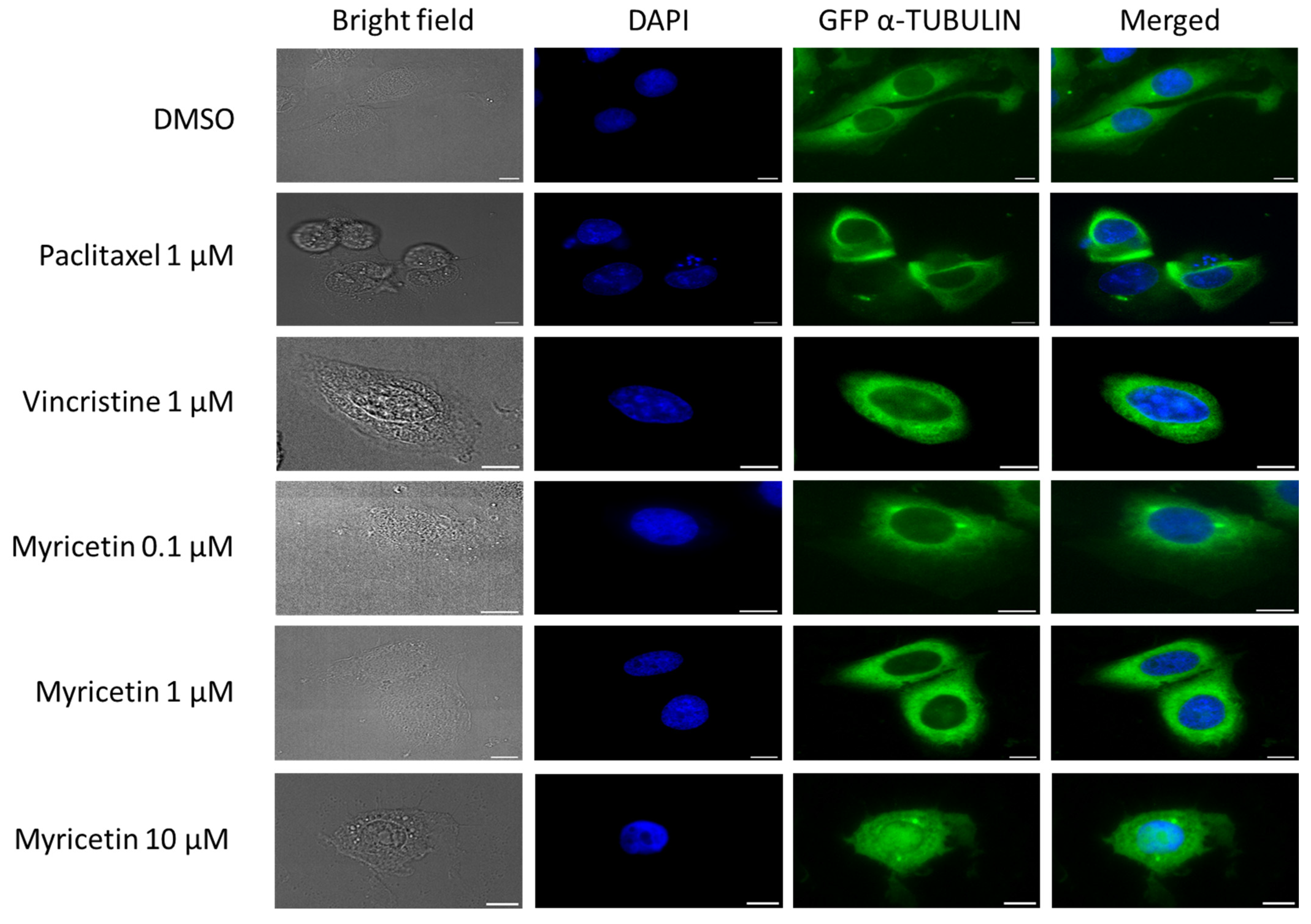
2.8. Immunofluorescence Microscopy of GFP Tagged α-Tubulin

Human U2OS osteosarcoma cells were left to attach for 24 h in μ-Slide 8 Well (30,000 cells/well) (ibidi, Gräfelfing, Germany). The next day, the cells were treated with myricetin (0.1, 1, 10 µM) (Sigma-Alderich Chemie GmbH, Taufkirchen, Germany) as well as paclitaxel (1 µM) and vincristine (1 µM) (both of the drugs were a gift from the pharmacy of the university hospital Johannes Gutenberg Mainz, Germany) as positive controls, along with DMSO as a negative control. After 24 h, cells were washed with PBS, fixed with 4% paraformaldehyde, and stained with 1 µg/mL of 4′6-diamidino-2-phenylindole (DAPI, Sigma Aldrich, Darmstadt, Germany) in the dark. Subsequently, the slides were immersed in Mounting Medium (ibidi, Gräfelfing, Germany). Widefield imaging was performed using a THUNDER Imager Live Cell (Leica Microsystems, Wetzlar, Germany) based on a Leica DMi8 microscope stand. Both transmission light and fluorescence images were acquired using a 63×/1.40 NA objective (HC PL APO CS2 63×/1.40 OIL UV). Fluorescence was excited with an LED light source (LED8, Leica) at 395 nm and 488 nm for imaging of DAPI and Tubulin–GFP, respectively. A quadband filter cube (DFT51010, Leica) split fluorescence excitation and emission light, and additional emission filters (460/80 for imaging of DAPI signals and 535/70 for imaging of GFP signals) were used to reduce bleed-through. The camera (Leica DFC9000 GTC) was operated in 2 × 2 binning mode, resulting in a pixel size of 206 nm measured in the object plane. Exposure times were set to 200 ms (GFP–Tubulin), 25 ms (DAPI), and 150 ms (differential interference contrast (DIC)). Images were analyzed with Image J software (National Institute of Health, Bethesda, MD, USA). The methodology of this experiment was described by us [51] and the microscopy techniques by Marton Gelléri (Institute of Molecular Biology gGmbH (IMB), Mainz, Germany).

3. Results

3.1. Phytochemical Analysis

The phytochemical analysis of chamomile extract was conducted using liquid chromatography–diode array detector–quadrupole time-of-flight mass spectrometry (LC–DAD–QToF). As shown in Figure 1, 21 phytochemicals were tentatively detected.

3.2. Cytotoxicity Assay

To examine the activity of chamomile extract against cancer cell lines, we have conducted growth inhibition assays. The extract revealed cytotoxicity against different cancer types. The IC50 values of the two hematopoietic cell lines CCRF-CEM and AMO1 (23.7 ± 4.7 µg/mL to 28.0 ± 2.2 µg/mL) were lower than those of the colon cancer cell lines. Wildtype HCT116 p53+/+ had an IC50 value of 28.0 ± 2.2 µg/mL, and HCT116 p53−/− knockout cells had an IC50 value of 74.3 ± 0.4 µg/mL. Hence, the p53 knockout cells were 2.65-fold more resistant to the chamomile extract than the p53 wild-type cells (Figure 2).

3.3. Molecular Docking In Silico

To investigate the full potential of chamomile tea, a total of 212 chamomile compounds were docked against human COX2 (PDB: 5F1A) using the PyRx program. Thirty of these 212 compounds were selected as the most abundant for further analysis. Table 1 shows the low binding energies (LBE, kcal/mol) and the predicted inhibition constants (pKi, µM) of these 30 ligands. The LBE and pKi values of these phytochemicals correlated significantly with each other (Pearson correlation test, p = 7.53 × 10 − 20; r = 0.574; Figure 1, 1st line left panel). The LBE values of 27 out of 30 substances were smaller than −6 kcal/mol (cut-off) and ranged from −11.73 (±0.44) kcal/mol (β-sitosterol) to −6.01 (±<0.01) kcal/mol (P-cymene). The pKi values of these 27 compounds were in a range from 0.003 (±0.002) µM to 39.57 (±<0.01) µM. Seven of the 27 compounds were selected because these substances were found in the NCI database (dtp.cancer.gov, accessed on 1 July 2023), whose data are required for further investigations. Celecoxib was used as a positive control drug since it is a well-known COX2 inhibitor. As shown in Figure 3 (1st line, middle, and right panel), the compounds were bound to three domains of COX2. Myricetin, daucosterol, and β-amyrin interacted with the first domain; β-sitosterol, apigenin, farnesol, and β-eudesmol with the second domain; and celecoxib with the third domain. Lines 2–5 of Figure 1 depict the three-dimensional binding poses of the compounds and the interacting amino acids.

3.4. Inhibition of COX2 Enzyme Activity In Vitro

To exemplarily verify the in silico predicted interaction of the compounds with COX2, we measured the enzymatic activity upon treatment with the selected seven phytochemicals. Figure 4A illustrates the rest activity of COX2 after treatment with β-sitosterol, β-amyrin, β-eudesmol, daucosterol, apigenin, myricetin, and farnesol at concentrations of 0.1 and 1 µM. β-Sitosterol exerted the highest inhibitory effect on COX2, with percentages of 92% and 98%, respectively. By contrast, apigenin had the lowest inhibitory activity (42% and 90%, respectively). Then, we corrected the percentages of COX2 rest activity upon treatment with 0.1 µM of the compounds in vitro with the LBE values in silico and found a significant correlation (Pearson collection test, p = 0.030; r = 0.736) (Figure 4B). We also observed a significant correlation upon treatment with 1 µM (p = 0.042; r = 0.692) (Figure 4C).

3.5. Cytotoxicity against Tumor Cells In Vitro

Since there is a mechanistic link between inflammation and carcinogenesis [52], we were interested in exploring the COX2-inhibitory activity of the selected compounds. The cytotoxicity of the selected compounds towards 60 cell lines of different tumor origins (leukemia, melanoma, brain tumors, carcinoma of the colon, ovary, breast, kidney, lung, or prostate) determined by a sulforhodamine B assay was mined in the NCI database (dtp.cancer.gov; accessed on 1 July 2023). The responsiveness of these cell lines is plotted as mean log10IC50 values for each tumor type in Figure 5. β-Sitosterol and daucosterol were minimally active against the tumor cell lines, while apigenin, myricetin, and farnesol were most active (Figure 5A). Celecoxib, the control drug, was the most active. β-Amyrin was not included in the NCI database. Therefore, we detected the cytotoxicity of this compound in CCRF-CEM cells which are also included in the NCI cell line panel and found that this compound did not affect these tumor cells. Since we previously investigated apigenin and farnesol in the NCI cell line panel [53,54,55,56], we focused on myricetin in the present study. The inhibitory activity of myricetin against the tumor cells shown in Figure 5B demonstrated that this compound was most active against colon and lung cancer cell lines. For comparison, celecoxib as a control drug was most active against leukemia and prostate cancer cell lines (Figure 5C).

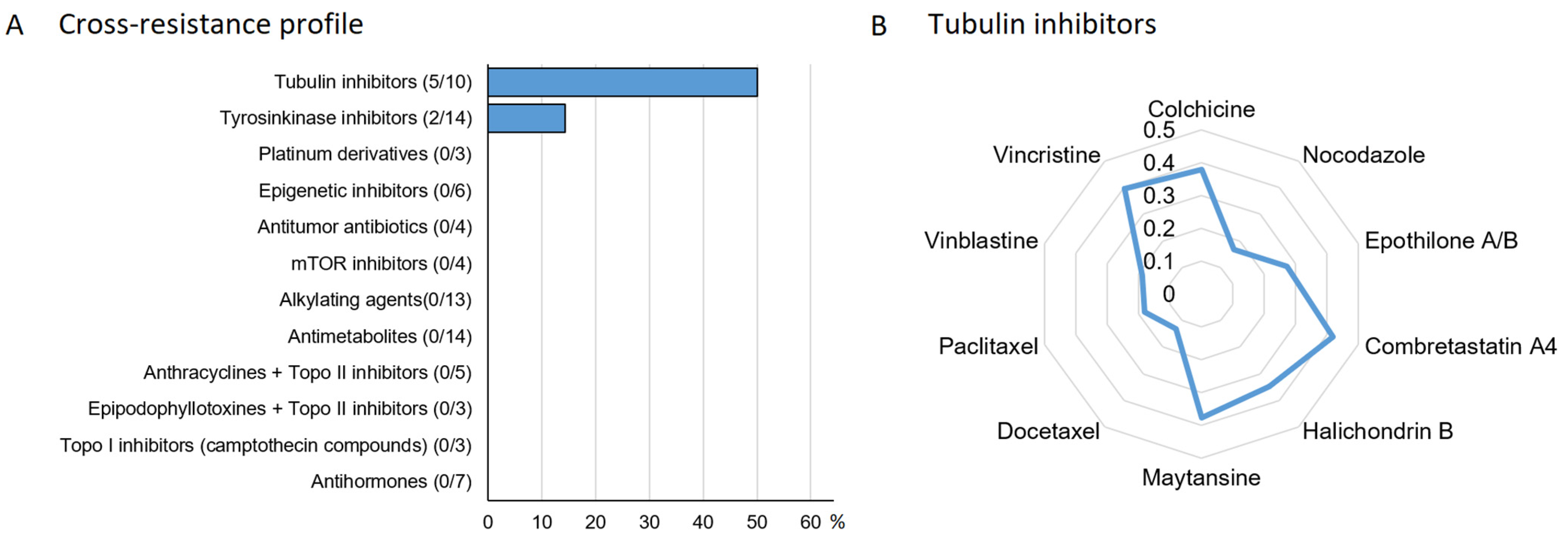
3.6. Oncobiogram Analysis

It is well accepted that natural products usually exert their bioactivity through multiple rather than single mechanisms [57]. Therefore, we assumed that myricetin might not only exert COX2-inhibitory activity. For this reason, we correlated the log10IC50 values for myricetin in 60 tumor cell lines to those of 91 standard anticancer compounds from diverse pharmacological classes. Significant correlations in cellular responsiveness were observed between myricetin and five of the ten tubulin inhibitors and two of the fourteen tyrosine kinase inhibitors. Correlations with drugs of other classes were not found (Figure 6A). A closer look at which tubulin inhibitor is correlated to myricetin is shown in Figure 6B.

3.7. Effect of Myricetin on α-Tubulin

To validate the results obtained from the oncobiogram analysis that myricetin might interact with tubulin, U2OS cells transfected with GFP α-tubulin were subjected to myricetin treatment at concentrations of 0.1, 1, and 10 µM for 24 h. The effect of myricetin on microtubules is depicted in Figure 7. Indeed, myricetin inhibits microtubules. In contrast to the well-organized microtubules seen in the untreated control cells, the myricetin-treated cells displayed shortened microtubule fragments that condensed around the nucleus, similar to vincristine-treated cells. Conversely, paclitaxel-treated cells exhibited extended and disorganized tubulin. Additionally, at the higher concentration of 10 µM myricetin, not only was the polymerization of microtubules significantly hindered (as evident from the reduced density of microtubules), but there were also multiple spindle poles observed in the area of the nucleus.

3.8. Proteome Analysis

To gain more insight into the determinants that define the sensitivity or resistance of cell lines to myricetin, the expression of a total of 3171 proteins in the 60 NCI cell lines deposited at the NCI database (dtp.cancer.gov accessed on 23 July 2023) was correlated to the log10IC50 values of myricetin using COMPARE analysis. A compilation of the top 40 proteins (20 directly correlating and 20 inversely correlating with myricetin) and their biological functions is compiled in Supplementary Table S2.
As a next step, we clustered the expression profiles using the hierarchical Ward cluster method within the first dimension and the log10IC50 values for myricetin in the second dimension. This two-dimensional clustering generated a color-coded heat map (Figure 8). Four major clusters were obtained for the 40 proteins (clusters A–D), and another three clusters appeared for the tumor cell lines tested (clusters 1–3). The cellular responsiveness of the cell lines to myricetin was categorized by defining the cell lines as being sensitive if their individual log10IC50 values were smaller than the median value across all cell lines and as being resistant if the individual log10IC50 value was higher than the median. By using the χ2 test, we calculated the statistical difference between the sensitive and resistant cell lines. Indeed, the distribution of clusters 1 and 2 (containing mainly myricetin-resistant cell lines) and cluster 3 (mainly containing sensitive cell lines) was statistically significant (p = 4.89 × 10⁻⁴).

3.9. Drug Resistance Profiling of Myricetin

Moreover, we addressed the question of whether myricetin is involved in classical drug resistance phenotypes of ABC-transporters (P-glycoprotein, ABCB5, ABCC1, and ABCG2), as well as oncogenes (EGFR), tumor suppressors (TP53, WT1), and others (heat shock protein HSP90, glutathione S-transferase π, and the proliferation rate of the cell lines) (Table 2). We did not observe statistically significant correlations between the log10IC50 values for myricetin and any of the resistance parameters except for GSTP and cellular proliferation rates. This indicates that the effectiveness of myricetin may be limited by these two drug resistance mechanisms, while all other resistance mechanisms to established anticancer drugs may not be relevant for myricetin.

3.10. Survival Analysis

Finally, we explored the relevance of COX2 expression for the survival prognosis of cancer patients. We speculated that a COX2 inhibitor (e.g., myricetin) should be more effective in tumors with high COX2 expression. A second assumption was that the connection between inflammation and COX2 expression is related to cancer growth, which might imply that high expression is related to short survival. Hence, COX2 inhibitors might contribute to the prolongation of the survival prognosis. Therefore, we performed Kaplan–Meier survival analyses using the KMPlotter database. The analysis of 21 tumor types revealed that high COX2 mRNA expression significantly correlated with shorter overall survival times in patients suffering from renal clear cell carcinoma than low COX2 expression (Figure 9A; p = 1.2 × 10⁻⁴). Refining the analyses within this group of patients showed that female patients with renal clear cell carcinoma with high COX2 expression died significantly earlier than those with low COX2 expression (Figure 9B; p = 3 × 10⁻⁵). Similarly, patients with low neoantigen load and high COX2 expression in their renal clear cell tumors had a worse survival prognosis than those with high COX2 expression (Figure 9C; p = 3 × 10⁻⁵). This data indicates that COX2-inhibiting compounds from chamomile tea might exert beneficial effects in the prevention and treatment of this tumor type.

4. Discussion

Chamomile tea has been widely utilized as a spice for food preservation and in traditional medicine. Its phenolic and flavonoid attributes offer anti-inflammatory and antioxidant properties [58]. Extensive research has been conducted on the cellular and molecular mechanisms of chamomile tea constituents against cancer, such as apigenin [59]. Since inflammation is related to carcinogenesis [60], we focused this study on the phytochemical constituents of chamomile tea and their effects against COX2, as well as their anti-inflammatory and cytotoxic activities on cancer cells.
In understanding the beneficial effect, phytochemistry plays a crucial role in characterizing the composition of secondary plant metabolites [61]. In chamomile tea, more than 200 substances have been identified from the classes of flavonoids, terpenoids, alkaloids, tannins, and polyphenols [62]. Other teas also contain compounds of these chemical classes, with differences in the specific types and quantities of their substances. For example, chamomile tea does not contain caffeine, while black, green, pu-erh, and oolong teas contain varying high amounts of caffeine [63]. Catechin is found in a very high quantity in green tea due to its minimized antioxidant capacity compared to chamomile, black, and peppermint tea [64,65], and chamomile tea is also the richest in apigenin in contrast to peppermint tea [66]. The question is whether or not the interaction between these different substances may be synergetic or additive. We suggest that the death of cancer cells is more probable if exposed to a combination of various substances in an additive manner rather than surrendering in a synergistic way, which means that the cell has an evolutionary mechanism predisposed to it. Moreover, the concept has been explored as to whether phytochemicals may act synergistically or additively as chemopreventive agents or if combined with cancer drugs for therapeutic intervention. This has been documented in several studies [67,68,69].
COX2 is an enzyme that plays a major role in inflammation and carcinogenesis. Since it is produced only during inflammation to catalyze the conversion of arachidonic acid to different prostaglandins [70]. Furthermore, arachidonic acid derivatives, including endocannabinoids such as 2-arachidonyl-glycerol, are capable of selectively binding to COX2 to undergo oxygenation and catalysis, resulting in the synthesis of hydroxyl-endoperoxide analogs. The prostaglandin H analog is then converted into glyceryl prostaglandins, including prostaglandin E2 and prostaglandin I2, which have anti-inflammatory and proliferative properties [71,72].
The positive feedback loop between COX2 expression and PGE2 production is involved in multiple cellular mechanisms and pathways, which, among other functions, can contribute to the promotion of tumor growth [73]. Hence, the development of COX2 inhibitors is crucial for treating both inflammation and cancer. Unlike traditional non-steroidal anti-inflammatory drugs (NSAIDs) that can have severe gastrointestinal side effects due to the suppression of COX1, COX2 selective inhibitors celecoxib, rofecoxib, and valdecoxib have fewer side effects. Nevertheless, it has been observed clinically that COXIBs can lead to cardiovascular toxicity [74]. Considering this, we investigated the potential of chamomile tea to inhibit COX2 and its cytotoxic effects against different cancer types. For that purpose, we conducted resazurin assays to measure the growth-inhibitory effects of chamomile extract against hematopoietic tumor cell lines (leukemia, multiple myeloma) and colon cancer cells. Hematopoietic cancer cells are frequently more sensitive to cytotoxic agents than solid cancer cells. This was also observed with our chamomile extract. We used two colon cancer cell lines: a p53 wildtype and a knockout line. The tumor suppressor p53 is not only a major driver of carcinogenesis if it is mutated but also causes resistance to anticancer drugs [75]. In previous investigations, we found that HCT116 p53−/− cells were resistant to the standard anticancer drug doxorubicin [76]. The p53 knockout cells were also moderately but significantly resistant to the chamomile extract compared to p53 wildtype cells (2.65-fold).
We have also performed virtual screening and molecular docking. By calculating the lowest binding energies of more than 200 compounds, we were able to focus on a few promising molecules to conduct a COX2 inhibitor screening assay for testing their activity against COX2. Subsequently, we narrowed our focus on myricetin to study its effect against cancer in addition to inflammation by conducting multiple advanced analyses such as oncobiogram, proteome, and Kaplan–Meier survival analyses.
Our results from molecular docking and the COX2 inhibition screening assay were significantly correlative. Seven of our compounds (β-sitosterol, β-amyrin, dausosterol, β-eudesmol, apigenin, farnesol, and myricetin) demonstrated high affinity binding to COX2. Moreover, the 60 NCI analyses have shown that β-sitosterol, β-eudesmol, and daucosterol were less effective on cancer cells compared to apigenin, myricetin, and farnesol. As we previously worked on apigenin and farnesol [55,56], we turned our focus to myricetin in the present investigation. The latter compound was more active against colon and lung cancer than other tumor types. This correlated with previous studies revealing that colon and lung cancers, among other tumor types, exhibited excessive COX2 expression [77,78]. More importantly, cell proliferation and anti-apoptosis effects were activated in colon cancer by the binding of prostaglandin E2 to its corresponding prostaglandin E2 receptor and to its respective G-protein, which stimulated the release of epidermal growth receptor (EGR) responsible for the activation of extracellular kinase ERK and the AKT/PI3kinase pathway, and in lung cancer, the PGE2-EP1R-Gq complex triggered cell growth through the activation of phospholipase C and ERK [79].
An important aspect is that the expression of target proteins such as COX2 may not be of equal prognostic value in all cancer types. We mined the KM Plotter database to associate COX2 expression with survival times in various tumor types. This approach was recently also applied to antioxidant response genes and tumor suppressor genes [48,80].
High COX2 expression was significantly associated with a shorter survival probability in renal clear cell carcinoma patients, indicating that COX2 inhibition by myricetin might positively affect the survival times for patients suffering from this tumor type. Here, we found that high COX2 expression in renal clear cell carcinoma was associated with shorter overall survival times for patients, and female patients with high COX2 expression died significantly earlier than those with low COX2 expression. Moreover, patients with a low neoantigen load and high COX2 expression had a worse survival prognosis than those with high COX2 expression. It can be speculated that patients with low neoantigen loads might be less susceptible to immunotherapy approaches than those with high neoantigen loads and, thus, might be more immunotherapy-resistant [47].
Our findings suggest that targeting COX2 expression with compounds such as myricetin could be a promising strategy for improving the survival prognosis of patients with renal clear cell carcinoma, especially tumors with low neoantigen loads. It may be hypothesized that myricetin might be effective in this subgroup of patients.
Furthermore, we also found from the oncobiogram analysis that myricetin may also inhibit tubulin and tyrosine kinases. Substantially, this finding has been reinforced with our obtained data from immunofluorescence microscopy of the effect of myricetin on α-tubulin. Myricetin at concentrations of 0.1 and 1 µM has blocked microtubule polymerization in a comparable manner as vincristine at 1 µM. Microtubules are composed of tubulin. They are responsible for the maintenance and stabilization of endothelial cells. The discovery of novel tubulin inhibitors plays an important role in tumor vasculature-based therapies [81]. Hence, the inhibition of tubulin by myricetin may not only be linked to the inhibition of COX2 but also to the inhibition of endothelial cell responses. Interestingly, many authors suggested that COX2 and PGE2 may work together to activate receptor tyrosine kinases, which are involved in regulating cell growth and division, e.g., in colon cancer [82,83]. In addition, COX2 overexpression in cancer cells elevated PGE2 production, which in turn activated vascular endothelial growth factor (VEGF) and/or other endothelial cell responses, promoting angiogenesis [73,79,84].
Additionally, in our analysis, we found that myricetin is not related to classical anticancer drug resistance mechanisms such as ATP-binding cassette (ABC) transporters (P-glycoprotein/ABCB1, ABCB5, ABCC1, ABCG2), tumor suppressors (TP53, WT1), or oncogenes (EGFR, RAS), except for glutathione S-transferase P (GSTP) and cellular proliferation rates.
COX2 overexpression contributed to P-glycoprotein-mediated multidrug resistance via regulation of c-Jun N-terminal protein kinase (JNK), which phosphorylates the transcription factor c-Jun on its N-terminus at Ser63/73 (pc-Jun) in colorectal cancer [85]. This could be an explanation for the absence of cross-resistance of P-glycoprotein-expressing cells towards myricetin.
WT1 is a tumor suppressor that produces different transcription factors that promote cell growth and survival. While its mutation has been initially linked to childhood kidney cancer (Wilms tumor), studies have suggested that WT1 can play either a promoting or inhibitory role in tumor formation and apoptosis in many tumor types [86,87,88]. A combined therapy targeting COX2 and WT1 has been proposed, as the two pathways have been shown to synergistically promote tumor cell proliferation in lung cancer. The authors found that suppressing the COX2 pathway with celecoxib increased WT1 expression, which may explain why myricetin did not appear to affect cross-resistance in WT1-expressing tumors [89]. Knocking down the WT1 gene led to an upregulation of COX2, particularly in Wilms’ tumors, suggesting that inhibiting COX2 in combination with other targeted treatments could be beneficial in treating cancer [90,91]. P53 is also a tumor suppressor that regulates cell growth and promotes apoptosis (controlling cell cycle arrest) but, in parallel, also plays a crucial role in the progression of many cancer types when it is mutated. The TP53 gene is mutated in several cancer types, including lung and colon cancer, and regaining its functions is an important therapeutic approach in cancer [92,93,94]. Genotoxic stress-induced p53 activated the Ras/Raf/MAPK pathway that led to COX2 expression downstream [95]. Moreover, COX2 promoted tumorigenesis by inhibiting the transcriptional activity of normal functional p53 and DNA damage-induced apoptosis. Further, The COX2-inhibitor NS-398 blocked the interaction between COX2 and p53, which activated apoptosis [96]. It has been discussed that the crosstalk between COX2 and p53 happens through different pathways [97].
There is a lot of published evidence that mutant p53 contributes to multidrug resistance [94]. It has been proposed to reduce drug resistance by increasing wild-type p53 in cancer. Myricetin increases the expression of the p53 protein and alters the function of Bcl-2 proteins, resulting in increased activity of apoptotic pathways such as Bax and Bak, which are also regulated by p53 [98]. This finding is particularly relevant because previous investigations showed that the PGE2 ligand–receptor complex induced by COX2 inhibited the activation of Bax [99]. Consequently, we hypothesize that the crosstalk between p53 and COX2 and the COX2 inhibition by myricetin may explain why p53-mutant cells were not cross-resistant to myricetin. Nevertheless, we also found HCT116p53−/− cells were resistant to our chamomile extract compared to HCT116p53+/+ cells. An explanation could be that other compounds, but not myricetin, in the chamomile extract were responsible for the resistance.
The epidermal growth factor receptor (EGFR) is a key factor in cell growth and is highly expressed in various carcinoma types, with lung and colon cancers being among the most affected. The activation of EGFR is essential for many cancer pathways, making it a prime target for cancer therapy. Despite the effectiveness of EGFR tyrosine kinase inhibitors in lung cancer treatment, resistance often develops through the activation of alternative pathways such as c-Met, HGF, and AXL, as well as divergent downstream pathways such as Ras/Raf/MAPK, Akt, and STAT. The Ras family, which includes H-Ras, N-Ras, and K-Ras, is one of the earliest discovered oncogenes and plays a crucial role in cell growth and proliferation by binding to the Raf and MAPK pathways controlled by EGFR. Mutations in Ras confer resistance to cancer drugs, making them a challenge in cancer therapy [100,101,102,103]. It is now well known that Ras mutant tumors are inherently resistant to anticancer agents, necessitating the development of alternative treatment strategies, and several agents targeting signaling components in the MAPK and PI3K pathways downstream of mutant K-Ras are currently undergoing clinical trials [104].
Myricetin has been reported to interact with and alter pathways such as AKT [105,106,107,108], which are linked to COX2. Myricetin-mediated COX2 inhibition might block EGFR- and Ras-related signaling cascades, potentially making tumor cells more sensitive to drugs and more susceptible to the blockade of cancer cell survival.
In the NCI cell line panel, there was no cross-resistance of myricetin to epirubicin, maytansine, vinblastine, pancratistatin (ABC transporters control drugs), erlotinib (EGFR inhibitor), melphalan (Ras control drug), 5-fluorouracil (p53 control drug), or ifosfamine (WT1 control drug). Nevertheless, there was a significant correlation with etoposide, which was used as the control drug for GSTP1. This is an enzyme mostly known for its detoxification, cytoprotective, and anti-apoptotic activities [109]. In aberrant crypt foci (ACF) in colonic adenoma, COX2 was inactive and GSTP1 was active, allowing ACF proliferation. ACF were also resistant to deoxycholic acid-induced apoptosis through this detoxification process rather than inflammation [110]. The activity of plant polyphenols and flavonoids against both GSTP1-1 and CS-X pumps on breast cancer cell lines has been investigated. Due to its multiple hydroxyl groups, myricetin had no inhibitory effect on GSTP1-1 and only a moderate effect on CS-X compared to two strongly active flavonoids, quercetin and luteolin [111]. GSTP1 plays an active role in cancer and contributes to drug resistance through its detoxification capability, and myricetin does not affect the function of GSTP1, which may explain the missing cross-resistance of GSTP1 to myricetin in the NCI cell line panel.
The main concept of our research efforts is to find cytotoxic compounds that can be used as functional food ingredients with negative side effects and that are able to bypass multidrug resistance or even provoke collateral sensitivity in resistant tumor cells [112,113,114,115]. Myricetin is a flavonoid present in tea, wine, fruits, and vegetables [116]. For decades, myricetin has been recognized for its antioxidant, anticancer, antidiabetic, and anti-inflammatory activities [117,118,119,120]. Furthermore, it has been reported that myricetin reduces resistance to anticancer drugs and other cytotoxic agents. Among them were camptothecin and podophyllotoxin by inducing topoisomerase I/II-DNA complex, cisplatin in ovarian and colon cancer cells by inducing apoptosis through Bax/Bcl-2 proteins, resveratrol in an additive manner by inhibiting 12-O-tetradecanoylphorbol-13-acetate (TPA) and EGF, as well as vincristine by blocking ABCC1 (MRP1) and ABCC2 (MRP2) [98,119,121,122].
There are findings that suggest that exposure to carcinogens may not only initiate carcinogenesis but also confer resistance to the very agents used to treat tumors [123,124,125,126]. Moreover, inflammation plays a significant role in the process of carcinogenesis, particularly in prostate and colon cancer. It is common sense that chronic inflammation leads to DNA damage, tissue damage, and abnormal cell growth that can ultimately result in cancer. Chemoprevention is a promising approach to preventing cancer by using natural or synthetic compounds to inhibit the development of cancer at its earliest stages. Several studies have demonstrated that anti-inflammatory agents prevent the initiation, promotion, and progression of cancer by modulating various molecular targets. Therefore, targeting inflammation through chemoprevention may represent a promising strategy for preventing cancer development and progression [127,128,129,130,131].
Therefore, it is straightforward to elaborate on chamomile, which contains not only myricetin but also various other bioactive compounds, including apigenin, farnesol, quercetin, and luteolin, to act as chemopreventive agents. This notion was supported by our growth inhibition assay, where the chamomile extract demonstrated significant activity against several cancer cell lines. It could, thus, be hypothesized that chamomile tea may help to prevent cancer and also suppress cancer cell growth at the early stages of tumor development.
For this reason, we used proteome analysis to gain more insights into the molecular mechanisms underlying the sensitivity and resistance of cancer cell lines to myricetin. The expression of 3171 proteins was determined in the NCI cell line panel [42] and correlated with the log10IC50 values of these cell lines for myricetin in the present investigation. The top 20 directly correlating proteins (indicating myricetin resistance) and the top 20 inversely correlating proteins (indicating myricetin sensitivity) were subjected to hierarchical cluster analysis. The distribution of the myricetin-resistant cell lines in clusters 1 and 2 and the myricetin-sensitive cell lines in cluster 3 was statistically significant (p = 0.00049). The 40 relevant proteins associated with cellular myricetin responsiveness are involved in a variety of multifactorial interactions.
The formation of clusters A–D has been performed to better illustrate the distribution of protein expression in clusters 1–4 (i.e., red and green fields). The assembly of proteins in clusters A–D occurred according to their degrees of protein up- or downregulation in the specific tumor cell lines. A preferential assembly of the molecular and cellular functions of these proteins was not observed in clusters A–D. However, many proteins with functions relevant to cancer were identified from this proteomic analysis (Supplementary Table S2), e.g., proteins involved in mRNA metabolism (DDX39B, CSNK2A, RPL18A, MRTO4, DDX21, BRIX1, HINT2, YTHDC1, and EEF1A1), proteins involved in programmed cell death (TP53I3, DDRGK, EIF5A, PDCD5, and PDCD6), proteins involved in cell growth, differentiation, and genome stability (CDK2, NELFA, PODXL, PKN2, THOC5, and BLM), transcription factors and signal transduction proteins (ADNP, ZNF428, SSRP1, TSNAX, DUSPP3, RAB38, and TCP1), and others (FBO2, COL4A3, DNAJA2, GSN, MRC2, UGP2, TPP1, NUCB2, CTSB, CLTA, CTSD, and VPS33B). The exact mechanisms by which these proteins influence sensitivity or resistance to myricetin are largely unknown at present but deserve further investigation in the future. Clearly, the sensitivity and resistance of cancer cells to myricetin are multi-factorially determined. The identified proteins might be used to predict, prior to treatment, the responsiveness of cancer cells to myricetin and could guide the development of new therapeutic strategies for individualized cancer treatment.
These results are in agreement with a multitude of previous observations that natural products rather act by multiple mechanisms than by single mechanisms [57]. Additionally, the pharmacological effects of phytotherapeutics may result from synergetic interactions among multiple phytochemicals. Therefore, it is important to better understand the synergistic effects of herbal mixtures to develop more effective multi-target drugs with fewer side effects [132]. The generation of expression profiles to predict the response to chemotherapy is an important step toward individualizing chemotherapy [133,134]. In this context, the role of natural products cannot be ignored. If the profiles of standard drugs are known, then we could look for phytochemicals with fundamentally different expression profiles. This could provide a rational basis to make resistant and refractory tumors responsive to therapy again [135,136,137]. In the past, we have mainly used transcriptomics and epigenomics [138,139,140,141,142,143]. The significance of such profiles for cancer prevention with tea is also worth noting, as it has a different profile than cytostatic drugs. Overall, the use of expression profiles from natural products offers promising avenues for improving cancer treatment and prevention.

5. Conclusions

In conclusion, the effects of tea on cancer prevention and therapy are not solely attributed to apigenin, as previously reported [144,145,146], but to a combination of many other natural substances. In the present work, we focused particularly on myricetin due to its potential cancer-inhibiting bioactivity. However, the inhibitory effect of myricetin on cancer cells may be moderate and weaker than conventional anticancer drugs, making it more appropriate for cancer prevention than therapy. Nonetheless, the potential of myricetin and other natural substances in cancer prevention and treatment cannot be ignored. Therefore, our findings provide valuable insights into the potential of natural substances for cancer prevention and as an additive to cancer therapy. Ultimately, the development of effective cancer prevention strategies is critical to reducing the burden of this disease on individual cancer patients. The potential of myricetin for individualized treatment is worth further exploring in the future.

Supplementary Materials

The following supporting information can be downloaded at: https://www.mdpi.com/article/10.3390/app13158935/s1, Table S1: Phyto chemical profiling of Chamomile (Matricaria chamomilla L.); Table S2: Correlation of constitutive mRNA expression of genes identified by COMPARE analysis with log10IC50 values for myricetin of the NCI tumor cell lines.

Author Contributions

A.I.D. performed the experiments and analyses; B.A. performed the phytochemical analysis; I.A.K. edited the manuscript; and T.E. supervised the project and wrote the manuscript. All authors have read and agreed to the published version of the manuscript.

Funding

This research received no external funding.

Institutional Review Board Statement

Not applicable.

Informed Consent Statement

Not applicable.

Data Availability Statement

Data are available upon reasonable request.

Acknowledgments

We thank the Microscope Core Facility at the Institute of Molecular Biology (Marton Gelléri, Institute of Molecular Biology gGmbH (IMB), Mainz, Germany) for their kind training and technical support for the microscopy-related experiment.

Conflicts of Interest

The authors declare that there are no conflict of interest.

References

  1. McKay, D.L.; Blumberg, J.B. The Role of Tea in Human Health: An Update. J. Am. Coll. Nutr. 2013, 21, 1–13. [Google Scholar] [CrossRef]
  2. Srivastava, J.K.; Shankar, E.; Gupta, S. Chamomile: A Herbal Medicine of the Past with a Bright Future (Review). Mol. Med. Rep. 2010, 3, 895–901. [Google Scholar]
  3. Hassan, D.M.A.; Salah-Eldin, A.A. Amerolative Influence of Chamomile (Matricaria recutita L.) on Synthetic Food Additive Induced Probable Toxicity in Male Albino Rats. J. Food Dairy Sci. J. 2021, 12, 161–170. [Google Scholar] [CrossRef]
  4. CFR—Code of Federal Regulations Title 21; Food and Drug Administration: Silver Spring, MD, USA, 2023; p. 3.
  5. Sándor, Z.; Mottaghipisheh, J.; Veres, K.; Hohmann, J.; Bencsik, T.; Horváth, A.; Kelemen, D.; Papp, R.; Barthó, L.; Csupor, D. Evidence Supports Tradition: The in Vitro Effects of Roman Chamomile on Smooth Muscles. Front. Pharmacol. 2018, 9, 323. [Google Scholar] [CrossRef] [PubMed]
  6. Pirouzpanah, S.; Mahboob, S.; Sanayei, M.; Hajaliloo, M.; Safaeiyan, A. The Effect of Chamomile Tea Consumption on Inflammation among Rheumatoid Arthritis Patients: Randomized Clinical Trial. Prog. Nutr. 2017, 19, 27–33. [Google Scholar] [CrossRef]
  7. Bayliak, M.M.; Dmytriv, T.R.; Melnychuk, A.V.; Strilets, N.V.; Storey, K.B.; Lushchak, V.I. Chamomile as a Potential Remedy for Obesity and Metabolic Syndrome. EXCLI J. 2021, 20, 1261–1286. [Google Scholar] [CrossRef]
  8. Srivastava, J. Antiproliferative and Apoptotic Effects of Chamomile Extract in Various Human Cancer Cells. J. Agric. Food Chem. 2007, 55, 9470–9478. [Google Scholar] [CrossRef]
  9. Srivastava, J.K.; Pandey, M.; Gupta, S. Chamomile, a Novel and Selective COX-2 Inhibitor with Anti-Inflammatory Activity. Life Sci. 2009, 85, 663–669. [Google Scholar] [CrossRef] [Green Version]
  10. Chang, S.M.; Chen, C.H. Effects of an Intervention with Drinking Chamomile Tea on Sleep Quality and Depression in Sleep Disturbed Postnatal Women: A Randomized Controlled Trial. J. Adv. Nurs. 2016, 72, 306–315. [Google Scholar] [CrossRef]
  11. Gh, D.; Kermanian, S.; Mozaffari-Khosravi, H.; Dastgerdi, G.; Zavar-Reza, J.; Rahmanian, M. The Effect of Chamomile Tea versus Black Tea on Glycemic Control and Blood Lipid Profiles in Depressed Patients with Type 2 Diabetes: A Randomized Clinical Trial. J. Nutr. Food Secur. 2018, 3, 157–166. [Google Scholar]
  12. Ghamchini, V.M.; Salami, M.; Mohammadi, G.R.; Moradi, Z.; Kavosi, A.; Movahedi, A.; Bidkhori, M.; Aryaeefar, M.R. The Effect of Chamomile Tea on Anxiety and Depression in Cancer Patients Treated with Chemotherapy. J. Young Pharm. 2019, 11, 309–312. [Google Scholar] [CrossRef] [Green Version]
  13. Paula Gardiner Chamomile (Matricaria Recutita, Anthemis Nobilis). The Longwood Herbal Task Force and The Center for Holistic Pediatric Education and Research. 1999, pp. 1–21. Available online: https://tratamientocelular.com/papers/cmran.pdf (accessed on 31 March 2023).
  14. El Joumaa, M.M.; Borjac, J.M. Matricaria Chamomilla: A Valuable Insight into Recent Advances in Medicinal Uses and Pharmacological Activities. Phytochem. Rev. 2022, 21, 1913–1940. [Google Scholar] [CrossRef]
  15. Wu, B.Y.; Liu, C.T.; Su, Y.L.; Chen, S.Y.; Chen, Y.H.; Tsai, M.Y. A Review of Complementary Therapies with Medicinal Plants for Chemotherapy-Induced Peripheral Neuropathy. Complement. Ther. Med. 2019, 42, 226–232. [Google Scholar] [CrossRef]
  16. Salehi, B.; Jornet, P.L.; López, E.P.F.; Calina, D.; Sharifi-Rad, M.; Ramírez-Alarcón, K.; Forman, K.; Fernández, M.; Martorell, M.; Setzer, W.N.; et al. Plant-Derived Bioactives in Oral Mucosal Lesions: A Key Emphasis to Curcumin, Lycopene, Chamomile, Aloe Vera, Green Tea and Coffee Properties. Biomolecules 2019, 9, 106. [Google Scholar] [CrossRef] [PubMed] [Green Version]
  17. Tai, Y.; Ling, C.; Wang, C.; Wang, H.; Su, L.; Yang, L.; Jiang, W.; Yu, X.; Zheng, L.; Feng, Z.; et al. Analysis of Terpenoid Biosynthesis Pathways in German Chamomile (Matricaria recutita) and Roman Chamomile (Chamaemelum nobile) Based on Co-Expression Networks. Genomics 2020, 112, 1055–1064. [Google Scholar] [CrossRef] [PubMed]
  18. Nováková, L.; Vildová, A.; Mateus, J.P.; Gonalves, T.; Solich, P. Development and Application of UHPLC-MS/MS Method for the Determination of Phenolic Compounds in Chamomile Flowers and Chamomile Tea Extracts. Talanta 2010, 82, 1271–1280. [Google Scholar] [CrossRef]
  19. Power, F.B.; Browning, H. CCXI.—The Constituents of the Flowers of Matricaria Chamomilla. J. Chem. Soc. Trans. 1914, 105, 2280–2291. [Google Scholar] [CrossRef] [Green Version]
  20. Lefort, É.C.; Blay, J. Apigenin and Its Impact on Gastrointestinal Cancers. Mol. Nutr. Food Res. 2013, 57, 126–144. [Google Scholar] [CrossRef]
  21. Xu, Y.; Xin, Y.; Diao, Y.; Lu, C.; Fu, J.; Luo, L.; Yin, Z. Synergistic Effects of Apigenin and Paclitaxel on Apoptosis of Cancer Cells. PLoS ONE 2011, 6, e29169. [Google Scholar] [CrossRef] [Green Version]
  22. Patel, D.; Shukla, S.; Gupta, S. Apigenin and Cancer Chemoprevention: Progress, Potential and Promise (Review). Int. J. Oncol. 2007, 30, 233–245. [Google Scholar] [CrossRef] [Green Version]
  23. Srivastava, J.K.; Gupta, S. Extraction, Characterization, Stability and Biological Activity of Flavonoids Isolated from Cham-omile Flowers. Mol. Cell. Pharmacol. 2009, 1, 138. [Google Scholar] [CrossRef]
  24. Bhaskaran, N.; Shukla, S.; Srivastava, J.K.; Gupta, S. Chamomile: An Anti-Inflammatory Agent Inhibits Inducible Nitric Oxide Synthase Expression by Blocking RelA/P65 Activity. Int. J. Mol. Med. 2010, 26, 935–940. [Google Scholar] [CrossRef] [PubMed] [Green Version]
  25. Rajakariar, R.; Yaqoob, M.M.; Gilroy, D.W. COX-2 in Inflammation and Resolution. Mol. Interv. 2006, 6, 199. [Google Scholar] [CrossRef] [PubMed]
  26. Williams, C.S.; Mann, M.; DuBois, R.N. The Role of Cyclooxygenases in Inflammation, Cancer, and Development. Oncogene 2000, 18, 7908–7916. [Google Scholar] [CrossRef] [PubMed] [Green Version]
  27. Vardeh, D.; Wang, D.; Costigan, M.; Lazarus, M.; Saper, C.B.; Woolf, C.J.; Fitzgerald, G.A.; Samad, T.A. COX2 in CNS Neural Cells Mediates Mechanical Inflammatory Pain Hypersensitivity in Mice. J. Clin. Investig. 2009, 119, 287–294. [Google Scholar] [CrossRef] [Green Version]
  28. Seibert, K.; Masferrer, J.L. Role of Inducible Cyclooxygenase (COX-2) in Inflammation. Receptor 1994, 4, 17–23. [Google Scholar]
  29. Avula, B.; Bae, J.Y.; Wang, Y.H.; Wang, M.; Ali, Z.; Khan, I.A. Chemical Profiling and Characterization of Anthraquinones from Two Bulbine Species and Dietary Supplements Using Liquid Chromatography–High Resolution Mass Spectrometry. J. AOAC Int. 2021, 104, 1394–1407. [Google Scholar] [CrossRef] [PubMed]
  30. Avula, B.; Sagi, S.; Masoodi, M.H.; Bae, J.Y.; Wali, A.F.; Khan, I.A. Quantification and Characterization of Phenolic Compounds from Northern Indian Propolis Extracts and Dietary Supplements. J. AOAC Int. 2020, 103, 1378–1393. [Google Scholar] [CrossRef] [PubMed]
  31. Supianto, A.A.; Nurdiansyah, R.; Weng, C.W.; Zilvan, V.; Yuwana, R.S.; Arisal, A.; Pardede, H.F.; Lee, M.M.; Huang, C.H.; Ng, K.L. Cluster-Based Text Mining for Extracting Drug Candidates for the Prevention of COVID-19 from the Biomedical Literature. J. Taibah Univ. Med. Sci. 2023, 18, 787–801. [Google Scholar] [CrossRef]
  32. PyRx (Version 0.8). Available online: https://pyrx.sourceforge.io/ (accessed on 31 March 2023).
  33. Lucido, M.J.; Orlando, B.J.; Vecchio, A.J.; Malkowski, M.G. Crystal Structure of Aspirin-Acetylated Human Cyclooxygenase-2: Insight into the Formation of Products with Reversed Stereochemistry HHS Public Access. Biochemistry 2016, 55, 1226–1238. [Google Scholar] [CrossRef] [Green Version]
  34. RCSB PDB—5F1A: The Crystal Structure of Salicylate Bound to Human Cyclooxygenase-2. Available online: https://www.rcsb.org/structure/5F1A (accessed on 31 March 2023).
  35. UCSF Chimera Home Page. Available online: https://www.cgl.ucsf.edu/chimera/ (accessed on 30 March 2023).
  36. Search|Scripps Research. Available online: https://www.scripps.edu/search/?s=autodocktools (accessed on 31 March 2023).
  37. Morris, G.M.; Goodsell, D.S.; Pique, M.E.; Huey, R.; Forli, S.; Hart, W.E.; Halliday, S.; Belew, R.; Olson, A.J. User Guide AutoDock Version 4.2 Updated for Version 4.2.6 Automated Docking of Flexible Ligands to Flexible Receptors; 1991. Available online: http://autodock.scripps.edu/ (accessed on 30 March 2023).
  38. AutoDock. Available online: https://autodock.scripps.edu/ (accessed on 31 March 2023).
  39. Yadav, T.C.; Kumar, N.; Raj, U.; Goel, N.; Vardawaj, P.K.; Prasad, R.; Pruthi, V. Exploration of Interaction Mechanism of Tyrosol as a Potent Anti-Inflammatory Agent. J. Biomol. Struct. Dyn. 2019, 38, 382–397. [Google Scholar] [CrossRef] [PubMed]
  40. Free Download: BIOVIA Discovery Studio Visualizer—Dassault Systèmes. Available online: https://discover.3ds.com/discovery-studio-visualizer-download (accessed on 30 March 2023).
  41. ab283401; COX-2 Inhibitor Screening Kit (Fluorometric). Abcam plc.: Singapore, 2020.
  42. About NCI—NCI. Available online: https://www.cancer.gov/about-nci (accessed on 30 March 2023).
  43. Alley, M.C.; Scudiere, D.A.; Monks, A.; Hursey, M.L.; Czerwinski, M.J.; Fine, D.L.; Abbott, B.J.; Mayo, J.G.; Shoemaker, R.H.; Boyd, M.R. Feasibility of drug screening with panels of human tumor cell lines using a microculture tetrazolium assay1. Cancer Res. 1988, 48, 89–90. [Google Scholar]
  44. Developmental Therapeutics Program (DTP). Available online: https://dtp.cancer.gov/ (accessed on 30 March 2023).
  45. Investigations of Anticancer Compounds from Camellia Sinensis: Green, Black, and White Tea—ProQuest. Available online: https://www.proquest.com/docview/1625976354?fromopenview=true&pq-origsite=gscholar (accessed on 22 July 2023).
  46. Kuete, V.; Wabo, H.K.; Eyong, K.O.; Feussi, M.T.; Wiench, B.; Krusche, B.; Tane, P.; Folefoc, G.N.; Efferth, T. Anticancer Activities of Six Selected Natural Compounds of Some Cameroonian Medicinal Plants. PLoS ONE 2011, 6, e21762. [Google Scholar] [CrossRef] [PubMed]
  47. Kaplan-Meier Plotter. Available online: https://kmplot.com/analysis/ (accessed on 30 March 2023).
  48. Lánczky, A.; Győrffy, B. Web-Based Survival Analysis Tool Tailored for Medical Research (KMplot): Development and Implementation. J. Med. Internet Res. 2021, 23, e27633. [Google Scholar] [CrossRef] [PubMed]
  49. Nagy, Á.; Munkácsy, G.; Győrffy, B. Pancancer Survival Analysis of Cancer Hallmark Genes. Sci. Rep. 2021, 11, 6047. [Google Scholar] [CrossRef]
  50. Benjamini, Y.; Hochberg, Y. Controlling the False Discovery Rate: A Practical and Powerful Approach to Multiple Testing. J. R. Stat. Soc. Ser. B Stat. Methodol. 1995, 57, 289–300. [Google Scholar] [CrossRef]
  51. Zhou, M.; Boulos, J.C.; Klauck, S.M.; Efferth, T.; Zhou, M.; Boulos, J.C.; Efferth, T.; Klauck, S.M. The Cardiac Glycoside ZINC253504760 Induces Parthanatos-Type Cell Death and G2/M Arrest via Downregulation of MEK1/2 Phosphorylation in Leukemia Cells. Cell Biol. Toxicol. 2023, 3, 1–27. [Google Scholar] [CrossRef]
  52. Vezzani, B.; Carinci, M.; Previati, M.; Giacovazzi, S.; Della Sala, M.; Gafà, R.; Lanza, G.; Wieckowski, M.R.; Pinton, P.; Giorgi, C. Epigenetic Regulation: A Link between Inflammation and Carcinogenesis. Cancers 2022, 14, 1221. [Google Scholar] [CrossRef]
  53. Saeed, M.; Efferth, T.; Kadioglu, O.; Khalid, H.; Sugimoto, Y. Activity of the Dietary Flavonoid, Apigenin, against Multidrug-Resistant Tumor Cells as Determined by Pharmacogenomics and Molecular Docking. J. Immunother. Cancer 2015, 3, 1. [Google Scholar] [CrossRef] [Green Version]
  54. Wei, Z.-F.; Luo, M.; Zhao, C.-J.; Li, C.-Y.; Gu, C.-B.; Wang, W.; Zu, Y.-G.; Efferth, T.; Fu, Y.-J. UV-Induced Changes of Active Components and Antioxidant Activity in Postharvest Pigeon Pea [Cajanus cajan (L.) Millsp.] Leaves. J. Agric. Food Chem. 2013, 61, 1165–1171. [Google Scholar] [CrossRef]
  55. Adham, A.N.; Abdelfatah, S.; Naqishbandi, A.M.; Mahmoud, N.; Efferth, T. Cytotoxicity of Apigenin toward Multiple Myeloma Cell Lines and Suppression of iNOS and COX-2 Expression in STAT1-Transfected HEK293 Cells. Phytomedicine 2021, 80, 153371. [Google Scholar] [CrossRef]
  56. Kuete, V.; Efferth, T. Molecular Determinants of Cancer Cell Sensitivity and Resistance towards the Sesquiterpene Farnesol. Pharmaceuticals 2011, 4, 567–580. [Google Scholar] [CrossRef] [Green Version]
  57. Efferth, T.; Koch, E. Complex Interactions between Phytochemicals. The Multi-Target Therapeutic Concept of Phytotherapy. Curr. Drug Targets 2011, 12, 122–132. [Google Scholar] [PubMed]
  58. Bhattacherjee, S.; Ray, A.; Chakraborty, K. Therapeutic and Ethnopharmacological Role of Chamomile (Matricaria chamomilla L.) and Its Holistic Impact on Genomics-A Comprehensive Review. World J. Pharm. Res. 2022, 11, 705. [Google Scholar] [CrossRef]
  59. Shukla, S.; Gupta, S. Apigenin: A Promising Molecule for Cancer Prevention. Pharm. Res. 2010, 27, 962–978. [Google Scholar] [CrossRef] [Green Version]
  60. Yasui, Y.; Tanaka, T. Protein Expression Analysis of Inflammation-Related Colon Carcinogenesis. J. Carcinog. 2009, 8, 10. [Google Scholar] [CrossRef] [PubMed]
  61. Singh, A. (Ed.) Herbalism, Phytochemistry and Ethnopharmacology; CRC Press: Boca Raton, FL, USA, 2011. [Google Scholar]
  62. Dai, Y.L.; Li, Y.; Wang, Q.; Niu, F.J.; Li, K.W.; Wang, Y.Y.; Wang, J.; Zhou, C.Z.; Gao, L.N. Chamomile: A Review of Its Traditional Uses, Chemical Constituents, Pharmacological Activities and Quality Control Studies. Molecules 2023, 28, 133. [Google Scholar] [CrossRef]
  63. Baek, G.H.; Yang, S.W.; Yun, C.I.; Lee, J.G.; Kim, Y.J. Determination of Methylxanthine Contents and Risk Characterisation for Various Types of Tea in Korea. Food Control 2022, 132, 108543. [Google Scholar] [CrossRef]
  64. Yoo, K.; Hwang, I.K.; Moon, B. Comparative Flavonoids Contents of Selected Herbs and Associations of Their Radical Scavenging Activity with Antiproliferative Actions in V79-4 Cells. J. Food Sci. 2009, 74, C419–C425. [Google Scholar] [CrossRef]
  65. Cleverdon, R.; Elhalaby, Y.; McAlpine, M.D.; Gittings, W.; Ward, W.E. Total Polyphenol Content and Antioxidant Capacity of Tea Bags: Comparison of Black, Green, Red Rooibos, Chamomile and Peppermint over Different Steep Times. Beverages 2018, 4, 15. [Google Scholar] [CrossRef] [Green Version]
  66. McKay, D.L.; Blumberg, J.B.; Research Labora-tory, A.; Mayer, J. A Review of the Bioactivity and Potential Health Benefits of Chamomile Tea (Matricaria recutita L.). Phytother. Res. 2006, 20, 519–530. [Google Scholar] [CrossRef] [PubMed]
  67. Rizeq, B.; Gupta, I.; Ilesanmi, J.; AlSafran, M.; Rahman, M.D.M.; Ouhtit, A. The Power of Phytochemicals Combination in Cancer Chemoprevention. J. Cancer 2020, 11, 4521–4533. [Google Scholar] [CrossRef] [PubMed]
  68. Tanos, V.; Brzezinski, A.; Drize, O.; Strauss, N.; Peretz, T. Synergistic Inhibitory Effects of Genistein and Tamoxifen on Human Dysplastic and Malignant Epithelial Breast Cells In Vitro. Eur. J. Obstet. Gynecol. Reprod. Biol. 2002, 102, 188–194. [Google Scholar] [CrossRef]
  69. Hemalswarya, S.; Doble, M. Potential Synergism of Natural Products in the Treatment of Cancer. Phytother. Res. 2006, 20, 239–249. [Google Scholar] [CrossRef] [PubMed]
  70. Chadwick, D.; Goode, J. Novartis Foundation. Cancer and Inflammation; John Wiley & Sons: Chichester, UK, 2004; ISBN 047085510X. [Google Scholar]
  71. Alhouayek, M.; Muccioli, G.G. COX-2-Derived Endocannabinoid Metabolites as Novel Inflammatory Mediators. Trends Pharmacol. Sci. 2014, 35, 284–292. [Google Scholar] [CrossRef]
  72. Rouzer, C.A.; Marnett, L.J. Non-Redundant Functions of Cyclooxygenases: Oxygenation of Endocannabinoids. J. Biol. Chem. 2008, 283, 8065–8069. [Google Scholar] [CrossRef] [Green Version]
  73. Hashemi Goradel, N.; Najafi, M.; Salehi, E.; Farhood, B.; Mortezaee, K. Cyclooxygenase-2 in Cancer: A Review. J. Cell Physiol. 2019, 234, 5683–5699. [Google Scholar] [CrossRef]
  74. Rouzer, C.A.; Marnett, L.J. Endocannabinoid Oxygenation by Cyclooxygenases, Lipoxygenases, and Cytochromes P450: Cross-Talk between the Eicosanoid and Endocannabinoid Signaling Pathways. Chem. Rev. 2011, 111, 5899–5921. [Google Scholar] [CrossRef]
  75. Hientz, K.; Mohr, A.; Bhakta-Guha, D.; Efferth, T. The Role of P53 in Cancer Drug Resistance and Targeted Chemotherapy. Oncotarget 2017, 8, 8921. [Google Scholar] [CrossRef] [Green Version]
  76. Mbaveng, A.T.; Damen, F.; Guefack, M.G.F.; Tankeo, S.B.; Abdelfatah, S.; Bitchagno, G.T.M.; Çelik, İ.; Kuete, V.; Efferth, T. 8,8-Bis-(Dihydroconiferyl)-Diferulate Displayed Impressive Cytotoxicity towards a Panel of Human and Animal Cancer Cells. Phytomedicine 2020, 70, 153215. [Google Scholar] [CrossRef]
  77. Eberhart, C.E.; Coffey, R.J.; Radhika, A.; Giardiello, F.M.; Ferrenbach, S.; Dubois, R.N. Up-Regulation of Cyclooxygenase 2 Gene Expression in Human Colorectal Adenomas and Adenocarcinomas. Gastroenterology 1994, 107, 1183–1188. [Google Scholar] [CrossRef] [PubMed]
  78. Hida, T.; Yatabe, Y.; Achiwa, H.; Muramatsu, H.; Kozaki, K.-I.; Nakamura, S.; Ogawa, M.; Mitsudomi, T.; Sugiura, T.; Takahashi, T. Increased Expression of Cyclooxygenase 2 Occurs Frequently in Human Lung Cancers, Specifically in Adenocarcinomas. Cancer Res. 1998, 58, 1761–3764. [Google Scholar]
  79. Rizzo, M.T. Cyclooxygenase-2 in Oncogenesis. Clin. Chim. Acta 2011, 412, 671–687. [Google Scholar] [CrossRef] [PubMed]
  80. Győrffy, B. Discovery and Ranking of the Most Robust Prognostic Biomarkers in Serous Ovarian Cancer. Geroscience 2023, submitted. [CrossRef]
  81. Banerjee, S.; Hwang, D.-J.; Li, W.; Miller, D.D. Molecules Current Advances of Tubulin Inhibitors in Nanoparticle Drug Delivery and Vascular Disruption/Angiogenesis. Molecules 2016, 21, 1468. [Google Scholar] [CrossRef] [Green Version]
  82. Shao, J.; Evers, B.M.; Sheng, H. Prostaglandin E2 Synergistically Enhances Receptor Tyrosine Kinase-Dependent Signaling System in Colon Cancer Cells. J. Biol. Chem. 2004, 279, 14287–14293. [Google Scholar] [CrossRef] [Green Version]
  83. Han, C.; Michalopoulos, G.K.; Wu, T. Prostaglandin E2 Receptor EP1 Transactivates EGFR/MET Receptor Tyrosine Kinases and Enhances Invasiveness in Human Hepatocellular Carcinoma Cells. J. Cell Physiol. 2006, 207, 261–270. [Google Scholar] [CrossRef]
  84. Brecht, K.; Weigert, A.; Hu, J.; Popp, R.; Fisslthaler, B.; Korff, T.; Fleming, I.; Geisslinger, G.; Brüne, B. Macrophages Programmed by Apoptotic Cells Promote Angiogenesis via Prostaglandin E2. FASEB J. 2011, 25, 2408–2417. [Google Scholar] [CrossRef]
  85. Sui, H.; Zhou, S.; Wang, Y.; Liu, X.; Zhou, L.; Yin, P.; Fan, Z.; Li, Q. COX-2 Contributes to P-Glycoprotein-Mediated Multidrug Resistance via Phosphorylation of c-Jun at Ser63/73 in Colorectal Cancer. Carcinogenesis 2011, 32, 667–675. [Google Scholar] [CrossRef] [Green Version]
  86. Yang, L.; Han, Y.; Saiz, S.; Minden, M.D. A Tumor Suppressor and Oncogene: The WT1 Story. Leukemia 2007, 21, 868–876. [Google Scholar] [CrossRef] [Green Version]
  87. Scharnhorst, V.; van der Eb, A.J.; Jochemsen, A.G. WT1 Proteins: Functions in Growth and Differentiation. Gene 2001, 273, 141–161. [Google Scholar] [CrossRef]
  88. Simpson, L.A.; Burwell, E.A.; Thompson, K.A.; Shahnaz, S.; Chen, A.R.; Loeb, D.M. The Antiapoptotic Gene A1/BFL1 Is a WT1 Target Gene That Mediates Granulocytic Differentiation and Resistance to Chemotherapy. Blood 2006, 107, 4695–4702. [Google Scholar] [CrossRef] [PubMed] [Green Version]
  89. Wu, C.; Li, X.; Zhang, D.; Xu, B.; Hu, W.; Zheng, X.; Zhu, D.; Zhou, Q.; Jiang, J.; Wu, C.; et al. IL-1β-Mediated Up-Regulation of WT1D via MiR-144-3p and Their Synergistic Effect with NF-ΚB/COX-2/HIF-1α Pathway on Cell Proliferation in LUAD. Cell. Physiol. Biochem. 2018, 48, 2493–2502. [Google Scholar] [CrossRef]
  90. Fridman, E.; Pinthus, J.H.; Kopolovic, J.; Ramon, J.; Mor, O.; Mor, Y. Expression of Cyclooxygenase-2 in Wilms Tumor: Immunohistochemical Study Using Tissue Microarray Methodology. J. Urol. 2006, 176, 1747–1750. [Google Scholar] [CrossRef] [PubMed]
  91. Maturu, P.; Jones, D.; Ruteshouser, E.C.; Hu, Q.; Reynolds, J.M.; Hicks, J.; Putluri, N.; Ekmekcioglu, S.; Grimm, E.A.; Dong, C.; et al. Role of Cyclooxygenase-2 Pathway in Creating an Immunosuppressive Microenvironment and in Initiation and Progression of Wilms’ Tumor. Neoplasia 2017, 19, 237–249. [Google Scholar] [CrossRef] [PubMed]
  92. George, D.P. P53 how crucial is its role in cancer? Int. J. Curr. Pharm. Res. 2011, 3, 19–25. [Google Scholar]
  93. Tomicic, M.T.; Dawood, M.; Efferth, T. Epigenetic alterations upstream and downstream of p53 signaling in colorectal carcinoma. Cancers 2021, 13, 4072. [Google Scholar] [CrossRef]
  94. Leber, M.F.; Efferth, T. Molecular Principles of Cancer Invasion and Metastasis (Review). Int. J. Oncol. 2009, 34, 881–895. [Google Scholar]
  95. Han, J.A.; Kim, J.I.; Ongusaha, P.P.; Hwang, D.H.; Ballou, L.R.; Mahale, A.; Aaronson, S.A.; Lee, S.W. p53-mediated induction of Cox-2 counteracts p53- or genotoxic stress-induced apoptosis. EMBO J. 2002, 21, 5635–5644. [Google Scholar] [CrossRef] [Green Version]
  96. Choi, E.M.; Heo, J.I.; Oh, J.Y.; Kim, Y.M.; Ha, K.S.; Kim, J.I.; Han, J.A. COX-2 regulates p53 activity and inhibits DNA damage-induced apoptosis. Biochem. Biophys. Res. Commun. 2005, 328, 1107–1112. [Google Scholar] [CrossRef]
  97. de Moraes, E.; Dar, N.A.; Gallo, C.V.D.M.; Hainaut, P. Cross-talks between cyclooxygenase-2 and tumor suppressor protein p53: Balancing life and death during inflammatory stress and carcinogenesis. Int. J. Cancer 2007, 121, 929–937. [Google Scholar] [CrossRef] [PubMed]
  98. Huang, H.; Chen, A.Y.; Ye, X.; Li, B.; Rojanasakul, Y.; Rankin, G.O.; Chen, Y.C. Myricetin inhibits proliferation of cisplatin-resistant cancer cells through a p53-dependent apoptotic pathway. Int. J. Oncol. 2015, 47, 1494–1502. [Google Scholar] [CrossRef] [PubMed] [Green Version]
  99. Greenhough, A.; Wallam, C.A.; Hicks, D.J.; Moorghen, M.; Williams, A.C.; Paraskeva, C. The proapoptotic BH3-only protein Bim is downregulated in a subset of colorectal cancers and is repressed by antiapoptotic COX-2/PGE2 signaling in colorectal adenoma cells. Oncogene 2010, 29, 3398–3410. [Google Scholar] [CrossRef] [PubMed] [Green Version]
  100. Normanno, N.; de Luca, A.; Bianco, C.; Strizzi, L.; Mancino, M.; Maiello, M.R.; Carotenuto, A.; de Feo, G.; Caponigro, F.; Salomon, D.S. Epidermal growth factor receptor (EGFR) signaling in cancer. Gene 2006, 366, 2–16. [Google Scholar] [CrossRef]
  101. Huang, L.; Fu, L. Mechanisms of resistance to EGFR tyrosine kinase inhibitors. Acta Pharm. Sin. B 2015, 5, 390–401. [Google Scholar] [CrossRef] [Green Version]
  102. Healy, F.M.; Prior, I.A.; MacEwan, D.J.; David MacEwan, C.J. The importance of Ras in drug resistance in cancer. Br. J. Pharmacol. 2022, 179, 2334–2355. [Google Scholar] [CrossRef]
  103. Fernández-Medarde, A.; Santos, E. Ras in cancer and developmental diseases. Genes Cancer 2011, 2, 344–358. [Google Scholar] [CrossRef] [Green Version]
  104. Weickhardt, A.J.; Tebbutt, N.C.; Mariadason, J.M. Strategies for overcoming inherent and acquired resistance to EGFR inhibitors by targeting downstream effectors in the RAS/PI3K pathway. Curr. Cancer Drug Targets 2010, 10, 824–833. [Google Scholar] [CrossRef]
  105. Lee, K.W.; Kang, N.J.; Rogozin, E.A.; Kim, H.G.; Cho, Y.Y.; Bode, A.M.; Lee, H.J.; Surh, Y.J.; Bowden, G.T.; Dong, Z. Myricetin is a novel natural inhibitor of neoplastic cell transformation and MEK1. Carcinogenesis 2007, 28, 1918–1927. [Google Scholar] [CrossRef]
  106. Jung, S.K.; Lee, K.W.; Kim, H.Y.; Oh, M.H.; Byun, S.; Lim, S.H.; Heo, Y.S.; Kang, N.J.; Bode, A.M.; Dong, Z.; et al. Myricetin suppresses UVB-induced wrinkle formation and MMP-9 expression by inhibiting Raf. Biochem. Pharmacol. 2010, 79, 1455–1461. [Google Scholar] [CrossRef] [Green Version]
  107. Jung, S.K.; Lee, K.W.; Byun, S.; Lee, E.J.; Kim, J.E.; Bode, A.M.; Dong, Z.; Lee, H.J. Myricetin inhibits UVB-induced angiogenesis by regulating PI-3 kinase in vivo. Carcinogenesis 2010, 31, 911–917. [Google Scholar] [CrossRef] [PubMed] [Green Version]
  108. Rosas-Martínez, M.; Gutiérrez-Venegas, G. Myricetin inhibition of peptidoglycan-induced COX-2 expression in H9c2 cardiomyocytes. Prev. Nutr. Food Sci. 2019, 24, 202. [Google Scholar] [CrossRef] [PubMed]
  109. Cui, J.; Li, G.; Yin, J.; Li, L.; Tan, Y.; Wei, H.; Liu, B.; Deng, L.; Tang, J.; Chen, Y.; et al. GSTP1 and cancer: Expression, methylation, polymorphisms and signaling (review). Int. J. Oncol. 2020, 56, 867–878. [Google Scholar] [CrossRef] [PubMed]
  110. Nobuoka, A.; Takayama, T.; Miyanishi, K.; Sato, T.; Takanashi, K.; Hayashi, T.; Kukitsu, T.; Sato, Y.; Takahashi, M.; Okamoto, T.; et al. Glutathione-S-transferase P1-1 protects aberrant crypt foci from apoptosis induced by deoxycholic acid. Gastroenterology 2004, 127, 428–443. [Google Scholar] [CrossRef]
  111. Van Zanden, J.J.; Geraets, L.; Wortelboer, H.M.; Van Bladeren, P.J.; Rietjens, I.M.C.M.; Cnubben, N.H.P. Structural requirements for the flavonoid-mediated modulation of glutathione S-transferase P1-1 and GS-X pump activity in MCF7 breast cancer cells. Biochem. Pharmacol. 2004, 67, 1607–1617. [Google Scholar] [CrossRef]
  112. Saeed, M.E.M.; Yücer, R.; Dawood, M.; Hegazy, M.E.F.; Drif, A.; Ooko, E.; Kadioglu, O.; Seo, E.J.; Kamounah, F.S.; Titinchi, S.J.; et al. In silico and in vitro screening of 50 curcumin compounds as EGFR and NF-ΚB inhibitors. Int. J. Mol. Sci. 2022, 23, 3966. [Google Scholar] [CrossRef]
  113. Romano, A.; Khalid, S.A.; Dawood, M.; Boulos, J.C.; Wasfi, M.; Drif, A.; Bahramimehr, F.; Shahhamzehei, N.; Shan, L.; Efferth, T. Molecules identification of gedunin from a phytochemical depository as a novel multidrug resistance-bypassing tubulin inhibitor of cancer cells. Molecules 2022, 27, 5858. [Google Scholar] [CrossRef]
  114. Saeed, M.E.M.; Drif, A.I.; Efferth, T. Biomarker Profiling Revealed Carcinoembryonic Antigen as a Target of Artesunate in a Ductal Breast Cancer Patient. Anticancer Res. 2022, 42, 3483–3494. [Google Scholar] [CrossRef]
  115. Yue, G.G.L.; Gomes, A.J.; Saeed, M.E.M.; Tsui, K.Y.; Dawood, M.; Drif, A.I.; Wong, E.C.W.; Lee, W.F.; Liu, W.; Chiu, P.W.Y.; et al. Identification of Active Components in Andrographis Paniculata Targeting on CD81 in Esophageal Cancer in Vitro and in Vivo. Phytomedicine 2022, 102, 154183. [Google Scholar] [CrossRef]
  116. Ong, K.C.; Khoo, H.-E. Biological Effects of Myricetin; Elsevier Science Inc.: Amsterdam, The Netherlands, 1997; Volume 29. [Google Scholar]
  117. Xie, Y.; Wang, Y.; Xiang, W.; Wang, Q.; Cao, Y. Mini-Reviews in Medicinal Chemistry Send Orders for Reprints to Reprints@benthamscience. Net Mini-Rev. Med. Chem. 2020, 20, 123–133. [Google Scholar] [CrossRef]
  118. Devi, K.P.; Rajavel, T.; Habtemariam, S.; Fazel Nabavi, S.; Nabavi, S.M. Molecular Mechanisms Underlying Anticancer Effects of Myricetin. Life Sci. 2015, 142, 19–25. [Google Scholar] [CrossRef] [PubMed]
  119. Semwal, D.K.; Semwal, R.B.; Combrinck, S.; Viljoen, A. Myricetin: A Dietary Molecule with Diverse Biological Activities. Nutrients 2016, 8, 90. [Google Scholar] [CrossRef] [PubMed] [Green Version]
  120. Song, X.; Tan, L.; Wang, M.; Ren, C.; Guo, C.; Yang, B.; Ren, Y.; Cao, Z.; Li, Y.; Pei, J. Myricetin: A Review of the Most Recent Research. Biomed. Pharmacother. 2021, 134, 111017. [Google Scholar] [CrossRef]
  121. Van Zanden, J.J.; De Mul, A.; Wortelboer, H.M.; Usta, M.; Van Bladeren, P.J.; Rietjens, I.M.C.M.; Cnubben, N.H.P. Reversal of in Vitro Cellular MRP1 and MRP2 Mediated Vincristine Resistance by the Flavonoid Myricetin. Biochem. Pharmacol. 2005, 69, 1657–1665. [Google Scholar] [CrossRef] [PubMed]
  122. López-Lázaro, M.; Willmore, E.; Austin, C.A. The Dietary Flavonoids Myricetin and Fisetin Act as Dual Inhibitors of DNA Topoisomerases I and II in Cells. Mutat. Res. Genet. Toxicol. Environ. Mutagen. 2010, 696, 41–47. [Google Scholar] [CrossRef]
  123. Kondo, S.; Carr, B.I.; Takagi, K.; Huang, T.H.; Chou, Y.-M.; Yokoyama, K.; Itakura, K. Expression of Rat Microsomal Epoxide Hydrolase Gene during Liver Chemical Carcinogenesis. Cancer Res. 1990, 50, 6222–6228. [Google Scholar]
  124. Carr, B.I.; Laishes, B.A. Carcinogen-Induced Drug Resistance in Rat Hepatocytes. Cancer Res. 1981, 41, 1715–1719. [Google Scholar]
  125. Carr, B.I. Pleiotropic Drug Resistance in Hepatocytes Induced by Carcinogens Administered to Rats. Cancer Res. 1987, 47, 5577–5583. [Google Scholar]
  126. Solt, D.B.; Shklar, G. Rapid Induction of γ-Glutamyl Transpeptidase-Rich Intraepithelial Clones in 7,12-Dimethylbenz(a)Anthracene-Treated Hamster Buccal Pouch. Cancer Res. 1982, 42, 285–291. [Google Scholar]
  127. Nakai, Y.; Nonomura, N. Inflammation and Prostate Carcinogenesis. Int. J. Urol. 2013, 20, 150–160. [Google Scholar] [CrossRef]
  128. Ohshima, H.; Tatemichi, M.; Sawa, T. Chemical Basis of Inflammation-Induced Carcinogenesis. Arch. Biochem. Biophys. 2003, 417, 3–11. [Google Scholar] [CrossRef] [PubMed]
  129. Ohnishi, S.; Ma, N.; Thanan, R.; Pinlaor, S.; Hammam, O.; Murata, M.; Kawanishi, S. DNA Damage in Inflammation-Related Carcinogenesis and Cancer Stem Cells. Oxid. Med. Cell. Longev. 2013, 2013, 387014. [Google Scholar] [CrossRef] [PubMed] [Green Version]
  130. Kawanishi, S.; Ohnishi, S.; Ma, N.; Hiraku, Y.; Murata, M. Crosstalk between DNA Damage and Inflammation in the Multiple Steps of Carcinogenesis. Int. J. Mol. Sci. 2017, 18, 1808. [Google Scholar] [CrossRef] [PubMed] [Green Version]
  131. Feagins, L.A.; Souza, R.F.; Spechler, S.J. Carcinogenesis in IBD: Potential Targets for the Prevention of Colorectal Cancer. Nat. Rev. Gastroenterol. Hepatol. 2009, 6, 297–305. [Google Scholar] [CrossRef]
  132. Malongane, F.; McGaw, L.J.; Mudau, F.N. The Synergistic Potential of Various Teas, Herbs and Therapeutic Drugs in Health Improvement: A Review. J. Sci. Food Agric. 2017, 97, 4679–4689. [Google Scholar] [CrossRef]
  133. Ma, J.; Shen, H.; Kapesa, L.; Zeng, S. Lauren Classification and Individualized Chemotherapy in Gastric Cancer (Review). Oncol. Lett. 2016, 11, 2959–2964. [Google Scholar] [CrossRef] [Green Version]
  134. Efferth, T.; Volm, M. Pharmacogenetics for Individualized Cancer Chemotherapy. Pharmacol. Ther. 2005, 107, 155–176. [Google Scholar] [CrossRef]
  135. Zhou, M.; Varol, A.; Efferth, T. Multi-Omics Approaches to Improve Malaria Therapy. Pharmacol. Res. 2021, 167, 105570. [Google Scholar] [CrossRef]
  136. Efferth, T.; Saeed, M.E.M.; Mirghani, E.; Alim, A.; Yassin, Z.; Saeed, E.; Khalid, H.E.; Daak, S. Integration of Phytochemicals and Phytotherapy into Cancer Precision Medicine. Oncotarget 2017, 8, 50284. [Google Scholar] [CrossRef] [Green Version]
  137. Poornima, P.; Kumar, J.D.; Zhao, Q.; Blunder, M.; Efferth, T. Network Pharmacology of Cancer: From Understanding of Complex Interactomes to the Design of Multi-Target Specific Therapeutics from Nature. Pharmacol. Res. 2016, 111, 290–302. [Google Scholar] [CrossRef]
  138. Kadioglu, O.; Saeed, M.; Mahmoud, N.; Azawi, S.; Mrasek, K.; Liehr, T.; Efferth, T. Identification of Potential Novel Drug Resistance Mechanisms by Genomic and Transcriptomic Profiling of Colon Cancer Cells with P53 Deletion. Arch. Toxicol. 2021, 95, 959–974. [Google Scholar] [CrossRef] [PubMed]
  139. Saeed, M.; Kuete, V.; Kadioglu, O.; Börtzler, J.; Khalid, H.; Greten, H.J.; Efferth, T. Cytotoxicity of the Bisphenolic Honokiol from Magnolia Officinalis against Multiple Drug-Resistant Tumor Cells as Determined by Pharmacogenomics and Molecular Docking. Phytomedicine 2014, 21, 1525–1533. [Google Scholar] [CrossRef] [PubMed]
  140. Lu, X.; Yan, G.; Dawood, M.; Klauck, S.M.; Sugimoto, Y.; Klinger, A.; Fleischer, E.; Shan, L.; Efferth, T. A Novel Moniliformin Derivative as Pan-Inhibitor of Histone Deacetylases Triggering Apoptosis of Leukemia Cells. Biochem. Pharmacol. 2021, 194, 114677. [Google Scholar] [CrossRef] [PubMed]
  141. Dawood, M.; Fleischer, E.; Klinger, A.; Bringmann, G.; Shan, L.; Efferth, T. Inhibition of Cell Migration and Induction of Apoptosis by a Novel Class II Histone Deacetylase Inhibitor, MCC2344. Pharmacol. Res. 2020, 160, 105076. [Google Scholar] [CrossRef] [PubMed]
  142. Kadioglu, O.; Saeed, M.E.M.; Mahmoud, N.; Hussein Azawi, S.S.; Rincic, M.; Liehr, T.; Efferth, T. Identification of Metastasis-Related Genes by Genomic and Transcriptomic Studies in Murine Melanoma. Life Sci. 2021, 267, 118922. [Google Scholar] [CrossRef]
  143. Dawood, M.; Ooko, E.; Efferth, T. Collateral Sensitivity of Parthenolide via NF-ΚB and HIF-α Inhibition and Epigenetic Changes in Drug-Resistant Cancer Cell Lines. Front. Pharmacol. 2019, 10, 542. [Google Scholar] [CrossRef] [Green Version]
  144. Magalhães, M.; Manadas, B.; Efferth, T.; Cabral, C. Chemoprevention and Therapeutic Role of Essential Oils and Phenolic Compounds: Modeling Tumor Microenvironment in Glioblastoma. Pharmacol. Res. 2021, 169, 105638. [Google Scholar] [CrossRef]
  145. Polier, G.; Ding, J.; Konkimalla, B.V.; Eick, D.; Ribeiro, N.; Köhler, R.; Giaisi, M.; Efferth, T.; Desaubry, L.; Krammer, P.H.; et al. Wogonin and Related Natural Flavones Are Inhibitors of CDK9 That Induce Apoptosis in Cancer Cells by Transcriptional Suppression of Mcl-1. Cell Death Dis. 2011, 2, e182. [Google Scholar] [CrossRef] [Green Version]
  146. Gao, Y.; Zhao, J.; Zu, Y.; Fu, Y.; Liang, L.; Luo, M.; Wang, W.; Efferth, T. Antioxidant Properties, Superoxide Dismutase and Glutathione Reductase Activities in HepG2 Cells with a Fungal Endophyte Producing Apigenin from Pigeon Pea [Cajanus cajan (L.) Millsp.]. Food Res. Int. 2012, 49, 147–152. [Google Scholar] [CrossRef]
Figure 1. Typical LC–DAD chromatogram (absorbance vs. retention time) of a tea sample (Matricaria chamomilla L.) at 254 nm (A), 280 nm (B) and 330 nm (C): chlorogenic acid (1), cis-GMCA [(Z)-2-β-d-glucopyranosyloxy-4-methoxycinnamic acid] (2), quercetagetin-7-O-β-d-glucopyranoside (3), trans-GMCA [(E)-2-β-d-glucopyranosyloxy-4-methoxycinnamic acid] (4), luteolin-7-O-β-d-glucoside (5), apigenin-7-O-β-d-glucoside (6), apigenin 7-O-(6″-O-acetyl-β-d-glucopyranoside) (7), apigenin (8), and isomers of tonghaosu [2-(2′,4′-hexadiynylidene)-1,6-dioxaspiro [4,4]-non-3-ene] (910), esculin (11), isoquercitrin (12), loliolide/isololiolide (1314), quercitrin (15), 3,5-di-caffeoylquinic acid (1618), isorhamnetin 3-O-β-d-glucopyranoside (19), quercetagetin 4′-methyl ether 7-(6-(E)-caffeoylglucoside) (20), glucuronolactone (21), and myricetin (22). All compounds were assigned based on mass spectrometry.
Applsci 13 08935 g001
Figure 2. Dose–response curve of the extract of chamomile in hematopoietic and colon cancer cell lines determined by a resazurin assay. (A) Growth inhibition of CCRF-CEM leukemia cells and AMO-1 multiple myeloma cells. (B) Growth inhibition of HCT116 p53+/+ wildtype and drug-resistant HCT116 p53−/− knockout cells. The IC50 values were determined from the dose–response curve, and the degree of resistance was obtained by dividing the IC50 value of HCT p53−/− by the IC50 value of HCT p53+/+. The mean values ± standard deviation values are from three independent experiments.
Applsci 13 08935 g002
Figure 3. Molecular docking analysis of 7 compounds derived from chamomile (Matricaria chamomilla) and celecoxib to COX2 (5F1A). They were bound to different pockets within the same domain. The three red circles indicate the different binding sites. The low binding energy (Kcal/mol) values significantly correlate with the predicted inhibition constant pki (µM) (r = 0.574; p = 7.53 × 10⁻²⁰), as shown on the left graph. The 2D and 3D illustrations (Lines 2–5) show the interaction of celecoxib, apigenin, β-amyrin, β-eudesmol, β-sitosterol, daucosterol, farnesol, and myricetin with different amino acids of COX2.
Applsci 13 08935 g003
Figure 4. (A). The percentage of the rest activity of COX2 after treatment with apigenin, β-amyrin, β-eudesmol, β-sitosterol, daucosterol, farnesol, and myricetin at 0.1 and 1 µM. (B,C). Pearson correlation of COX2 rest activity (%) at 0.1 and 1 µM treatment, respectively, vs. low binding energy (kcal/mol). Blue points represent COX2 activity in vitro vs. lowest binding energy (LBE in silico. Red line represents linear regression of blue points.
Applsci 13 08935 g004
Figure 5. (A) Cytotoxicity of the six selected chamomile derivates with celecoxib (positive control) to 60 NCI tumor cell lines was plotted as mean log10IC50 values for each tumor type. (B,C) the inhibitory activity of myricetin and celecoxib (control drugs), respectively, against the NCI 60 cell line panel plotted as mean log10IC50.
Applsci 13 08935 g005
Figure 6. Cross-resistance profiling of myricetin to standard anticancer drugs. (A) Percentage of standard anticancer drugs from different pharmacological classes that significantly correlated to the responsiveness of NCI tumor cell lines to myricetin. It shows that 5 of the 10 tubulin inhibitors and 2 of the 14 tyrosine kinase inhibitors significantly correlate to myricetin. (B) The tubulin inhibitors are correlated to myricetin.
Applsci 13 08935 g006
Figure 7. Disruption of the microtubule network in U2OS cells treated with myricetin. Fixed U2OS cells were treated with DMSO and various concentrations of Myricetin at 0.1, 1, 10 µM, vincristine at 1 µM, and paclitaxel at 1 µM for 24 h. Micrographs of the cells were captured at 63 × 1.40 NA objective (HC PL APO CS2 63×/1.40 OIL UV) magnification using the Thunder Imager Live Cell microscope. The microtubules were visualized with green fluorescence, and the images were merged with DAPI (blue) to represent the nucleus. Scale bars indicate 10 µm.
Applsci 13 08935 g007
Figure 8. A 2D color-coded heat map and agglomerative cluster analysis of protein expression in the response of 60 NCI tumor cell lines to myricetin (log10IC50). On the right side of the heat map are the responsive cell lines to myricetin and their respective tumor types. Clusters A–D represent the 40 top proteins, and on the left are the 3 major clusters appearing for the tumor cell lines. The cellular responsiveness of various cell lines to myricetin was classified based on their individual log10IC50 values. Cell lines were classified as “sensitive” if their log10IC50 values were lower than the median value of all cell lines and as “resistant” if their log10IC50 values were higher than the median value. The χ2 test shows statistical significance (p = 4.89 × 10⁻⁴) by comparing the three different clusters of protein expression in the cell lines, where clusters 1 and 2 contained mainly myricetin-resistant cell lines, and cluster 3 contained mainly sensitive cell lines.
Applsci 13 08935 g008
Figure 9. Kaplan–Meier statistics of overall survival time (months) for renal clear cell carcinoma correlating with the expression of COX2 mRNA obtained from the KMPlotter database. (A) All patient profiles: COX2 mRNA expression correlates significantly with overall survival time, p = 1.2 × 10⁻⁴. (B) Female patients’ profile: COX2 mRNA expression correlates significantly with overall survival time, p = 3 × 10⁻⁵. (C) Patients with low neoantigen load and high COX2 expression in their renal clear cell tumors had a worse survival prognosis than those with high COX2 expression, p = 3 × 10⁻⁵.
Applsci 13 08935 g009
Table 1. Molecular docking of 30 chamomile compounds binds to COX2 (PDB ID: 5F1A). Shown are the lowest binding energies (kcal/mol), predicted inhibition constants (µM), and the pharmacophores interacting between the ligands and COX2. Bold indicates hydrogen bonds.
No.CompoundsLBE (kcal/mol)pKi (µM)Pharmacophore
1.β-Sitosterol−11.73 ± 0.330.003 ± 0.002ALA199, ALA202, GLN203, THR206, HIS207, PHE210, THR212, HIS214, ASN382, TYR385, HIS386, TRP387, HIS388, LEU390, LEU391
2Celecoxib−10.18 ± 0.030.03 ± <0.01HIS90, GLN192, LEU352, SER353, TYR355, LEU359, TRP387, VAL523, ALA527
3β-Amirin−9.59 ± 0.010.09 ± <0.01LEU145, GLY225, HIS226, GLY227, VAL228, ASN375, ARG376, GLY533, ASN537, VAL538
4(+)-Catechin−9.43 ± <0.010.12 ± <0.01ALA199, ALA202, GLN203, THR206, HIS207, PHE210, ASN382, HIS386, TRP387, HIS388, LEU390, LEU391
5α-Bisabolol−9.30 ± 0.030.15 ± 0.01ALA199, THE206, HIS207, PHE210, ASN382, TYR385, HIS386, TRP387, HIS388, LEU390, LEU391
6Daucosterol−9.26 ± 0.010.17 ± <0.01LEU145, LEU224, GLY225, HIS226, GLY227, VAL228, ASP229, GLY235, GLU236, THR237, LU238, ARG333, GLN374, ASN375, ARG376, ASN537, VAL538
7β-Eudesmol−9.18 ± 0.010.19 ± <0.01ALA199, ALA202, GLN203, THR206, HIS207, TYR385, HIS386, TRP387, HIS388, LEU390, LEU391
8Bisabelol oxide B−9.02 ± 0.010.24 ± <9.91ALA199, ALA202, GLN203, THR206, PHE210, TYR385, HIS386, TRP387, HIS388, LEU390, LEU391
9Kaempferol−8.93 ± 0.02 0.28 ± 0.01ALA199, ALA202, GLN203, THR206, HIS205, ASN382, HIS386, TRP387, HIS388, LEU390, LEU391
10Luteolin-7-O-glucoside−8.92 ± 0.070.29 ± 0.03PHE200, GLN203, HIS207, PHE210, ASN382, TYR385, TRP387, HIS388, LEU390, LEU391, TYR404, VAL444
11(-)-Epicatechin−8.89 ± 0.070.31 ± 0.04ALA199, ALA202, GLN203, HIS207, PHE210, ASN382, TYR385, HIS386, TRP387, HIS388, LEU390, LEU391
12Apigenin−8.84 ± 0.060.33 ± 0.03ALA199, ALA202, GLN203, HIS207, PHE210, THR212, HIS214, ASN382, TYR385, HIS386, TRP387, HIS388, LEU390
13Quercitin hydrate−8.82 ± 0.080.34 ± 0.04ALA199, ALA202, GLN203, THR206, HIS207, PHE210, ASN382, HIS386, TRP387, HIS388, LEU390, LEU391
14Chlorogenic acid−8.67 ± 0.100.44 ± 0.08ALA199, ALA202, GLN203, THR206, PHE210, TYR385, HIS386, TRP387, HIS388, LEU390, LEU391
15Luteolin−8.67 ± 0.120.45 ± 0.09ALA202, GLN203, HIS207, PHE210, ASN382, TYR385, HIS386, TRP387, HIS388, LEU390, LEU391
16Lupeol−8.47 ± <0.010.62 ± <0.01GLY225, HIS226, GLY227, VAL228, ASP229, ARG333, ILE337, TYR373, GLN374, ASN375, GLY536, ASN537, VAL538
17Bisabolol oxide A−8.38 ± <0.010.71 ± 0.01ALA199, PHE200, ALA202, GLN203, THR206, HIS207, TYR385, TRP387, LEU390, LEU391
18Guaiazulene−8.33 ± <0.010.79 ± <0.01ALA199, ALA202, GLN203, HIS207, PHE210, ASN382, TYR385, HIS386, TRP387, HIS388, LEU390, LEU391
19Myrecitin−8.32 ± 0.410.94 ± 0.71GLY225, GLY227, VAL228, GLN374, ASN375, ARG376, GLY533, ASN537
20Quercitrin−8.29 ± 0.060.85 ± 0.08ILE124, ASP125, PRO128, THR129, THR149, ALA151, ASN375, ARG376, ALA378, PHE529
21Farnesol−7.98 ± 0.101.42 ± 0.23ALA202, THR206, TYR385, HIS386, TRP387, HIS388, LEU390, LEU391
22Bisabolone oxide A−7.92 ± <0.01 1.57 ± <0.01ALA199, ALA202, GLN203, THR206, PHE210, TYR385, TRP387, HIS388, LEU390, LEU391
23Chamazulene−7.74 ± 0.012.10 ± 0.01ALA199, ALA202, GLN203, THR206, HIS207, PHE210, ASN382, TYR385, HIS386, TRP387, HIS388, LEU390, LEU391
24Caffeic acid−7.09 ± 0.086.41 ± 0.87ALA202, THR206, TYR385, HIS386, TRP387, HIS388, LEU391
25(+)-Terpinen-4-ol−6.92 ± <0.018.47 ± 0.01ALA202, GLN203, HIS207, PHE210, THR212, ASN382, TYR385, HIS386, TRP387, LEU390
26Citronellol−6.01 ± 0.0139.50 ± 6.52ILE124, ASP125, THR129, THR149, ARG150, ASN375, ARG376, ILE377, ALA378, PHE529
27P-Cymene−6.01 ± <0.0139.57 ± <0.01ALA202, THR206, TYR385, HIS388, LEU390, LEU391, ALA199, GLN203, THR206, HIS207, PHE210, TYR385, TRP387, HIS388, LEU390, LYS97, ASN104, GLN350, TYR355, HIS356, LYS358
Table 2. Correlation between the log10IC50 values for myricetin and various mechanisms of multidrug resistance in the NCI panel of tumor cell lines, including ABC-transporter-mediated (P-glycoprotein/ABCB1, ABCB5, ABCC1, and ABCG2), EGFR, RAS, TP53, WT1, HSP90, GST, and the proliferative rate. Bold and * p < 0.05 and r > 0.3 (or r < −0.3).
Myricetin (log10 IC50, M)Control Drug (log10 IC50, M)
ABCB1 Expression Epirubicin
7q21 (Chromosomalr-value−0.1200.447 *
Locus of ABCB1 Gene)p-value0.2073.55 × 10−4 *
ABCB1 Expressionr-value−0.1240.533 *
(Microarray)p-value0.186* 6.82 × 10−6
ABCB1 Expressionr-value0.118* 0.410
(RT-PCR)p-value0.215* 1.54 × 10−3
ABCB5 Expression Maytansine
ABCB5 Expressionr-value−0.0400.454 *
(Microarray)p-value0.3846.67 × 10−4 *
ABCB5 Expressionr-value0.0600.402 *
(RT-PCR)p-value0.3300.0026 *
ABCC1 Expression Vinblastine
DNA Gener-value0.0590.429 *
Copy Numberp-value0.3330.001 *
ABCC1 Expressionr-value−0.0350.398 *
(Microarray)p-value0.4020.003 *
ABCC1 Expressionr-value0.1490.299
(RT-PCR)p-value0.1700.036 *
ABCG2 Expression Pancratistatin
ABCG2 Expressionr-value0.1630.329 *
(Microarray)p-value0.1200.006 *
ABCG2 Expressionr-value−0.1270.346 *
(Western Blot)p-value0.1770.004 *
EGFR Expression Erlotinib
EGFR Gene r-value0.135−0.245
Copy Numberp-value0.1600.029 *
EGFR Expressionr-value0.133−0.458 *
(Microarray)p-value0.1641.15 × 10−4 *
EGFR Expressionr-value0.077−0.379 *
(PCR Slot Blot)p-value0.2910.002 *
EGFR Expressionr-value0.166−0.376 *
(Protein Array)p-value0.1130.001 *
N-/K-/H-RAS Mutations Melphalan
TP53 Mutationr-value0.0520.367 *
(cDNA Sequencing)p-value0.3540.002 *
TP53 Mutation 5-Fluorouracil
TP53 Mutationr-value0.077−0.502 *
(cDNA Sequencing)p-value0.2903.50 × 10−5 *
TP53 Functionr-value0.211−0.436 *
(Yeast Functional Assay)p-value0.0715.49 × 10−4 *
WT1 Expression Ifosfamide
WT1 Expressionr-value0.064−0.316 *
(Microarray)p-value0.3200.007 *
GSTP1 Expression Etoposide
GSTP1 Expressionr-value−0.2570.399
(Microarray)p-value0.0289.58 × 10−4 *
GST Expressionr-value−0.2250.509
(Northern Blot)p-value0.0482.24 × 10−5 *
HSP90 Expression Geldanamycin
HSP90 Expressionr-value−0.172−0.392 *
(Microarray)p-value0.1050.001 *
Proliferation 5-Fluorouracil
Cell Doublingr-value0.2580.627 *
p-value0.0317.14 × 10−6 *
Disclaimer/Publisher’s Note: The statements, opinions and data contained in all publications are solely those of the individual author(s) and contributor(s) and not of MDPI and/or the editor(s). MDPI and/or the editor(s) disclaim responsibility for any injury to people or property resulting from any ideas, methods, instructions or products referred to in the content.

