Abstract
The central objective of this research was to examine the color consistency of three materials, Biodentine (Septodont, France), Angelus MTA (Angelus, Brasil), and BIOfactor MTA (Imicryl, Turkey), when exposed to various irrigation solutions and to observe their impact on tooth discoloration. Each material was used to make sample cylinders (n = 18). After hardening, the samples were immersed for 24 h in either distilled water, 2.5% sodium hypochlorite (NaOCl), or 2% chlorhexidine gluconate (CHX). A spectrophotometer was used to measure the color changes. On the mandibular molars, access cavities were made in order to assess the discoloring impact of calcium silicate cements. In the pulp chambers of the teeth, Biodentine, Angelus MTA, and BIOfactor MTA were inserted (n = 17). Glass ionomer cement was used to seal the samples. Spectrophotometric measurements were made at five different intervals (the beginning, one week, one month, three months, and six months), after which color variance values were computed. The resulting information was statistically evaluated. In all solutions, Biodentine and BIOfactor MTA displayed similar ΔE values. When Angelus MTA was soaked in NaOCl, it discolored more than in CHX or distilled water. Distilled water and NaOCl both caused identical discoloration on all material groups when solutions were examined separately. With CHX, Biodentine changed colors more significantly. After a week and a month, all substances caused comparable tooth discoloration. Biodentine produced the most significant color shifts on teeth at the third and sixth months. Angelus MTA exhibited less color stability in NaOCl and Biodentine in CHX. While Biodentine induced significant discoloration, BIOfactor MTA only showed a moderate amount.
1. Introduction
Endodontic practices have changed greatly over time, and research is constantly looking for ways to make the materials and procedures utilized even better. Success in endodontic therapy is demonstrated by healthy, functional teeth devoid of clinical complaints and periapical inflammation as seen on radiographs [1]. The most prevalent worry today in regard to endodontic therapy is tooth structure discoloration brought on by the use of endodontic cements [2,3,4]. For a variety of restorative and endodontic treatment procedures, including indirect and direct pulp capping, pulpotomy, apexification, regenerative endodontics, sealing of perforations, and retrograde root fillings, materials like calcium silicate-based cements and mineral trioxide aggregates (MTAs) are frequently used [4,5].
According to the European Society of Endodontology [6], pulpotomy is the procedure of removal of an inflamed vital pulp tissue followed by the application of a biomaterial onto the remaining pulp tissue prior to the placement of a permanent restoration. In this pulpotomy implementation, the exposed area is typically covered with calcium silicate-based materials. Materials similar to MTA are generated from the Portland cement’s parent component; it is a bioactive silicate that includes hydrophilic elements like bismuth, iron, tricalcium silicate, tricalcium aluminate, and dicalcium silicate [7,8]. When subjected to physiologic solutions, MTA has been shown to be a biocompatible material that can generate hydroxyapatite [9]. Moreover, high clinical success rates were reported with the use of MTA [10]. Despite its positive properties, MTA has disadvantages such as tooth discoloration, long setting time, and difficult processing [11].
Grey MTA, the first commercial calcium silicate-based cement to be present on the market, was made of calcium, silica, and bismuth oxide [12]. Grey MTA has been linked to teeth discoloration, according to various studies [13,14,15]. White MTA (wMTA), which has aluminum, magnesium, and ferrous oxides reduced, has been created to navigate around this restriction. The aluminoferrite phase, which gives grey MTA its gray hue, was eliminated as a result of the reduction of ferrous oxide [16]. However, it has been demonstrated that bismuth oxide, in particular, can lead to tooth discoloration, with potential causes including iron content oxidation [17], bismuth oxide oxidation [18,19], and bismuth oxide’s interference with dentin collagen [16]. More recent calcium silicate materials were created specifically to lessen the negative effects of MTA. For instance, wMTA was developed especially for use in aesthetic areas, and the literature has reported various research on the discoloration that is observed after clinical use [20,21]. The manufacturer recommends using wMTA Angelus (Angelus Solucoes Odontologicas, Lodrina, Brazil) in aesthetically demanding regions due to its color stability. However, there are also other clinical issues related to the clinical usage of wMTA Angelus, such as its handling characteristics and tooth discoloration effects [22]. Additionally, due to the bismuth oxide content, teeth filled with wMTA Angelus have been reported to show a grayish discoloration with dentin staining [16].
Biodentine (Septodont, France), another calcium silicate-based material originally produced as a pulp coating and pulpotomy material, can also be used in endodontic conditions similar to MTA [23,24] and is a calcium silicate-based cement that is claimed to overcome some of the disadvantages of MTA [25]. Calcium carbonate, zirconium oxide, tricalcium silicate, and liquid calcium chloride are components of Biodentine [26]. With improved qualities such as short setting time, homogeneity, sealing ability, and high compressive strength, Biodentine offers a high biocompatibility and bioactivity [12]. Rather than bismuth oxide that is found in MTA, Biodentine contains zirconium oxide to provide radiopacity. Because zirconium oxide does not discolor or change the material’s hydration, it has been asserted that Biodentine produces less coronal discoloration than MTA [27,28]. Biodentine, as opposed to MTA, has the advantage of looking more like natural tooth color and reducing discoloration brought on by material translucency through hard tissues of the teeth [3]. Previous studies comparing color stability in Biodentine have reported a higher color stability compared to ProRoot MTA [29,30]. The one-year follow-up finding of another study reported that Biodentine placed on extracted bovine teeth had a lower staining potential than bioaggregate and Angelus MTA [15].
BIOfactor MTA (Imicryl Dental, Konya, Turkey) is a new type of MTA that has recently been introduced to clinical use in pulp capping, pulpotomy, apexification, retrograde filling, apical plug procedures, and root perforation repair, as with other calcium silicate-based materials. The material powder contains tricalcium and dicalcium silicate, tricalcium aluminate, and ytterbium oxide, which acts as a radiopacifier. Demineralized water and a 0.5–3% water-soluble carboxylated polymer are the components of BIOfactor MTA fluid. The consistency of the material can be varied to suit the treatment protocol [31]. BIOfactor MTA has previously been studied for bond strength, vital pulpotomies of primary molars [32], and shear bond strength compared to compomer [33]. The manufacturer claims that BIOfactor MTA does not cause discoloration.
Color change that follows as a result of irrigation solutions and calcium silicate-based materials interacting or the tooth discoloration efficacy of the materials will have a substantial impact on clinical outcomes and treatment planning. The objective of this study is to evaluate the color stability of Angelus MTA, Biodentine, and BIOfactor MTA interacting with various irrigation solutions (chlorhexidine gluconate, sodium hypochlorite, and distilled water) and to compare the coronal discoloration effect at varying time points following the application of these materials on teeth.
2. Materials and Methods
The ethics committee approved this study, which consisted of two parts. Figure 1 shows the schematic experimental view of the investigation.
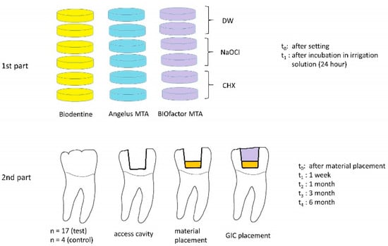
Figure 1.
Schematic view of experimental design.
2.1. Color Stability of Materials in Different Solutions
In the first part of the study, three different biomaterials (Biodentine, Angelus MTA, and BIOfactor MTA) and three different irrigation solutions (2% chlorhexidine gluconate, 2.5% sodium hypochlorite, and distilled water) were used. Biodentine, Angelus MTA, and BIOfactor MTA were mixed homogeneously as the manufacturer recommended and put into 8 mm diameter by 2 mm height Teflon cylindrical molds. The samples were stored at a 37 °C and 100% humidity level while the materials reached their ideal mechanical characteristics. Color measurements on both sides of the samples were performed after each sample was removed from the mold. Six samples from each group were prepared and left in each of the 3 irrigants for 24 h (n = 2). Following this period, the samples were dried and the colors of the two sides of each sample were measured. The same operator measured the color under steady lab lighting using a spectrophotometer (VITA Easyshade Advance; Vita Zahnfabrik, Bad Sackingen, Germany). Before each measurement, the instrument was calibrated three times. Color differences (ΔE) were computed using the Commission Internationale de l’Eclairage (CIE) system L * a * b * values using the following formula:
(ΔE) L*a*b* = [(ΔL)2 + (Δa)2 + (Δb)2]1/2
2.2. Preparation of Tooth Samples to Determine the Discoloration Effect of the Materials on Teeth
Fifty-five extracted mandibular first molars without caries, restoration, root filling, and coronal discoloration were used in this study. We collected and kept these teeth in distilled water until processing. An ultrasonic cleaning procedure was used to remove debris and exterior stains from crown surfaces. A fissure bur was used to create an access cavity and the buccal enamel–dentin thickness was standardized to 3 mm using spring calipers. The pulp was removed and 2.5% sodium hypochlorite, 17% EDTA, and then saline solution were used to rinse the prepared cavities. Using paper points and an air–water spray, samples were dried.
The samples were randomly distributed into three experimental groups (n = 17) and one control group (n = 4). Biomaterials were prepared according to the manufacturer’s instructions and the pulp chambers were filled with one of the following: Biodentine, Angelus MTA, or BIOfactor MTA. The access cavities were covered with glass ionomer cement (Nova Glass LC, Imicryl, Konya, Turkey) and then all samples were incubated for 48 h at 37 °C and 100% humidity. The control group samples did not include Angelus MTA, Biodentine, or BIOfactor MTA, and they were not sealed with glass ionomer cement. The color of the specimens was measured by a single operator with a spectrophotometer device after material placement (t0) and at the 1st week (t1), 1st month (t2), 3rd month (t3), and 6th month (t4) thereafter. Measurements were performed under constant light conditions by positioning the spectrophotometer device from the buccal surface of the tooth. The device was calibrated before each material group. Color changes between initial (immediately after material and glass ionomer cement placement) and other time intervals were recorded.
2.3. Statistical Analysis
The outcomes were examined using IBM SPSS Statistics for Windows version 22.0 (IBM Corp., Armonk, NY, USA). The data for the first step of the investigation did not have a normal distribution, according to the Kolmogrov–Smirnov and Shapiro–Wilk tests, so a Kruskal–Wallis test was carried out. An analysis of variance and post hoc Bonferroni tests were used to statistically analyze the color change values for the second step of the investigation because the Shapiro–Wilk test revealed that the data were normally distributed. The level of statistical significance was adjusted at p = 0.05.
3. Results
3.1. Color Stability of Materials in Different Solutions
Mean color change values of the materials according to the material groups are given in Table 1 and the solutions are given in Table 2. When immersed in NaOCl, Angelus MTA discolored more than when it was in CHX or distilled water (p: 0.008). In all solutions, Biodentine and BIOfactor MTA displayed similar ΔE values (p: 0.097 and p: 0.232, respectively). Distilled water and NaOCl both caused identical discoloration on all material groups when solutions were examined separately (p: 0.643 and p: 0.258, respectively). CHX significantly changed the color of Biodentine (p: 0.001).

Table 1.
The average values of changes in material color.
Table 2.
According to irrigation solutions, the average color change values of calcium silicate-based materials.
3.2. Coloring Effect of Materials on Teeth
The tooth color change values obtained in the second part of our study are presented in Table 3. When comparing the tooth discoloration values of materials after one week and one month, no statistically significant differences were found (p > 0.05). The Biodentine group’s ΔE value was shown to be the highest in the third and sixth months, whereas the control group displayed the lowest color change value (p < 0.05).
Table 3.
Measurement of color change (ΔE) values of all study groups.
In the sixth month, the Biodentine and BIOfactor MTA groups displayed the highest tooth color change values (p < 0.05). In the third month, MTA Angelus displayed the lowest tooth color change values (p < 0.05). In the first month, the control group displayed the greatest color change (p < 0.05).
4. Discussion
This study was designed to investigate the color stability of calcium silicate cements in contact with irrigation solutions and to present information about the effect of calcium silicate cements on tooth discoloration.
The Vita Easyshade color measurement tool was employed in this study to deter-mine color. One of the most reliable spectrophotometers with high repeatability is the Vita Easyshade color measurement tool. The device measures the amount of light that reaches a detector after passing through a sample using a light beam. Changes in a material’s reflectance characteristics imply changes in the color parameters, or the lightness values L*, a*, and b*, as well as the visually perceived color. A three-dimensional Cartesian coordinate system is used to define a color’s location in the CIELAB color space. L* denotes a color’s brightness or darkness. The a* and b* values indicate the location on the green (–) red (+) and blue (–) yellow (+) axes, respectively [18]. The CIE system is a framework for universal color standardization and is approved by the ISO [34].
This study utilized instrumental color measurement through spectrophotometry. This approach offers advantages by eliminating subjective aspects of color measurement and providing numerical expressions of shade differences related to visual perception and clinical significance [35]. The visual determination of tooth color is considered highly subjective due to various factors like external light conditions, human eye fatigue, color blindness, and there is limited standardized verbal communication for color characteristics. Despite these limitations, the human eye is efficient in detecting small color differences [36,37,38,39]. On the contrary, spectrophotometry offers several benefits, including high standardization, sensitivity to small color changes, repeatability, and objectivity. This method has been widely used and validated in dental research, enabling precise and reliable color comparisons [40,41,42,43,44,45].
In the current investigation, it was revealed that all calcium silicate-based materials had discoloration as a result of irrigation with distilled water, NaOCl, and CHX. According to this investigation, when exposed to NaOCl, Angelus MTA displayed the most pronounced discoloration. In a previous study, discoloration resulting from exposure of wMTA to a NaOCl solution was attributed to the reaction between bismuth oxide in MTA and NaOCl. The mechanism of the discoloration was the reduction of NaOCl resulting from the contact between bismuth oxide and sodium chloride [18]. In this study, Angelus MTA containing bismuth oxide as a radiopacifier agent showed similar results. It is thought that Biodentine and BIOfactor MTA do not include bismuth oxide and therefore may have low interaction with NaOCl.
It was determined that CHX had the greatest impact on the color of Biodentine. Previously, it has been reported that there was a significant color change when Biodentine and wMTA came into contact with the commonly used irrigation agents NaOCl and CHX [46]. The mechanism of CHX-caused material discoloration is still not clear. However, some research suggests that the micro-surface porosities of calcium silicate-based materials may be responsible for this discoloration [19,47]. It has been determined that the color change values of the materials in distilled water are very similar and that there is not a statistically significant difference.
The outcomes of our investigation agree with those of earlier research that examined the color stability of calcium silicate-based materials when they came into contact with various irrigation solutions [48,49]. In conjunction with our findings, all calcium silicate-based compounds showed discoloration in these investigations, and NaOCl showed greater discoloration in bismuth oxide-containing materials. In a recent study that investigated the color stability of calcium silicate-based materials in endodontic irrigation solutions, BIOfactor MTA showed more color change than Biodentine [23]. Another major finding from that study was the lack of statistically significant color changes caused by various irrigation agents [50]. This finding supports the current body of research in which we confirmed that various irrigation solutions did not make noteworthy color changes on both BIOfactor MTA and Biodentine.
We selected a 24 h contact time based on previous research [48,51]. Although the materials may be exposed to different solutions and varying durations, we chose a 24 h duration during which the set specimens were submerged in irrigation solutions. This duration does not fully reflect clinical conditions; however, this decision was made to replicate prolonged contact between the solutions and calcium silicate-based materials [48]. Additionally, we applied the same duration for every material to standardize the method. Another limitation regarding the contact duration is that the residual effect of the irrigation solution, which may influence the long-term color stability of the material, was ignored in this study. Further research with larger sample sizes would enhance the statistical power and generalizability of our findings. Including a broader range of irrigation solutions and longer evaluation periods would provide a more thorough assessment of the materials’ color stability over extended exposure times.
The findings of the second part show that the tested materials stained the tooth structure to varying degrees, as seen by the variation in ∆Ε values. Biodentine did not cause any color change on the teeth at the 1-week and 1-month point. The ΔE value then increased both at the 3-month and 6-month points, with the 6-month point showing the highest color change value. BIOfactor MTA, on the other hand, showed no color change on teeth except at the 6-month period. An unexpected finding was that both Biodentine and BIOfactor MTA groups had more color changes than Angelus MTA. This outcome is also consistent with the results of the earlier color research which showed less discoloration in bismuth oxide containing ProRoot MTA than Endosequence and Biodentine [52]. Additionally, ProRoot MTA showed lower ΔE during measurements over a longer span of time [52]. Although MTA Angelus showed lower discoloration than Biodentine in our study, it is not visible to the naked eye. In our study, the ΔE value of the Angelus MTA group was measured at the lowest at 3 months and was different during other time points (1 week and 1 and 6 months).
The irrigation protocol included washing with 2.5% NaOCl and 17% EDTA followed by saline solution for the removal of the smear layer. For maximum material penetration into the dentinal tubules, this irrigation protocol was chosen. According to Davis et al. [41], if the smear layer is left, discoloration is less common or lasts longer. Irrigation with NaOCl has been shown to cause dark brown discoloration when it reacts with materials containing bismuth oxide [18]. Although Angelus MTA was the only material containing bismuth oxide, the color change in the Angelus MTA group did not differ significantly compared to Biodentine or BIOfactor MTA. The interaction of Angelus MTA with NaOCl was likely minimal because the teeth had extensively been dried off.
NaOCl is frequently used to irrigate root canals in endodontics. NaOCl residues have been demonstrated to penetrate into dentin and cannot be easily removed from root canals. A black precipitate is produced when NaOCl is exposed to bismuth and other heavy metal oxides. Although the effect is potent, hypochlorite is not necessary for discoloration to occur [53]. Ramos et al. [29] did not use any irrigation solution to chemically remove pulp tissue or the smear layer from dentinal walls, and discoloration caused by WMTA was seen but lower than that reported in other studies. This demonstrates that irrigation solutions cannot be the only factor responsible for the discoloration. Unlike Ramos et al. [29], we used an irrigation solution and observed discoloration in our study, but we are unable to distinguish whether this is a result of the irrigation solution or the material itself.
In the 1st week and 1st month, the experimental materials showed similar tooth discoloration. However, tooth discoloration after six months was similar in BIOfactor MTA and Angelus MTA, but less so in Biodentine. To the best of our knowledge, this is the first research that investigates the tooth discoloration effect of BIOfactor MTA. The findings of this study over 6 months reveal that BIOfactor MTA showed more favorable results than Biodentine. Here, we would note that Biodentine and BIOfactor MTA are categorized differently from Angelus MTA according to the classification depending on the material chemistry [54]. Less discoloration of BIOfactor MTA could be attributed to the ytterbium oxide content.
Regarding the limitations of the second step of the study, clinical trials are required in the patient’s mouth for any vital pulp therapy. This in vitro study did not entirely mimic clinical conditions. Other factors were not considered in this study, such as the contact with irrigation fluids, presence of blood, and homeostatic temperature changes, which may all play a role in tooth discoloration. To gain a comprehensive understanding of the tooth discoloration potential of calcium silicate-based dental materials, extensive clinical research is essential to elucidate the underlying mechanisms. Additionally, conducting assessments in a more clinically relevant context, such as in vivo studies or simulations that better replicate real clinic conditions, would offer valuable insights into the practical implications of the tooth discoloration potential of dental materials.
5. Conclusions
Overall, the color of MTA Angelus was altered in contact with NaOCl, CHX induced severe discoloration on Biodentine, and BIOfactor MTA showed moderate color stability when submerged in irrigants. The tested calcium silicate-based materials caused discoloration on the teeth to varying degrees. Biodentine caused the most severe tooth discoloration and BIOfactor MTA seemed more suitable for use in relation to tooth discoloration. Finally, it should be noted that none of the discolorations on the teeth could be detected macroscopically.
Author Contributions
Conceptualization, M.B.A.; data curation, Ş.N.M.; formal analysis, Ş.N.M.; investigation, Ş.N.M. and M.B.A.; methodology, Ş.N.M. and M.B.A.; project administration, Ş.N.M. and M.B.A.; resources, Ş.N.M. and M.B.A.; software, Ş.N.M. and M.B.A.; supervision, M.B.A.; validation, Ş.N.M.; visualization, Ş.N.M. and M.B.A.; writing—original draft, Ş.N.M. and M.B.A.; writing—review and editing, Ş.N.M. and M.B.A. All authors have read and agreed to the published version of the manuscript.
Funding
This research was funded by Necmettin Erbakan University Scientific Research Projects Coordination, grant number 221224004.
Institutional Review Board Statement
This study was conducted in accordance with the Declaration of Helsinki and approved by the Institutional Review Board (or Ethics Committee) of Necmettin Erbakan University, Faculty of Dentistry (protocol code 2020/04 and date of 10.05.2020).
Informed Consent Statement
Not applicable.
Data Availability Statement
The data set used in the current study will be made available at any reasonable request.
Acknowledgments
The authors thank Imicryl for the donation of some of the materials used in this project. This study was presented at the 20th European Society of Endodontology (ESE) Biennial Congress (7–10 September 2022, Budapest, Hungary) as an oral presentation.
Conflicts of Interest
The authors declare no conflict of interest.
References
- Chugal, N.; Mallya, S.M.; Kahler, B.; Lin, L.M. Endodontic treatment outcomes. Dent. Clin. 2017, 61, 59–80. [Google Scholar] [CrossRef] [PubMed]
- Gönder, H.Y.; Elmacı, İ.; Karaköy, H. The effect of whitening toothpastes on the color change of the discolored composite material. NEU Dent. J. 2022, 4, 48–54. [Google Scholar]
- Kahler, B.; Rossi-Fedele, G. A review of tooth discoloration after regenerative endodontic therapy. J. Endod. 2016, 42, 563–569. [Google Scholar] [CrossRef] [PubMed]
- Malkondu, Ö.; Kazandağ, M.K.; Kazazoğlu, E. A review on biodentine, a contemporary dentine replacement and repair material. Bıomed Rest.Int. 2014, 2014, 160951. [Google Scholar] [CrossRef] [PubMed]
- Torabinejad, M.; Parirokh, M.; Dummer, P.M. Mineral trioxide aggregate and other bioactive endodontic cements: An updated overview–part II: Other clinical applications and complications. Int. Endod. J. 2018, 51, 284–317. [Google Scholar] [CrossRef]
- Duncan, H.F.; Galler, K.M.; Tomson, P.L.; Simon, S.; El-Karim, I.; Kundzina, R.; Krastl, G.; Dammaschke, T.; Fransson, H.; Markvart, M.; et al. European Society of Endodontology position statement: Management of deep caries and the exposed pulp. Int. Endod. J. 2019, 52, 923–934. [Google Scholar]
- Asgary, S.; Kamrani, F.A.; Taheri, S. Evaluation of antimicrobial effect of MTA, calcium hydroxide, and CEM cement. Iran. Endod. J. 2007, 2, 105–109. [Google Scholar]
- Parirokh, M.; Torabinejad, M. Mineral trioxide aggregate: A comprehensive literature review—Part III: Clinical applications, drawbacks, and mechanism of action. J. Endod. 2010, 36, 400–413. [Google Scholar] [CrossRef]
- Torabinejad, M.; Hong, C.; McDonald, F.; Ford, T.P. Physical and chemical properties of a new root-end filling material. J. Endod. 1995, 21, 349–353. [Google Scholar] [CrossRef]
- Krastl, G.; Allgayer, N.; Lenherr, P.; Filippi, A.; Taneja, P.; Weiger, R. Tooth discoloration induced by endodontic materials: A literature review. Dent. Traumatol. 2013, 29, 2–7. [Google Scholar] [CrossRef]
- Sarkar, N.; Caicedo, R.; Ritwik, P.; Moiseyeva, R.; Kawashima, I. Physicochemical basis of the biologic properties of mineral trioxide aggregate. J. Endod. 2005, 31, 97–100. [Google Scholar] [CrossRef]
- Kaur, M.; Singh, H.; Dhillon, J.; Batra, M.; Saini, M. MTA versus Biodentine: Review of Literature with a Comparative Analysis. J. Clin. Diagn. Res. 2017, 11, ZG01. [Google Scholar] [CrossRef]
- Kahler, B.; Chugal, N.; Lin, L.M. Alkaline materials and regenerative endodontics: A review. J. Mater. 2017, 10, 1389. [Google Scholar] [CrossRef] [PubMed]
- Nagas, E.; Ertan, A.; Eymirli, A.; Uyanik, O.; Cehreli, Z.C. Tooth discoloration induced by different calcium silicate–based cements: A two-year spectrophotometric and photographic evaluation in vitro. JOCPD 2021, 45, 112–116. [Google Scholar] [CrossRef] [PubMed]
- Yoldaş, S.E.; Bani, M.; Atabek, D.; Bodur, H. Comparison of the potential discoloration effect of bioaggregate, biodentine, and white mineral trioxide aggregate on bovine teeth: In vitro research. J. Endod. 2016, 42, 1815–1818. [Google Scholar] [CrossRef]
- Marciano, M.A.; Costa, R.M.; Camilleri, J.; Mondelli, R.F.L.; Guimarães, B.M.; Duarte, M.A.H. Assessment of color stability of white mineral trioxide aggregate angelus and bismuth oxide in contact with tooth structure. J. Endod. 2014, 40, 1235–1240. [Google Scholar] [CrossRef]
- Felman, D.; Parashos, P. Coronal tooth discoloration and white mineral trioxide aggregate. J. Endod. 2013, 39, 484–487. [Google Scholar] [CrossRef]
- Camilleri, J. Color stability of white mineral trioxide aggregate in contact with hypochlorite solution. J. Endod. 2014, 40, 436–440. [Google Scholar] [CrossRef]
- Vallés, M.; Mercadé, M.; Duran-Sindreu, F.; Bourdelande, J.L.; Roig, M. Color stability of white mineral trioxide aggregate. Clin. Oral. Investig. 2013, 17, 1155–1159. [Google Scholar] [CrossRef]
- Naik, S.; Hegde, A.M. Mineral trioxide aggregate as a pulpotomy agent in primary molars: An in vivo study. J. Indian. Soc. Pedod. Prev. Dent. 2005, 23, 13–16. [Google Scholar] [PubMed]
- Percinoto, C.; De Castro, A.M.; Pinto, L. Clinical and radiographic evaluation of pulpotomies employing calcium hydroxide and trioxide mineral aggregate. Gen. Dent. 2006, 54, 258–261. [Google Scholar]
- Butt, N.; Talwar, S.; Chaudhry, S.; Nawal, R.R.; Yadav, S.; Bali, A. Comparison of physical and mechanical properties of mineral trioxide aggregate and Biodentine. IJDR 2014, 25, 692. [Google Scholar] [CrossRef] [PubMed]
- Nowicka, A.; Lipski, M.; Parafiniuk, M.; Sporniak-Tutak, K.; Lichota, D.; Kosierkiewicz, A.; Kaczmarek, W.; Buczkowska-Radlińska, J. Response of human dental pulp capped with biodentine and mineral trioxide aggregate. J. Endod. 2013, 39, 743–747. [Google Scholar] [CrossRef] [PubMed]
- Tran, X.; Gorin, C.; Willig, C.; Baroukh, B.; Pellat, B.; Decup, F.; Opsahl Vital, S.; Chaussain, C.; Boukpessi, T. Effect of a calcium-silicate-based restorative cement on pulp repair. J. Dent. Res. 2012, 91, 1166–1171. [Google Scholar] [CrossRef]
- Katge, F.A.; Patil, D.P. Comparative analysis of 2 calcium silicate–based cements (Biodentine and Mineral Trioxide Aggregate) as direct pulp-capping agent in young permanent molars: A split mouth study. J. Endod. 2017, 43, 507–513. [Google Scholar] [CrossRef]
- Camilleri, J. Investigation of Biodentine as dentine replacement material. J. Dent. 2013, 41, 600–610. [Google Scholar] [CrossRef]
- Marconyak Jr, L.J.; Kirkpatrick, T.C.; Roberts, H.W.; Roberts, M.D.; Aparicio, A.; Himel, V.T.; Sabey, K.A. A comparison of coronal tooth discoloration elicited by various endodontic reparative materials. J. Endod. 2016, 42, 470–473. [Google Scholar] [CrossRef]
- Palma, P.J.; Marques, J.A.; Santos, J.; Falacho, R.I.; Sequeira, D.; Diogo, P.; Caramelo, F.; Ramos, J.C.; Santos, J.M. Tooth discoloration after regenerative endodontic procedures with calcium silicate-based cements—An ex vivo study. Appl. Sci. 2020, 10, 5793. [Google Scholar] [CrossRef]
- Ramos, J.C.; Palma, P.J.; Nascimento, R.; Caramelo, F.; Messias, A.; Vinagre, A.; Santos, J.M. 1-year in vitro evaluation of tooth discoloration induced by 2 calcium silicate–based cements. J. Endod. 2016, 42, 1403–1407. [Google Scholar] [CrossRef] [PubMed]
- Vallés, M.; Roig, M.; Duran-Sindreu, F.; Martínez, S.; Mercadé, M. Color stability of teeth restored with Biodentine: A 6-month in vitro study. J. Endod. 2015, 41, 1157–1160. [Google Scholar] [CrossRef]
- Akbulut, M.B.; Bozkurt, D.A.; Terlemez, A.; Akman, M. The push-out bond strength of BIOfactor mineral trioxide aggregate, a novel root repair material. Restor. Dent. Endod. 2019, 44, e5. [Google Scholar] [CrossRef] [PubMed]
- Öznurhan, F.; Kayabaşı, M.; Keskus, B. Evaluation of Long-Term Results of Two Different Calcium Silicate Based Materials in Primary Molar Teeth Vital Pulpotomies: An Invivo Study. Cumhur. Dent. J. 2020, 23, 45–51. [Google Scholar] [CrossRef]
- Buldur, B.; Öznurhan, F.; Kayabaşı, M.; Şahin, F. Shear bond strength of two calcium silicate-based cements to compomer. Cumhur. Dent. J. 2018, 21, 18–23. [Google Scholar] [CrossRef]
- Ioannidis, K.; Mistakidis, I.; Beltes, P.; Karagiannis, V. Spectrophotometric analysis of coronal discolouration induced by grey and white MTA. Int. Endod. J. 2013, 46, 137–144. [Google Scholar] [CrossRef]
- Douglas, R.D.; Steinhauer, T.J.; Alvin, G.W. Intraoral determination of the tolerance of dentists for perceptibility and acceptability of shade mismatch. J. Prosthet. Dent. 2007, 97, 200–208. [Google Scholar] [CrossRef]
- Berns, R.S. Billmeyer and Saltzman’s Principles of Color Technology, 4th ed.; John Wiley & Sons: New York, NY, USA, 2000. [Google Scholar]
- Hunter, R.S.; Harold, R.W. The Measurement of Appearance, 2nd ed.; John Wiley & Sons: New York, NY, USA, 1987. [Google Scholar]
- Seghi, R.; Johnston, W.; O’Brien, W. Performance assessment of colorimetric devices on dental porcelains. J. Dent.Res. 1989, 68, 1755–1759. [Google Scholar] [CrossRef]
- Wyszecki, G.; Stiles, W.S. Color Science: Concepts and Methods, Quantitative Data and Formulae, 2nd ed.; John Wiley & Sons: New York, NY, USA, 1982. [Google Scholar]
- Fidan, M.; Yeşilirmak, N. Tuncdemir, Evaluation of the effect of coloration with coffee on color stability and translucency parameter in composite resins. NEU Dent. J. 2021, 3, 26–32. [Google Scholar]
- Gungor, A.S.; Orcun, M.E.; Donmez, N. In vitro evaluation of the effect of commercial whitening toothpastes on enamel color change and the surface roughness. NEU Dent. J. 2023, 5, 25–34. [Google Scholar]
- Ioannidis, K.; Beltes, P.; Lambrianidis, T.; Kapagiannidis, D.; Karagiannis, V. Validation and spectrophotometric analysis of crown discoloration induced by root canal sealers. Clin.Oral. Investig. 2013, 17, 1525–1533. [Google Scholar] [CrossRef]
- Khokhar, Z.; Razzoog, M.; Yaman, P. Color stability of restorative resins. J. Dent. Res. 1991, 22, 733–737. [Google Scholar]
- Kirchhoff, A.; Raldi, D.; Salles, A.; Cunha, R.; Mello, I. Tooth discolouration and internal bleaching after the use of triple antibiotic paste. Int. Endod. J. 2015, 48, 1181–1187. [Google Scholar] [CrossRef]
- Paul, S.; Peter, A.; Pietrobon, N. Visual and Spectrophotometric Shade Analysis of Human Teeth. J. Dent. Res. 2002, 81, 578–582. [Google Scholar] [CrossRef] [PubMed]
- Trusha, S.; Banga, K.S. Effect of commonly used irrigants on the colour stabilities of two calcium-silicate based material. Eur. Oral. Res. 2019, 53, 141–145. [Google Scholar]
- Eghbal, M.J.; Torabzadeh, H.; Bagheban, A.A.; Shamszadeh, S.; Marvasti, L.A.; Asgary, S. Color stability of mineral trioxide aggregate and calcium enriched mixture cement. J. Investig. Clin. Dent. 2016, 7, 341–346. [Google Scholar] [CrossRef]
- Keskin, C.; Demiryurek, E.O.; Ozyurek, T. Color stabilities of calcium silicate–based materials in contact with different irrigation solutions. J. Endod. 2015, 41, 409–411. [Google Scholar] [CrossRef] [PubMed]
- Yılmaz, K.; Tüfenkçi, P.M.A. The influence of different irrigation solutions on the color stability of several calcium silicate–based materials. Selcukdentj 2020, 7, 22–26. [Google Scholar] [CrossRef]
- Atas, O.; Aras, A. Review. Investigation of the change in color caused by the contact of calcium silicate-based materials with endodontic irrigation solutions. J. Dent. Res. Rev. 2021, 8, 82–85. [Google Scholar]
- Camilleri, J. Staining potential of Neo MTA Plus, MTA Plus, and Biodentine used for pulpotomy procedures. J. Endod. 2015, 41, 1139–1145. [Google Scholar] [CrossRef]
- Beatty, H.; Svec, T. Quantifying coronal tooth discoloration caused by biodentine and endosequence root repair material. J. Endod. 2015, 41, 2036–2039. [Google Scholar] [CrossRef]
- Camilleri, J.; Borg, J.; Damidot, D.; Salvadori, E.; Pilecki, P.; Zaslansky, P.; Darvell, B.W. Colour and chemical stability of bismuth oxide in dental materials with solutions used in routine clinical practice. PLoS ONE 2020, 15, e0240634. [Google Scholar] [CrossRef]
- Camilleri, J. Classification of hydraulic cements used in dentistry. Front. Dent. Med. 2020, 1, 9. [Google Scholar] [CrossRef]

Disclaimer/Publisher’s Note: The statements, opinions and data contained in all publications are solely those of the individual author(s) and contributor(s) and not of MDPI and/or the editor(s). MDPI and/or the editor(s) disclaim responsibility for any injury to people or property resulting from any ideas, methods, instructions or products referred to in the content. |
© 2023 by the authors. Licensee MDPI, Basel, Switzerland. This article is an open access article distributed under the terms and conditions of the Creative Commons Attribution (CC BY) license (https://creativecommons.org/licenses/by/4.0/).