Article

DNA Barcoding and Molecular Phylogenetics Revealed a New Cryptic Bamboo Aphid Species of the Genus Takecallis (Hemiptera: Aphididae)

by
Karina Wieczorek
1,* and
Natalia Sawka-Gądek
2
1
Institute of Biology, Biotechnology and Environmental Protection, Faculty of Natural Sciences, University of Silesia in Katowice, Bankowa 9, 40-007 Katowice, Poland
2
Institute of Systematics and Evolution of Animals, Polish Academy of Sciences, Sławkowska 17, 31-016 Krakow, Poland
*
Author to whom correspondence should be addressed.
Appl. Sci. 2023, 13(13), 7798; https://doi.org/10.3390/app13137798
Submission received: 22 May 2023 / Revised: 29 June 2023 / Accepted: 29 June 2023 / Published: 1 July 2023
(This article belongs to the Special Issue Sustainable Strategies for Pest Management in Plants and Animals)

Abstract

:
Takecallis nigroantennatus Wieczorek sp. nov. (Hemiptera: Aphididae), associated with the cold hardy bamboo variety Fargesia spp. (Bambusoideae), is described and illustrated along with a key to species of the genus Takecallis. The results of a mitochondrial COI (DNA barcoding) and nuclear elongation factor 1 (EF1α) gene sequences, which confirm the genetic difference of the new taxon from the other congeneric species, are provided. The possible way of introduction of this species to Europe is discussed with its new locality from Belgium.

1. Introduction

Aphids (Hemiptera, Aphididae), due to the seasonal polymorphism and complex life cycles, are insects difficult to identify. Thus, the usability of DNA barcoding, a fragment of the mitochondrial cytochrome oxidase subunit I (COI) gene, has been used for additional diagnostic data in species identification [1,2,3,4,5]. Moreover, DNA barcoding enables the detection of cryptic species and the reassessment of species diversity delimitation on various taxonomic levels [6,7,8,9].
The bamboo feeding aphid genus Takecallis Matsumura contains seven taxa of oriental origin: T. affinis affinis Ghosh, T. affinis niitakayamensis Quednau, T. alba Lee, T. arundicolens (Clarke), T. arundinariae (Essig), T. assumenta Qiao and Zhang, T. sasae (Matsumura), and T. taiwana (Takahashi) [10]. Matsumura (1917) [11] erected Takecallis for Callipterus arundicolens Clarke (= T. bambusae Matsumura, 1917). The genus was revised by Higuchi (1969) [12] and later reviewed by Quednau (2003) [13]. The author synonymized T. takahashii Hsu with T. arundinariae, described T. affinis niitakayamensis, and provided keys to species and illustrations of all available morphs. Qiao and Zhang (2004) [14] reviewed the genus Takecallis from China and described T. assumenta. Recently, T. alba from Korea was distinguished based on morphological and molecular evidence [15]. Moreover, comprehensive assessment of DNA barcodes for the subfamily Calaphidinae revealed deep intraspecific divergence within the species complex of T. arundicolens, which indicates that there is more than one species under this name [6]. In their natural range, all known species of this genus are restricted to East Asia (China, India, Japan, Korea, Manchurian subregion, Taiwan). Three species—T. arundicolens, T. arundinariae and T. taiwana are now widely distributed and introduced to other continents, including Europe [16]. All of them are treated as a pest on Bambusoideae, i.e., Arundinaria spp., Bambusa spp., Phyllostachys spp., and Sasa spp. [13,17].
Aphids, as herbivorous sap-sucking insects, can cause considerable damage to crops and ornamental plants, and also as virus vectors. Therefore, it is very important to use sustainable strategies for their management. In order to use them, it is important to identify the pest correctly, and choose the right methods of its control. An examination of aphid samples collected in 2022 from the cold hardy bamboo variety Fargesia rufa T.P. Yi revealed the first detection of the genus Takecallis from Poland. These specimens were firstly classified as belonging to the T. arundicolens complex [18]. In order to exclude whether the material collected in Poland is a color variant of T. arundicolens or an unknown cryptic species of this genus, detailed morphometric studies together with analyses of a mitochondrial COI (DNA barcoding) and nuclear elongation factor 1 (EF1α) gene sequences were undertaken, and the results are presented in this paper. In addition, the material collected from Poland was compared with aphids feeding on various species of bamboo, collected in 2023 in the Meise Botanic Garden, Belgium.

2. Materials and Methods

2.1. Specimen Collection and Identification

The aphids (adult winged viviparous females and immature alatoid nymphs) were collected directly from the host plant using a fine brush and placed into Eppendorf tubes containing 70% and 98% ethanol. Insects were slide-mounted using the method of Wieczorek [19], examined using a Nikon Ni-U light microscope equipped with a phase contrast system and photographed using a Nikon SMZ 25 stereoscopic microscope with a DS-Fi2 camera. Field photographs were taken using an iPhone 7 camera with the OlloClip Macro Pro Lens Set. The figures were prepared using Corel Draw 2021, Corel Corporation. The measurements were taken according to Ilharco and van Harten [20] and are given in millimeters.

2.2. DNA Sequencing and Phylogenetic Analysis

DNA was extracted from seven specimens (isolates 1BAN, 2BAN, 3BAN, 4BAA, 5BAN, 6BAA, 7BAN, Poland locality). Genomic DNA was isolated without modifying the protocol using the Bio-Trace DNA Purification Kit (EURx, Gdańsk, Poland). To elute the purified DNA, we applied 50 μL of an Elution Buffer onto the silica membrane. Two genes were amplified, mitochondrial COI and nuclear EF1α. To amplify a fragment of the mitochondrial cytochrome c oxidase I (COI) gene, the primer pair LCO-1490 and HCO1850 [21] was used. For the amplification of the Elongation factor 1-alpha, alpha (EF1α) primers EF2 [22] and EF3 [23] were used. Polymerase chain reaction (PCR) amplification for all DNA fragments was performed in a final volume of 20 μL containing 30 ng of DNA, 10 μL of 2× Phanta Master Mix (Vazyme Biotech, Nanjing, China), 0.4 μL of 20 μM of each primer, in a Mastercycler ep system (Eppendorf, Germany). The cycling profile for the PCR was: 95 °C for 3 min, 35 cycles of 95 °C for 15 s, Tm of oligos for 15 s, 72 °C for 1 min and a final extension period of 72 °C for 5 min.
In order to assess the quality of the amplification, the PCR products were electrophoresed in 1% agarose gel for 45 min at 85 V with a DNA molecular weight marker (Mass Ruler Low Range DNA Ladder, Thermo-Scientific, Waltham, MA, USA). The PCR products were purified using Clean-Up Concentrator (A & A Biotechnology, Gdańsk, Poland).
Samples were sequenced in both directions using the same primers as for the PCR reactions, except EF1α products where two or more internal primers, selected from: EF4 (5′-GAACCACCATACAGCGAAG) or EF4a (5′-GAACCACCGTACAGTGAAG), EF5 (5′-TTGATGTAACTGCTGACTTC), EF7 (5′-ATTGGAGGTATTGGAACAGT) and EF8 (5′-GGGACTGTTCCAATACCTCC) [24] were used combined with a BigDye Terminator 3.1 Cycle Sequencing Kit (Applied Biosystems, Waltham, MA, USA) in the chain termination reaction method [25]. The sequencing reaction was carried out with the PCR product at a total volume of 20 μL, containing 2 μL of BigDye Terminator Reaction Ready Mix v. 3.1, 2 μL 5×sequencing buffer, 3.2 mol/μL of the primer solution, and 6 μL of the purified PCR product. The cycle-sequencing profile was 3 min at 94 °C followed by 30 cycles of 10 s at 96 °C, 5 s at 50 °C, and 2 min at 60 °C.
Sequencing products were precipitated using CleanDTR (CleanNa, Waddinxveen, The Netherlands), and were separated on an ABI PRISM 3130xl DNA Sequencer (Applied Biosystems, Waltham, MA, USA).
Raw chromatograms were evaluated and corrected in Geneious v10. 2.6 (https://www.geneious.com, accessed on 15 February 2023) [26]. In order to identify the numts [27,28], the mitochondrial COI and EF1 sequences were translated into amino acid sequences with Geneious v R10.2.6 using the respectively standard invertebrate mitochondrial genetic code and standard code. All of the nucleotide sequences were verified using BLAST searches of NCBI (http://blast.ncbi.nlm.nih.gov/Blast.cgi, accessed on 20 Febuary 2023). Protein coding data were aligned using the MAFFT versions 7.263 [29] plugin within Geneious v R10.2.6 and introns were removed from the EF1 α alignment. All sequences used in the phylogenetic analysis are presented in Table 1.
Models for Bayesian (BI) and maximum likelihood (ML) analyses were calculated in MrModeltest [34] using the Akaike information criterion (AIC). BI analyses were performed in MrBayes v3.2.6 [35] with four independent runs, each having three heated and one cold chain. Analyses were run for 2 million generations with trees sampled every 1000 generations. The first 25% of each run was discarded as burn-in. Convergence among the runs was assessed using Tracer [36], and examination of the potential scale reduction factor (PSRF) values and average standard deviation of the split frequencies in the MrBayes output was performed. The ML analyses were performed using IQ-TREE v.1.6.8 [37] with the ultrafast bootstrap approximation for branch support with 1000 replicates [38]. All trees were visualized using FigTree (http://tree.bio.ed.ac.uk/software/figtree/, accessed on 20 February 2023). Trees were edited and annotated in Corel Draw 17.1.0.572, 2014 Corel Corporation. Pairwise distances were calculated using MEGA X with Kimura-2 parameter model [39].

2.3. Species Delimitation

Three DNA-based species delimitation methods, Poisson tree processes (bPTP) model with Bayesian implementation [40], Automatic Barcode Gap Discovery (ABDG) [41], and Assemble Species by Automatic Partitioning (ASAP) [42] were used to confirm independently evolved linage in the Takecallis genus. Poisson tree process modeling is a method intended for delimitating species in single-locus molecular phylogenies. ABDG and ASAP use pairwise distances to group intra-sequences into potential species based on detecting gaps in the variation between supposed intra- and interspecific groups (barcode thresholds). bPTP analysis was performed with PTP web server (https://species.h-its.org, accessed on 12 June 2023) using the IQ-TREE result from the previous analysis as an input. Automatic Barcode Gap Discovery (ABDG) was performed using ABDG web server (https://bioinfo.mnhn.fr/abi/public/abgd/abgdweb.htm, accessed on 12 June 2023) with default parameters using the alignment of sequences as an input file. Assemble Species by Automatic Partitioning (ASAP) was performed using the ASAP web server (https://bioinfo.mnhn.fr/abi/public/asap/, accessed on 12 June 2023) with alignment of sequences as an input file.

3. Results

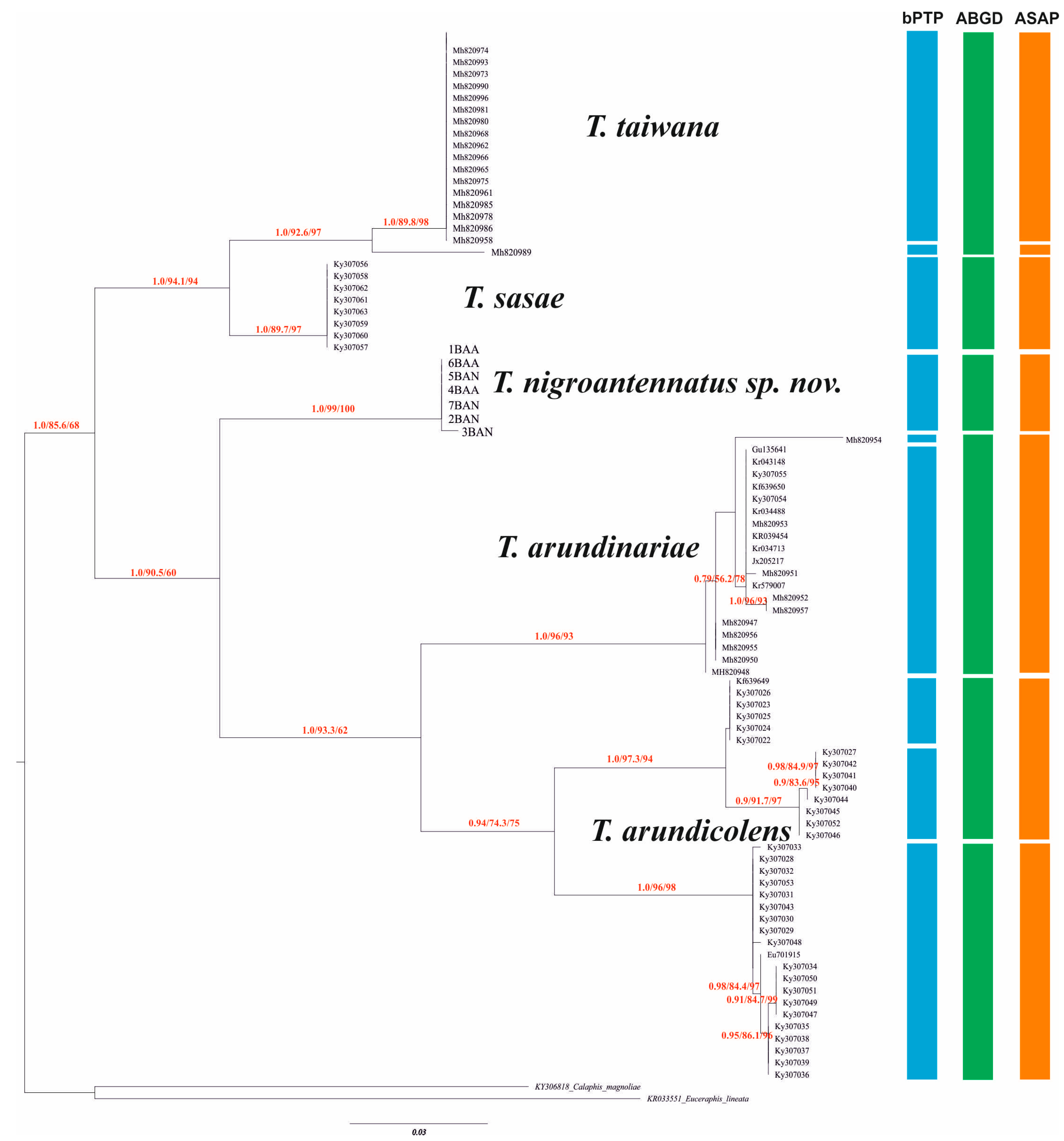
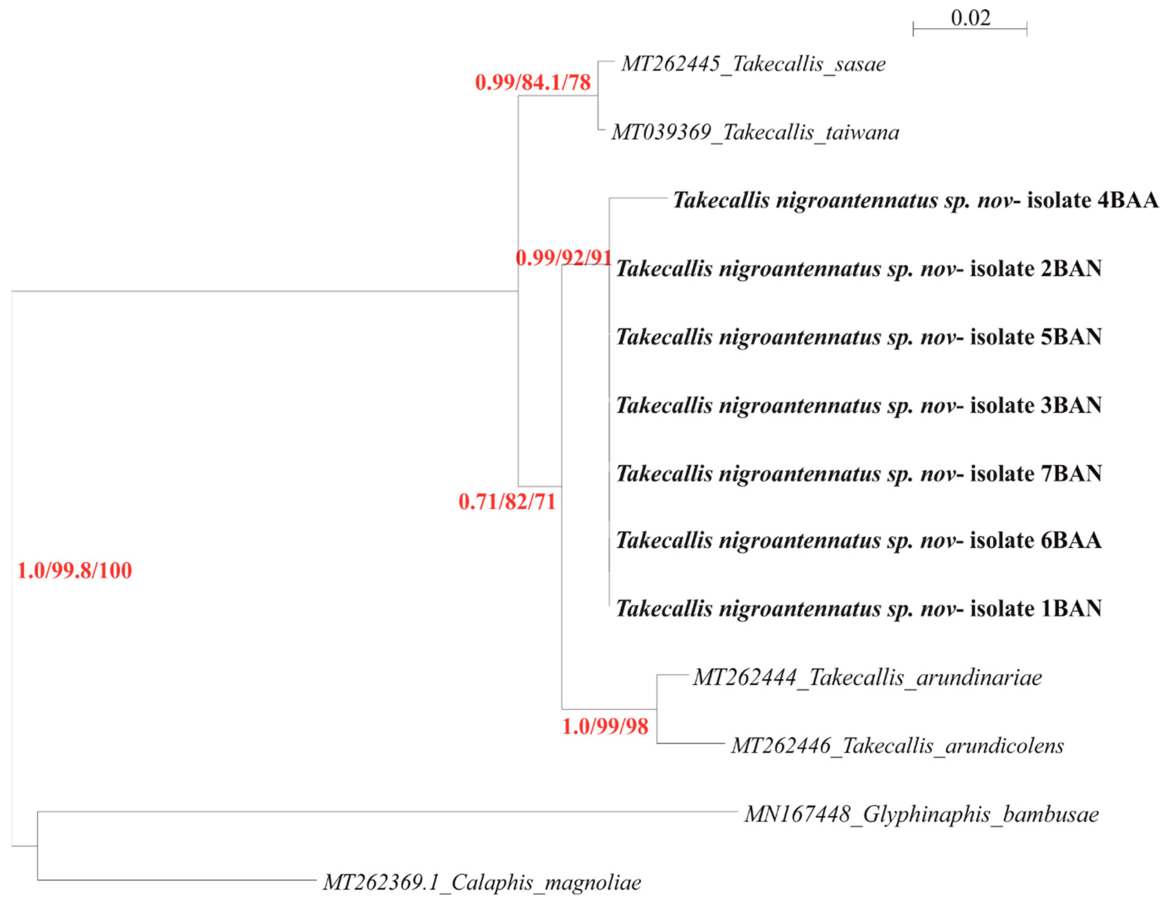
3.1. Molecular Phylogeny

Phylogenetic inferences were obtained for COI and EF1α genes using Bayesian inference (BI) and Maximum likelihood (ML).
For the COI, the jModelTest estimated GTR + G + I as the best fitting model. ML and BI analysis resulted in phylograms with the same topologies. The ESS values reported for BI analysis were >200, and the observed PRSF was 1.0. The phylogenies wereresolved and the main splits were supported by high bootstrap values and posterior probabilities. Calaphis magnoliae Essig and Kuwana and Euceraphis lineata Baker were used to root the topologies. Phylogram shows two main clades, first formed by two sister subclades; one with species belonging to the T. taiwana, and the second by species belonging to the T. sasae. The second clade is formed by two subclades. The first subclade is formed by the T. nigroantennatus sp. nov., described in the present paper (Figure 1). The second subclade consists of two groups, first formed by species belonging to the T. arundinariae. The second group is formed by two sister phylogenetic lines formed by species belonging to the T. arundicolens.
For the EF1α, the jModelTest estimated GTR + I as the best fitting model. ML and BI analysis resulted in trees with the same topologies. The ESS values reported for BI analysis were >200, and the observed PRSF was 1.0. The phylogenies were resolved and the main splits were supported by high bootstrap values and posterior probabilities. Calaphis magnoliae and Glyphinaphis bambusae van der Goot were used to root the topologies. The phylogram shows two main clades. First is formed by two subclades, one represented by T. sasae and the second by T. taiwana. The second clade is formed by two subclades; one formed by two phylogenetic lines, T. arundicolens and T. arundinariae. The second subclade is formed by the T. nigroantennatus sp. nov., described in the present paper (Figure 2).
p-distance between investigated specimens and group of species of T. taiwana, T. sasae, T. arundicolens and T. arundinariae was 9.3%, 7.8%, 10.2%, and 9.3%, respectively.

3.2. Species Delimitation

The different species delimitation methods (bPTP, ABDG and ASAP) resulted in different numbers of molecular operational taxonomic units (MOTUs) in the analyzed representative of the Takecallis genus (Figure 1). ASAP analysis identified 5–21 hypothetical species. The first ASAP-score (1.0) was chosen, which provides the best-fit scenario at the threshold distance of 2.78% (JC69) with five hypothetical species. The pairwise distance gap approach (ABDG) with default settings (X = 0.5) suggested six species with a barcode gap distance of 4.9%. The bPTP showed nine hypothetical species. All performed analyses indicated that Takecallis nigroantennatus sp. nov may be a species.

3.3. Taxonomy

Family Aphididae Latreille, 1802
Subfamily Calaphidinae Oestlund, 1919
Tribe Therioaphidini Börner, 1944
Genus Takecallis Mastumura, 1917
Species Takecallis nigroantennatus Wieczorek sp. nov.
Materials examined: 28 adult viviparous females and 14 alatoid nymphs (collected in the type locality, Poland); specimens were mounted on microscope slides (20 adult viviparous females + 6 alatoid nymphs) and processed for DNA sequencing (8 + 8). Additional fresh material for comparative morphological study was collected by the first author on 4 May 2023 in the Meise Botanic Garden, Belgium: 10 adult viviparous females of T. arundicolens from Pseudosasa japonica (Siebold and Zucc. ex Steud.) Makino ex Nakai; 10 adult viviparous females of T. arundinariae from Sasa megalophylla Makino and Uchida; and five adult viviparous females of T. nigroantennatus sp. nov. from Fargesia nitida x murielae.
Type of locality and host plant: 54°27′52.19″ N, 17°02′01.55″ ES; Słupsk, Poland; coll. from Fargesia rufa T.P.Yi on 9 July, 1 August, and 3 September 2022 by Karina Wieczorek.
Type of repository: Holotype (Słupsk, Poland, Fargesia rufa 1 August 2022, leg. K. Wieczorek, slide DZUS 1/8.22_7) and three paratypes (slides DZUS 9/7.22_8, DZUS 1/8.22_9, DZUS 3/10.22_10 same data as holotype) were deposited in the Entomology Collection of the University of Silesia in Katowice, Poland (DZUS).
Etymology: The species name refers to the antennae black, except for the antennal segments I–II, and the very base of segment III, clearly visible in live, fresh fixed, and slide-mounted adult viviparous females.

3.3.1. Differential Diagnosis

T. nigroantennatus sp. nov. is morphologically close to T. affinis niitakayamensis. However, this species can be distinguished from the latter by the following characteristics: body length 1.40–1.95 mm, antennae 0.95–1.14 times body length, antennal segment I with 4 setae, URS with 4 accessory setae, cauda with 8–11 setae, each lobe of anal plate with 5–9 setae, spinal abdominal setae barely visible. On the contrary, body length 2.45 mm, antennae 1.3 times body length, antennal segment I with 5–7 setae, URS with 3 accessory setae, cauda with 18 setae, each lobe of anal plate with 15 setae, spinal abdominal setae on low elevations are characteristic for T. affinis niitakayamensis.

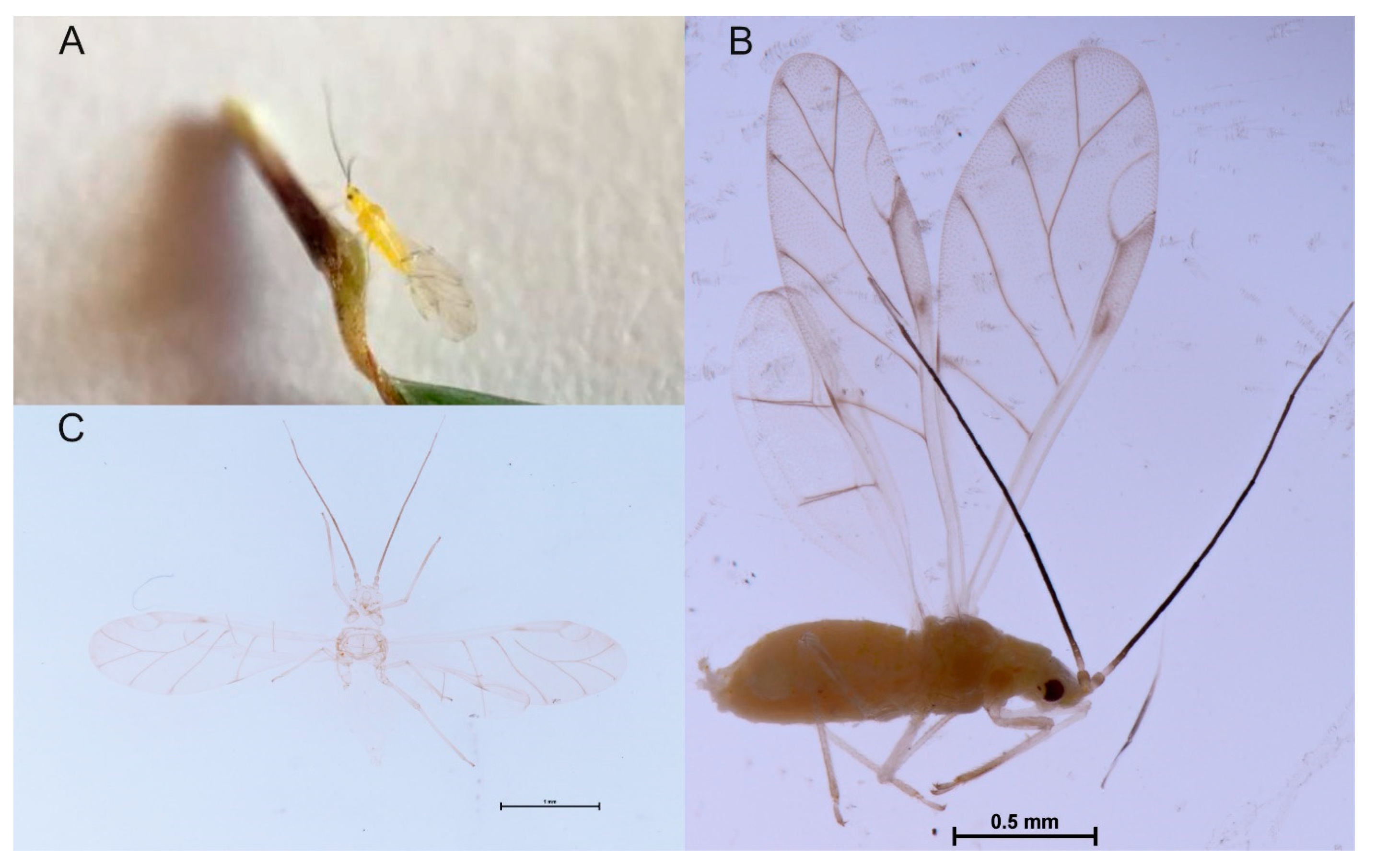
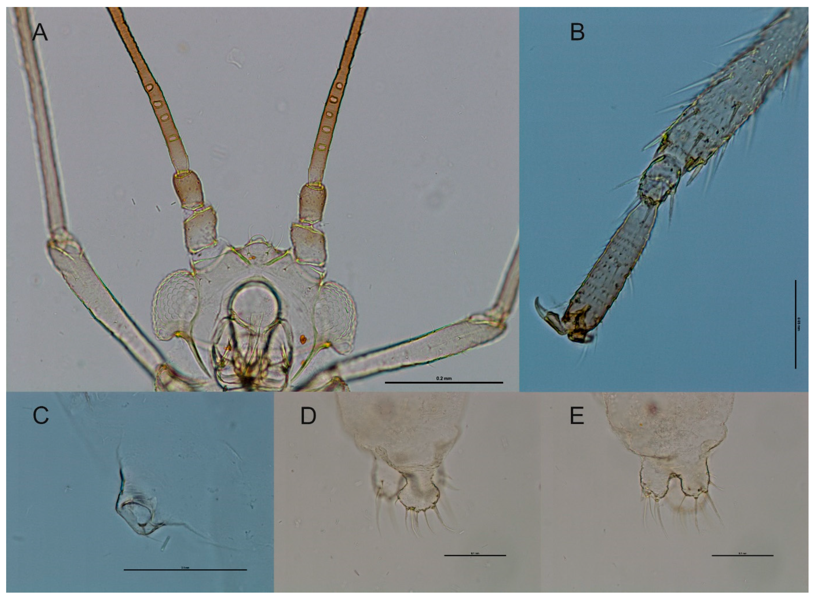
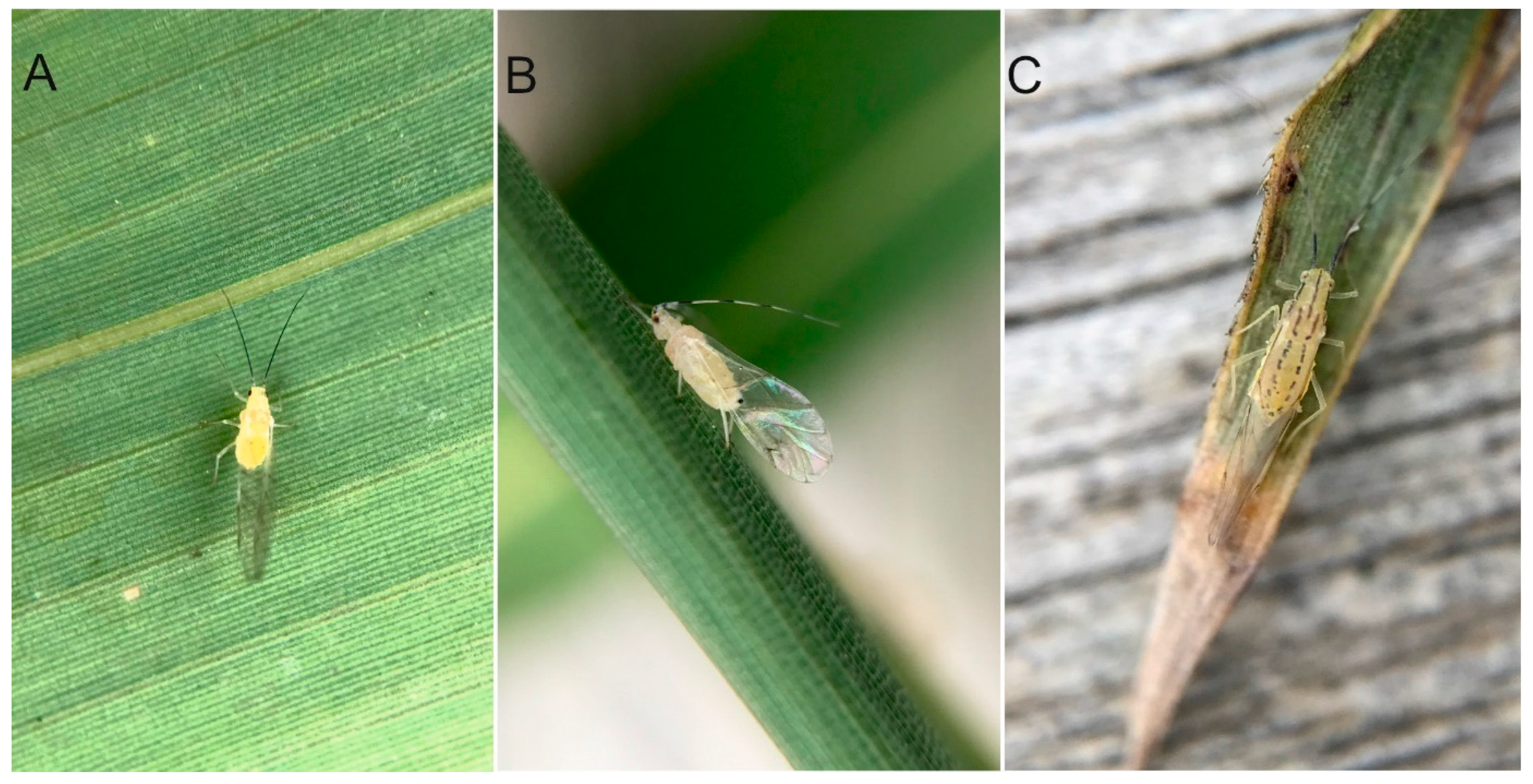
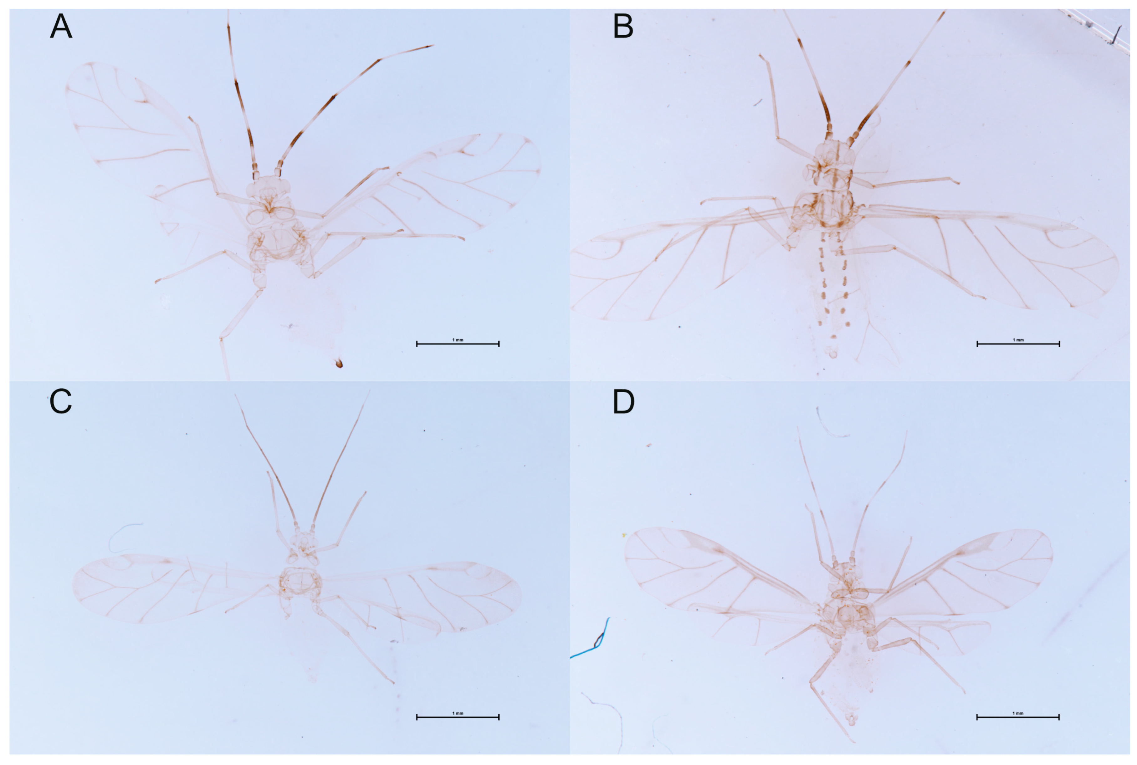
3.3.2. Species Description

Winged viviparous female (Table 2).
Color in life: uniformly yellow, with antennae black, except for the antennal segments I–II, and the very base of segment III, which are pale (Figure 3A). This same coloration is clearly visible in the fresh fixed specimen (Figure 3B).
Pigmentation of cleared specimens on slide: uniformly pale with tarsi dusky and antennae black, except for the antennal segments I–II and the very base of segment III which are concolorous with head. Wing veins dusky (Figure 3C).
Morphometric characters: Body oval. Head with three pairs of anterior and two pairs of posterior short and pointed discal setae about 0.01–0.04 mm, median protrusion on frons developed, epicranial suture and antennal tubercle developed, head dorsum without tubercles (Figure 4A). Antennae 6-segmented, 0.95–1.14 × body length. Antennal segment III, the longest; antennal segment IV slightly longer than antennal segment V; antennal segment V always shorter than antennal segment VI; processus terminalis 0.95–1.05 × base; other antennal ratios: VI:III 0.70–0.86, V:III 0.51–0.56, IV:III 0.49–0.57. Antennal segment III with 3–6 transversely elliptical ciliated secondary rhinaria in a row arranged rather near the base of the segment (Figure 4A); antennal segment IV without secondary rhinaria; base of antennal segment VI with 3 accessory rhinaria. Antennal segments IV–VI and partly antennal segment III imbricated. Antennal segments: I with 4 setae; II with 3 setae; III with 13–16 setae; IV with 5–8 setae; V with 3–5 setae; base with a single seta; processus terminalis with 5 apical setae. Antennal setae pointed, fine and very short, longest seta on antennal segment III 0.30 × basal articular diameter of this segment. Clypeus with large nose-like processus. Rostrum very short, reaching to fore coxae, ultimate rostral segment short, blunted, 0.07–0.08 mm long with 4 accessory setae, 0.33–0.40 × base, 0.61–0.70 × second segment of the hind tarsus. Thorax smooth, without tubercles. Tibiae setose, with numerous stout, pointed setae and rastral organ on its distal part (Figure 4B). First tarsal segment with 4 setae. The forewings typical with a normal venation; the radius strongly curved, the media with three branches. The hind wings have two oblique veins. Pterostigma and wing veins slightly pigmented. Dorsal abdominal tergites I–VII with a pair of spinal and marginal seta on low elevations; spinal setae barely visible. Siphunculus short, cylindrical, with 0.02–0.04 mm of single marginal seta at its base (Figure 4C). Cauda knobbed with 8–11 setae (Figure 4D). Anal plate bilobed, each lobe with 5–9 setae (Figure 4E).

3.3.3. Key to Species of the Genus Takecallis

  • Antennae longer than body..................................................................................2
    Antennae shorter than body.............................................................................8
  • Ultimate rostral segment without any accessory setae. Abdominal tergites 1–7 with paired dark circular spots around spinal setae...............................................T. assumenta
    Ultimate rostral segment with 3–4 accessory setae. Abdominal tergites 1–7 with or without dark spots....................................................................................3
  • Antennal segment I with (4-) 5–7 setae. Antennal segments III–VI dark, except for basal part of antennal segment III. Antennal segment III with 4–15 secondary rhinaria................................................................................................4
    Antennal segment I usually with 4, rarely with 3 or 5, setae. Antennal segments III–VI or IV–VI base usually pale basally and dark apically. Antennal segment III with 3–9 secondary rhinaria.....................................................................................6
  • Antennal segment III with 10–15 secondary rhinaria, marginal seta on abdominal tergite VI not positioned on base of siphunculus.................................................T. affinis s. str.
    Antennal segment III with 4–7 (often 5) secondary rhinaria, marginal seta on abdominal tergite VI positioned on base of siphunculus............................................5
  • Antennae 1.3 times body length, antennal segment I with 5–7 setae, cauda with 18 setae, each lobe of anal plate with 15 setae, spinal abdominal setae on low elevations..........................................................T. affinis ssp. niitakayamensis
    Antennae 0.95–1.14 times body length, antennal segment I with 4 setae, cauda with 8–11 setae, each lobe of anal plate with 5–9 setae, spinal abdominal setae barely visible................................................................ T. nigroantennatus sp. nov.
  • Thorax and abdomen usually without dark dorsal markings, never with paired elongate dark abdominal patches. Cauda slightly dusky or blackish...............................................7
    Thorax with variably-developed longitudinal dark stripes, and abdomen with a pair of elongate dark patches on each tergite. Cauda pale or dusky....................T. arundinariae
  • Antennae 3.36–4.00 mm, secondary rhinaria densely concentrated on very short dark
    Section of proximal third of antennal segment III...........................................T. alba
    Antennae 2.36–2.51 mm, secondary rhinaria spread over longer dark section oc cupying most of proximal third of antennal segment III...........................................T. arundicolens
  • Secondary rhinaria confined to basal third of antennal segment III. Each abdominal tergite bearing only 2 spinal setae .................................................................T. taiwana
    Secondary rhinaria extending about half of length of antennal segment III. Each abdominal tergite bearing at least 4 setae besides the marginal ones...................T. sasae

4. Discussion

In the present study, T. nigroantennatus sp. nov. was identified as separate from the T. arundicolens complex by either morphology or molecular analysis. This species (Figure 5A) is uniformly yellow, with antennae black, except for the antennal segments I–II, and the very base of segment III, which are pale. Three to six secondary rhinaria of antennal segment III are arranged rather near base of the segment. The body length is 1.4–1.9 mm. T. arundicolens (Figure 5B) is characterized by a lack of dorsal abdominal markings, the black cauda and variegated antennae with 5–10 elliptical secondary rhinaria, distributed on the proximal quarter of the III antennal segment. The body length is 1.8–2.8 mm. The third similar species introduced to Europe and occupying the same trophic niche, i.e., the underside of mature leaves of different bamboos species is T. arundinariae (Figure 5C). However, winged females of this species are whitish, pale yellow or greyish-yellow, with longitudinal dark stripes on the thorax, paired black elongate spots on abdominal tergite 1–7, variegated antennae with 5–10 elliptical secondary rhinaria, and a pale cauda. The body length is 1.7–2.6 mm.
It can, therefore, be assumed that the Takecallis forms with black antennae discovered in the United Kingdom [16,43,44] belong to the newly described species T. nigroantennatus sp. nov., and are not a color variant of T. arundicolens. This is also supported by the same host plant—both T. nigroantennatus sp. nov. and ‘black antenna form’ from the United Kingdom were collected from Fargesia bamboo. At the same time, no other species belonging to the genus Takecallis have so far been reported from Fargesia spp. Thus, the discovery of a new species may be related to the recent introduction of frost-resistant varieties of bamboo (i.e., Fargesia spp.), frequently found in European garden centers. Until now, the species has probably been confused with T. arundicolens or overlooked, and its distribution is wider, as evidenced by the finding of winged adult females and immatures of this species on Fargesia nitida x murielae in the Meise Botanic Garden in Belgium.
Relatively obscure insects that are moved in commerce can become better known in areas where they are non-native. This has happened in the case of Cinara curvipes Path or Appendiseta robiniae Gillette. Their bionomics are better known in Europe [45,46] than in North America. As all species of the genus Takecallis (Figure 6A–D) introduced to Europe are treated as pests, knowledge of their biology and distribution as well as correct identification is needed, especially due to the unique ornamental and aesthetic values of bamboo. Chemical control is still the most important measure to combat populations of aphids. However, it currently is replaced by the plant-pest-biological control agent interactions as a more sustainable pest management method. Thus, the tritrophic bamboo-aphid-parasitoid interactions should be the cost-effective aphid management measures and significant part of bamboo protections, in particular, in their plantations, botanic gardens, arboretums, parks, or public and private gardens.
DNA barcoding provides a significant tool to aid in the determination of species boundaries and the discovery of new taxa, evolutionary biology, and conservation. Although sequence variation in traditional DNA barcodes is sometimes insufficient for species-level discrimination in many large clades, the advances in computational and sequencing technology are changing the concept of DNA barcodes, from just a few loci to large, genome-scale sequences from organelles [47]. In particular, with the development of the next-generation sequencing (NGS) technologies, increasing numbers of insect complete mitogenomes (mitochondrial DNA sequence, mtDNA) have been extensively used in species identification for insects [48]. Therefore, in the integrative taxonomy, the use of mtDNA is also promoted as an effective tool in a broad taxonomic toolkit.

Author Contributions

Conceptualization, K.W. and N.S.-G.; methodology and investigation, K.W. and N.S.-G.; project supervision and administration, resources and funding acquisition, K.W.; writing, original draft preparation, review and editing, K.W. and N.S.-G. All authors have read and agreed to the published version of the manuscript.

Funding

This research received internal financial assistance from the Deputy Dean for Research, Faculty of Natural Sciences, University of Silesia in Katowice, Poland. This financial assistance is gratefully acknowledged.

Institutional Review Board Statement

Not applicable.

Informed Consent Statement

Not applicable.

Data Availability Statement

The data presented in this study are available in insert article.

Acknowledgments

The first author is very grateful to Ann Bogaerts and Kenneth Bauters (Meise Botanic Garden, Belgium) for help and hospitality during the visit. This research received support from the SYNTHESYS Project http://www.synthesys.info/BE-TAF (accessed on 28 September 2022), which is financed by the European Community Research Infrastructure Action under the H2020 SYNTHESYS+ Programme. We would also like to thank Marzena Zmarzły and Dominik Chłond for assistance with preparing the images of aphids.

Conflicts of Interest

The authors declare no conflict of interest.

References

  1. Foottit, R.G.; Maw, H.E.; von Dohlen, C.D.; Hebert, P.D. Species identification of aphids (Insecta: Hemiptera: Aphididae) through DNA barcodes. Mol. Ecol. Resour. 2008, 8, 1189–1201. [Google Scholar] [CrossRef]
  2. Lozier, J.D.; Foottit, R.G.; Miller, G.L.; Mills, N.J.; Roderick, G.K. Molecular and morphological evaluation of the aphid genus Hyalopterus Koch (Insecta: Hemiptera: Aphididae), with a description of a new species. Zootaxa 2008, 1688, 1–9. [Google Scholar] [CrossRef] [Green Version]
  3. Théry, T.; Kanturski, M.; Favret, C. Molecular data and species diagnosis in Essigella Del Guercio, 1909 (Sternorrhyncha, Aphididae, Lachninae). ZooKeys 2018, 765, 103–122. [Google Scholar] [CrossRef] [PubMed]
  4. Barbagallo, S.; Cocuzza, G.E.M. Description of a new Myzocallis (Hemiptera Aphididae) living on Valonia oak in Southern Italy with DNA barcoding accounts on allied species-group. Zootaxa 2022, 5183, 187–202. [Google Scholar] [CrossRef] [PubMed]
  5. Barjadze, S.; Halbert, S.E.; Ben-Schlomo, R. A new gall-producing species of Geoica Hart, 1894 (Hemiptera: Aphididae: Eriosomatinae) from Israel. Zootaxa 2022, 5183, 343–354. [Google Scholar] [CrossRef]
  6. Lee, Y.; Lee, W.; Kanturski, M.; Foottit, R.G.; Akimoto, S.I.; Lee, S. Cryptic diversity of the subfamily Calaphidinae (Hemiptera: Aphididae) revealed by comprehensive DNA barcoding. PLoS ONE 2017, 12, e0176582. [Google Scholar] [CrossRef] [Green Version]
  7. Kanturski, M.; Lee, Y.; Choi, J.; Lee, S. DNA barcoding and a precise morphological comparison revealed a cryptic species in the Nippolachnus piri complex (Hemiptera: Aphididae: Lachninae). Sci. Rep. 2018, 8, 1–16. [Google Scholar] [CrossRef]
  8. Li, Q.; Wu, L.; Cheng, Z.; Liu, Z.; Huang, X. Two new cryptic species within Periphyllus koelreuteriae (Hemiptera: Aphididae) feeding on Koelreuteria (Sapindaceae) in China. Zootaxa 2022, 5183, 220–238. [Google Scholar] [CrossRef]
  9. Lee, Y.; Kanturski, M.; Foottit, R.G.; Kim, S.; Lee, S. Molecular phylogeny and evolution of Calaphidinae (Hemiptera: Aphididae). Cladistics 2022, 38, 159–186. [Google Scholar] [CrossRef]
  10. Favret, C. Aphid Species File. Available online: http://aphid.speciesfile.org/HomePage/Aphid/HomePage.aspx (accessed on 28 March 2023).
  11. Matsumura, S. A list of the Aphididae of Japan, with description of new species and genera. J. Coll. Agric. Tohoku Imp. Univ. 1917, 7, 351–414. [Google Scholar]
  12. Higuchi, H. A revision of the genus Takacallis Matsumura. Insecta Matsumurana 1968, 31, 25–34. [Google Scholar]
  13. Quednau, F.W. Atlas of the Drepanosiphine aphid Part II: Panaphidini Oestlund, 1923 Panaphidina Oestlund, 1923 (Hemiptera: Aphididae: Calaphidinae); American Entomological Institute: Gainesville, FL, USA, 2003; 301p. [Google Scholar]
  14. Qiao, G.-X.; Zhang, G.-X. Review of the genus Takecallis Matsumura (Homoptera: Aphididae: Myzocallidinae) from China and description of one new species. Raffles Bull. Zool. 2004, 52, 373–378. [Google Scholar]
  15. Lee, S.; Lee, Y.; Kim, S. Insects Fauna of Korea. Calaphidinae (Arthropoda: Insecta: Hemiptera: Aphididae: Calaphidinae); National Institute of Biological Resources: Incheon, Republic of Korea, 2018; Volume 9, pp. 1–246. [Google Scholar]
  16. Blackman, R.L.; Eastop, V.F. Aphids of the World’s Plants: An Online Identification and Information Guide. 2023. Available online: http://www.aphidsonworldsplants.info (accessed on 3 April 2023).
  17. Alford, D.V. Pests of Ornamental Trees, Shrubs and Flowers: A Color Handbook, 2nd ed.; Academic Press: London, UK, 2012. [Google Scholar]
  18. Wieczorek, K. The first detection of the alien, invasive bamboo aphid species of the genus Takecallis (Hemiptera: Aphididae) in Poland. J. Plant Prot. Res. 2023, 63, 233–238. [Google Scholar] [CrossRef]
  19. Wieczorek, K.; Chłond, D. Hop-on, hop-off: The first record of the alien species crescent-marked lily aphid (Neomyzus circumflexus) (Insecta, Hemiptera, Aphididae) in Greenland. Polar Res. 2020, 39. [Google Scholar] [CrossRef]
  20. Ilharco, F.A.; van Harten, A. Systematics. In Aphids: Their Biology, Natural Enemies and Control; Minks, A.K., Harrewijn, P., Eds.; Elsevier Science Publishers: Amsterdam, The Netherlands, 1987; pp. 51–77. [Google Scholar]
  21. Folmer, O.; Black, M.; Hoeh, W.; Lutz, R.; Vrijenhoek, R. DNA primers for amplification of mitochondrial cytochrome c oxidase subunit I from diverse metazoan invertebrates. Mol. Mar. Biol. Biotechnol. 1994, 3, 294–299. [Google Scholar] [PubMed]
  22. Palumbi, S.R. Nucleic acids, II: The polymerase chain reaction. In Molecular Systematics; Hillis, D.M., Moritz, C., Mable, B.K., Eds.; Sinauer Associates, Inc.: Sunderland, MA, USA, 1996; pp. 205–247. [Google Scholar]
  23. von Dohlen, C.D.; Kurosu, U.; Aoki, S. Phylogenetics and evolution of the eastern Asian-eastern North American disjunct aphid tribe, Hormaphidini (Hemiptera: Aphididae). Mol. Phylogenet. Evol. 2002, 23, 257–267. [Google Scholar] [CrossRef]
  24. Choi, H.; Shin, S.; Jung, S.; Clarke, D.J.; Lee, S. Molecular phylogeny of Macrosiphini (Hemiptera: Aphididae): An evolutionary hypothesis for the Pterocomma-group habitat adaptation. Mol. Phylogenet. Evol. 2018, 121, 12–21. [Google Scholar] [CrossRef] [PubMed]
  25. Sanger, F.; Nicklen, S.; Coulson, A.R. DNA sequencing with chain-terminating inhibitor. Proc. Natl. Acad. Sci. USA 1977, 74, 5463–5467. [Google Scholar] [CrossRef]
  26. Geneious R10.2.6. Available online: https://www.geneious.com (accessed on 15 February 2023).
  27. Bensasson, D.; Zhang, D.X.; Hartl, D.L.; Hewitt, G.M. Mitochondrial pseudogenes: Evolution’s misplaced witnesses. Trends Ecol. Evol. 2001, 16, 314–321. [Google Scholar] [CrossRef]
  28. Song, H.; Buhay, J.E.; Whiting, M.F.; Crandall, K.A. Many species in one: DNA barcoding overestimates the number of species when nuclear mitochondrial pseudogenes are coamplified. Proc. Natl. Acad. Sci. USA 2008, 105, 13486–13491. [Google Scholar] [CrossRef]
  29. Katoh, K.; Misawa, K.; Kuma, K.I.; Miyata, T. MAFFT: A novel method for rapid multiple sequence alignment based on fast Fourier transform. Nucleic Acids Res. 2002, 30, 3059–3066. [Google Scholar] [CrossRef] [Green Version]
  30. Li, Q.; Deng, J.; Chen, C.; Zeng, L.; Lin, X.; Cheng, Z.; Qiao, G.-X.; Huang, X. DNA Barcoding subtropical aphids and implications for population differentiation. Insects 2019, 11, 11. [Google Scholar] [CrossRef] [PubMed] [Green Version]
  31. Gwiazdowski, R.A.; Foottit, R.G.; Maw, H.E.; Hebert, P.D. The Hemiptera (Insecta) of Canada: Constructing a reference library of DNA barcodes. PLoS ONE 2015, 10, e0125635. [Google Scholar] [CrossRef] [Green Version]
  32. Hebert, P.D.; Ratnasingham, S.; Zakharov, E.V.; Telfer, A.C.; Levesque-Beaudin, V.; Milton, M.A.; Pedersen, S.; Jannetta, P.; de Waard, J.R. Counting animal species with DNA barcodes: Canadian insects. Phil. Trans. R Soc. B 2016, 371, 20150333. [Google Scholar] [CrossRef] [Green Version]
  33. Valenzuela, I.; Boulton, A.; Malipatil, M.B. First record of Takecallis arundinariae (Essig) (Hemiptera: Aphidididae) from Australia. Gen. Appl. Ent. 2010, 39, 23–25. [Google Scholar]
  34. Nylander, J.A.A. MrModeltest; Version 2; Program Distributed by the Author; Evolutionary Biology Centre, Uppsala University: Uppsala, Sweden, 2004. [Google Scholar]
  35. Ronquist, F.; Teslenko, M.; van der Mark, P.; Ayres, D.L.; Darling, A.; Höhna, S.; Larget, B.; Liu, L.; Suchard, M.A.; Huelsenbeck, J.P. MrBayes 3.2: Efficient Bayesian phylogenetic inference and model choice across a large model space. Syst. Biol. 2012, 61, 539–542. [Google Scholar] [CrossRef] [Green Version]
  36. Rambaut, A.; Drummond, A.J.; Xie, D.; Baele, G.; Suchard, M.A. Posterior summarisation in Bayesian phylogenetics using Tracer 1.7. Syst. Biol. 2018, 67, 901–904. [Google Scholar] [CrossRef] [PubMed] [Green Version]
  37. Nguyen, L.T.; Schmidt, H.A.; von Haeseler, A.; Minh, B.Q. IQ-TREE: A Fast and Effective Stochastic Algorithm for Estimating Maximum-Likelihood Phylogenies. Mol. Biol. Evol. 2015, 32, 268–274. [Google Scholar] [CrossRef] [PubMed]
  38. Hoang, D.T.; Chernomor, O.; von Haeseler, A.; Minh, B.Q.; Vinh, L.S. UFBoot2: Improving the Ultrafast Bootstrap Approximation. Mol. Biol. Evol. 2018, 35, 518–522. [Google Scholar] [CrossRef]
  39. Kumar, S.; Stecher, G.; Li, M.; Knyaz, C.; Tamura, K. MEGA X: Molecular Evolutionary Genetics Analysis across computing platforms. Mol. Biol. Evol. 2018, 35, 1547–1549. [Google Scholar] [CrossRef]
  40. Zhang, J.; Kapli, P.; Pavlidis, P.; Stamatakis, A. A general species delimitation method with applications to phylogenetic placements. Bioinformatics 2013, 29, 2869–2876. [Google Scholar] [CrossRef] [PubMed] [Green Version]
  41. Puillandre, N.; Lambert, A.; Brouillet, S.; Achaz, G.J.M.E. ABGD, Automatic Barcode Gap Discovery for primary species delimitation. Mol. Ecol. 2012, 21, 1864–1877. [Google Scholar] [CrossRef]
  42. Puillandre, N.; Brouillet, S.; Achaz, G. ASAP: Assemble species by automatic partitioning. Mol. Ecol. Resour. 2021, 21, 609–620. [Google Scholar] [CrossRef] [PubMed]
  43. Dransfield, R.D.; Brightwell, R. InfluentialPoints. Takecallis arundicolens. 2023. Available online: https://influentialpoints.com/Gallery/Takecallis_arundicolens_Black-tailed_bamboo_aphid.htm (accessed on 2 April 2023).
  44. Stapleton, C. An Online Bamboo Identification. Pests of Bamboos Cultivated in Western Gardens. 2023. Available online: http://bamboo-identification.co.uk/html/pests.html (accessed on 21 March 2023).
  45. Scheurer, S.; Binazzi, A. Notes on bio-ecology and ethology of Cinara curvipes (Patch), a newly introduced species into Europe (Aphididae Lachninae). Redia 2004, 87, 61–65. [Google Scholar]
  46. Borowiak-Sobkowiak, B.; Durak, R. Biology and ecology of Appendiseta robiniae (Hemiptera: Aphidoidea)—An alien species in Europe. Cent. Eur. J. Biol. 2012, 7, 487–494. [Google Scholar] [CrossRef] [Green Version]
  47. Gostel, M.R.; Kress, W.J. The Expanding Role of DNA Barcodes: Indispensable Tools for Ecology, Evolution, and Conservation. Diversity 2022, 14, 213. [Google Scholar] [CrossRef]
  48. Song, N.; Lin, X.; Zhao, T. Description of the Three Complete Mitochondrial Genomes of Click Beetles (Coleoptera, Elateridae) with Phylogenetic Implications. Taxonomy 2023, 3, 204–220. [Google Scholar] [CrossRef]
Figure 1. Phylogenetic tree showing the phylogenetic position of Takecallis nigroantennatus sp. nov. among Takecallis species, inferred from maximum likelihood (ML) and Bayesian Interference (BI) analysis based on cytochrome oxidase I (COI) gene sequences with results of species delimitations (bPTP, ABGD and ASAP). The first number on the branch is posterior probabilities from MrBayes; the second, SH-aLRT support (%); and third, ultrafast bootstrap support (%) from IQ-TREE. Scale bar: expected substitutions per site.
Applsci 13 07798 g001
Figure 2. Phylogenetic tree showing the phylogenetic position of Takecallis nigroantennatus sp. nov. among Takecallis species, inferred from maximum likelihood (ML) and Bayesian Interference (BI) analysis based on elongation factor 1 (EF1α) gene sequences. The first number on the branch is posterior probabilities from MrBayes; the second, SH-aLRT support (%); and third, ultrafast bootstrap support (%) from IQ-TREE. Scale bar: expected substitutions per site.
Applsci 13 07798 g002
Figure 3. Takecallis nigroantennatus sp. nov. winged viviparous female. (A)—life; (B)—fresh fixed; (C)—slide-mounted specimen (holotype DZUS 1/8.22_7).
Applsci 13 07798 g003
Figure 4. Takecallis nigroantennatus sp. nov. winged viviparous female. (A)—head and antennal segments I–III; (B)—distal part of hind tibia with rastral organ and tarsus; (C)—siphunculus with single marginal seta at its base; (D)—cauda; (E)—anal plate.
Applsci 13 07798 g004
Figure 5. Comparison of yellow-colored winged viviparous female of the genus Takecallis introduced to Europe, feeding on the underside of mature leaves of bamboo. Live specimens. (A)—T. nigroantennatus sp. nov.; (B)—T. arundicolens; (C)—T. arundinariae.
Applsci 13 07798 g005
Figure 6. Comparison of winged viviparous female of the genus Takecallis introduced to Europe. Slide-mounted specimens. (A)—T. arundicolens; (B)—T. arundinariae; (C)—T. nigroantennatus sp. nov.; (D)—T. taiwana.
Applsci 13 07798 g006
Table 1. List of species and GenBank accession numbers used in the phylogenetic analysis.
Sample IDCOI EF1Reference
Takecallis nigroantennatus sp. nov—isolate 1BAAOR001758Present StudyOR039806Present Study
Takecallis nigroantennatus sp. nov—isolate 2BANOR001759Present StudyOR039807Present Study
Takecallis nigroantennatus sp. nov—isolate 3BANOR001760Present StudyOR039808Present Study
Takecallis nigroantennatus sp. nov—isolate 4BAAOR001761Present StudyOR039809Present Study
Takecallis nigroantennatus sp. nov—isolate 5BANOR001762Present StudyOR039810Present Study
Takecallis nigroantennatus sp. nov—isolate 6BAAOR001763Present StudyOR039811Present Study
Takecallis nigroantennatus sp. nov—isolate 7BANOR001764Present StudyOR039812Present Study
Takecallis taiwanaMH820965[30]MT039369Direct submission
MH820989[30]
MH820958[30]
MH820978[30]
MH820985[30]
MH820961[30]
MH820975[30]
MH820966[30]
MH820962[30]
MH820968[30]
MH820980[30]
MH820981[30]
MH820996[30]
MH820990[30]
MH820973[30]
MH820993[30]
MH820974[30]
MH820977[30]
MH820970[30]
MH820987[30]
MH820964[30]
MH820988[30]
MH820979[30]
MH820963[30]
MH820984[30]
MH820991[30]
MH820967[30]
MH820982[30]
MH820976[30]
MH820997[30]
MH820992[30]
MH820969[30]
MH820995[30]
MH820959[30]
MH820951[30]
Takecallis sasaeKY307057[6]MT262445Direct submission
KY307060[6]
KY307059[6]
KY307063[6]
KY307061[6]
KY307062[6]
KY307058[6]
KY307056[6]
Takecallis aroundicolensEU701915[1]MT262446Direct submission
KY307036[6]
KY307039[6]
KY307037[6]
KY307038[6]
KY307035[6]
KY307047[6]
KY307049[6]
KY307051[6]
KY307050[6]
KY307034[6]
KY307048[6]
KY307029[6]
KY307030[6]
KY307043[6]
KY307031[6]
KY307053[6]
KY307032[6]
KY307028[6]
KY307033[6]
KY307046[6]
KY307052[6]
KY307045[6]
KY307044[6]
KY307040[6]
KY307041[6]
KY307042[6]
KY307027[6]
KY307022[6]
KY307024[6]
KY307025[6]
KY307023[6]
KY307026[6]
KY307049[6]
Takecallis arundinariaeKR039454[31]MT262444Direct submission
MH820948[30]
MH820950[30]
MH820955[30]
MH820956[30]
MH820947[30]
MH820954[30]
MH820957[30]
MH820952[30]
KR579007[32]
MH820951[30]
JX205217Direct submission
KR034713[31]
MH820953[30]
KR034488[31]
KY307054[6]
KF639650Direct submission
KY307055[6]
KR043148[31]
GU135641[33]
Euceraphis lineataKR033551[31]
Calaphis magnoliaeKY306818[6]MT262369[9]
Glyphinaphis bambusae- MN167448Direct submission
Table 2. Measurements of winged viviparous females of T. nigroantennatus sp. nov.
No.BodyAntennaAntennal SegmentsUltimate Rostral SegmentSecond Segment of Hind Tarsus
IIIIVVVIbase + VI Processus Terminalis
1 Holotype1.791.880.640.390.340.21 + 0.200.070.10
21.421.620.520.350.300.19 + 0.200.070.11
31.401.570.470.350.290.19 + 0.190.070.12
41.751.890.590.410.350.22 + 0.210.080.12
51.741.870.640.450.400.21 + 0.220.070.10
61.701.870.620.390.330.20 + 0.210.080.10
71.741.850.630.560.330.22 + 0.210.080.11
81.951.870.640.450.380.22 + 0.220.070.10
91.741.900.630.430.360.25 + 0.200.070.09
101.861.890.630.480.330.20 + 0.210.080.11
Disclaimer/Publisher’s Note: The statements, opinions and data contained in all publications are solely those of the individual author(s) and contributor(s) and not of MDPI and/or the editor(s). MDPI and/or the editor(s) disclaim responsibility for any injury to people or property resulting from any ideas, methods, instructions or products referred to in the content.

