
Clinical Observation of Choroidal Osteoma Using Swept-Source Optical Coherence Tomography and Optical Coherence Tomography Angiography

Abstract
1. Introduction
2. Materials and Methods
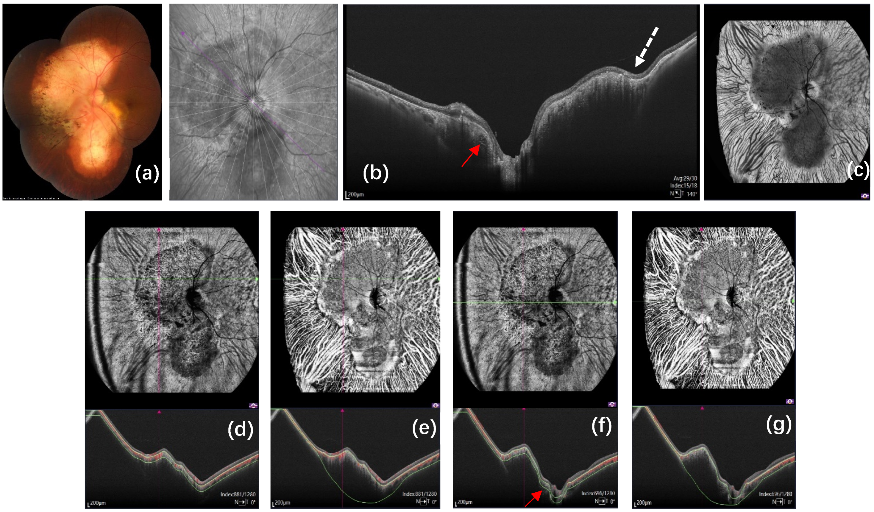
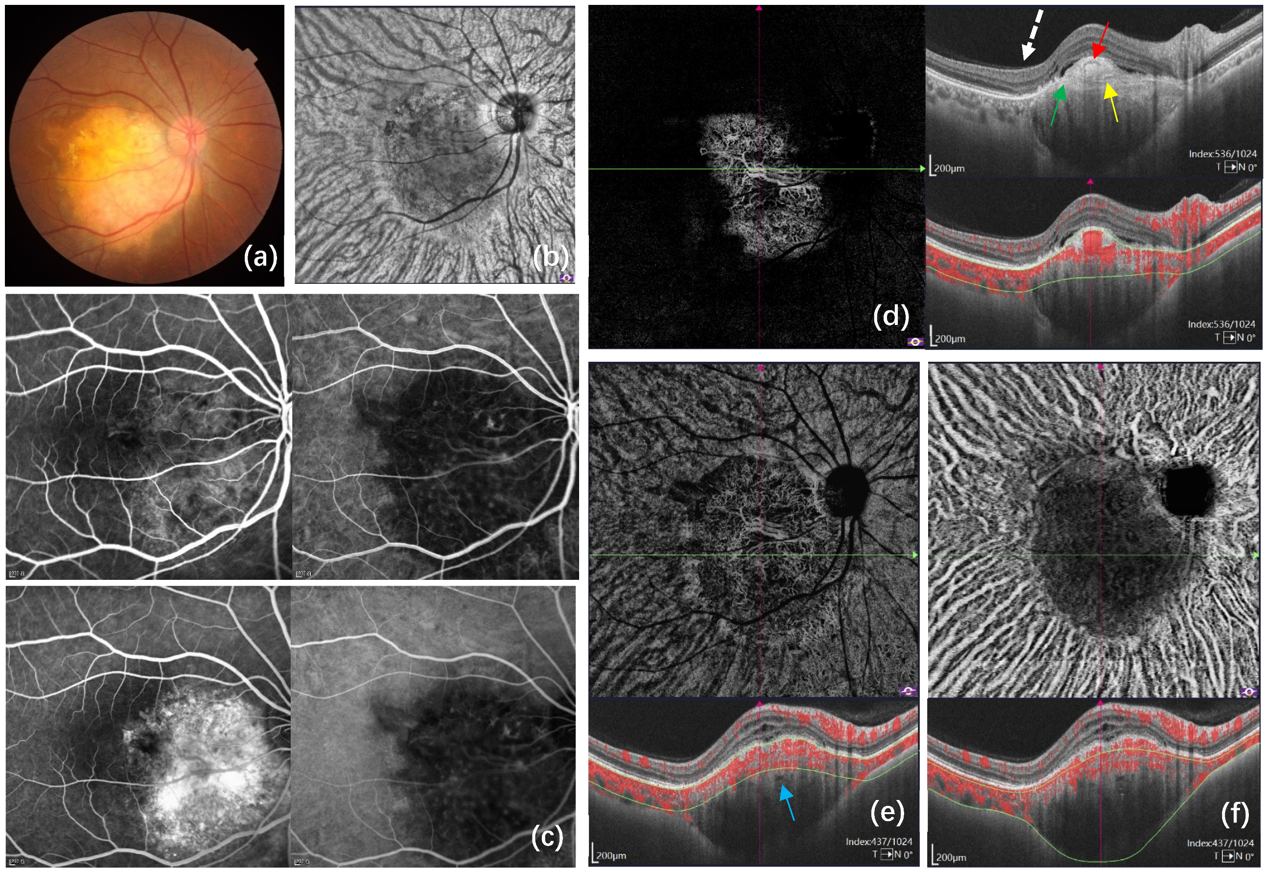
3. Results
4. Discussion
5. Conclusions
Author Contributions
Funding
Institutional Review Board Statement
Informed Consent Statement
Data Availability Statement
Conflicts of Interest
References
- Gass, J.D.; Guerry, R.K.; Jack, R.L.; Harris, G. Choroidal osteoma. Arch. Ophthalmol. 1978, 96, 428–435. [Google Scholar] [CrossRef] [PubMed]
- Shields, C.L.; Sun, H.; Demirci, H.; Shields, J.A. Factors predictive of tumor growth, tumor decalcification, choroidal neovascularization, and visual outcome in 74 eyes with choroidal osteoma. Arch. Ophthalmol. 2005, 123, 1658–1666. [Google Scholar] [CrossRef]
- Chen, J.; Lee, L.; Gass, J.D. Choroidal osteoma: Evidence of progression and decalcification over 20 years. Clin. Exp. Optom. 2006, 89, 90–94. [Google Scholar] [CrossRef] [PubMed]
- Shields, C.L.; Shields, J.A.; Augsburger, J.J. Choroidal osteoma. Surv. Ophthalmol. 1988, 33, 17–27. [Google Scholar] [CrossRef]
- Szelog, J.T.; Bonini Filho, M.A.; Lally, D.R.; de Carlo, T.E.; Duker, J.S. Optical coherence tomography angiography for detecting choroidal neovascularization secondary to choroidal osteoma. Ophthalmic Surg. Lasers Imaging Retin. 2016, 47, 69–72. [Google Scholar] [CrossRef] [PubMed]
- Shields, C.L.; Perez, B.; Materin, M.A.; Mehta, S.; Shields, J.A. Optical coherence tomography of choroidal osteoma in 22 cases: Evidence for photoreceptor atrophy over the decalcified portion of the tumor. Ophthalmology 2007, 114, e53–e58. [Google Scholar] [CrossRef] [PubMed]
- Freton, A.; Finger, P.T. Spectral domain-optical coherence tomography analysis of choroidal osteoma. Br. J. Ophthalmol. 2012, 96, 224–228. [Google Scholar] [CrossRef] [PubMed]
- Cennamo, G.; Romano, M.R.; Iovino, C.; Velotti, N.; Breve, M.A.; de Crecchio, G.; Cennamo, G. OCT angiography in choroidal neovascularization secondary to choroidal osteoma. Acta Ophthalmol. 2017, 95, e152–e154. [Google Scholar] [CrossRef]
- Grisolia, A.B.D.; de França Martins, M.; Demirci, H. Imaging of neovascular membrane over a choroidal osteoma by OCT angiography. Ophthalmology 2018, 125, 236. [Google Scholar] [CrossRef]
- Sagar, P.; Shanmugam, M.; Ramanjulu, R.; Konana, V.K. OCT angiography characteristics of choroidal osteoma. Ophthalmol. Retina 2018, 2, 77–79. [Google Scholar] [CrossRef]
- Pellegrini, M.; Invernizzi, A.; Giani, A.; Staurenghi, G. Enhanced depth imaging optical coherence tomography features of choroidal osteoma. Retina 2014, 34, 958–963. [Google Scholar] [CrossRef]
- Erol, M.K.; Coban, D.T.; Ceran, B.B.; Bulut, M. Enhanced depth imaging optical coherence tomography and fundus autofluorescence findings in bilateral choroidal osteoma: A case report. Arq. Bras. Oftalmol. 2013, 76, 189–191. [Google Scholar] [CrossRef]
- Shields, C.L.; Arepalli, S.; Atalay, H.T.; Ferenczy, S.R.; Fulco, E.; Shields, J.A. Choroidal osteoma shows bone lamella and vascular channels on enhanced depth imaging optical coherence tomography in 15 eyes. Retina 2015, 35, 750–757. [Google Scholar] [CrossRef]
- Dinah, C.; Sandinha, T. Enhanced depth imaging as an adjunctive tool in the diagnosis of decalcified choroidal osteoma. Eye 2014, 28, 356–358. [Google Scholar] [CrossRef][Green Version]
- Laíns, I.; Wang, J.C.; Cui, Y.; Katz, R.; Vingopoulos, F.; Staurenghi, G.; Vavvasa, D.G.; Millera, J.W.; Millerab, J.B. Retinal applications of swept source optical coherence tomography (OCT) and optical coherence tomography angiography (OCTA). Prog. Retin. Eye Res. 2021, 84, 100951. [Google Scholar] [CrossRef]
- Azad, S.V.; Takkar, B.; Venkatesh, P.; Kumar, A. Swept source: Optical coherence tomography angiography features of choroidal osteoma with choroidal neovascular membrane. BMJ Case Rep. 2016, bcr2016215899. [Google Scholar] [CrossRef]
- Hayashi, Y.; Mitamura, Y.; Egawa, M.; Semba, K.; Nagasawa, T. Swept-source optical coherence tomographic findings of choroidal osteoma. Case Rep. Ophthalmol. 2014, 5, 195–202. [Google Scholar] [CrossRef]
- Azad, S.V.; Kumar, V.; Chawla, R.; Kashyap, B.; Temkar, S.; Kumar, A.; Venkatesh, P.; Vohra, R.; Molla, K.; Sharma, A. In vivo optical biopsy of choroidal osteoma: A swept source optical coherence tomography-based tumor characterization. Ther. Adv. Ophthalmol. 2020, 12, 2515841420922740. [Google Scholar] [CrossRef]
- Chehab, H.E.; Dot, C.; Mathis, T.; Agard, E.; Kodjikian, L. Contribution of swept-source OCT-angiography in analysis of choroidal osteoma and its quiescent neovascular complications: A case study. Am. J. Ophthalmol. Case Rep. 2020, 19, 100769. [Google Scholar] [CrossRef]
- Zhou, N.; Xu, X.; Liu, Y.; Wei, W.; Peng, X. Appearance of tumor vessels in patients with choroidal osteoma using swept-source optical coherence tomographic angiography. Front. Oncol. 2021, 11, 762394. [Google Scholar] [CrossRef]
- Olguin-Manríquez, F.; Enríquez, A.B.; Crim, N.; Meraz-Gutierrez, M.; Soberón-Ventura, V.; Ávila, I.; Morales-Canton, V.; Jimenez-Sierra, J.M. Multimodal imaging in choroidal osteoma. Int. J. Retin. Vitreous 2018, 4, 30. [Google Scholar] [CrossRef] [PubMed]
- Shen, C.; Yan, S.; Du, M.; Zhao, H.; Shao, L.; Hu, Y. Assessment of choroidal osteoma complicating choroidal neovascularization by optical coherence tomography angiography. Int. Ophthalmol. 2018, 38, 787–792. [Google Scholar] [CrossRef] [PubMed]
- Wang, F.; Zhang, Q.; Deegan, A.J.; Chang, J.; Wang, R.K. Comparing imaging capabilities of spectral domain and swept source optical coherence tomography angiography in healthy subjects and central serous retinopathy. Eye Vis. 2018, 5, 19. [Google Scholar] [CrossRef] [PubMed]
- Williams, A.T.; Font, R.L.; Van Dyk, H.J.; Riekhof, F.T. Osseous choristoma of the choroid simulating a choroidal melanoma. Association with a positive 32P test. Arch. Ophthalmol. 1978, 96, 1874–1877. [Google Scholar] [CrossRef]
- Aylward, G.W.; Chang, T.S.; Pautler, S.E.; Gass, J.D. A long-term follow-up of choroidal osteoma. Arch. Ophthalmol. 1998, 116, 1337–1341. [Google Scholar] [CrossRef]
- Lafaut, B.A.; Mestdagh, C.; Kohno, T.; Gaudric, A.; De Laey, J.J. Indocyanine green angiography in choroidal osteoma. Graefes Arch. Clin. Exp. Ophthalmol. 1997, 235, 330–337. [Google Scholar] [CrossRef]
- Toledo, J.J.; Asencio, M.; García, J.R.; Morales, L.A.; Tomkinson, C.; Cajigal, C. OCT Angiography: Imaging of choroidal and retinal tumors. Ophthalmol. Retin. 2018, 2, 613–622. [Google Scholar] [CrossRef]
- Basavaraj, T.M.; Galiyugavaradhan, S. Sequential imaging of a case of choroidal osteoma using swept-source OCT and optical coherence tomography angiography: A 4-year follow-up study. Indian J. Ophthalmol. 2019, 67, 2097–2100. [Google Scholar]
- Furino, C.; Di Antonio, L.; Grassi, M.O.; Rispoli, M.; Reibaldi, M.; Niro, A.; Alessio, G. Choroidal neovascularization due to choroidal osteoma treated with anti-vascular endothelial growth factor therapy: An optical coherence tomography angiography study. Eur. J. Ophthalmol. 2019, 29, 323–329. [Google Scholar] [CrossRef]
- Pierro, L.; Marchese, A.; Gagliardi, M.; Introini, U.; Battaglia Parodi, M.; Casalino, G.; Bandello, F. Choroidal excavation in choroidal osteoma complicated by choroidal neovascularization. Eye 2017, 31, 1740–1743. [Google Scholar] [CrossRef]
- Navajas, E.V.; Costa, R.A.; Calucci, D.; Hammoudi, D.S.; Simpson, E.R.; Altomare, F. Multimodal fundus imaging in choroidal osteoma. Am. J. Ophthalmol. 2012, 153, 890–895. [Google Scholar] [CrossRef]
- Yi, X.; Min, W.; Qing, C.; Yongjin, Z. Swept-source optical coherence tomography analysis of choroidal osteoma. Chin. J. Ocul. Fundus Dis. 2020, 36, 435–441. [Google Scholar]
- Leitão Guerra, R.L.; Arantes, R.C.; Marback, E.F.; Shields, C.L. Novel OCT findings in choroidal osteoma: Brief report. Int. J. Retin. Vitreous 2021, 7, 46. [Google Scholar] [CrossRef]
- Spaide, R.F.; Ryan, E.H., Jr. Loculation of fluid in the posterior choroid in eyes with central serous chorioretinopathy. Am. J. Ophthalmol. 2015, 160, 1211–1216. [Google Scholar] [CrossRef]
- Xu, H.; Zeng, F.; Shi, D.; Sun, X.; Chen, X.; Bai, Y. Focal choroidal excavation complicated by choroidal neovascularization. Ophthalmology 2014, 121, 246–250. [Google Scholar] [CrossRef]
- Lee, J.H.; Lee, W.K. Choroidal neovascularization associated with focal choroidal excavation. Am. J. Ophthalmol. 2014, 157, 710–718. [Google Scholar] [CrossRef]
- Gan, Y.; Ji, Y.; Zuo, C.; Su, Y.; Liao, N.; Zhang, X.; Zeng, Y.; Wen, F. Correlation between focal choroidal excavation and underying retinochoridal disease: A pathological hypothesis from clinical obervation. Retina 2022, 42, 348–356. [Google Scholar] [CrossRef]
- Jampol, L.M.; Shankle, J.; Schroeder, R.; Tornambe, P.; Spaide, R.F.; Hee, M.R. Diagnostic and therapeutic challenges. Retina 2006, 26, 1072–1076. [Google Scholar] [CrossRef]
- Margolis, R.; Mukkamala, S.K.; Jampol, L.M.; Spaide, R.F.; Ober, M.D.; Sorenson, J.A.; Gentile, R.C.; Miller, J.A.; Sherman, J.; Freund, K.B. The expanded spectrum of focal choroidal excavation. Arch. Ophthalmol. 2011, 129, 1320–1325. [Google Scholar] [CrossRef]
- Introini, U.; Casalino, G.; Parodi, M.B.; Bandello, F.; London, N.J. Diagnostic and therapeutic challenges. Retina 2016, 36, 422–427. [Google Scholar] [CrossRef]
- Kamalden, T.A.; Lingam, G.; Sundar, G. Bone remodeling in choroidal osteoma monitored by fundus photography and spectral-domain optical coherence tomography. Ocul. Oncol. Pathol. 2014, 1, 13–18. [Google Scholar] [CrossRef] [PubMed]


| Patient Number | Age, Years | Gender | Affected Eye | Baseline BCVA (logMAR) | Number of Tumors | Tumor Greatest Linear Dimension, mm | Tumor Location | Previous Treatment | CNV | Grow over Bruch Membrane | FCE |
|---|---|---|---|---|---|---|---|---|---|---|---|
| 1 | 32 | F | OD | 0.8 | Single | 4.63 | Macula | None | Y | Y | Y |
| 2 | 42 | F | OS | 0.3 | Single | 5.78 | Juxtapapillary superior | Anti-VEGF injections | Y | Y | Y |
| 3 | 32 | M | OS | 1.3 | Single | 10.99 | Circumpapillary with macular involvement | None | Y | Y | |
| 4 | 29 | F | OS | 0.2 | Single | 16.36 | Circumpapillary with macular involvement | Anti-VEGF injections | Y | Y | Y |
| 5 | 37 | F | OD | 1.4 | Single | 14.07 | Circumpapillary with macular involvement | Anti-VEGF injections | Y | Y | Y |
| 6 | 50 | M | OD | 0.1 | Single | 2.83 | Macula | Anti-VEGF injections | Y | Y | |
| 7 | 23 | F | OU | 0.1/0.0 | Single | 5.40/4.11 | Macula/Juxtapapillary inferior | Anti-VEGF injections OD | Y OD | Y OD | |
| 8 | 38 | F | OS | 0.4 | Single | 5.7 | Macula | Anti-VEGF injections | Y | Y | |
| 9 | 21 | M | OS | 0.3 | Multifocal | 10.71/5.65 | Circumpapillary | None | Y | ||
| 10 | 46 | F | OS | 1.0 | Single | 8.18 | Macula | Anti-VEGF injections | Y | Y | Y |
| 11 | 42 | F | OS | 1.0 | Single | 2.61 | Juxtapapillary temporal | None | |||
| 12 | 23 | M | OS | 0.3 | Single | 6.14 | Macula | None | Y | ||
| 13 | 26 | F | OD | 1.3 | Single | 8.91 | Macula | Anti-VEGF injections | Y | Y | |
| 14 | 34 | F | OD | 0.1 | Single | 8.31 | Macula | Anti-VEGF injections | Y | Y | Y |
| 15 | 25 | F | OU | 0.7/0.8 | Multifocal | 3.54/2.95/2.23/4.18/1.96 | Juxtapapillary superior/superior/temporal/superior/nasal | None | |||
| 16 | 28 | F | OS | 0.7 | Single | 6.7 | Macula | Anti-VEGF injections | Y | Y | Y |
| 17 | 33 | F | OS | 0.2 | Single | 11.29 | Circumpapillary with macular involvement | Anti-VEGF injections | Y | Y | Y |
| 18 | 37 | F | OS | 0.0 | Single | 8.21 | Circumpapillary with macular involvement | None | Y | Y | |
| 19 | 34 | F | OD | 0.7 | Single | 8.01 | Macula | Anti-VEGF injections | Y | Y | Y |
| 20 | 16 | F | OD | 1.0 | Single | 14.1 | Circumpapillary with macular involvement | None | Y | Y | |
| 21 | 37 | F | OD | 0.0 | Single | 6.37 | Juxtapapillary temproal | None |
Publisher’s Note: MDPI stays neutral with regard to jurisdictional claims in published maps and institutional affiliations. |
© 2022 by the authors. Licensee MDPI, Basel, Switzerland. This article is an open access article distributed under the terms and conditions of the Creative Commons Attribution (CC BY) license (https://creativecommons.org/licenses/by/4.0/).
Share and Cite
Xuan, Y.; Chang, Q.; Zhang, Y.; Ye, X.; Liu, W.; Li, L.; Wang, K.; Zhou, J.; Wang, M. Clinical Observation of Choroidal Osteoma Using Swept-Source Optical Coherence Tomography and Optical Coherence Tomography Angiography. Appl. Sci. 2022, 12, 4472. https://doi.org/10.3390/app12094472
Xuan Y, Chang Q, Zhang Y, Ye X, Liu W, Li L, Wang K, Zhou J, Wang M. Clinical Observation of Choroidal Osteoma Using Swept-Source Optical Coherence Tomography and Optical Coherence Tomography Angiography. Applied Sciences. 2022; 12(9):4472. https://doi.org/10.3390/app12094472
Chicago/Turabian StyleXuan, Yi, Qing Chang, Yongjin Zhang, Xiaofeng Ye, Wei Liu, Lei Li, Keyan Wang, Jian Zhou, and Min Wang. 2022. "Clinical Observation of Choroidal Osteoma Using Swept-Source Optical Coherence Tomography and Optical Coherence Tomography Angiography" Applied Sciences 12, no. 9: 4472. https://doi.org/10.3390/app12094472
APA StyleXuan, Y., Chang, Q., Zhang, Y., Ye, X., Liu, W., Li, L., Wang, K., Zhou, J., & Wang, M. (2022). Clinical Observation of Choroidal Osteoma Using Swept-Source Optical Coherence Tomography and Optical Coherence Tomography Angiography. Applied Sciences, 12(9), 4472. https://doi.org/10.3390/app12094472