
Analysis of Uncertainties in Clinical High-Energy Photon Beam Calibrations Using Absorbed Dose Standards

 ,
,
Abstract
:1. Introduction
1.1. Dosimetry Formalism
1.2. Correction Factors for Influence Quantities
2. Materials and Methods
2.1. Dosimetry Equipment
2.2. Determination of the Beam Quality Specifiers
2.3. Absorbed Dose Measurements
2.4. Measurement Uncertainty
3. Results
4. Discussion
5. Conclusions
Author Contributions
Funding
Institutional Review Board Statement
Informed Consent Statement
Data Availability Statement
Conflicts of Interest
References
- Almond, P.R.; Biggs, P.J.; Coursey, B.M.; Hanson, W.F.; Huq, M.S.; Nath, R.; Rogers, D.W. AAPM’s TG-51 protocol for clinical reference dosimetry of high-energy photon and electron beams. Med. Phys. 1999, 26, 1847–1870. [Google Scholar] [CrossRef] [PubMed] [Green Version]
- Dosismessverfahren Nach der Sondenmethode für Photonen—Und Elektronenstrahlung Teil 2: Ionisationsdosimetrie DIN 6800-2; Institut für Normung: Berlin, German, 1998.
- Lillicrap, S.C.; Owen, B.; Williams, J.R.; Williams, P.C. Code of practice for high-energy photon therapy dosimetry based on the NPL absorbed dose calibration service. Phys. Med. Biol. 1990, 35, 1355–1360. [Google Scholar] [CrossRef]
- The Netherlands Commission on Radiation Dosimetry (NCS). Code of Practice for the Absorbed Dose Determination in High Energy Photon and Electron Beams. In NCS Report No. 18; The Netherlands Commission on Radiation Dosimetry: Delft, The Netherlands, 2008. [Google Scholar]
- Andreo, P.; Burns, D.T.; Hohlfeld, K.; Huq, M.S.; Kanai, T.; Laitano, F.; Smyth, V.; Vynckier, S. Absorbed dose determination in external beam radiotherapy: An international code of practice for dosimetry based on standards of absorbed dose to water. In IAEA TRS Report. 398; International Atomic Energy Agency: Vienna, Austria, 2000. [Google Scholar]
- International Atomic Energy Agency. Implementation of the International Code of Practice on Dosimetry in Radiotherapy (TRS 398): Review of Testing Results. In IAEA Technical Document (TECDOC-1455); International Atomic Energy Agency: Vienna, Austria, 2005. [Google Scholar]
- Swanpalmer, J. Reference dose determination in 60Co and high-energy radiotherapy photon beams by using Farmer-type cylindrical ionization chambers—An experimental investigation. Biomed. Phys. Eng. Express 2020, 6, 045003. [Google Scholar] [CrossRef] [PubMed]
- Al-Ahbabi, S.S.; Bradley, D.A.; Beyomi, M.; Alkatib, Z.; Adhaheri, S.; Darmaki, M.; Nisbet, A. A comparison of protocols for external beam radiotherapy beam calibrations. Appl. Radiat. Isot. 2012, 70, 1331–1336. [Google Scholar] [CrossRef] [PubMed] [Green Version]
- Castrillón, S.V.; Henríquez, F.C. Comparison of IPSM 1990 photon dosimetry code of practice with IAEA TRS-398 and AAPM TG-51. J. Appl. Clin. Med. Phys. 2009, 10, 136–146. [Google Scholar] [CrossRef] [PubMed]
- Rogers, D.W. ICRU Report 64: Dosimetry of High-Energy Photon Beams Based on Standards of Absorbed Dose to Water. ICRU Rep. 2001, 47, 20. [Google Scholar] [CrossRef]
- Mayles, P.; Nahum, A.; Rosenwald, J.C. Handbook of Radiotherapy Physics: Theory and Practice; CRC Press: Boca Raton, FL, USA, 2007. [Google Scholar]
- Institute of Physics and Engineering in Medicine (IPEM). Physics Aspects of Quality Control in Radiotherapy. In IPEM Report No.81; Institute of Physics and Engineering in Medicine (IPEM): York, UK, 1999. [Google Scholar]
- Brahme, A.; Andreo, P. Dosimetry and quality specification of high energy photon beams. Acta Radiol. Oncol. 1986, 25, 213–223. [Google Scholar] [CrossRef] [PubMed]
- Hohlfeld, K. The standard DIN 6800: Procedures for absorbed dose determination in radiology by the ionization method (IAEA-SM-298/31). Vienna Austria Dosim. Radiother. 1988, 1, 13–22. [Google Scholar]
- IAEA. Accuracy Requirements and Uncertainties in Radiotherapy; IAEA Human Health Series; IAEA: Vienna, Austria, 2016. [Google Scholar]
- ISO. International Organization for Standardization. In Guide to the Expression of Uncertainty in Measurement; ISO: Geneva, Switzerland, 1995. [Google Scholar]
- Zakaria, A.; Schuette, W.; Younan, C. Reference Dosimetry according to the New German Protocol DIN 6800-2 and Comparison with IAEA TRS 398 and AAPM TG 51. Biomed. Imaging Interv. J. 2011, 7, e15. [Google Scholar] [PubMed]
- Zakaria, G.A.; Schuette, W. Determination of absorbed dose to water for high-energy photon and electron beams-comparison of the standards DIN 6800-2 (1997), IAEA TRS 398 (2000) and DIN 6800-2 (2006). J. Med. Phys. Assoc. Med. Phys. India 2007, 32, 3. [Google Scholar] [CrossRef] [PubMed]
- Kinoshita, N.; Oguchi, H.; Adachi, T.; Shioura, H.; Kimura, H. Uncertainty in positioning ion chamber at reference depth for various water phantoms. Rep. Pract. Oncol. Radiother. 2018, 23, 199–206. [Google Scholar] [CrossRef] [PubMed]
- Kinoshita, N.; Kohno, R.; Oguchi, H. Influence of entrance window deformation on reference dosimetry measurement in various beam modalities. Med. Phys. 2019, 46, 1037–1043. [Google Scholar] [CrossRef] [PubMed]

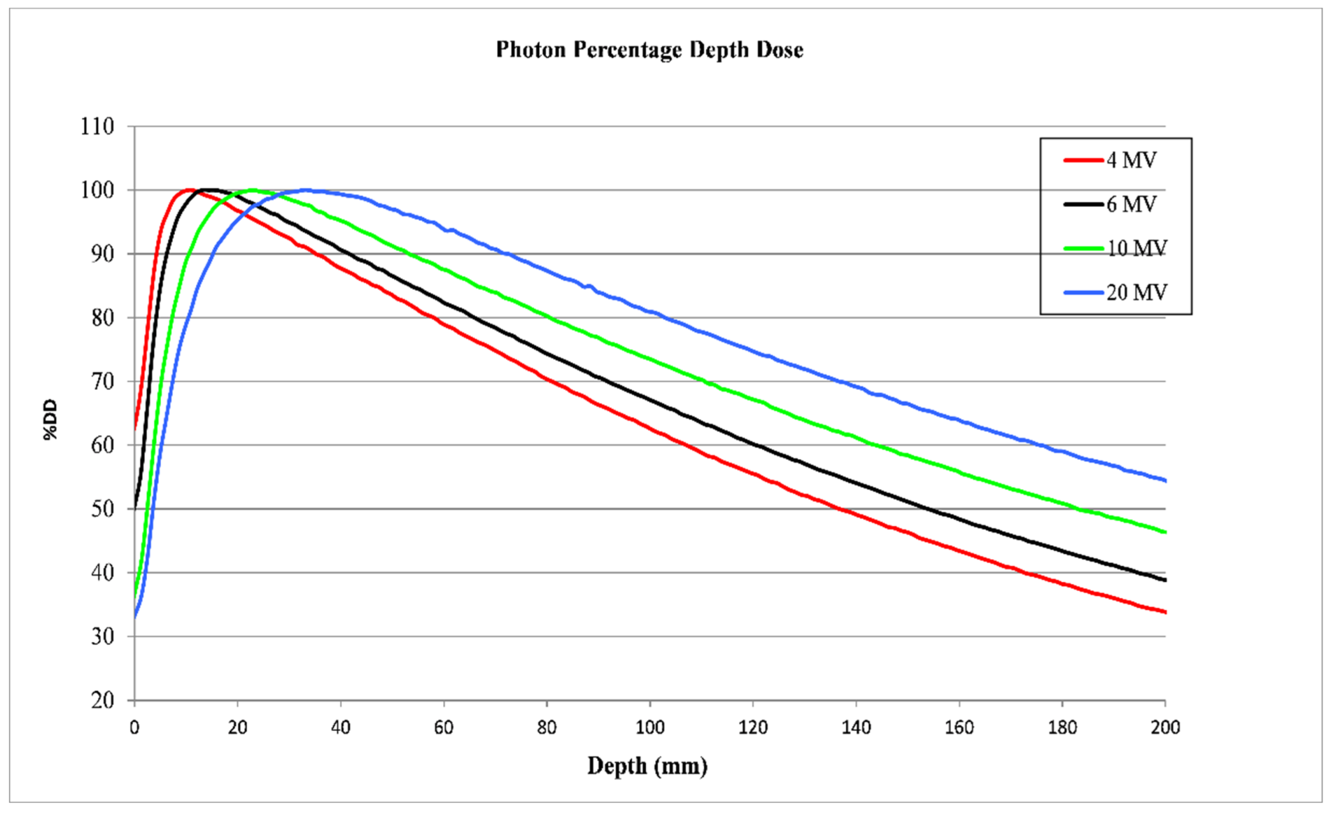
| IAEA TRS-398 | 4 MV | 6 MV | 10 MV | 20 MV |
|---|---|---|---|---|
| R100 (mm) | 11.10 | 13.20 | 23.20 | 33.00 |
| D100 (%) | 62.60 | 67.09 | 73.34 | 80.90 |
| D200 (%) | 33.58 | 38.85 | 46.39 | 54.50 |
| TPR 20/10 | 0.628 | 0.677 | 0.745 | 0.797 |
| MV | TPR20,10 | %dd (10)x. | Sw,air | |
|---|---|---|---|---|
| Measurements | From PDD | |||
| 4 | 0.619 | 0.628 | 62.66 | 1.127 |
| 6 | 0.675 | 0.677 | 67.27 | 1.119 |
| 10 | 0.738 | 0.745 | 72.60 | 1.106 |
| 20 | 0.793 | 0.797 | 82.50 | 1.084 |
| Chamber | Beam Energy (MV) | kQ | ||
|---|---|---|---|---|
| TRS-398 | TG-51 | DIN 6800-2 | ||
| NACP-02 | 4 | 0.9991 | 1.0003 | 0.996 |
| 10 | 0.9665 | 0.9633 | 0.9607 | |
| 6 | 0.9991 | 1.0032 | 0.9960 | |
| 20 | 0.9665 | 0.9633 | 0.9607 | |
| CC15 | 4 | 0.9996 | 0.9991 | 0.9981 |
| 10 | 0.9854 | 0.9836 | 0.9815 | |
| 6 | 0.9953 | 0.9913 | 0.9933 | |
| 20 | 0.9981 | 0.9656 | 0.9622 | |
| FC65-G | 4 | 0.9981 | 0.9991 | 0.9971 |
| 10 | 0.9854 | 0.9914 | 0.9815 | |
| 6 | 0.9953 | 0.9913 | 0.9925 | |
| 20 | 0.9685 | 0.9656 | 0.9638 | |
| Chamber | 6 MV | 20 MV | 4 MV | 10 MV | ||||||||
|---|---|---|---|---|---|---|---|---|---|---|---|---|
| TRS | TG-51 | DIN | TRS | TG-51 | DIN | TRS | TG-51 | DIN | TRS | TG-51 | DIN | |
| kpol | ||||||||||||
| NACP | 0.9981 | 0.9981 | 0.9982 | 0.999 | 0.999 | 0.999 | 0.999 | 0.9971 | 0.999 | 0.9978 | 0.9978 | 0.9978 |
| CC15 | 0.9994 | 0.9995 | 0.9994 | 0.9994 | 0.9995 | 0.9994 | 0.9988 | 1.0002 | 0.9988 | 0.9998 | 0.9998 | 0.9998 |
| FC65-G | 0.9989 | 0.9987 | 0.9989 | 0.9989 | 0.9989 | 0.9989 | 0.9989 | 0.9987 | 0.9989 | 0.9989 | 0.9989 | 0.9989 |
| ks | ||||||||||||
| NACP | 1.0062 | 1.0062 | 1.0033 | 1.0071 | 1.0073 | 1.0045 | 1.0036 | 1.0032 | 1.0028 | 1.0026 | 1.0027 | 1.0023 |
| CC15 | 1.0043 | 1.0043 | 1.0031 | 1.0043 | 1.0043 | 1.0031 | 1.0026 | 1.0017 | 1.0023 | 1.0027 | 1.0028 | 1.0023 |
| FC65-G | 1.0025 | 1.0027 | 1.0022 | 1.0033 | 1.0035 | 1.0027 | 1.0025 | 1.0027 | 1.0022 | 1.0033 | 1.0035 | 1.0027 |
| kTP | ||||||||||||
| NACP | 1.0041 | 1.0041 | 0.9973 | 1.0041 | 0.9973 | 1.0041 | 1.0127 | 1.0058 | 1.0127 | 1.0127 | 1.0058 | 1.0127 |
| CC15 | 1.0135 | 1.0066 | 1.0135 | 1.0135 | 1.0066 | 1.0135 | 1.0128 | 1.006 | 1.0128 | 1.0128 | 1.006 | 1.0128 |
| FC65-G | 1.0130 | 1.0061 | 1.013 | 1.013 | 1.0061 | 1.0130 | 1.0130 | 1.0061 | 1.0130 | 1.0130 | 1.0061 | 1.0130 |
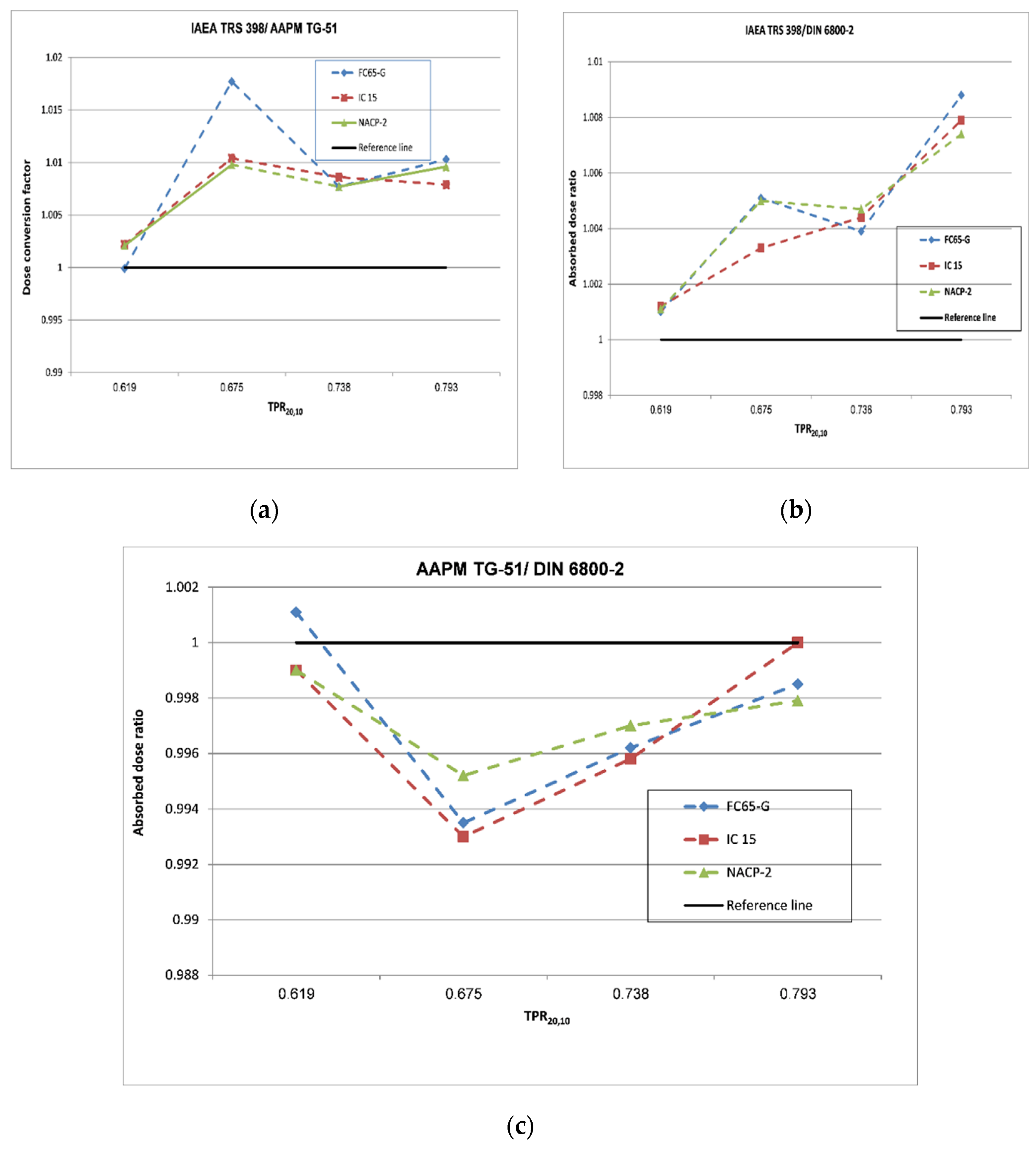
| MV | Ionization Chamber | TRS-398/ TG-51 | TRS-398/DIN 6800-2 | TG-51/DIN 6800-2 |
|---|---|---|---|---|
| 4 | FC65-G | 0.9999 | 1.0010 | 1.0011 |
| CC15 | 1.0022 | 1.0012 | 0.9990 | |
| NACP-02 | 1.0021 | 1.0011 | 0.9990 | |
| 6 | FC65-G | 1.0177 | 1.0051 | 0.9935 |
| CC15 | 1.0104 | 1.0033 | 0.9930 | |
| NACP-02 | 1.0098 | 1.0050 | 0.9952 | |
| 10 | FC65-G | 1.0077 | 1.0039 | 0.9962 |
| CC15 | 1.0086 | 1.0044 | 0.9958 | |
| NACP-02 | 1.0077 | 1.0047 | 0.9970 | |
| 20 | FC65-G | 1.0103 | 1.0088 | 0.9985 |
| CC15 | 1.0079 | 1.0079 | 1.0000 | |
| NACP-02 | 1.0096 | 1.0074 | 0.9979 |
| Influence Quantities | Source | Evaluation Type | Cylindrical Chamber | Plane-Parallel Chamber | ||
|---|---|---|---|---|---|---|
| TRS-398 | DIN 6800-2 | TRS-398 | DIN 6800-2 | |||
| ND,W | Chamber certificate | B | 0.55 | 0.55 | 0.55 | 0.55 |
| Depth of measurement | Calculated | B | 0.33 | 0.33 | 0.33 | 0.33 |
| kpol | Calculated | A/B | 0.04 | 0.04 | 0.12 | 0.12 |
| kTP | Calculated | A/B | 0.01 | 0.01 | 0.01 | 0.01 |
| kS | Calculated | B | 0.04 | 0.04 | 0.12 | 0.12 |
| kQ | IAEA TRS-398 | B | 1 | 1 | ||
| kE | DIN 6800-2 | B | 1 | 1 | ||
| Interaction coefficient | IAEA TRS 277 | B | ||||
| Dosimeter stability | Dosimeter manual | B | 0.28 | 0.28 | 0.28 | 0.28 |
| Dosimeter reading | Calculated | A | 0.20 | 0.20 | 0.20 | 0.20 |
| Combined uncertainty (k = 1) | 1.24 | 1.24 | 1.25 | 1.25 | ||
Publisher’s Note: MDPI stays neutral with regard to jurisdictional claims in published maps and institutional affiliations. |
© 2022 by the authors. Licensee MDPI, Basel, Switzerland. This article is an open access article distributed under the terms and conditions of the Creative Commons Attribution (CC BY) license (https://creativecommons.org/licenses/by/4.0/).
Share and Cite
Elbashir, F.E.M.; Ksouri, W.; Habbani, F.; El-Khayatt, A.M.; Eisa, M.H.; Suliman, I.I. Analysis of Uncertainties in Clinical High-Energy Photon Beam Calibrations Using Absorbed Dose Standards. Appl. Sci. 2022, 12, 3857. https://doi.org/10.3390/app12083857
Elbashir FEM, Ksouri W, Habbani F, El-Khayatt AM, Eisa MH, Suliman II. Analysis of Uncertainties in Clinical High-Energy Photon Beam Calibrations Using Absorbed Dose Standards. Applied Sciences. 2022; 12(8):3857. https://doi.org/10.3390/app12083857
Chicago/Turabian StyleElbashir, Fawzia E. M., Wassim Ksouri, Farouk Habbani, Ahmed M. El-Khayatt, Mohamed Hassan Eisa, and Ibrahim I. Suliman. 2022. "Analysis of Uncertainties in Clinical High-Energy Photon Beam Calibrations Using Absorbed Dose Standards" Applied Sciences 12, no. 8: 3857. https://doi.org/10.3390/app12083857
APA StyleElbashir, F. E. M., Ksouri, W., Habbani, F., El-Khayatt, A. M., Eisa, M. H., & Suliman, I. I. (2022). Analysis of Uncertainties in Clinical High-Energy Photon Beam Calibrations Using Absorbed Dose Standards. Applied Sciences, 12(8), 3857. https://doi.org/10.3390/app12083857