Article

Pregnancy Achievement by Medical Assisted Reproduction Is Correlated to the G Protein-Coupled Receptor 30 mRNA Abundance in Human Spermatozoa

1
Endocrine and Metabolic Research, Unit for Multidisciplinary Research in Biomedicine, Department of Anatomy, School of Medicine and Biomedical Sciences (ICBAS), University of Porto, 4050-313 Porto, Portugal
2
Laboratory for Integrative and Translational Research in Population Health (ITR), University of Porto, 4099-002 Porto, Portugal
3
Department of Pathology, Faculty of Medicine, University of Porto, 4200-319 Porto, Portugal
4
QOPNA & LAQV, Department of Chemistry, Campus Universitario de Santiago, University of Aveiro, 3810-193 Aveiro, Portugal
5
Colégio Internato dos Carvalhos, Pedroso, 4415-133 Vila Nova de Gaia, Portugal
6
Centre for Reproductive Genetics Professor Alberto Barros, 4100-012 Porto, Portugal
7
Reproductive Genetics and Embryo-Fetal Development, i3S–Instituto de Investigação e Inovação em Saúde, University of Porto, 4200-135 Porto, Portugal
8
Laboratory of Cell Biology, Unit for Multidisciplinary Research in Biomedicine, Department of Microscopy, School of Medicine and Biomedical Sciences (ICBAS), University of Porto, 4050-313 Porto, Portugal
9
Biotechnology of Animal and Human Reproduction (TechnoSperm), Institute of Food and Agricultural Technology, University of Girona, ES-17003 Girona, Spain
10
Unit of Cell Biology, Department of Biology, Faculty of Sciences, University of Girona, ES-17003 Girona, Spain
*
Author to whom correspondence should be addressed.
Appl. Sci. 2022, 12(7), 3240; https://doi.org/10.3390/app12073240
Submission received: 12 January 2022 / Revised: 17 March 2022 / Accepted: 21 March 2022 / Published: 22 March 2022
(This article belongs to the Topic Human Anatomy and Pathophysiology)

Abstract

:
Estrogens, specifically 17β-estradiol (E2), play an important role in male health, including male fertility. The G protein-coupled receptor for estrogen 30 (GPR30) is essential for mediating the rapid non-genomic effects of E2 on a variety of testicular cells, including spermatozoa, although its molecular effects remain largely unknown. In this work, we hypothesized that the GPR30 mRNA abundance in spermatozoa could be correlated to sperm quality. Sperm GPR30 mRNA could also be carried into the oocyte, potentially impacting embryo development and the success of a pregnancy. For this study, 81 sperm samples were collected from couples seeking fertility treatment and undergoing medically assisted reproduction treatments (ART), following the World Health Organization guidelines. GPR30 mRNA abundance in spermatozoa was assessed with a quantitative polymerase chain reaction. The resulting data show that there is no correlation between the abundance of the GPR30 transcript with paternal BMI, age, or sperm quality parameters. Interestingly, we observed that higher levels of GPR30 mRNA abundance in spermatozoa were related to the achievement of biochemical pregnancy and clinical pregnancy (p < 0.05) by couples undergoing treatment. These results highlight the role of the sperm’s RNA cargo in offspring development, suggesting that spermatozoa mRNA content can influence ART success.

1. Introduction

For a long time, the role of estrogens in male physiology, particularly in reproductive health, remained unknown and was a matter of intense debate. Nowadays, it is known that estrogens, specifically 17β-estradiol (E2), play an important role in male health, including male fertility [1,2]. E2 can mediate genomic effects through specific estrogen receptors (ERα and ERβ), which are widely distributed in different tissues and particularly in the cells of the male reproductive system [3]. Recently, a non-classic receptor, known as the G protein-coupled receptor 30 (GPR30), was also found to be essential for mediating the non-genomic rapid effects of E2 on the human testis, being present in Sertoli cells, Leydig cells, spermatogonia, spermatocytes, round spermatids, and spermatozoa [4,5]. As androgens, E2 can induce a negative regulation of the hypothalamus-pituitary-gonadal (HPG) axis, promoting the down-expression of the gonadotropin-releasing hormone, as well as the release of luteinizing hormone (LH) and follicle-stimulating hormone (FSH). The pituitary sex hormones, LH and FSH, play crucial roles in the maintenance of both steroidogenesis and spermatogenesis [6]. However, the effects of E2 on spermatozoa, particularly the ones mediated by GPR30, remain to be elucidated.
As a haploid cell, sperm DNA is highly condensed, allowing for its safe delivery into the oocyte. In addition, the closed conformation of the genetic material leads to the arrest of almost all transcription activity [7]. Interestingly, mature spermatozoa carry into the oocyte a pool of mRNAs. The origin and function of these mRNAs are still unknown [7,8]. Nonetheless, recent studies suggest that the male metabolic state can influence the offspring’s health. Fullston and colleagues reported that, in mice, the induction of obesity by the consumption of a high-fat diet by F0 males altered the germ cell DNA methylation pattern, the expression of testicular mRNAs, and the miRNA sperm content in the males of subsequent generations, even though these animals were fed with a standard diet [9]. Several studies from Crisóstomo and colleagues came to a similar conclusion [10,11,12]. The authors reported high-fat-feeding male mice (F0) affected the testicular metabolome and function of their sons (F1) and grandsons (F2) [10]. Sperm quality was affected in all generations, even though F1 and F2 males were fed with a standard diet [10,11]. Interestingly, with a reversion in the diet of the F0 animals from a high-fat to a standard diet, the testicular content in fatty acids was still irreversibly affected, as well as sperm defects [12]. This means that the effects promoted by the high-fat diet could be irreversible and still passable to subsequent generations [12]. Although these studies did not identify RNAs as being responsible for the transmission of these metabolic defects through the generations, another study reported solid evidence of such a finding. Chen and colleagues isolated transfer-RNA-derived small RNAs (tsRNAs) from obese mice sperm. These tsRNAs were later injected into healthy developing embryos, which resulted in the alteration of the expression of genes associated with metabolic regulation [13]. Furthermore, other factors, such as paternal age and obesity, are known to have several detrimental effects on males’ fertility (for further details, see [14,15]). Several studies have already linked high paternal body mass indexes (BMI) and decreased live birth outcomes after medically assisted reproduction treatments (ART) [16,17,18]. Other studies have reported a similar trend for advanced paternal age and ART outcomes [19,20].
The present study aimed to detect and quantify the abundance of GPR30 mRNA in human spermatozoa and to determine its impact on the outcome of ART. Furthermore, we aimed to elucidate if the abundance of this transcript in spermatozoa is associated with paternal BMI and age.

2. Materials and Methods

2.1. Patients’ Characterization

Eighty-one couples seeking fertility treatment and undergoing ART at the Center for Reproductive Genetics, Alberto Barros (Porto, Portugal) were included in this study. Sperm samples were collected from October 2018 to January 2019. Patients’ anthropometric and demographic data, which includes the age and BMI measurements of participating couples and oocyte donors, can be consulted in Table S1 in the Supplementary Materials. The same selection criteria for male patients were applied as in [21]. Semen analysis was performed according to the World Health Organization guidelines [22], along with the protocol discussed by the Joint Ethics Committee of CHUP/ICBAS, receiving the approval number 2019/CE/P017(266/CETI/ICBAS). Of the current group, 66% of the male individuals were considered normozoospermic (normal concentration, normal motility, normal morphology of sperm), according to the WHO nomenclature.

2.2. Sample Preparation for ART

The samples were washed and subjected to seminal processing via the swim-up technique [23]. Fertilization protocols were performed by certified embryologists at the Center for Reproductive Genetics, which include a total of 14 cycles of in vitro fertilization (IVF) and 67 cycles of intracytoplasmic sperm injection (ICSI). Sperm quality parameters and embryo development parameters were assessed to ensure and monitor the embryo quality before embryo transfer, namely, the fertilization rate, embryo cleavage rate, high-quality embryo rate, and blastocyst rate. Embryo transfer was performed 2 or 5 days after fertilization.
After embryo transfer, pregnancy was evaluated at two different stages. Women were classified as having a biochemical pregnancy when β-HCG concentration surpassed the value of 20 mIU/mL (positive serum β-HCG levels), 12 days after embryo transfer [24]. All women with negative serum β-HCG levels ([β-HCG] < 20 mIU/mL) were not considered pregnant. Biochemical pregnancies evolved into clinical pregnancies when a fetal heartbeat was detected.

2.3. ART Data Analysis

Embryo development was evaluated through embryo quality parameters, which include the fertilization rate, embryo cleavage rate, high-quality embryo rate, and blastocyst rate. Embryo quality parameters were calculated with the following formulas:
Fertilization   Rate = N º   2 pronuclear   embryos N º   Mature   Oocytes   ( MII )
Embryo   Cleavage   Rate = N º   4 cell   stage   embryos N º   Mature   Oocytes   ( MII )
High quality   Embryo   Rate = N º   High quality   8 cell   stage   embryos N º   Mature   Oocytes   ( MII )
Blastocyst   Rate = N º   blastocyst   stage   embryos N º   Mature   Oocytes   ( MII )
Embryo development after embryo transference was evaluated using the biochemical pregnancy rate (BP), which was calculated with the following formula:
BP = [ Serum   β human   chorionic   gonadotropin ] ( mIU · mL 1 ) N º   Embryo   Transfer  

2.4. Reverse Transcriptase-Polymerase Chain Reaction and Quantitative Real-Time PCR

The extraction of total RNA (RNAt) from spermatozoa was performed using the RNeasy Mini isolation kit from Qiagen (Hilden, Germany), following the manufacturer’s instructions. The extracted RNAt was reversely transcribed into complementary deoxyribonucleic acid (cDNA) (NZY M-MuLV Reverse Transcriptase, random hexamer primers, and dNTPs were obtained from NZYTech, Lisboa, Portugal). The resulting cDNA was used to perform quantitative polymerase chain reaction (qPCR) assays. We used an exon-exon spanning primers set designed by Rago and collaborators (Rago, et al., 2011 [5]) to amplify human GPR30 for a product size of 155 base pairs (Primer Forward: CTGGGGAGTTTCCTGTCTGA; Primer Reverse: GCTTGGGAAGTCACATCCAT). qPCR experiments were performed with a CFX96 Connect Real-Time PCR detection system (Bio-Rad, Hercules, California, USA). All qPCR reagents were purchased from NZYTech, Lisboa, Portugal. A cDNA-free sample was used as a negative control [25]. The β2-microglobulin mRNA abundance was used to normalize GPR30 transcript abundance (Primer Forward: AGATGAGTATGCCTGCCGTG; Primer Reverse: TCATCCAATCCAAATGCGGC). The fold variation of the target gene abundance was calculated using the following mathematical formula: 2−ΔΔCt [26].

2.5. Statistical Analysis

The associations between GPR30 mRNA abundance in spermatozoa, BMI, age, and sperm quality, as well as with biochemical pregnancy, were evaluated by computing the Pearson correlation coefficients (r), with a confidence interval of 95%. Normality was tested through the Shapiro–Wilk and Kolmogorov–Smirnov tests. Patients were also classified into two groups, based on the achievement or not of pregnancy. Statistical analysis between pregnancy and non-pregnancy groups was performed by a two-tailed Student’s t-test (for parametric data). A two-tailed Student’s t-test (for nonparametric data, the Kolmogorov–Smirnov test) was performed to evaluate the differences between fertilization procedures (ICSI and IVF) and the achievement of pregnancy. Values of p < 0.05 were considered to be statistically significant. The statistical analysis was performed using GraphPad Prism 8 (GraphPad Software Inc., San Diego, CA, USA).

3. Results

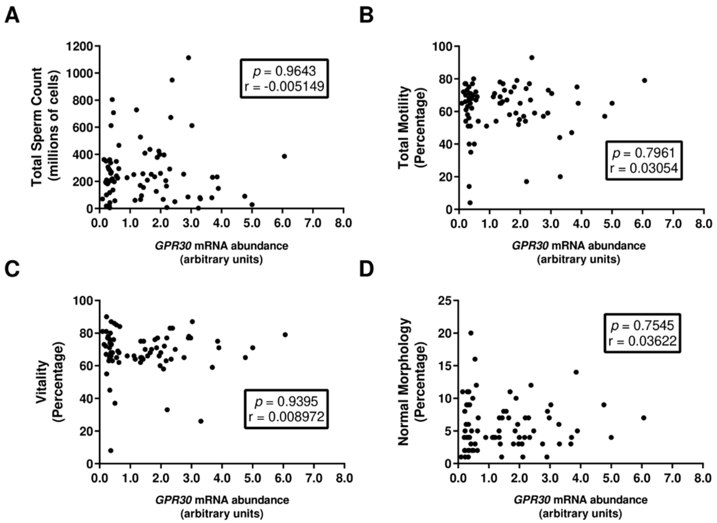
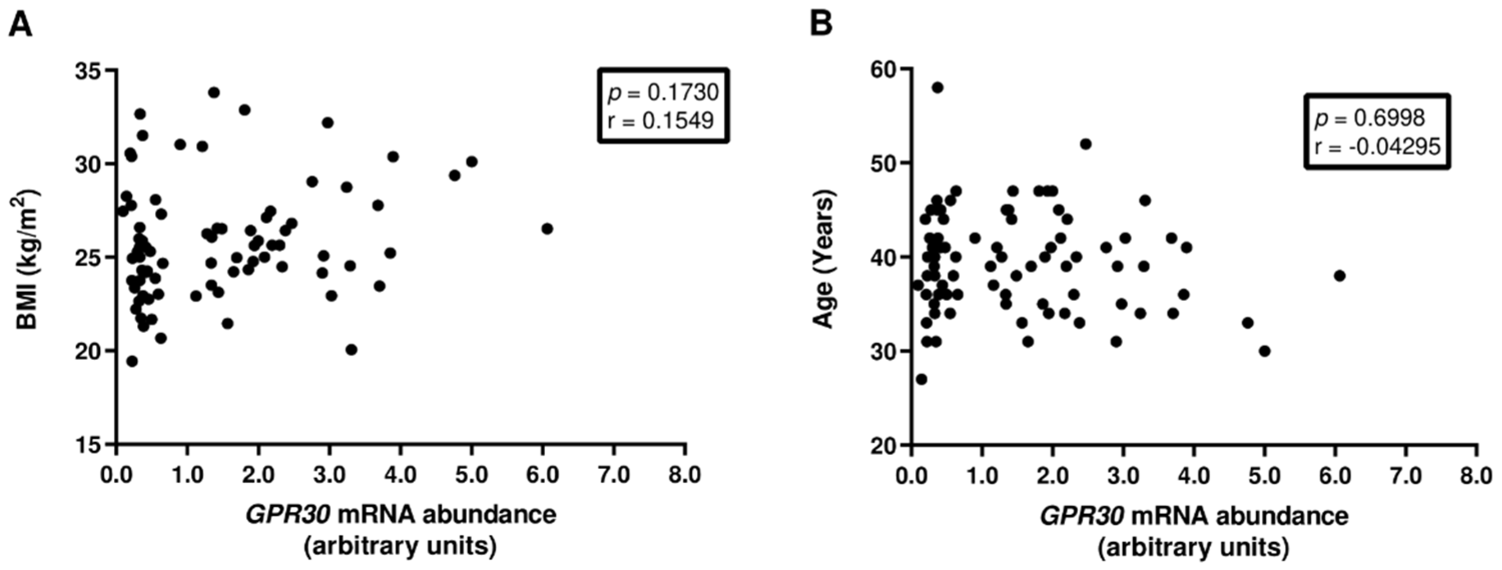
3.1. GPR30 mRNA Abundance in Human Spermatozoa Is Not Associated with Decreased Sperm Quality

The GPR30 transcript and protein were previously identified in human spermatozoa by Rago and collaborators [27]. In our work, we further confirmed that GPR30 mRNA is present in human spermatozoa by a conventional PCR (Figure S1). Since sperm parameters are used as reliable biomarkers for male fertility potential, we analyzed the association of GPR30 mRNA abundance with different sperm parameters, including total sperm count, total motility, normal morphology, and vitality. We could not identify any correlation between GPR30 mRNA abundance and the sperm parameters studied (Figure 1), suggesting that GPR30 abundance in spermatozoa is not associated with sperm quality.
The study of our cohort revealed that total motility and progressive motility, as well as normal morphology, were negatively correlated with advanced paternal age (data not shown), in accordance with previous reports [28,29]. However, we were not able to find any correlation between GPR30 transcript abundance in spermatozoa and paternal BMI (Figure 2A) nor with paternal age (Figure 2B).

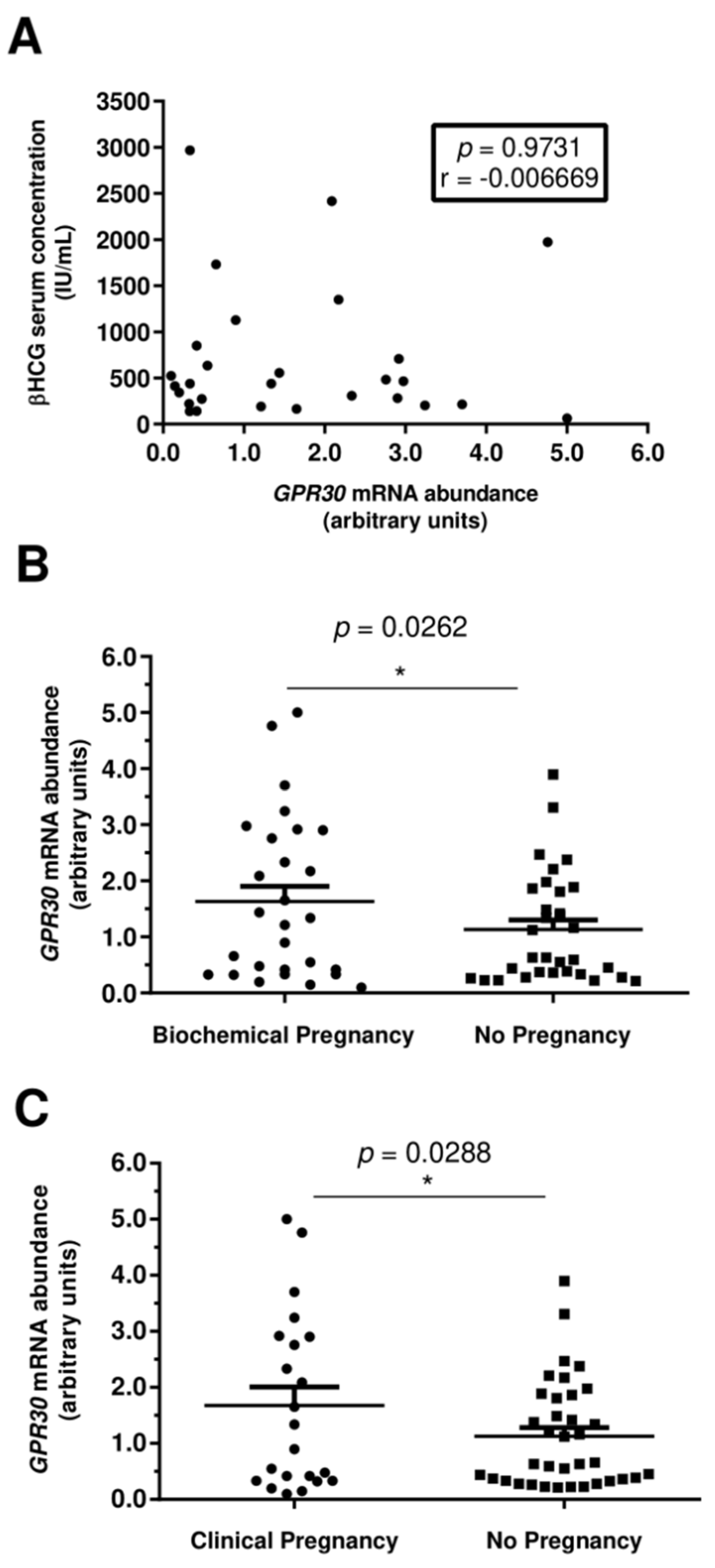
3.2. Increased GPR30 Abundance Levels in Human Spermatozoa Are Associated with Clinical Pregnancies

From the 81 couples seeking medical reproductive treatments at the beginning of this study, embryo transfer (ET) (of one or two embryos) was performed in 60 women. This decision was made by the medical team working with each couple. The reasons for ET not being performed were: the women’s physical condition was not ideal for ET and the embryos were cryopreserved (n = 15); lack of fertilization (n = 2); bad embryo development (n = 2); and lack of good-quality oocytes (n = 2). The following analysis concerns only the 60 couples that were allowed to continue with treatment and performed ET. The fertilization rate and embryo development indicators (embryo cleavage rate, high-quality embryo rate, and blastocyst rate) were assessed before embryo transfer. Oocyte donations were used in 16 cases. We could not find any correlation between the GPR30 transcript abundance in human spermatozoa and the fertilization rate or with the embryo development indicators (Figure 3).
Women were classified with a biochemical pregnancy when the serum beta-human chorionic gonadotropin (β-HCG) concentration surpassed the value of 20 mIU/mL (positive serum β-HCG levels), 12 days after embryo transfer [24]. Pregnancy was not detected in 32 of the initial 60 women (negative serum β-HCG levels). The remaining 28 cases were classified as biochemical pregnancies (positive serum β-HCG levels). After this point, clinicians consider that the embryo has started the process of implantation. Notwithstanding this fact, the rise of β-HCG can be transient and pregnancy can still fail to progress [24]. We could not identify a direct correlation between GPR30 mRNA abundance in human spermatozoa and serum β-HCG concentration (Figure 4A). Nevertheless, our data show that higher levels of GPR30 mRNA abundance were present in those spermatozoa that originated biochemical pregnancies (1.63 ± 0.27 arbitrary units). Concomitantly, lower levels of GPR30 transcript abundance were found in spermatozoa where the resulting embryos failed to implant in the uterus (1.13 ± 0.17 arbitrary units), p = 0.0262 (Figure 4B).
In our cohort, up until April 2019, 4 women who were previously classified as biochemically pregnant suffered an abortion. Furthermore, we lost track of 2 women who were previously classified with biochemical pregnancies. The remaining pregnancies evolved into clinical pregnancies. To summarize, from the 60 embryo transfers performed, only 22 cases evolved into clinical pregnancies. As before, our data show that the mean of GPR30 transcript abundance in human spermatozoa associated with clinical pregnancies (1.68 ± 0.33 arbitrary units) was higher than the mean of GPR30 transcript abundance in human spermatozoa that were not associated with clinical pregnancies (1.13 ± 0.16 arbitrary units), where p = 0.0288 (Figure 3C). No correlation between GPR30 transcript abundance in spermatozoa and paternal age was found (Figure 2B).
It is noteworthy that no statistical difference was found regarding the mean maternal age nor the BMI between the clinical pregnancy and non-pregnancy groups (Figure S2). The same finding is true regarding the age and BMI of the oocyte donors for the same groups. Likewise, the fertilization procedure of ICSI or IVF selected for each couple appears not to affect the achievement of biochemical pregnancy in our population (Figure S3). A cut-of-values analysis was performed with several other parameter analyses in this work concerning pregnancy (biochemical and clinical) and non-pregnancy (Table S2). From the parameters analyzed, only the males’ age was statistically different from the pregnancy groups (biochemical and clinical) and the non-pregnancy groups (p = 0.0008 and p = 0.0048, respectively). Nonetheless, since no correlation was found between the paternal age (Figure 2 and Table S3), we can conclude that the difference in the GPR30 mRNA abundance that was found between the clinical pregnancy group and the non-pregnancy group (Figure 4) is not related to differences in male age.

4. Discussion

GPR30 is a transmembrane receptor that induces rapid non-genomic responses upon E2 stimulation. Along with classical E2 receptors, evidence suggests that GPR30 has a crucial role regarding the testis, where it appears to participate in the regulation of the proliferation/apoptosis balance of germ cells [30], along with the downregulation of steroidogenesis [31]. The work performed by Bernardino and colleagues further confirmed the importance of the GPR30 role for the testis, by proposing that this receptor could be responsible for the modulation of the detrimental effects of excess E2 on the testis of Klinefelter individuals [32]. However, the role of GPR30 in spermatozoa remains to be elucidated. We hypothesized that the GPR30 transcript was present in human spermatozoa and that its abundance in spermatozoa could be related to the fertility potential of males and have an impact on the outcome of ART.
A previous study proposed that GPR30 can modulate the E2 effects on immature rat epididymal epithelium cells [33]. The authors, Cao and colleagues, hypothesized that the role of GPR30 on the epididymal cells could be essential for sperm maturation [33]. Nonetheless, in our population, sperm quality was not correlated with GPR30 mRNA abundance in spermatozoa. From a different viewpoint, the present study likewise could not establish any correlation between paternal BMI and GPR30 mRNA abundance in spermatozoa. Paternal age was also not correlated to GPR30 mRNA abundance in spermatozoa, even though the strong hormonal dysregulation found in older men is known to have severe detrimental effects on male fertility potential in general [28,34]. However, our results suggest that the GPR30 transcript may not have an important role in the modulation of E2 effects on human spermatozoa formation nor on maturation.
Some evidence suggests that the GPR30 transcript may be important for pregnancy, specifically during embryo implantation. A study performed by Yu and colleagues reported that the stimulation of GPR30 with E2 induces a rapid Ca2+ intracellular flux in mouse blastocyst cells. This stimulation promoted the implantation of the mouse blastocysts in a uterine epithelial cell line (Ishikawa cell line) [35]. Furthermore, GPR30 appears to be important in the regulation of proliferation/apoptosis balance between trophoblast cells (the cells forming the outer layer of the blastocyst) [36]. The deficient trophoblast invasion of the endometrium, which results in shallow implantation, is suggested to be one of the causes of preeclampsia development [37]. This condition is characterized by a generalized systemic maternal inflammatory response during pregnancy [36]. Human trophoblasts from normal placentas revealed higher levels of GPR30 expression than human trophoblasts from preeclampsia placentas [36]. GPR30, activated by E2, appears to regulate trophoblast proliferation by the extracellular signal-regulated kinases (ERK) phosphorylation pathway, while the protein kinase B (Akt) pathway, usually associated with apoptosis, is downregulated [36]. Our results further emphasize the importance of GPR30 to pregnancy, since we demonstrated that increased GPR30 transcript abundance in human spermatozoa is associated with pregnancy during ART. Classically, spermatozoa are described as transcriptionally inactive, being responsible only for the delivery of the paternal genetic cargo to the oocyte. However, in recent years, it has become evident that spermatozoa also carry different mRNAs, although the significance of this cargo is still enigmatic [38]. Here, we advance a novel suggestion for the function of a specific sperm mRNA. The GPR30 transcript from the spermatozoa seems to be an important factor for embryo implantation and/or post-implantation development. Notwithstanding this suggestion, we must acknowledge the female factor’s influence on our results. Regarding the female reproductive system, it is known that the Gs signaling pathway is involved in meiotic arrest in mammal oocytes [39,40]. Furthermore, in fish oocytes, it has already been demonstrated that GPR30 plays an important role during this process [41]. However, due to the lack of human biological material, it is difficult to study the role of GPR30 in the female reproductive system, or even in the development of human embryos. Along with this, we must also acknowledge that this study is composed of a small number of patients, due to the short period within which it was conducted. Although we were able to find an interesting correlation between the GPR30 transcript abundance in human spermatozoa and pregnancy during ART, our conclusions need to be further explored in future studies. Nevertheless, we did not find any correlation between the achievement of pregnancy and female age/BMI for both mothers and oocyte donors. These results demonstrate that ET was only performed in women whose physical condition was considered ideal for pregnancy by the medical team. Likewise, the achievement of a biochemical pregnancy (defined by positive β-HCG serum levels, as reported in women after embryo transfers) was not correlated to the fertilization procedure selected, meaning that pregnancy achievement appears not to be affected by clinical protocols for fertilization.

5. Conclusions

To summarize, our results suggest that the abundance of GPR30 mRNA in human spermatozoa may play an important role during pregnancy. Our results are in accordance with previous works [42,43] that suggest that the sperm RNA pool is transferred into the oocyte, where it plays an important role in embryonic development. However, similarly to those works, our results fail to elucidate the molecular mechanisms regarding the role of spermatozoa transcripts on fertilization and embryo development. Our results demonstrate the urgent need for new studies that seek to understand how the paternal phenotype can induce sperm RNA modifications and how it can shape offspring development, from the early stages of life to adulthood.

Supplementary Materials

The following supporting information can be downloaded at: https://www.mdpi.com/article/10.3390/app12073240/s1.

Author Contributions

S.C.P. provided a substantial contribution to the conception and design of the article, from the acquisition of data to bibliographic search, analysis, writing, illustration, data interpretation, and critical discussion. I.F.E. provided a substantial contribution to the acquisition of data, data analysis, and critical interpretation of the results. S.P. provided a substantial contribution to the acquisition of clinical data, providing a clinical perspective to the discussion of this article. A.B. and M.S. provided a medical perspective to the discussion of the article. M.G.A. and P.F.O. contributed to article conception and design, writing, bibliographic enrichment, data interpretation, and critical discussion. All authors have read and agreed to the published version of the manuscript.

Funding

This work was supported by “Fundação para a Ciência e a Tecnologia”—FCT to Sara C. Pereira (2021.05487.BD); Marco G. Alves (IFCT2015 and PTDC/MEC-AND/28691/2017); Pedro F. Oliveira (Scientific Employment Stimulus-Institutional Call-reference CEECINST/00026/2018); LAQV-REQUIMTE (UIDB/50006/2020); UMIB (UIDB/00215/2020, and UIDP/00215/2020); ITR—Laboratory for Integrative and Translational Research in Population Health (LA/P/0064/2020); and by the Portuguese Society of Reproductive Medicine (SPMR) through the “Prémio Laboratório 2020” research grant.

Institutional Review Board Statement

The study was conducted in accordance with the Declaration of Helsinki and approved by the Joint Ethics Committee CHUP/ICBAS with the approval number 2019/CE/P017(266/CETI/ICBAS).

Informed Consent Statement

Informed consent was obtained from all subjects involved in the study.

Acknowledgments

We thank Luís Crisóstomo for the statistical review of this work.

Conflicts of Interest

The authors declare no conflict of interest.

References

  1. Bernardino, R.L.; Carrageta, D.F.; Silva, A.M.; Calamita, G.; Alves, M.G.; Soveral, G.; Oliveira, P.F. Estrogen Modulates Glycerol Permeability in Sertoli Cells through Downregulation of Aquaporin-9. Cells 2018, 7, 153. [Google Scholar] [CrossRef] [PubMed]
  2. O’Donnell, L.; Robertson, K.M.; Jones, M.E.; Simpson, E.R. Estrogen and Spermatogenesis. Endocr. Rev. 2001, 22, 289–318. [Google Scholar] [CrossRef] [PubMed] [Green Version]
  3. Cavaco, J.E.B.; Laurentino, S.S.; Barros, A.; Sousa, M.; Socorro, S. Estrogen Receptors α and β in Human Testis: Both Isoforms are Expressed. Syst. Biol. Reprod. Med. 2009, 55, 137–144. [Google Scholar] [CrossRef] [PubMed]
  4. Oliveira, P.F.; Alves, M.G.; Martins, A.D.; Correia, S.; Bernardino, R.L.; Silva, J.; Barros, A.; Sousa, M.; Cavaco, J.E.; Socorro, S. Expression pattern of G protein-coupled receptor 30 in human seminiferous tubular cells. Gen. Comp. Endocrinol. 2014, 201, 16–20. [Google Scholar] [CrossRef] [PubMed]
  5. Rago, V.; Romeo, F.; Giordano, F.; Maggiolini, M.; Carpino, A. Identification of the estrogen receptor GPER in neoplastic and non-neoplastic human testes. Reprod. Biol. Endocrinol. 2011, 9, 135. [Google Scholar] [CrossRef] [Green Version]
  6. Katib, A. Mechanisms linking obesity to male infertility. Cent. Eur. J. Urol. 2015, 68, 79–85. [Google Scholar] [CrossRef] [Green Version]
  7. Kramer, J.A.; Krawetz, S.A. RNA in spermatozoa: Implications for the alternative haploid genome. Mol. Hum. Reprod. 1997, 3, 473–478. [Google Scholar] [CrossRef]
  8. Abraham, K.A.; Bhargava, P.M. Nucleic acid metabolism of mammalian spermatozoa. Biochem. J. 1963, 86, 298–307. [Google Scholar] [CrossRef] [Green Version]
  9. Fullston, T.; Teague, E.M.C.O.; Palmer, N.O.; DeBlasio, M.J.; Mitchell, M.; Corbett, M.; Print, C.G.; Owens, J.A.; Lane, M. Paternal obesity initiates metabolic disturbances in two generations of mice with incomplete penetrance to the F2 generation and alters the transcriptional profile of testis and sperm microRNA content. FASEB J. 2013, 27, 4226–4243. [Google Scholar] [CrossRef]
  10. Crisóstomo, L.; Jarak, I.; Rato, L.P.; Raposo, J.F.; Batterham, R.L.; Oliveira, P.F.; Alves, M.G. Inheritable testicular metabolic memory of high-fat diet causes transgenerational sperm defects in mice. Sci. Rep. 2021, 11, 1–15. [Google Scholar] [CrossRef]
  11. Crisóstomo, L.; Rato, L.; Jarak, I.; Silva, B.M.; Raposo, J.F.; Batterham, R.L.; Oliveira, P.F.; Alves, M.G. A switch from high-fat to normal diet does not restore sperm quality but prevents metabolic syndrome. Reproduction 2019, 158, 377–387. [Google Scholar] [CrossRef] [PubMed]
  12. Crisóstomo, L.; Videira, R.A.; Jarak, I.; Starčević, K.; Mašek, T.; Rato, L.; Raposo, J.F.; Batterham, R.L.; Oliveira, P.F.; Alves, M.G. Diet during early life defines testicular lipid content and sperm quality in adulthood. Am. J. Physiol. Endocrinol. Metab. 2020, 319, E1061–E1073. [Google Scholar] [CrossRef]
  13. Chen, Q.; Yan, M.; Cao, Z.; Li, X.; Zhang, Y.; Shi, J.; Feng, G.-h.; Peng, H.; Zhang, X.; Zhang, Y.; et al. Sperm tsRNAs contribute to intergenerational inheritance of an acquired metabolic disorder. Science 2016, 351, 397. [Google Scholar] [CrossRef] [PubMed] [Green Version]
  14. Oliveira, P.; Sousa, M.; Silva, B.; Monteiro, M.; Alves, M. Obesity, energy balance and spermatogenesis. Reproduction 2017, 153, R173–R185. [Google Scholar] [CrossRef] [PubMed] [Green Version]
  15. Almeida, S.; Rato, L.; Sousa, M.; Alves, M.; Oliveira, P. Fertility and Sperm Quality in the Aging Male. Curr. Pharm. Des. 2017, 23, 4429–4437. [Google Scholar] [CrossRef]
  16. Bakos, H.W.; Henshaw, R.C.; Mitchell, M.; Lane, M. Paternal body mass index is associated with decreased blastocyst development and reduced live birth rates following assisted reproductive technology. Fertil. Steril. 2011, 95, 1700–1704. [Google Scholar] [CrossRef]
  17. Colaci, D.S.; Afeiche, M.; Gaskins, A.J.; Wright, D.L.; Toth, T.L.; Tanrikut, C.; Hauser, R.; Chavarro, J.E. Men’s body mass index in relation to embryo quality and clinical outcomes in couples undergoing in vitro fertilization. Fertil. Steril. 2012, 98, 1193–1199. [Google Scholar] [CrossRef] [Green Version]
  18. Anifandis, G.; Dafopoulos, K.; Messini, C.I.; Polyzos, N.; Messinis, I.E. The BMI of men and not sperm parameters impact on embryo quality and the IVF outcome. Andrology 2013, 1, 85–89. [Google Scholar] [CrossRef]
  19. Frattarelli, J.; Miller, K.; Miller, B.; Hirsch, K.; Scott, R. Male age negatively impacts embryo development and reproductive outcome in donor oocyte art cycles. Fertil. Steril. 2007, 88, 97–103. [Google Scholar] [CrossRef]
  20. Klonoff-Cohen, H.S.; Natarajan, L. The effect of advancing paternal age on pregnancy and live birth rates in couples undergoing in vitro fertilization or gamete intrafallopian transfer. Am. J. Obstet. Gynecol. 2004, 191, 507–514. [Google Scholar] [CrossRef]
  21. Pereira, S.C.; Martins, A.D.; Monteiro, M.P.; Pinto, S.; Barros, A.; Oliveira, P.F.; Alves, M.G. Expression of obesity-related genes in human spermatozoa affects the outcomes of reproductive treatments. F&S Sci. 2021, 2, 164–175. [Google Scholar]
  22. WHO Organization. WHO Laboratory Manual for the Examination and Processing of Human Semen; World Health Organization: Geneva, Switzerland, 2010. [Google Scholar]
  23. Jameel, T. Sperm swim-up: A simple and effective technique of semen processing for intrauterine insemination. J. Pak. Med. Assoc. 2008, 58, 71. [Google Scholar]
  24. Annan, J.J.K.; Gudi, A.; Bhide, P.; Shah, A.; Hombrug, R. Biochemical pregnancy during assisted conception: A little bit pregnant. J. Clin. Med. Res. 2013, 5, 269–274. [Google Scholar] [CrossRef] [PubMed] [Green Version]
  25. Alves, M.; Martins, A.; Vaz, C.; Correia, S.; Moreira, P.; Oliveira, P.; Socorro, S. Metformin and male reproduction: Effects on Sertoli cell metabolism. Br. J. Pharmacol. 2014, 171, 1033–1042. [Google Scholar] [CrossRef] [Green Version]
  26. Pfaffl, M.W. A new mathematical model for relative quantification in real-time RT-PCR. Nucleic Acids Res. 2001, 29, e45. [Google Scholar] [CrossRef] [PubMed]
  27. Rago, V.; Giordano, F.; Brunelli, E.; Zito, D.; Aquila, S.; Carpino, A. Identification of G protein-coupled estrogen receptor in human and pig spermatozoa. J. Anat. 2014, 224, 732–736. [Google Scholar] [CrossRef] [Green Version]
  28. Pasqualotto, F.F.; Sobreiro, B.P.; Hallak, J.; Pasqualotto, E.B.; Lucon, A.M. Sperm concentration and normal sperm morphology decrease and follicle-stimulating hormone level increases with age. BJU Int. 2005, 96, 1087–1091. [Google Scholar] [CrossRef]
  29. Verón, G.L.; Tissera, A.D.; Bello, R.; Beltramone, F.; Estofan, G.; Molina, R.I.; Vazquez-Levin, M.H. Impact of age, clinical conditions, and lifestyle on routine semen parameters and sperm kinematics. Fertil. Steril. 2018, 110, 68–75.e64. [Google Scholar] [CrossRef]
  30. Chimento, A.; Sirianni, R.; Delalande, C.; Silandre, D.; Bois, C.; Andò, S.; Maggiolini, M.; Carreau, S.; Pezzi, V. 17β-Estradiol activates rapid signaling pathways involved in rat pachytene spermatocytes apoptosis through GPR30 and ERα. Mol. Cell Endocrinol. 2010, 320, 136–144. [Google Scholar] [CrossRef]
  31. Vaucher, L.; Funaro, M.G.; Mehta, A.; Mielnik, A.; Bolyakov, A.; Prossnitz, E.R.; Schlegel, P.N.; Paduch, D.A. Activation of GPER-1 estradiol receptor downregulates production of testosterone in isolated rat Leydig cells and adult human testis. PLoS ONE 2014, 9, e92425. [Google Scholar] [CrossRef]
  32. Bernardino, R.L.; Alves, M.; Silva, J.; Barros, A.; Ferraz, L.; Sousa, M.; Sá, R.; Oliveira, P. Expression of estrogen receptors alpha (ER-α), beta (ER-β), and G protein-coupled receptor 30 (GPR30) in testicular tissue of men with Klinefelter syndrome. Horm. Metab. Res. 2016, 48, 413–415. [Google Scholar] [CrossRef]
  33. Cao, X.; Huang, J.; Zhang, G.; Zuo, W.; Lan, C.; Sun, Q.; Yang, D.; Gao, D.; Cheng, C.H.; Zhou, W.L. Functional expression of G protein-coupled receptor 30 in immature rat epididymal epithelium. Cell Biol. Int. 2017, 41, 134–146. [Google Scholar] [CrossRef] [PubMed]
  34. Jarak, I.; Almeida, S.; Carvalho, R.A.; Sousa, M.; Barros, A.; Alves, M.G.; Oliveira, P.F. Senescence and declining reproductive potential: Insight into molecular mechanisms through testicular metabolomics. Biochim. Biophys. Acta 2018, 1864, 3388–3396. [Google Scholar] [CrossRef] [PubMed]
  35. Yu, L.; Qu, T.; Zhang, S.; Yuan, D.; Xu, Q.; Zhang, J.; He, Y.; Yue, L. GPR30 Mediates the Fast Effect of Estrogen on Mouse Blastocyst and its Role in Implantation. Reprod. Sci. 2015, 22, 1312–1320. [Google Scholar] [CrossRef]
  36. Li, J.; Chen, Z.; Zhou, X.; Shi, S.; Qi, H.; Baker, P.; Zhang, H. Imbalance between proliferation and apoptosis−related impaired GPR30 expression is involved in preeclampsia. Cell Tissue Res. 2016, 366, 499–508. [Google Scholar] [CrossRef] [PubMed] [Green Version]
  37. Nandi, P.; Siddiqui, M.F.; Lala, P.K. Restraint of Trophoblast Invasion of the Uterus by Decorin: Role in Pre-eclampsia. Am. J. Reprod. Immunol. 2016, 75, 351–360. [Google Scholar] [CrossRef]
  38. Boerke, A.; Dieleman, S.; Gadella, B. A possible role for sperm RNA in early embryo development. Theriogenology 2007, 68, S147–S155. [Google Scholar] [CrossRef]
  39. DiLuigi, A.; Weitzman, V.N.; Pace, M.C.; Siano, L.J.; Maier, D.; Mehlmann, L.M. Meiotic arrest in human oocytes is maintained by a Gs signaling pathway. Biol. Reprod. 2008, 78, 667–672. [Google Scholar] [CrossRef] [Green Version]
  40. Liu, W.; Xin, Q.; Wang, X.; Wang, S.; Wang, H.; Zhang, W.; Yang, Y.; Zhang, Y.; Zhang, Z.; Wang, C.; et al. Estrogen receptors in granulosa cells govern meiotic resumption of pre-ovulatory oocytes in mammals. Cell Death Dis. 2017, 8, e2662. [Google Scholar] [CrossRef] [Green Version]
  41. Thomas, P. Role of G-protein-coupled estrogen receptor (GPER/GPR30) in maintenance of meiotic arrest in fish oocytes. J. Steroid Biochem. Mol. Biol. 2017, 167, 153–161. [Google Scholar] [CrossRef]
  42. Chen, Q.; Yan, W.; Duan, E. Epigenetic inheritance of acquired traits through sperm RNAs and sperm RNA modifications. Nat. Rev. Genet. 2016, 17, 733–743. [Google Scholar] [CrossRef] [PubMed]
  43. Sharma, U.; Conine, C.C.; Shea, J.M.; Boskovic, A.; Derr, A.G.; Bing, X.Y.; Belleannee, C.; Kucukural, A.; Serra, R.W.; Sun, F.; et al. Biogenesis and function of tRNA fragments during sperm maturation and fertilization in mammals. Science 2016, 351, 391–396. [Google Scholar] [CrossRef] [PubMed] [Green Version]
Figure 1. Association of GPR30 mRNA expression with sperm parameters. The figure shows the distribution of the GPR30 mRNA abundance of spermatozoa with classic sperm parameters, including total sperm count, n = 78 (A), total motility, n = 74 (B), vitality, n = 71 (C), and normal morphology, n = 74 (D). Sperm parameter data were collected from 81 patients with an average age of 39.7 years (age range 27–58 years) and different BMI values (weight range 19.4–33.8 kg/m2). The relationship between GPR30 mRNA abundance and the different sperm parameters was evaluated by computing the Pearson correlation coefficients (r), assuming a Gaussian distribution (confidence interval of 95%). No correlations between the studied parameters were found to be statistically significant.
Applsci 12 03240 g001
Figure 2. Association of GPR30 mRNA expression with paternal body mass index and age. The figure shows the distribution of GPR30 mRNA abundance in spermatozoa according to paternal BMI, (range 19.4–33.8 kg/m2), n = 79 (A); and age, (range 27–58 years), n = 81 (B). The association between GPR30 mRNA abundance, paternal BMI, and age was evaluated by computing the Pearson correlation coefficients (r), assuming a Gaussian distribution (confidence interval of 95%). No correlations between the studied parameters were found to be statistically significant.
Applsci 12 03240 g002
Figure 3. Association between GPR30 mRNA abundance in spermatozoa and embryo quality parameters. Association between GPR30 mRNA abundance in human spermatozoa and (A) fertilization rate (n = 54), (B) embryo cleavage rate (n = 54), (C) high-quality embryo rate (n = 53), and (D) blastocyst rate (n = 54) was evaluated by computing the Pearson correlation coefficients (r), assuming a Gaussian distribution (confidence interval of 95%). No correlations between the studied parameters were found to be statistically significant.
Applsci 12 03240 g003
Figure 4. The association between GPR30 mRNA abundance in spermatozoa and pregnancy. The figure shows the association of GPR30 mRNA abundance in spermatozoa with biochemical pregnancy, n = 28. (A) Serum β-HCG concentrations were collected from women who were reported to achieve a biochemical pregnancy after embryo transfer. The association between GPR30 mRNA expression and serum β-HCG concentration was evaluated by computing the Pearson correlation coefficients (r), assuming a Gaussian distribution (confidence interval of 95%). Correlations between the studied parameters were not found to be statistically significant. (B) From a total of 60 embryo transfers, 28 women were classified as biochemically pregnant. The remaining 32 women did not become pregnant after embryo transfer. Results are expressed as the mean ± standard error mean (biochemical pregnancy mean = 1.63 ± 0.27 arbitrary units, n = 28; no pregnancy mean = 1.13 ± 0.17 arbitrary units, n = 32), p = 0.0262. (C) Of these, 22 women were reported to be clinically pregnant. The remaining 36 women had not become pregnant after embryo transfer or had a spontaneous abortion. Results are expressed as mean ± standard error mean (clinical pregnancy mean = 1.68 ± 0.33 arbitrary units, n = 22; no pregnancy mean = 1.13 ± 0.16 arbitrary units, n = 36). Statistical analysis was performed with a two-tailed Student’s t-test for parametric data (confidence interval of 95%). Values of * p < 0.05 were considered to be statistically significant.
Applsci 12 03240 g004
Publisher’s Note: MDPI stays neutral with regard to jurisdictional claims in published maps and institutional affiliations.

