Article

Enhancing Antioxidant Activities and Anti-Aging Effect of Rice Stem Cell Extracts by Plasma Treatment

1
Plasma Bio Team, Plasma Bio Research Division, Institute of Plasma Technology, Korea Institute of Fusion Energy, 37 Dongjangsan-ro, Gunsan 54004, Korea
2
Plasma Bioscience Research Center, Kwangwoon University, Seoul 01897, Korea
3
Department of Food Science and Biotechnology of Animal Resources, College of Sanghuh Bioscience and Tehnology, Konkuk University, Seoul 05029, Korea
4
Department of Crop Science, College of Sanghuh Life Science, Konkuk University, Seoul 05029, Korea
*
Authors to whom correspondence should be addressed.
These authors contributed equally to this work.
Appl. Sci. 2022, 12(6), 2903; https://doi.org/10.3390/app12062903
Submission received: 13 December 2021 / Revised: 11 February 2022 / Accepted: 8 March 2022 / Published: 11 March 2022
(This article belongs to the Special Issue Plasma Medicine Technologies: Volume II)

Abstract

:
Plant-derived substances exhibit antioxidant and antibacterial activities and have been proven to have beneficial effects in wound healing and skin regeneration. Plant stem cells have recently received much attention as research materials in cosmetic development because they promote regeneration after damage. In this paper, we demonstrate for the first time that the plasma treatment of stem cells obtained from rice-seed embryos can be effective in enhancing antioxidant activity and in regenerating human skin. We investigated this potential utilizing micro-DBD (Dielectric Barrier Discharge) plasma as a pretreatment technique to enhance the vitality and functional activity of rice stem cells. The results of the cell culture experiments show that plasma-treated rice stem cell extracts (RSCE) have promising antioxidant and anti-skin aging activities. The results of quantitative real-time PCR (qRT-PCR) for major antioxidant enzymes and anti-aging genes confirm that the plasma technique used in the pretreatment of RSCE was able to enhance cell activities in skin regeneration, including cell survival, proliferation, and collagen enhancement for Human Fibroblast (HFB) degraded by oxidative stress. These results show that the relatively low energy of less than 300 W and an amount of NOx-based reactive nitrogen species (RNS) from plasma discharge of about 3 μL/L were the key factors and that RSCE, of which the antioxidant activity was enhanced by plasma treatment, appeared to be a major contributor to the protective effect of HFB against oxidative stress. Plasma-treated RSCE induced excellent anti-aging properties by stimulating HFB to promote collagen synthesis, thereby promoting skin regeneration. These properties can protect the skin from various oxidative stresses. This study demonstrates that plasma-treated extracts of stem cells derived from rice-seed embryos have an excellent regenerative effect on aging-treated HFB. Our results demonstrate the potential utility of plasma-treated RSCE as a skin anti-aging agent in cosmeceutical formulations for the first time.

1. Introduction

Human skin consists largely of two layers of tissue. The outer layer is the epidermis, which contains keratinocytes, and the inner layer is the dermis, which contains fibroblasts and an extracellular matrix. Skin aging is a complex process involving both layers. The cause of skin aging is the degradation of the structure and physiological functions of the skin by various exogenous (exterior) and endogenous (interior) factors. Exogenous aging is caused by environmental factors such as ultraviolet (UV) radiation, alcohol ingestion, pollutants, and severe physical stress [1,2]. These factors cause oxidative damage to skin lipids, proteins, and DNA through the formation of free radicals, leading to loss of skin elasticity. UV accounts for up to 80% of exogenous aging. Endogenous skin aging is caused by accumulated intrinsic damage due to the persistent formation of reactive oxygen species (ROS) [3,4]. Free radicals damage the connective tissue of the skin, leading to premature skin aging (photoaging) [3,4,5]. Previous studies have indicated that sustained ROS exposure can stimulate skin aging through antioxidant system destruction, wrinkling, and melanin overproduction [5]. To delay such aging of the skin, methods of strengthening and protecting skin cells have been studied as an important issue in the cosmetics industry. Innovative formulations of cosmetic raw materials are being developed. In particular, nanotechnology in the cosmetic industry has a very small particle size, so it is possible to increase the surface area to increase skin absorption of active ingredients [6,7,8].
Plant-derived active ingredients such as polyphenols have been used to prevent skin aging [9] because they have strong antioxidant activity against ROS. Bioactive compounds such as isoflavones, anthocyanins, and catechins protect the epidermis of the skin from external stimuli and oxidative stress. These plant-derived active ingredients with antioxidant activity were mostly obtained through plant cultivation. However, plant cultivation generally requires a great deal of space and a long period of time. In addition, it is difficult to obtain consistent plant extracts because control of the cultivation conditions is relatively difficult. As an alternative, growing plant calluses in an environment where cultivation conditions are easy to control allows for management of changes in the external environment, thus enabling high productivity and a fast turnover rate.
Calluses, parts of which have the same roles as animal stem cells, is a mass of plant stem cells with properties that help stimulate and regenerate plants after injury [10]. A plant stem cell is an undifferentiated cell that has the genetic potential to produce an entire mature plant, strong proliferative capacity, and high differentiation potential, so it is possible to produce a mature plant from a specific plant callus [11,12,13]. Plant stem cells exhibit excellent anti-aging properties because fibroblasts stimulate collagen synthesis and promote skin regeneration, among other activities. These unique properties of plant stem cells are focused on the development of new cosmetics. Although active research is being conducted on the use of plant callus materials called plant stem cells as functional natural materials with excellent physiological activity in the cosmetic field, plant stem cell materials have not been developed [14]. The anti-aging effects of grapes, raspberries, tomatoes, medicinal material, and many plant stem cells have been previously reported. However, the anti-aging efficacy of stem cells obtained from rice embryonic cells has hardly been reported [15,16,17]. Rice (Oryza sativa) is composed of many phytochemicals such as γ-oryzanol, vitamin E homologues, and phenolic acids that produce beneficial health properties [18,19]. Rice Stem Cell Extract (RSCE) is a callus extract derived from embryonic cells of rice seeds. It has a high vitamin content and excellent antioxidant effect.
Plasma is a state of matter in which partially ionized gas leads to a dynamic mix of charged particles, reactive nitrogen and oxygen species (RONS), excited atoms, free radicals, ultraviolet photons, and electric fields [20,21]. As an environmentally friendly and sustainable new technology, applications of plasma have provided a variety of reaction mechanisms for bio-targets by changing the combination of parameters generated by various feed gases.
Plasma medicine is a fusion of research involving both plasma physics and biology, and it has been studied as a new field in medical research for more than ten years worldwide. Plasma medical research using cold atmospheric plasma has been actively performed for the purpose of therapeutic treatment of the human body. The purpose of this study is to confirm the effect of plasma-treated rice stem cell extracts (RSCE) in promoting cell regeneration of fibroblasts.
Herein, we report the first RSCE-based cosmetics work focused on the effects of plasma-treated rice stem cells on the aging of human skin.

2. Materials and Methods

2.1. Plant Materials

De-husked mature rice seeds of the Japonica rice variety Dongjin were placed in a 50 mL conical tube and then sterilized using 100% ethanol for 30 s with shaking. Afterward, the ethanol was discarded and 50% Clorox (4% sodium hypochlorite) was added for a second sterilization for 20 min with shaking (Supplementary Figure S1A). The sterilized seeds were rinsed 4–5 times with sterile double-distilled water (ddH2O) with shaking and then were placed on sterilized filter paper on a clean bench and dried. The medium N6 is widely used for the culture of callus tissue from many plants [22,23]. Chu N6 Medium supplemented with vitamins was used to induce the growth of calluses, which contained the stem cells of the plant. The composition of the rice stem-cell induction medium is shown in Table 1. The pH of the medium was adjusted to 5.8 using KOH before autoclaving began. Then, 25 mL of the autoclaved culture medium was poured into a 90 mm Petri dish under sterile conditions. The sterilized rice seeds were planted on the culture medium with the rice embryo side up and then maintained in darkness in a growth chamber at 25 ± 2 °C for callus induction. Approximately 20 rice seeds were planted in each Petri dish. The callus induction frequency was obtained after 3 to 6 weeks of culture by repeatedly recording the number of calluses induced in each of 20 Petri dishes (with three repetitions). The following equation was used to determine the callus induction frequency [24,25]:
%F = Ncallus/Nseeds × 100
where F = callus induction frequency, Ncallus = number of calluses produced from seeds, and Nseeds = number of planted seeds.

2.2. Plasma Device

In this study, a μ-DBD (dielectric barrier discharge) plasma device with a dimming-type high-voltage inverter and duty ratio of on/off time of the discharge method was used (Figure 1). The electrode structure of the plasma device is well described in previous studies [26]. The DBD plasma device consists of electrodes, a dielectric layer (silicon dioxide (SiO2)), hydration-prevention layers, aluminum oxide (Al2O3), and a magnesium oxide (MgO) layer. The thickness of the SiO2 dielectric layer is about 30 μm, and the diameter of the plasma discharge area is about 60 mm. To prevent hydration during plasma discharge, Al2O3 was added onto the MgO layer. Plasma was generated using nitrogen gas with a flow rate of 1500 Standard Cubic Centimeter per Minute (sccm) and electrical current of 50–63 mA current at 1.2 kV input voltage. The optical emission spectra of the plasmas generated were captured using spectrometry (Ocean Optics, HR4000, FL, USA) with a charge-coupled device (CCD).

2.3. Treatment of Rice Stem Cells with Plasma

The rice stem cells induced by 3 weeks in culture media were then treated with plasma. Rice stem cells on culture medium were exposed to μ-DBD plasma at a distance of 1 mm from the source. The time of each plasma treatment was 1, 3, or 5 min. The plasma treated rice stem cells were incubated for one day under the same culture conditions as before (25 ± 2 °C in the dark). After one day, the rice stem cells were collected and ground in liquid nitrogen using a pestle and mortar.

2.4. Reactive Species Measurement in Gas Phase

The generated O3 and NOx were measured and recorded using an O3 analyzer (GM-6000PRO, Anseros, Germany) and NOx analyzer (T200, Teledyne, Thousand Oaks, CA, USA), respectively. These analyzers used a Urethane tube (Sang-A Pneumatic co., LTD., Daegu, Korea; OD: Φ6 mm, ID: Φ4 mm) as the sample gas inlet, which were located 10 mm away from the DBD electrode. The flow of sample gas that two analyzers took in were 1000 sccm in total. The measurements were performed for 30 min, and the measured data were recorded as analogue outputs (4–20 mA) by a data logger (GL840-M, Graphtec, Japan).

2.5. Extraction of Rice Stem Cells

A sample of approximately 1 g of ground rice stem cells was mixed with 20 mL of methanol and incubated for 3 h with shaking at room temperature (RT). After centrifugation several times (3000 rpm for 15 min at 4 °C), the collected supernatant was transferred to a new tube. Then, the collected supernatant was concentrated using a vacuum evaporator (Jeiotech, Daejeon, Korea). After the solvent was completely evaporated, the remaining pellet was re-dissolved in extraction solvent at the final concentration of 1 mg/mL.

2.6. Assay for the Amount of Total Polyphenol

For extraction of the total polyphenols, harvested rice stem cells (3 weeks old) were ground in liquid nitrogen. The ground samples were suspended in methanol solvent and incubated for 4 h with shaking at room temperature (RT). The total phenolic content (TPC) was determined using the modified Folin–Ciocalteu method [27]. After two cycles of centrifugation (3000 rpm for 15 min), the supernatant was filtered and transferred to a new tube. Then, the collected supernatant was concentrated using a vacuum evaporator. After the solvent was completely evaporated, the pellet was re-dissolved in 0.2 mL of the same extraction solvent. The dissolved solution was mixed with 0.5 mL of the Folin–Ciocalteau reagent (Sigma, St. Louis, MO, USA) and incubated for 5 min at RT in the dark. Then, 1 mL of 7.5% sodium carbonate (w/v) was added, thoroughly mixed, and incubated for 1 h at RT in the dark. Absorbance was measured at 765 nm using a UV-vis spectrophotometer using microplate reader (Biotek, Seattle, Washington, DC, USA). A standard curve was obtained using the absorbance values of gallic acid (reference polyphenol), and the total phenol amount in each sample was estimated as gallic acid equivalents (mg GAE/g).

2.7. Total RNA Isolation and qRT-PCR

Total RNA isolation was performed based on the MMY method [28]. Rice stem cells (30 mg) were ground in liquid nitrogen using a pestle and mortar. Total RNA was then extracted from the ground powder using TRIzol reagent (Invitrogen, Carlsbad, CA, USA) with chloroform. The complementary DNA (cDNA) was synthesized using SMART® MMLV Reverse Transcriptase (Clontech, Mountain View, CA, USA) following the manufacturer instructions. A portion (1 mL) of synthesized cDNA was used as the template for the qRT-PCR. The analysis was performed using iQ SYBR Green Supermix (Bio-Rad, Hercules, CA, USA) and a Real-Time PCR detection system (CFX96, Bio-Rad, Hercules, CA, USA). The relative expression of mRNA for major genes associated with skin aging on HFB cells were analyzed by qRT-PCR. Primer sequences are listed in Table 2. The qRT-PCR was performed by normalization to the mRNA level of GAPDH. GAPDH was used as a housekeeping gene for the qRT-PCR. The PCR thermal cycles involved a three-step protocol: activation at 95 °C for 3 min, followed by 40 cycles of denaturation at 95 °C for 10 s, and an annealing/extension at 60 °C for 30 s.

2.8. Gas Chromatography–Mass Spectrometery (GC-MS) Analysis

Plasma-treated 3-week-old calluses were subjected to extraction with methanol solvent and analyzed by GC-MS to compare changes in metabolites. GC-MS was performed with a Shimadzu GC-2010 gas chromatography instrument coupled with a Shimadzu QP2010 mass spectrometer. Compounds were separated on a fused silica capillary column Rtx-5MS (100% polymethylsiloxane, 30 cm × 0.25 mm i.d., 0.25 mm film thickness). The oven temperature program was initiated at 70 °C, held for 10 min, increased at a rate of 5 °C min−1 to 195 °C, and then increased again at 10 °C min−1 to 300 °C, and held there for 15 min. The spectrometers were operated in electron-ionization mode. The scan range was 35–500 amu, the ionization energy was 70 eV, and the scan rate was 0.20 s per scan. The injector, interface, and ion source were kept at 250, 250, and 200 °C, respectively. Split injection (1 mL) was conducted with a split ratio of 1:10, and helium was used as the carrier gas at a flow rate of 1.0 sccm.

2.9. Cell Culture

Normal human primary dermal fibroblasts from neonatal foreskin (HDFn) (ATCC PCS-201-010) were obtained from the American Type Culture Collection (ATCC, Manassas, VA, USA). Fibroblasts were cultured in DMEM-high glucose medium (Invitrogen, Carlsbad, CA, USA, No. 10569-044). The medium was supplemented with 10% FBS (Thermo HyClone, Logan, UT, USA, No. SH30406.02) and 1% glutamine, 1% penicillin (100 IU/mL), and streptomycin (100 mg/mL) (HyClone, Promega, Madison, WI, USA). The cells were cultured at 37 °C, 95% relative humidity, and 5% CO2.

2.10. Cell Viability Assay

To investigate the proliferation activity, we used the CellTiter-Glo cell viability assay kit (Promega, Madison, WI, USA; Cat no. G7570). This is one kind of luminescent cell viability assay with a procedure used to determine the number of viable cells in a culture based on the quantization of the ATP present. ATP indicates the presence of metabolically active cells. For this assessment, the cells were prepared with various concentrations of the callus extract and chemicals on the human fibroblast cells; then, the cells were treated with the CellTiter-Glo® substrate at room temperature. Plates with 96 wells were prepared with HFB in 100 μL of the culture medium per well, along with control wells containing the medium without cells to measure the background luminescence. After 30 min of incubation, the luminescence signal of the plate was read at 490 nm using a microreader.
HFB was first treated with 1 mM of tert-butyl hydroperoxide (t-BHP) to artificially induce oxidative stress. To investigate cytotoxicity after t-BHP treatment, HFB cells were exposed to t-BHP at 200 mM for 1 h. In order to observe the t-BHP-induced cytotoxicity prevention activity of plasma-treated RSCE, HFB cells were pretreated with RSCE at a concentration of 0.1 mg/mL and plasma-treated for 24 h before t-BHP treatment.

2.11. Statistical Analysis

All percentages and relative values of the data are represented as the mean ± standard deviation (S.D.) of the indicated number of replicates (≥3). Statistical analyses of the data were performed using the student’s t test to establish the significance between data points, and the significance of differences were based on either p < 0.05 (*) or p < 0.01 (**).

3. Results and Discussion

3.1. Preparation of Stem Cells from Rice Seeds

Rice Stem Cell Extract (RSCE) is a callus extract derived from embryonic cells of rice seeds, with high vitamin content and excellent antioxidant effects. However, the anti-aging efficacy of stem cells harvested from rice embryonic cells has rarely been reported. Here, we utilized plasma as a pretreatment technique for the activation of RSCE and confirmed that, in particular, the antioxidant and skin aging regeneration effects of RSCE were enhanced by plasma.
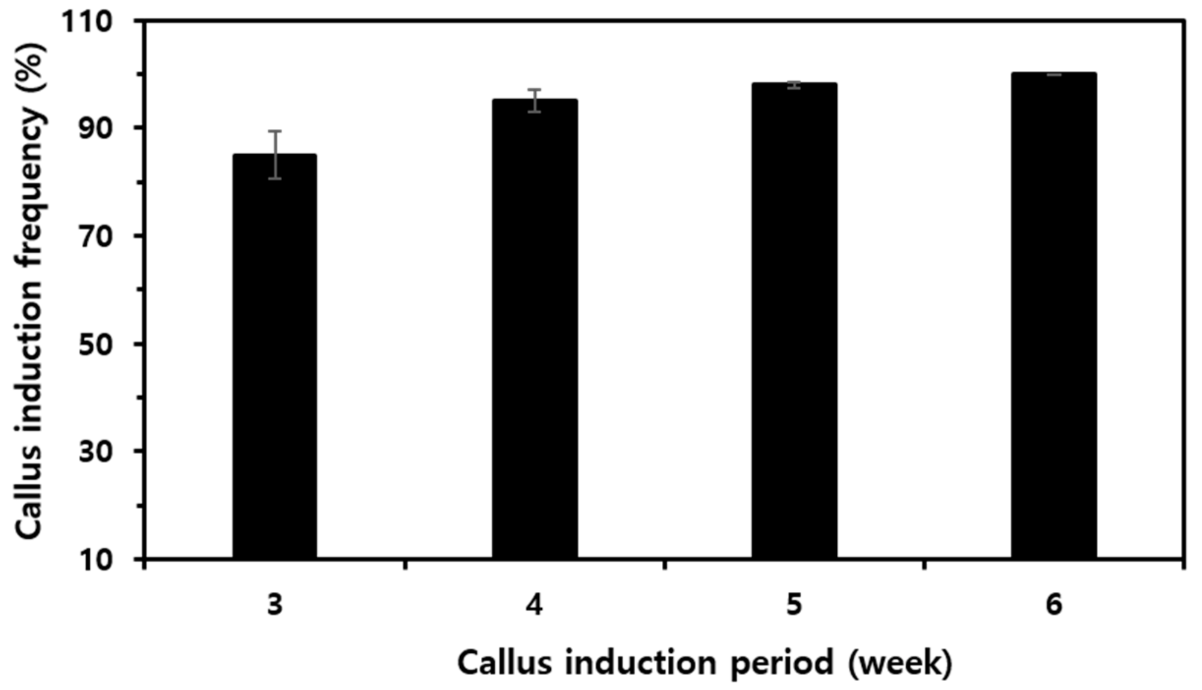
As shown in Supplementary Figure S1B, rice stem cells were obtained from the peeled embryos of sterilized rice seeds. Three-week-old calluses obtained from approximately 20 seeds in one Petri dish were plasma-treated (Supplementary Figure S1C). Three-week-old rice stem cells were ground in a mortar with liquid nitrogen and then were subjected to solvent extraction to obtain a rice stem cell extract (Supplementary Figure S1D,E). The various analyses of the callus extracts extracted from plasma-treated calluses were compared. The acquired callus induction frequency was made at a constant rate (about 90% or more) for 3–6 weeks, as shown in Figure 2. An additional experiment was performed by freeze-drying the harvested 3-week-old rice stem cells.
Chu N6 Medium supplemented with vitamins was used to induce the growth of calluses, which contain the stem cells of the plant. The callus induction frequency was obtained after 3 to 6 weeks of culture by repeatedly recording the number of calluses induced in each of the 20 Petri dishes (with three repetitions). The acquired callus induction frequency was made at a constant rate (about 90% or more) for 3–6 weeks. All measurements were made in three or more replicates. * p < 0.05; ** p < 0.01.

3.2. Ozone and NOx Analysis

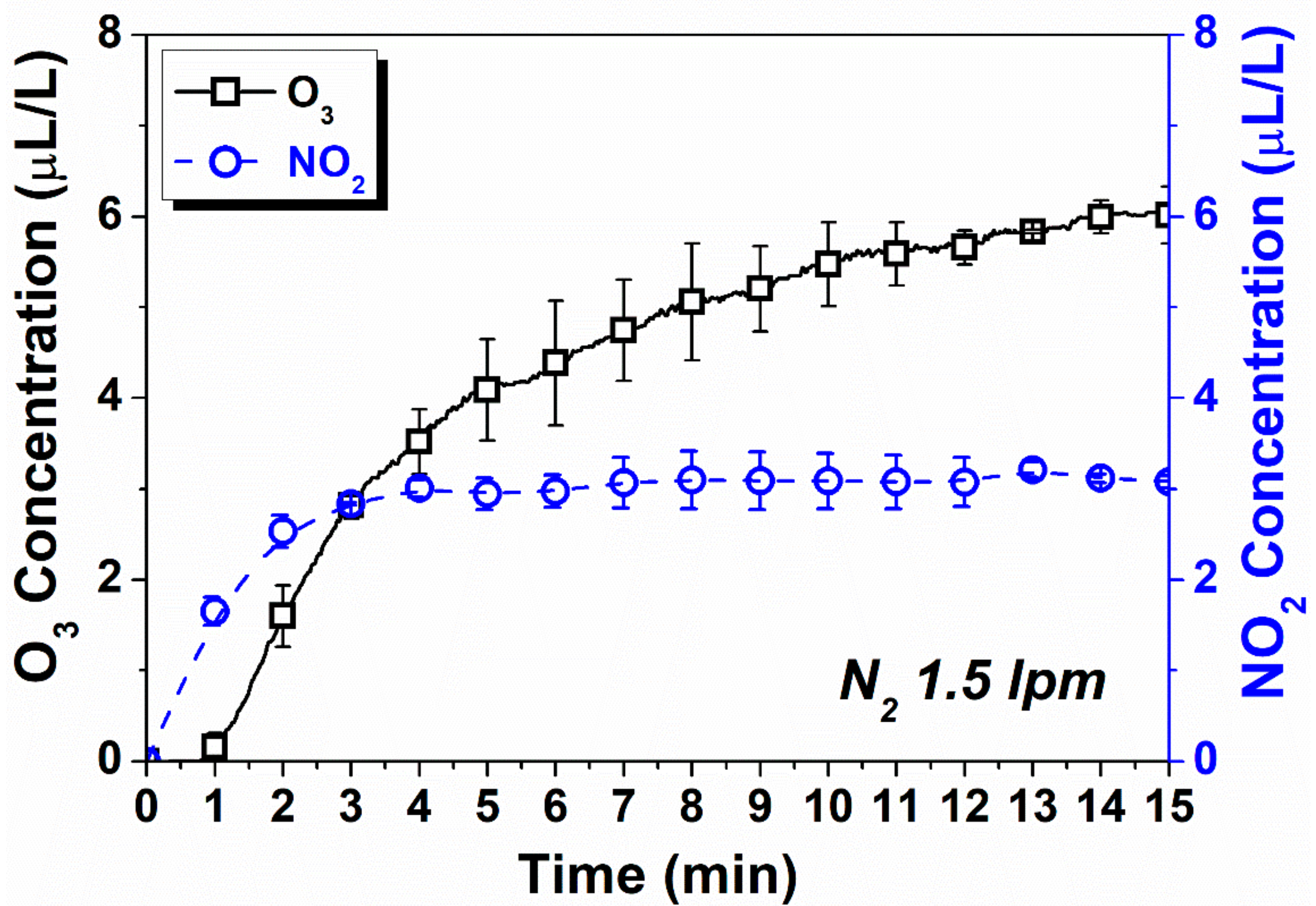
The measured O3 and NO2 concentrations according to the time variant at the N2 flow rate condition of 1500 sccm are depicted in Figure 3. The concentration of O3 was increased up to about 6 μL/L at 15 min after the discharge was ignited and sustained until the end of measurement. The NO2 concentration was increase up to about 3 μL/L at 4 min and sustained. The averaged concentrations of O3 and NO2 after 10 min were 6.24 and 3.1 μL/L, respectively. Until the plasma discharge time of 3 min, NO2 was dominant as the active species around the plasma electrode. However, when plasma discharge was more than 3 min, the concentration of NO2 was saturated and O3 was increased. It was found that, after 3 min of plasma discharge, the active species concentration was traded off.

3.3. Antioxidant Activities of Rice Stem Cell Extracts

The diameter of the discharge region of the μ-DBD plasma device with the duty cycle used in this study was 60 mm; the plasma characteristics were reported in a previous paper [26]. Through the previous studies, we were able to confirm that the plasma device in this study used about 1.27 kV and 50 mA of root mean square voltage and current and that the electrical discharge energy delivered to the rice stem cells in a 1 s treatment was 1.55 W [26].
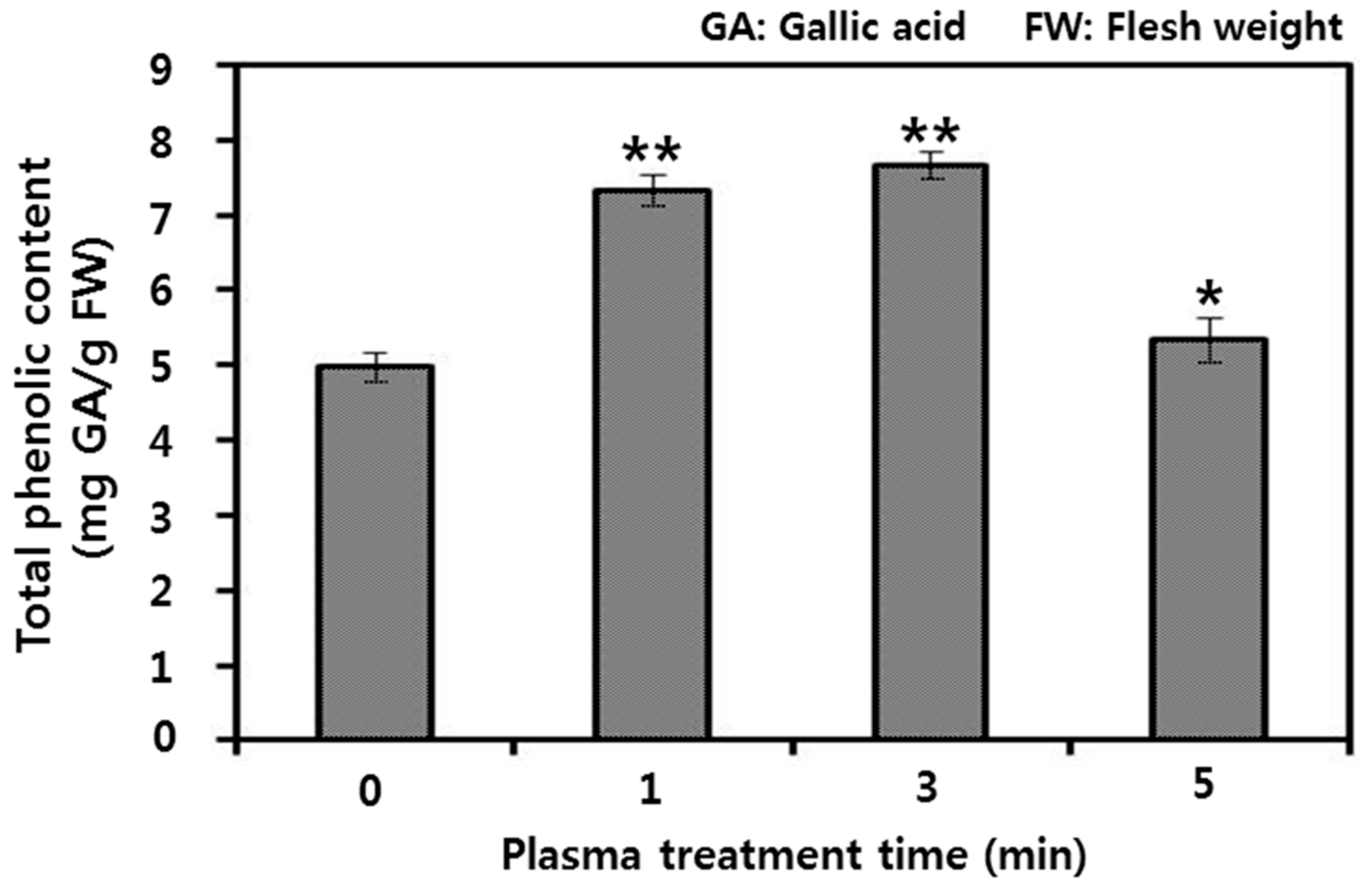
To test whether plasma treatment could affect the physiological aspects of RSCE, we analyzed the total phenolic compound content of rice stem cells. The content of phenolic compounds is closely related to their antioxidant activity [29,30,31]. The total phenol content of the rice stem cell extract was analyzed using the Folin–Ciocalteu reagent method, and the TPC content was compared according to the plasma treatment time (Figure 4). The concentrations of total phenolic compounds in the rice stem cells showed a tendency to increase with plasma treatment (Figure 4). As the N2 plasma treatment time increased, the polyphenol concentration also increased. The total antioxidant activity of the callus extract treated with plasma for 1 min (7.2 mg/g) and for 3 min (7.6 mg/g) was increased by about 1.5 times compared with the control (5 mg/g). Antioxidant activity increased in proportion to plasma treatment time up to 3 min, but in the 5 min (5.2 mg/g) treatment, antioxidant activity decreased compared with the 1 and 3 min treatments and the content was similar to the untreated control. Additionally, there was no significant difference between the 1 and 3 min plasma treatments.
The total phenol content (TPC) of the rice stem cell extract was analyzed using the Folin–Ciocalteu reagent method, and the TPC content was compared according to the plasma treatment time. Absorbance was measured at 765 nm using a UV-vis spectrophotometer. A standard curve was obtained using the absorbance values of gallic acid (reference polyphenol), and the total phenol amount in each sample was estimated as gallic acid equivalents (mg GAE/g).

3.4. GC-MS Analysis of Rice Stem Cell Extracts

To compare the changes in the RSCE components induced by plasma treatment, we confirmed the chemical composition of the RSCE according to the plasma treatment time using GC-MS analysis (Table 3 and Supplementary Figure S2). When the retention times (RT) of untreated RSCE and plasma-treated RSCE were compared, five types of candidate metabolites were detected as flows, with the highest peak in those RSCE treated with plasma for 3 min: fructose (RT = 34.805 min), galactopyranoside (RT = 36.690 min), melibiose (RT = 47.830 min), glycoside (RT = 49.325), and sucrose (RT = 49.620 min) (Table 3). These candidate metabolites were mainly primary metabolites and showed a slight difference in peak height according to the duration of plasma treatment (Supplementary Figure S2). Melibiose metabolites showed similar peak heights in the remaining treatment groups except for the RSCE plasma treated for 3 min. In the remainder, fructose, galactopyranoside, glycoside, and sucrose all increased in RSCE plasma treated for 3 min and 1 min; however, they decreased in the 5 min plasma-treated RSCE when compared with the untreated control. Primary metabolites, such as carbohydrates, fats, and proteins that play major roles in basal metabolic processes, are substances directly involved in general growth, development, and reproduction in living organisms. An increase in the contents of these candidate metabolites can be associated with an increase in cell viability and vitality.

3.5. Effects of Rice Stem Cell Extracts against Oxidative Damage on HFB

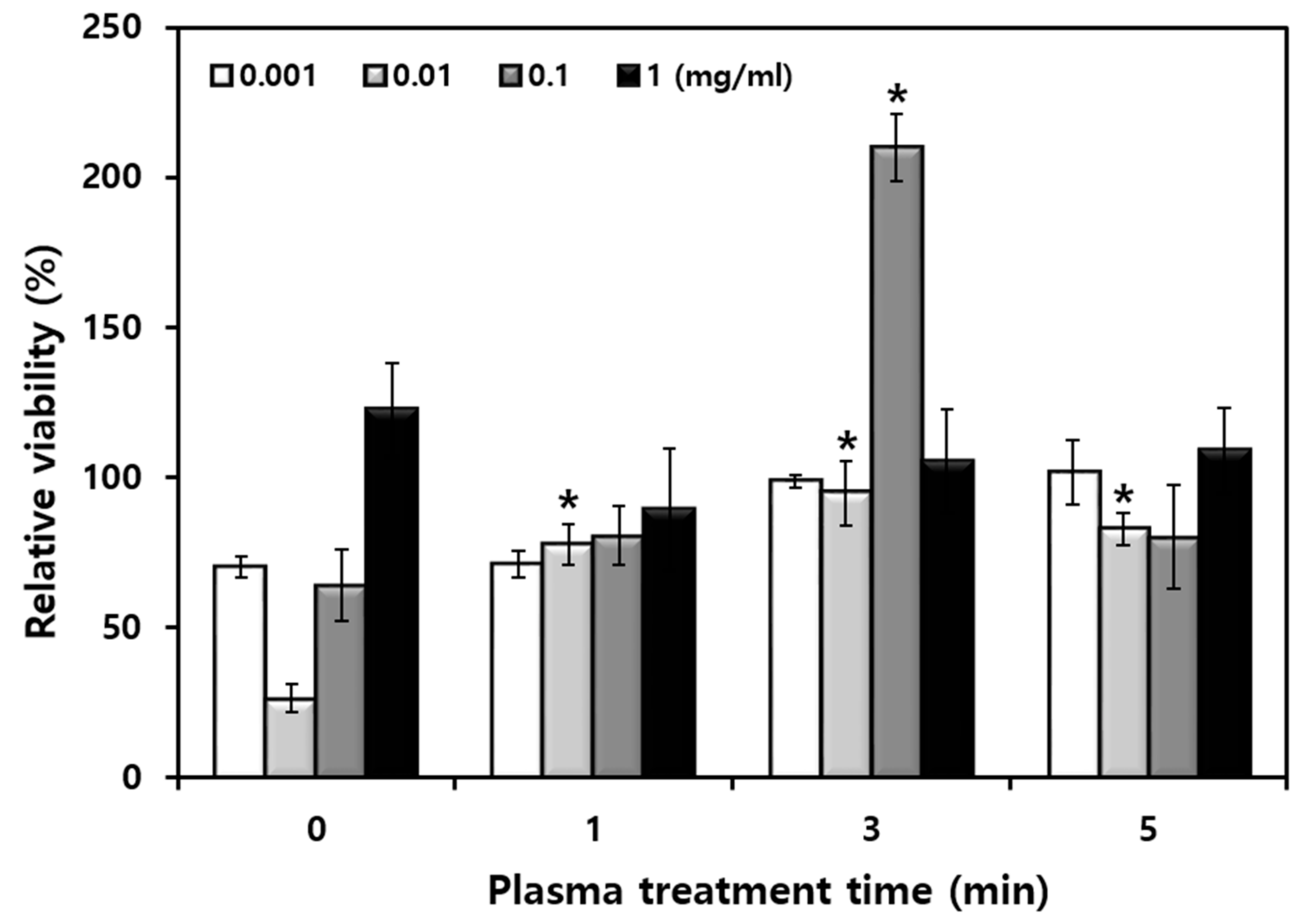
To confirm the regenerative effect of HFB on N2 μ-DBD plasma-treated RSCE, HFB was first treated with 1 mM tert-butyl hydroperoxide (t-BHP) to artificially induce oxidative stress. Tert-butyl hydroperoxide (tBHP) is a well-known oxidative stress inducer used to induce cellular senescence [32,33]. This substance depletes cellular antioxidant defense mechanisms, leading to lipid peroxidation [34,35]. For this experiment, we used a human skin fibroblast (HFB) cell line. We used the various concentrations of callus extracts to observe the proliferation effects of HFB cells. To investigate the effects on the proliferation assay, cells were incubated with 10% FBS in DMEM. Various t-BHP concentrations (12.5, 25, 50, 100, 300, and 500 µM) were tested, and cell proliferation was evaluated (Supplementary Figure S3). As shown in Figure 5, plasma-treated RSCE was treated for each concentration (0.001, 0.01, 0.1, and 1 mg/mL), and the cell proliferation rate of HFB was compared. HFB cells were pretreated with RSCE that was plasma-treated for 24 h before t-BHP treatment. In Figure 5, the number of viable cells in HFB under t-BHP treatment increased the most using RSCE treated with a 0.1 mg/mL concentration of plasma for 3 min. Through the comparison of HFB viability using the concentration for plasma-treated RSCE in a state in which t-BHP was not treated, it was confirmed that 0.1 mg/mL was the optimal concentration (Supplementary Figure S4). The concentration of the extract from the 0.1 mg/mL group was shown to have higher effects than the other three groups (0.001, 0.01, and 1 mg/mL) after 24 h (Supplementary Figure S4), and it still consists of the effects until 48 h (data not shown). Plasma-treated RSCE at a concentration of 0.1 mg/mL showed a significant difference and increased the proliferation of HFB cells. RSCE at a concentration of 0.1 mg/mL treated for 1 min and 3 min was increased compared with RSCE without plasma treatment, and RSCE at a concentration of 0.1 mg/mL treated for 5 min showed a similar cell viability to that of the untreated control (Supplementary Figure S4). This trend showed a marked difference when treated with t-BHP oxidative stress. The 3 min plasma treated with 0.1 mg/mL RSCE showed an excellent ability to overcome the oxidative stress of t-BHP.
In order to confirm the cell proliferation of plasma-treated rice stem cell extracts (RSCE), HFB was pretreated with plasma-treated RSCE at each concentration for 24 h. The plasma treatment lasted 1, 3, and 5 min, and nitrogen gas (1500 sccm) was used as the feeding gas. The RSCE was used in concentrations of 0.001, 0.01, 0.1, and 1 mg/mL. All measurements were made in three or more replicates. * p < 0.05; ** p < 0.01.
As a result of measuring the concentration of active species generated by plasma discharge, the concentration of NO2 increased to about 3 μL/L at 4 min of discharge and continued. NO2 was dominant until 3 min of plasma discharge, but when plasma discharge was more than 3 min, the concentration of NO2 was saturated and O3 increased. Based on the results of offsetting the concentration of active species after 3 min of plasma discharge, it was speculated that NOx-type RNS including NO2 played an important role in RSCE activity. In addition, the micro DBD plasma electrode used in this study is characterized by a relatively weak energy intensity. The discharge energy value according to the nitrogen supply gas was 1.55 W per second. Therefore, the energy values according to the plasma treatment time were 93 (1 min), 279 (3 min), and 465 (5 min) W. Therefore, the threshold value that can activate cells is 300 W, and at energy intensities higher than that, it would have been in the range of energy that could cause damage rather than activation of the cells.
Various studies have suggested that ROS play critical roles in cell differentiation [36]. We suggest that plasma-generated RONS could enhance the antioxidant activity of RSCE, which is involved in inducing the differentiation of HFB cells. Reactive species are well-known signaling molecules [37,38,39]. ROS and RNS produced by the plasma can be key factors in the expression of genes involved in plant cell proliferation. In our study, RNS generated from plasma were just the possible triggers for increasing the proliferation rates of rice callus cell.
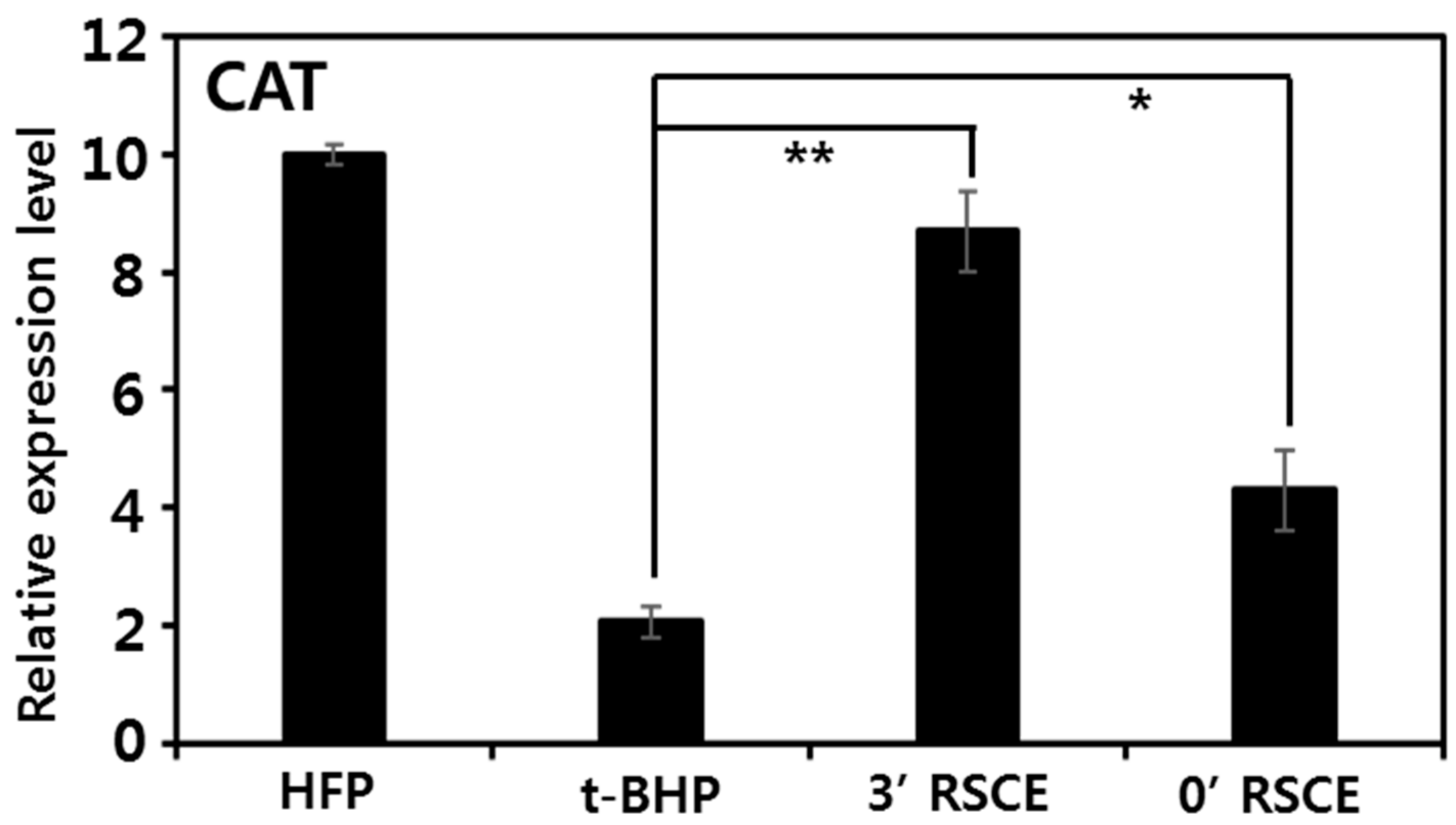
The beneficial effect of plasma-treated RSCE on oxidative damage may be due to the induction of the ability of fibroblasts to inhibit ROS. Burdon et al. [40] demonstrated that low concentrations (10–6 to 10–8 M) of H2O2 and t-butyl hydroperoxide could stimulate growth in both primary fibroblast cultures and transformed baby hamster kidney fibroblasts (BHK-21). Centella extract from the callus culture induced the expression of CAT exposed to H2O2. It has been clearly shown that Centella extract can prevent H2O2 cytotoxicity by enhancing the ability of fibroblasts to scavenge harmful ROS [41]. To support this fact, the mRNA expression levels for catalase (CAT), a major antioxidant enzyme, were compared. Our qRT-PCR results showed that plasma-treated RSCE had excellent efficacy on the induction of antioxidants in fibroblasts (Figure 6). Plasma-treated RSCE improved the CAT expression level in cells damaged by oxidative stress after t-BHP treatment with a significant four times greater difference. This was improved by a factor of two compared with RSCE without plasma treatment. These qRT-PCR data suggested that the upregulation of cellular antioxidant enzymes is a major factor for the protective effect of RSCE, in which the antioxidant activity was enhanced by plasma treatment. Taken together, these results clearly demonstrated that the increased antioxidant capacity of plasma-treated RSCE could prevent oxidative stress by enhancing the ability of HFB to scavenge harmful ROS. It is considered that the relatively low energy and RONS generated from the plasma were at an appropriate level for activating RSCE rather than damaging it. Plasma treatment was able to enhance the antioxidant ability of RSCE and lowered the intracellular ROS level of HFB damaged by oxidative stress.
The mRNA expression levels for catalase (CAT), a major antioxidant enzyme, were compared, and 1 mM of tert-butyl hydroperoxide (t-BHP) was used to artificially induce oxidative stress. Plasma-treated RSCE improved the CAT expression level in cells damaged by oxidative stress after t-BHP treatment, with a significant difference (* p < 0.05; ** p < 0.01). 3′ RSCE, extract of rice stem cells treated with plasma for 3 min. 0′ RSCE, extract of rice stem cells without plasma treatment. In order to observe the t-BHP-induced cytotoxicity-prevention activity of plasma-treated RSCE, HFB cells were pretreated with RSCE (3′ RSCE vs. 0′ RSCE) at a concentration of 0.1 mg/mL and plasma-treated for 24 h before t-BHP treatment.

3.6. Antiaging Effects of Rice Stem Cell Extracts

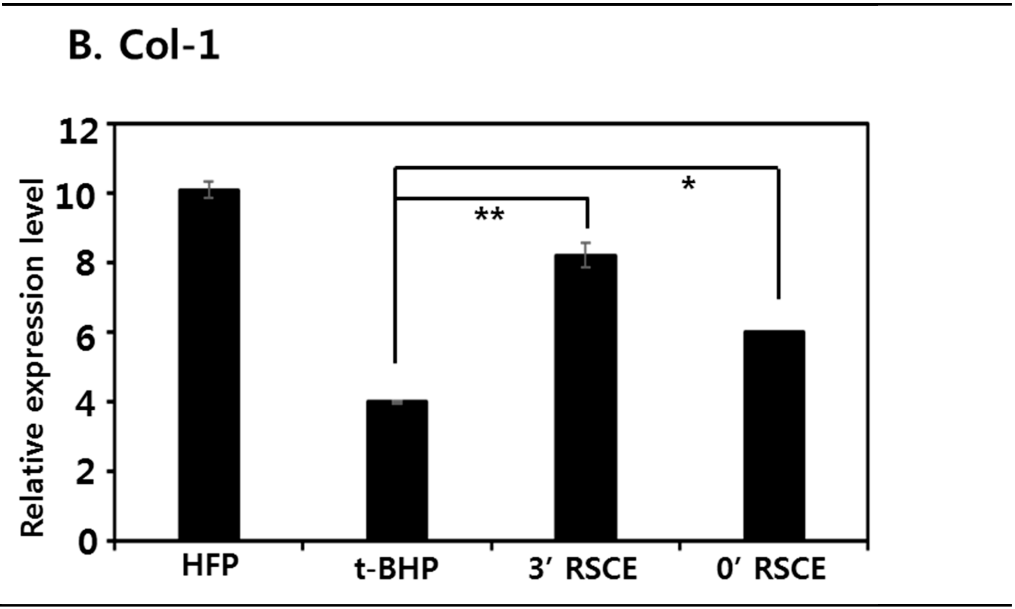
The beneficial effect of plasma-treated RSCE on oxidative damage may be due to the induction of the ability of fibroblasts to inhibit senescence. To support this hypothesis, we compared the mRNA expression levels of skin aging-related genes in fibroblasts with and without RSCE treated with plasma after oxidative stress treatment. The formation of oxidative stress increases the amount of matrix metalloproteinase (MMP)-9 expression in fibroblasts, which in turn leads to the degradation of collagen. Increased collagen breakdown by MMP-9 may eventually be a key factor in the aging process of skin tissue [42,43]. Here, we confirmed that the oxidative stress treatment of t-BHP significantly increased the MMP-9 expression level of HFB, suggesting that RSCE may have anti-aging activity by suppressing the MMP-9 mRNA expression level. The levels of gene expression were quantified as fold changes in expression using the GAPDH as a housekeeping gene. Plasma-treated RSCE reduced the expression level of MMP-9 by 2.8-fold. This expression level showed about 0.8-fold inhibition efficiency compared with RSCE not treated with plasma (Figure 7A). Buranasudja et al. demonstrated that the Centella callus extract significantly inhibited the upregulation of MMP-9 mRNA expression after H2O2 exposure [41]. A decrease in MMP levels may be associated with increased collagen production after plasma-treated RSCE treatment. Moreover, collagen has anti-aging properties, so when the skin is exposed to UV rays for a long time, proteolytic enzymes increase and break down collagen and elastin fibers (elastic fibers) that make up the skin, causing wrinkles that reduce skin elasticity [44]. Therefore, we compared the mRNA expression patterns of HFB for collagen synthesis-related genes in conditions with and without plasma-treated RSCE. Our qRT-PCR results showed that RSCE treated with N2 plasma for 3 min had a regenerative effect on the oxidative damage of HFB (Figure 7B). In HFB, in which the COl-1 expression level was decreased by t-BHP oxidative stress, the COl-1 mRNA expression level was increased again by RSCE. The plasma-treated RSCE (2-fold) showed a significant difference in the expression level of COl-1 and slightly enhanced it compared with the non-plasma-treated RSCE (1.5-fold). The mRNA expression level for COl-1 showed a profile in contrast with that of MMP-9. By analyzing the gene expression of RSCE on fibroblasts, we found common molecular pathway activation and response to the stresses connected to anti-aging. This might involve that skin fibroblasts responded correctly to the anti-aging effects of RSCE. Our findings give novel insight into utilizing RSCE for anti-aging effects and its possible mechanism. Although more molecular mechanism studies are required to develop anti-aging strategies with RSCE, our results suggest that plasma may be a potential tool used to enhance the activity of callus extracts to prevent oxidative stress.
All data points represent mean ± standard deviation of three replicate experiments. The significance of difference between the control and treatment was examined by Student’s t-test; * p < 0.05; ** p < 0.01. In order to observe the t-BHP-induced cytotoxicity-prevention activity of plasma-treated RSCE, HFB cells were pretreated with RSCE (3′ RSCE vs. 0′ RSCE) at a concentration of 0.1 mg/mL and plasma-treated for 24 h before t-BHP treatment.

4. Conclusions

Rice stem cells were induced in vitamin-containing Chu N6 medium from embryos of peeled mature rice seeds. The well-induced 3-week-old rice stem cells were treated with plasma, cultured for one day, and then extracted to make RSCE. The total phenolic content in 3 min plasma-treated RSCE was increased by about 1.5 times compared with the control, and 0.1 mg/mL of RSCE after 3 min plasma treatment had rapidly increasing cell proliferation rates of HFB compared with untreated RSCE under conditions of oxidative stress. This indirectly provides evidence that plasma treatment not only induces cell proliferation but also can increase the cell vitality by reducing cell damage. As a result of comparing the proliferation of HFB and the expression of collagen synthesis protein-related genes after oxidative stress treatment used to evaluate the skin regeneration efficacy of plasma-treated RSCE, plasma-treated RSCE showed a significant difference compared with untreated RSCE and increased by about more than four times.
The human body has a balance of substances that promote and inhibit oxidation. However, when this balance is disrupted, the cells produce excess oxidative-promoting substances, resulting in oxidative stress. Oxidative stress is primarily caused by ROS, such as superoxide anion radicals, hydroxyl radicals, singlet oxygen, and oxygen-derived H2O2. When the balance between ROS generation and the antioxidant response that removes it is disrupted, ROS generation becomes dominant, causing oxidative stress. However, a low concentration of ROS plays an important role in cell proliferation, differentiation, and signal transduction. ROS can be produced at low levels in response to the activation of certain signaling pathways, such as the epidermal growth factor receptor (EGFR) pathway. Therefore, our results suggest that plasma-generated RONS could enhance the antioxidant activity of RSCE and that plasma-generated RONS could be a key factor for the expression of genes involved in cell proliferation. In addition, our results suggest that the low energy of plasma delivered to rice stem cells can increase the antioxidant activity of rice stem cells. RSCE, in which antioxidant activity was increased with plasma treatment, restored HFB damaged by oxidative stress; this suggests that cellular vitality was enhanced by enhancing the energy metabolism of HFB using antioxidants and other metabolites of RSCE increased by plasma treatment. To support this conclusion, it is necessary to compare quantitative analyses of major metabolites related to the antioxidant activity of plasma-treated RSCE.
Unlike previous studies that use dedifferentiated callus from plant wounds, plant embryonic callus using rice seed embryos is similar to plant stem cells, so it is superior to studies using dedifferentiated callus because it can obtain various physiologically active substances. There is a possibility of developing the material. In the future, we aim to prepare and characterize aqueous extracts of plasma-treated RSCE as an anti-aging cosmetic ingredient to produce various bioactive compounds.

Supplementary Materials

The following supporting information can be downloaded at: https://www.mdpi.com/article/10.3390/app12062903/s1, Figure S1: Experimental flow chart; Figure S2: Individual GC-MS total ion chromatogram (TIC) of samples in full scale; Figure S3: Viability of HFB with t-BHP treatment; Figure S4: Viabillity of HFB with plasma treated RSCE.

Author Contributions

S.H.J. and M.A. performed experiments. S.H.J. and I.H. did conceptualization, methodology, data interpretation and wrote the main manuscript text. E.Y.K. and Y.S.K. did data interpretation, and validation. E.H.C., S.H.J. and I.H. did project administration, funding acquisition, conception and design of the study. All authors have read and agreed to the published version of the manuscript.

Funding

This work was funded by the ‘Cooperative Research Program for Agri-culture Science and Technology Development (PJ01505201)’ Rural Development Administration, which was funded by the National Research Foundation of Korea (NRF) funded by the Ministry of science (2020R1I1A1A01073071, 2021R1A2C1094162).

Institutional Review Board Statement

Not applicable.

Informed Consent Statement

Not applicable.

Data Availability Statement

Not applicable.

Acknowledgments

This work was carried out with support of the R&D Program of (EN2225–9) through the Korea Institute of Fusion Energy (KFE) funded by the Government funds. We would like to thank Sangheum Eom for measurement of active species generated from plasma discharge.

Conflicts of Interest

The authors declare no conflict of interest.

References

  1. Chung, J.H.; Seo, J.Y.; Choi, H.R.; Lee, M.K.; Youn, C.S.; Rhie, G.E.; Cho, K.H.; Kim, K.H.; Park, K.C.; Eun, H.C. Modulation of skin collagen metabolism in aged and photoaged human skin in vivo. J. Investig. Dermatol. 2001, 117, 1218–1224. [Google Scholar] [CrossRef] [PubMed] [Green Version]
  2. Yoon, Y.M.; Yoon, J.Y.; Lim, K.M.; Hahn, H.J.; Kim, Y.R.; Ahn, K.J.; An, S.K. Effects of the complex containing Centella asiaticaand folic acid-fermented extracts, acetyl glutamine, and nicotinic acid adenine dinucleotide phosphate on the inhibition of senescence and melanogenesis, promotion of collagen expression, cellular regeneration, and keratinocyte differentiation, and anti-inflammation. Korean J. Aesthet. Cosmetol. 2013, 11, 675–684. [Google Scholar]
  3. Yasui, K.; Sakurai, H. Chemiluminescent detection and imaging of reactive oxygen species in live mouse skin exposed to UVA. Biochem. Biophys. Res. Commun. 2000, 269, 131–136. [Google Scholar] [CrossRef] [PubMed]
  4. Park, J.S. Walnut husk ethanol extract possess antioxidant activity and inhibitory effect of matrix metalloproteinase-1 expression induced by tumor necrosis factor alpha in human keratinocyte. Korean J. Aesthet. Cosmetol. 2013, 11, 715–719. [Google Scholar]
  5. Kim, D.S.; Jeon, B.K.; Mun, Y.J.; Kim, Y.M.; Lee, Y.E.; Woo, W.H. Effect of Dioscorea aimadoimo on anti-aging and skin moisture capacity. J. Orient. Physiol. Pathol. 2011, 25, 425–430. [Google Scholar]
  6. Mohammad, A.E.; Ali, N.; Sarvin, M.A.; Hossein, K.; Seyedeh, M.G.; Sobhan, M.D. Enhanced catalytic and antibacterial efficiency of biosynthesized Convolvulus fruticosus extract capped gold nanoparticles (CFE@AuNPs). J. Photochem. Photobiol. B Biol. 2020, 209, 111949. [Google Scholar] [CrossRef]
  7. Pardis, M.; Mehdi, S.A.; Sobhan, M.D.; Ahmad, B.R.; Seyedeh, M.G. PEG-Citrate dendrimer second generation: Is this a good carrier for imaging agents In Vitro and In Vivo? IET Nanobiotechnol. 2019, 13, 560–564. [Google Scholar] [CrossRef]
  8. Lucia, S.; Lucia, M.; Evelyn, O.; Massimo, L.; Luisa, F.; Luca, P.; Davide, P.; Miriam, C. The emerging role of nanotechnology in skincare. Adv. Colloid Interface Sci. 2021, 293, 102437–102460. [Google Scholar] [CrossRef]
  9. Kaur, C.; Kapoor, H.C. Anti-oxidant activity and total phenolic content of some Asian vegetables. Int. J. Food Sci. Technol. 2002, 37, 153–161. [Google Scholar] [CrossRef]
  10. Trehan, S.; Michniak-Kohn, B.; Beri, K. Plant stem cells in cosmetics: Current trends and future directions. Future Sci. 2017, 3, FSO226. [Google Scholar] [CrossRef] [Green Version]
  11. Vasil, I.K.; Vasil, V. Totipotency and embryogenesis in plant cell and tissue cultures. In Vitro 1972, 8, 117–125. [Google Scholar] [CrossRef]
  12. Fay, M.F. Conservation of rare and endangered plants using in vitro methods. In Vitro Cell. Dev. Biol.-Plant 1992, 28, 1–4. [Google Scholar] [CrossRef]
  13. Irvani, N.; Solouki, M.; Omidi, M.; Zare, A.; Shahnazi, S.J.P.C. Callus induction and plant regeneration in Dorem ammoniacum D., an endangered medicinal plant. Plant Cell Tissue Organ Cult. 2010, 100, 293–299. [Google Scholar] [CrossRef]
  14. Oh, S.T.; Jung, H.S.; Cho, M.J.; Song, M.Y.; Moh, S.H.; Seo, H.H. Effect of Artemisia annua Linne callus induced by plant cell culture technology on wound healing. J. Korea Acad.-Ind. Coop. Soc. 2014, 15, 5628–5636. [Google Scholar] [CrossRef] [Green Version]
  15. Saewan, N.; Vichit, W.; Prinyarux, T. Anti-aging efficacy of thai red rice callus cosmetic product. J. Appl. Sci. 2018, 17, 63–72. [Google Scholar] [CrossRef]
  16. Mousumee, K.; Soyeon, P.; Hyeon-Jin, K.; Kui-Jae, L.; Dea-Heon, K.; So-Hyeon, B.; Seong-Tshool, H. The Resveratrol Rice DJ526 Callus Significantly Increases the Lifespan of Drosophila (Resveratrol Rice DJ526 Callus for Longevity). Nutrients 2019, 11, 983. [Google Scholar] [CrossRef] [Green Version]
  17. Widowati, W.; Fauziah, N.; Herdiman, H.; Afni, M.; Afifah, E.; Kusuma, H.S.W.; Nufus, H.; Arumwardana, S.; Rihibiha, D.D. Antioxidant and anti-aging assays of Oryza Sativa extracts, vanillin and coumaric Acid. J. Nat. Remedies 2016, 16, 88–99. [Google Scholar] [CrossRef] [Green Version]
  18. Finocchiaro, F.; Ferrari, B.; Gianinetti, A.; Dall’asta, C.; Galaverna, G.; Scazzina, F.; Nicoletta, P. Characterization of antioxidant compounds of red and white rice and changes in total antioxidant capacity during processing. Mol. Nutr. Food Res. 2007, 51, 1006–1019. [Google Scholar] [CrossRef]
  19. Yawadio, R.; Tanimori, S.; Morita, N. Identification of phenolic compounds isolated from pigmented rices and their aldose reductase inhibitory activities. Food Chem. 2007, 101, 1616–1625. [Google Scholar] [CrossRef]
  20. Dezest, M.; Chavatte, L.; Bourdens, M.; Quinton, D.; Camus, M.; Garrigues, L.; Descargues, P.; Arbault, S.; Burlet-Schiltz, O.; Casteilla, L.; et al. Mechanistic insights into the impact of Cold Atmospheric Pressure Plasma on human epithelial cell lines. Sci. Rep. 2017, 7, 41163. [Google Scholar] [CrossRef] [Green Version]
  21. Cheng, X.; Sherman, J.; Murphy, M.; Ratovitski, E.; Canady, J.; Keidar, M. The effect of tuning cold plasma composition on glioblastoma cell viability. PLoS ONE 2014, 9, e98652. [Google Scholar] [CrossRef] [Green Version]
  22. Chu, C.C. The N6 medium and its applications to anther culture of cereal crops. In Proceedings of the Symposium on Plant Tissue Culture, Beijing, China, 25–30 May 1978; Science Press: Beijing, China, 1978; pp. 45–50. [Google Scholar]
  23. Tabei, Y. (Ed.) Transformation Protocol. In Plants; Kagaku-Doujin: Tokyo, Japan, 2012. (In Japanese) [Google Scholar]
  24. Islam, M.M.; Ahmed, M.; Mahaldar, D. In vitro callus induction and plant regeneration in seed explants of rice (Oryza sativa L.). Res. J. Agric. Biol. Sci. 2005, 1, 72–75. [Google Scholar]
  25. Shahsavari, E.; Maheran, A.A.; Akmar, A.S.N.; Hanafi, M.M. The effect of plant growth regulators on optimization of tissue culture system in Malaysian upland rice. Afr. J. Biotechnol. 2010, 9, 2089–2094. [Google Scholar]
  26. Ji, S.H.; Kim, J.S.; Lee, C.H.; Seo, H.S.; Chun, S.C.; Oh, J.S.; Choi, E.H.; Park, G.S. Enhancement of vitality and activity of a plant growth promoting bacteria (PGPB) by atmospheric pressure non-thermal plasma. Sci. Rep. 2019, 9, 1044. [Google Scholar] [CrossRef]
  27. Ikawa, M.; Schafer, T.; Dollard, C.; Sasner, J. Utilization of Folin-Ciocalteu reagent for the detection of certain nitrogen compounds. J. Agric. Food Chem. 2003, 51, 1811–1815. [Google Scholar] [CrossRef]
  28. Ma, X.B.; Yang, J. An optimized preparation method to obtain high-quality RNA from dry sunflower seeds. Genet. Mol. Res. 2011, 10, 160–168. [Google Scholar] [CrossRef]
  29. Scalbert, A.; Johnson, I.T.; Saltmarsh, M. Polyphenols: Antioxidants and beyond. Am. J. Clin. Nutr. 2005, 81, 215S–217S. [Google Scholar] [CrossRef]
  30. Pandey, K.B.; Rizvi, S.I. Plant polyphenols as dietary antioxidants in human health and disease. Oxid. Med. Cell. Longev. 2009, 2, 270–278. [Google Scholar] [CrossRef] [PubMed] [Green Version]
  31. Hider, R.C.; Liu, Z.D.; Khodr, H.H. Metal chelation of polyphenols. Methods Enzymol. 2001, 335, 190–203. [Google Scholar] [CrossRef] [PubMed]
  32. Dumont, P.; Burton, M.; Chen, Q.M.; Gonos, E.S.; Frippiat, C.; Mazarati, J.B.; Eliaers, F.; Remacle, J.; Toussaint, O. Induction of replicative senescence biomarkers by sublethal oxidative stresses in normal human fibroblast. Free Radic. Biol. Med. 2000, 28, 361–373. [Google Scholar] [CrossRef]
  33. Kučera, O.; Endlicher, R.; Roušar, T.; Lotkova, H.; Garnol, T.; Drahota, A.; Cervinkova, Z. The effect of tert-butyl hydroperoxide-induced oxidative stress on lean and steatotic rat hepatocytes in vitro. Oxid. Med. Cell. Longev. 2014, 2014, 752506. [Google Scholar] [CrossRef] [Green Version]
  34. Davies, M.J. Detection of peroxyl and alkoxyl radicals produced by reaction of hydroperoxides with rat liver microsomal fractions. Biochem. J. 1989, 257, 603–606. [Google Scholar] [CrossRef] [Green Version]
  35. Crane, D.; Haussinger, D.; Graf, P.; Sies, H. Decreased flux through pyruvate dehydrogenase by thiol oxidation during t-butyl hydroperoxide metabolism in perfused rat liver. Hoppe-Seyler’s Z. Physiol. Chem. Bd. 1983, 364, 977–987. [Google Scholar] [CrossRef]
  36. Sohal, R.S.; Allen, R.G.; Nations, C. Oxygen free radicals play a role in cellular differentiation: An hypothesis. J. Free Radic. Biol. 1986, 2, 175–181. [Google Scholar] [CrossRef]
  37. Finkel, T. Signal transduction by reactive oxygen species. J. Cell Biol. 2011, 194, 7–15. [Google Scholar] [CrossRef] [Green Version]
  38. Thannickal, V.J.; Fanburg, B.L. Reactive oxygen species in cell signaling. Am. J. Physiol.-Lung Cell. Mol. Physiol. 2000, 279, L1005–L1028. [Google Scholar] [CrossRef] [Green Version]
  39. Ismail, T.J. ROS and RNS: Key signaling molecules in plants. J. Exp. Bot. 2018, 69, 3313–3315. [Google Scholar] [CrossRef] [Green Version]
  40. Burdon, R.H.; Gill, V.; Rice-Evans, C. Cell proliferation and oxidative stress. Free Radic. Res. Commun. 1989, 7, 149–159. [Google Scholar] [CrossRef]
  41. Buranasudja, V.; Rani, D.; Malla, A.; Kobtrakul, K.; Vimolmangkang, S. Insights into antioxidant activities and anti-skin-aging potential of callus extract from Centella asiatica (L.). Sci. Rep. 2021, 11, 13459–13475. [Google Scholar] [CrossRef]
  42. Pittayapruek, P.; Meephansan, J.; Prapapan, O.; Komine, M.; Ohtsuki, M. Role of matrix metalloproteinases in photoaging and photocarcinogenesis. Int. J. Mol. Sci. 2016, 17, 868. [Google Scholar] [CrossRef] [Green Version]
  43. Fussell, J.C.; Kelly, F.J. Oxidative contribution of air pollution to extrinsic skin ageing. Free Radic. Biol. Med. 2020, 151, 111–122. [Google Scholar] [CrossRef] [PubMed]
  44. Imokawa, G.; Nakajima, H.; Ishida, K. Biological mechanisms underlying the ultraviolet radiation-induced formation of skin wrinkling and sagging II: Overexpression of neprilysin plays as essential role. Int. J. Mol. Sci. 2015, 16, 7776–7795. [Google Scholar] [CrossRef] [PubMed] [Green Version]
Figure 1. µ-DBD plasma. Plasma discharge appearance and schematic diagram.
Applsci 12 02903 g001
Figure 2. Frequency of callus induction from rice embryonic cells.
Applsci 12 02903 g002
Figure 3. Measured O3 and NO2 concentrations according to the time variant. Changes in O3 (black line) and NO2 (blue line) concentrations during plasma discharge 15 min.
Applsci 12 02903 g003
Figure 4. Total antioxidant capacity in rice stem cell extracts (RSCE) according to the plasma treatment time. All measurements were made in 3 or more than 3 replicates. * p < 0.05; ** p < 0.01.
Applsci 12 02903 g004
Figure 5. Cell viability of HFB according to the plasma treatment time. All measurements were made in 3 or more than 3 replicates. * p < 0.05.
Applsci 12 02903 g005
Figure 6. Antioxidant enzymes expression of RSCE. All measurements were made in 3 or more than 3 replicates. * p < 0.05; ** p < 0.01.
Applsci 12 02903 g006
Figure 7. Quantitative RT-PCR analysis of anti-aging-related genes MMP-9 and Col-1 after treatment of RSCE in HFB. (A) MMP-9 (B) Col-1. All measurements were made in more than 3 replicates. * p < 0.05; ** p < 0.01.
Applsci 12 02903 g007aApplsci 12 02903 g007b
Table 1. The composition of the medium inducing rice stem cells.
Total (ddH2O)1 L
Sucrose30 g
Casamino acid0.3 g
Proline2.878 g
CHU N63.956 g
Myo-inosiitol0.1 g
2.4-D (mg/mL)2 mL
pH5.8
Gellan Gum4 g
Table 2. Primer sequence for qRT-PCR.
PrimerTemperature (°C)Sequence (5′→3′)
CAT60AACTGTCCCTACCGTGCTCG
ATTGGCAGTGTTGAATCTCCGC
Col-160GAGGGCCAAGACGAAGACATC
CAGATCACGTCATCGCACAAC
MMP-960ACTCGGGTGGCAGAGATGC
AGGTGATGTTGTGGTGGTGC
GAPDH60ATGAGAAGTATGACAACAGCC
AGTCCTTCCACGATACCAAA
Table 3. Candidate metabolites in rice stem cells.
Retention Time (min)Metabolites 1Peak Height Changed 2
34.805Fructose3 > 1 > C = 5
36.690Galactopyranoside3 > 1 > C > 5
47.830Melibiose3 > C = 1 = 5
49.325Glycoside3 > 1 > C > 5
49.620Sucrose3 > 1 > C > 5
1 Candidate metabolites according to the retention time. 2 The difference in the height of the changed peaks in order from highest to lowest. C, untreated control; 1, 3, 5; each plasma treatment time.
Publisher’s Note: MDPI stays neutral with regard to jurisdictional claims in published maps and institutional affiliations.