Article

Study of the Intra-Coronal Pressure Generated by Internal Bleaching Agents and Its Influence on Temporary Restoration

by
Alba Pallarés-Serrano
1,2,*,
Antonio Pallarés-Serrano
1,
Sandra Pallarés-Serrano
1 and
Antonio Pallarés-Sabater
1,2
1
Doctoral School, Catholic University of Valencia, 46001 Valencia, Spain
2
Department of Endodontics and Restorative Dentistry, School of Medicine and Dentistry, Catholic University of Valencia, Quevedo 2, 46001 Valencia, Spain
*
Author to whom correspondence should be addressed.
Appl. Sci. 2022, 12(6), 2799; https://doi.org/10.3390/app12062799
Submission received: 9 February 2022 / Revised: 26 February 2022 / Accepted: 7 March 2022 / Published: 9 March 2022
(This article belongs to the Special Issue Restorative and Endodontic Materials for Clinical Dentistry)

Abstract

:
Intra-coronal bleaching is a treatment that whitens non-vital teeth from within the pulp chamber, a procedure by which oxygen and free radicals are released. This in vitro study analyzed and compares the oxygen expansion produced when different bleaching agents encounter dental tissues during this type of bleaching. Here, 120 lower incisors were included and prepared to achieve conditions as close as practicable to internal bleaching with the walking bleach technique. The access cavity of the lingual surface was prepared to seal glass tubes with oil inside them by adhesive restoration once the bleach was placed inside the pulp chamber. The following bleaching groups were used: hydrogen peroxide (HP) 30% (n = 30), sodium Perborate (SP) with distilled water (n = 30), a mixture of HP 30% with SP (n = 30) and a control group (n = 30). After 10 days, the oil displacement that took place through the tube due to oxygen release was measured daily. Displacement of the oil was observed in all groups except the control group. The final mean expansion at 10 days was 335.24 ± 76.85 mm for the HP 30% group, 8.40 ± 1.74 mm for the SP group and 183.07 ± 49.93 mm for the HP30% + SP mixture. Significant statistical differences were observed between the three groups using the Games–Howell post hoc test, where HP30% caused the greatest expansion and SP the least expansion. These results suggest that the higher the amount of HP in the sample, the more oxygen expansion is observed, so that temporary restoration must be performed more carefully to avoid displacement.

1. Introduction

Treatments that improve dental aesthetics increase the patient’s quality of life and psychological state, as a harmonious smile is positively related to high tests of social, intellectual and occupational competence [1,2,3,4,5,6,7,8].
Dental dyschromias vary in aetiology, location and severity, and their origin may be related to extrinsic causes, intrinsic causes or a combination of both [9,10,11,12,13]. When tooth discoloration occurs, a detailed study of the case must be carried out in order to make an accurate diagnosis, since the success of the treatment and the accuracy of the results will depend on it [9,14,15].
Cases in which only a single tooth in the anterior sector is affected are relatively frequent, and when this occurs the negative effect becomes even more pronounced because the color does not match the rest [2,5,16].
Internal or intra-coronal whitening is a therapeutic option that involves the application of an oxidizing chemical agent, which removes intrinsic stains via chromogenic degradation, and by breaking down the larger pigments into smaller ones the color of the teeth is lightened [17,18].
The most widely used bleaching agents for decades in this type of bleach for devitalized teeth were hydrogen peroxide (HP) and sodium perborate (SP). Recently, carbamide peroxide (CP), which decomposes into HP itself and urea, has also been used.
HP is a powerful, unstable oxidizing agent that causes irritation to the skin, eyes and mucous membranes upon exposure to high concentrations. It decomposes into water and reactive oxygen radicals. The maximum reported concentration of HP without causing mucosal irritation has been estimated at 5%, and harmful effects have been observed from 8% onward [19]. SP is an effective and relatively inexpensive intra-coronary whitening agent used on devitalized teeth in cases where there is intrinsic discoloration [20,21,22]. It is a white powder that is usually mixed with HP 30% or distilled water when used for internal bleaching purposes [23,24].
The walking bleach technique is generally used by placing the bleaching agent inside the pulp chamber of the devitalized and darkened tooth [10,11]. The cavity is temporarily sealed, leaving it to act for about 7 days between sessions, where the bleaching agent is changed and renewed until the tooth reaches the desired shade.
The reactive oxygen released during the whitening process has been associated with both the whitening capacity and the toxicity of the agents, depending on the concentration of HP and the duration of the treatment [25]. It has been shown in some studies that there is microleakage of the temporary coronal sealant after internal bleaching, which has certain drawbacks, such as conditioning the desired results and the leakage of HP and reactive oxygen into the oral cavity [26,27,28]. If the expected success is not achieved during the first session, the treatment time is prolonged and the possibility of coronal leakage may increase due to inserting another temporary restoration when renewing the bleaching agent. This may compromise the endodontic treatment and the viability of the teeth with related aesthetic consequences, since it is generally performed on the teeth of the anterior sector [29]. Furthermore, it has been shown that leakage of HP in high concentrations into the oral cavity can easily cause burns to mucosal and periodontal tissues [30]. The aim of this in vitro study was to evaluate and compare the oxygen expansion that takes place after the reaction of the bleaching agents HP 30% and SP with dental tissues from inside the pulp chamber.

2. Materials and Methods

This experimental study was approved by the Ethics Committee of the Catholic University of Valencia (Valencia, Spain) (Ref.: UCV/2019-2020/037). It was focused on the whitening of non-vital teeth, so we used higher concentrations of these products than would be used in vital teeth, in accordance with the European Directive 93/42/EEC.

2.1. Sample Preparation

For this experimental in vitro study, a total of 120 human lower incisors were extracted for periodontal reasons. Patients were informed about the further use of the teeth for research purposes and gave their consent prior to tooth extraction was performed. All lower incisors free of caries, cracks, restorations, endodontic treatment, vertical fractures and resorptions were included. Any remaining biofilm was removed with sterile gauze and a prophylaxis brush. All teeth were stored in Hank’s balanced salt solution (HBSS), and were used within three months of extraction. The teeth were endodontically treated. Access cavities were made on the lingual surface with a turbine diamond bur (FG ML 200442AA ISO198016, Diatech; Coltene/Whaledent, Altstätten, Switzerland) at 300,000 rpm and cooled under water irrigation.
The teeth were prepared by a single operator to working length using a ProTaper® system (Dentsply Maillefer, Ballaigues, Switzerland) and X-SmartTM endodontic micromotor (Dentsply, UK). After each instrument, the canals were irrigated with 5 mL of 2.5% NaOCl and saline solution with a 30-G needle and were filled using gutta-percha in combination with AH Plus sealing cement (Dentsply Sirona, York, PA, USA). A 3 mm endodontic filling was removed apical to the cementoenamel junction (CEJ) with a tungsten carbide bur (Tungsten bur, FG 330, Kerr, Bolzano, Italy), then a 2 mm seal was applied using a resin-reinforced glass ionomer cement (Vitrebond, 3M ESPE, Maplewood, MN, USA).
The cement was cured for 60 s at approximately 1 cm, with a 1200 mW lamp intensity.

2.2. Experimental Design

The experimental model used to assess the expansion capacity of the bleaching agents when interacting with hard dental tissues consisted of a tooth that underwent endodontic treatment, then after placing the bleaching agent, a 1 mm diameter glass tube (2940211, Paul Marienfeld Superior, Lauda-Konigshofen, Germany) was assembled with composite in the endodontic access cavity. Previously, the enamel on the palatal surface of the tooth was etched with 37% ortho-phosphoric acid (ScotchbondEtchant, 3M ESPE, Saint Paul, MN, USA) and adhesive (excite, Ivoclar Vivadent, Schaan, Liechtenstein) was applied in order to fix the tube.
A syringe was used to inject a volume of oil occupying a length of 10 mm into the tube, with the purpose of containing the fluids.
The expansion was measured through the displacement of the oil towards the opposite end with a vernier caliper due to the released oxygen. If necessary, in order to monitor the continuous displacement of the oil, tubes were joined together with composite to allow the oil to rise through the tubes and the expansion to be measured for 10 days. The experimental model is shown in Figure 1.

2.3. Experimental Groups

The teeth were divided into groups according to the bleaching agent used. The following bleaching agents were used: liquid containing HP 30% (H2O2) (Foret, Peroxfarma, Barcelona, Spain) and SP powder (NaBO3) (Acofarma Distribution, S.A., Madrid, Spain).
  • Group 1: HP 30%
  • Group 2: SP (2 g) (mixed with 1 mL of distilled water)
  • Group 3: HP 30% (1 mL) + SP (2 g)
The mixtures were made with the help of a cement spatula and a dappen glass. A control group of 30 tubes was established, in which no bleaching agent was introduced to check that the oil did not displace on its own.

2.4. Statistical Analysis

All data on the expansion that was observed due to the interaction of the bleaching agent with the dental tissues were collected in an Excel spreadsheet for subsequent statistical analysis. The statistical analysis of the data collected for the present study was carried out using the SPSS 23 computer program (IBM Corp., Armonk, NY, USA).

3. Results

In the control group the oil was not displaced, so only the study groups were compared. The process consisted of first using a robust test for equality of means (Welch’s ANOVA), and subsequently by performing a Games–Howell post hoc test.
The final average expansion after 10 days was 335.24 ± 76.85 mm for HP 30%, 8.40 ± 1.74 mm for the SP agent and 183.07 ± 49.93 mm for the HP 30% + SP mixture (Table 1).
Figure 2 shows the evolution of the average expansion observed in each bleaching agent over the first 10 days. This figure shows the discreet, stable and irrelevant progress of oxygen expansion in the SP group, which contrasts with the HP group, where a significant and strong expansion is observed from the first day. Additionally, the average expansion of the HP 30% + SP group sits between the aforementioned two groups, with neither reaching the levels of HP 30% nor evolving as weakly as in the SP group (Figure 2).
The p-values of the Games–Howell contrast statistic were less than 0.05 in all comparisons, corroborating that there were statistically significant differences between the final mean expansion of the three bleaching agents.
Furthermore, the mean final expansion of the HP 30% agent was significantly higher than the expansion of the other two agents, and the final mean expansion of the HP 30% + SP agent was significantly greater than the expansion of the SP agent (Games–Howell, p < 0.05). The data are summarized in Table 2.

4. Discussion

This study was designed and conducted in order to objectively measure and compare the expansion resulting from the reactions between hard dental tissues and HP 30%, SP and HP 30% combined with SP during internal bleaching using the walking bleach technique. Theoretical aspects of the bleaching reaction are shown in a directly visible way.
During the first 24 h, a strong expansion was observed in the HP 30% and HP 30% + SP groups, which decreased as the days went by. However, in the SP group, the aforementioned strong expansion did not continue to grow after 10 days, but a discrete, constant and homogeneous expansion, and was significantly lower than the other two groups. These results coincide with the findings observed in an in vitro study, in which expansion in enamel and dentin separately was also measured [31].
The analyzed data analyzed showed significant differences between the three groups, with the HP 30% group showing the greatest expansion after 10 days and the SP group showing the least expansion.
Nutting and Poe introduced the walking bleach technique, modifying the technique used by Spasser, in which SP was mixed with water to whiten teeth, and SP with was combined with HP 30% instead of distilled water [23,24]. They thought that this would make the treatment more effective by releasing more oxygen in their reaction since, oxygen and free radicals establish their main mechanism of action in whitening [32,33,34]. Some studies have supported this theory by showing more effective and faster bleaching results when mixing SP with HP 30% instead of mixing it with water [35,36,37,38].
Internal whitening is a relatively simple procedure for dentists that is comfortable for patients, restoring the aesthetics of darkened non-vital teeth, which is one of the main causes of loss of self-esteem among the population. However, the lack of predictability is one of the main disadvantages of internal whitening, as its effectiveness can be affected by the misalignment of the temporary restoration due to the reactions that occur inside the tooth when the whitening agent is introduced [28].
In this study, a strong force with which oxygen is released by the bleaching reaction after the rapid displacement of the oil was observed, especially during the first 24 h, which continued for at least 10 days. This gas can cause a significant increase in intra-cameral pressure, which increases the possibility of pushing on the temporary restoration, affecting its marginal integrity and causing the seal to be lost [39].
This can be a cause of failure due to internal bleaching, because if the temporary restoration is misaligned in the first days, oxygen and free radicals involved in the bleaching mechanism can escape through the open space between the restoration and the teeth from the beginning, meaning their effect is likely to be null.
It should be noted that an unsealed access cavity allows bacteria to penetrate the dentine through the microscopic space that is caused, which may affect the endodontic treatment and the survival of the tooth [26]. In addition, intra-coronal bleaching has also been linked to the occurrence of external cervical root resorption, which is a severe pathology associated with an inflammatory process that is invasive in nature, involving losses of cementum and dentine, potentially leading to tooth loss [9,10]. Studies show that internal bleaching and trauma are the main predisposing factors for its appearance. Although the mechanism for this effect is still unclear, these studies support the idea that the bleaching agent or products derived from it reach the periodontal tissue through the dentinal tubules, initiating an inflammatory process.
Therefore, quality restorations are important even though they are temporary. In non-vital bleaching, restorations are generally made with the same materials that are used for provisional restorations in endodontics to temporarily seal the tooth and prevent the loss of medication [40,41,42]. In the walking bleach technique, the aim is to avoid loss of the bleaching agent. However, the oxygen expansion reaction that occurs inside the chamber with the bleaching agents must be taken into account. Consequently, it would be reasonable to perform the provisional sealing with composite resin material and adhesives to ensure efficacy, so as to avoid bacterial leakage into the tooth and to prevent the bleaching agent from coming into contact with the oral tissues, since at high concentrations reactive oxygen radicals can cause genotoxicity and cytotoxicity [33,43]. Care must be taken in choosing the proper temporary materials for the walking bleach technique.
Waite et al. already reported that the walking bleach technique requires a solid seal around the access cavity using a restorative composite to ensure its effectiveness and to avoid the aforementioned leaks, which cannot be guaranteed if endodontic provisional restorative materials are used. The clinical choice of a coronal temporary restorative material is of paramount importance to achieve a hermetic seal and for the success of the whitening treatment [33,44].
Furthermore, temporary coronal restorative materials in endodontics are not indicated for periods of time longer than 1–2 weeks. In a previous study, gas expansion due to the bleaching agent was observed after 10 days [41]. Teixeira et al. conducted an in vitro study with the aim of evaluating the effects of bleaching agents on the microleakage of coronal access restorations, observing microleakage in all groups except the control group [27]. The observations made in the current study coincide with and justify the results obtained by Teixeira et al.
Traviglia et al. demonstrated the influence of a temporary adhesive restoration on the effectiveness of the whitening reaction with the walking bleach technique compared to a temporary “mechanical retention” filling, which is usually achieved with coronal temporary endodontic materials [44]. “Mechanical retention” cannot prevent oxygen leakage, which forces the patient to perform more sessions and increases the risk of complications. The observations made in this study are consistent with those made by Travigila et al., who state that maintaining an adequate coronal seal maintains the rapid dissociation of HP exclusively within the pulp chamber, inducing an increase in internal pressure and consequent penetration of oxygen into the dentinal tubules, whitening the tooth from the inside out without the need for repeated sessions.
This study highlights the clinical importance of understanding the bleaching reaction inside the tooth with each agent in order to choose the best possible temporary sealing material for each condition. Since oxygen expansion increases with higher HP, sealing with high concentrations of HP must be thorough.
However, although the study was carried out very thoroughly and meticulously, it has certain limitations inherent to an in vitro experimental study, such as the temperature (since the oral temperature is higher). The degree of coronal filtration should be checked with other studies for each bleaching agent and the actual effect on the bleaching capacity should be evaluated.

5. Conclusions

The results of the present in vitro experimental study show that oxygen release occurs with all bleaching agents.
However, significant differences were observed between the different agents used, with HP 30% showing the highest oxygen expansion and SP with distilled water showing the lowest expansion.
The HP 30% + SP group showed significantly lower expansion than the HP 30% group but significantly higher expansion than the SP group. It is suggested that the degree of oxygen expansion depends on the amount of HP available in the preparation, as it is the active agent in all samples. Therefore, the provisional restoration should provide a more effective seal with higher concentrations of HP to avoid mismatch and the resulting problems it could cause.

Author Contributions

Conceptualization, A.P.-S. (Antonio Pallarés-Sabater) and A.P.-S. (Alba Pallarés-Serrano); methodology, A.P.-S. (Antonio Pallarés-Sabater); validation, A.P.-S. (Alba Pallarés-Serrano), A.P.-S. (Antonio Pallarés-Serrano), S.P.-S. and A.P.-S. (Antonio Pallarés-Sabater); formal analysis, A.P.-S. (Alba Pallarés-Serrano) and A.P.-S. (Antonio Pallarés-Sabater); investigation, A.P.-S. (Alba Pallarés-Serrano), S.P.-S., A.P.-S. (Antonio Pallarés-Serrano) and A.P.-S. (Antonio Pallarés-Sabater). All authors have read and agreed to the published version of the manuscript.

Funding

This research received no external funding.

Institutional Review Board Statement

The study was conducted according to the guidelines of the Declaration of Helsinki and approved by the Ethics Committee of Catholic University of Valencia (protocol code UCV/2019-2020/037; date of approval: February 2020).

Informed Consent Statement

Informed consent was obtained from all subjects involved in the study.

Data Availability Statement

Data are reported within this article.

Conflicts of Interest

The authors declare no conflict of interest.

References

  1. Greta, D.C.; Colosi, H.A.; Gasparik, C.; Dudea, D. Color comparison between non-vital and vital teeth. J. Adv. Prosthodont. 2018, 10, 218–226. [Google Scholar] [CrossRef] [PubMed] [Green Version]
  2. Bersezio, C.; Martin, J.; Peña, F.; Rubio, M.; Estay, J.; Vernal, R.; Junior, O.O.; Fernández, E. Effectiveness and Impact of the Walking Bleach Technique on Esthetic Self-perception and Psychosocial Factors: A Randomized Double-blind Clinical Trial. Oper. Dent. 2017, 42, 596–605. [Google Scholar] [CrossRef] [PubMed]
  3. John, M.; Slade, G.; Szentpétery, A.; Setz, J. Oral health-related quality of life in patients treated with fixed, removable, and complete dentures 1 month and 6 to 12 months after treatment. Int. J. Prosthodont. 2004, 17, 503–511. [Google Scholar] [CrossRef] [PubMed]
  4. Grossmann, A.; Hassel, A.; Schilling, O.; Lehmann, F.; Koob, A.; Rammelsberg, P. Treatment with double crown-retained removable partial dentures and oral health-related quality of life in middle and high-aged patients. Int. J. Prosthodont. 2007, 20, 576–578. [Google Scholar] [PubMed]
  5. Samorodnitzky-Naveh, G.R.; Geiger, S.B.; Levin, L. Patients’ satisfaction with dental esthetics. J. Am. Dent. Assoc. 2007, 138, 805–808. [Google Scholar] [CrossRef]
  6. Van Der Geld, P.; Oosterveld, P.; Van Heck, G.; Kuijpers-Jagtman, A.M. Smile attractiveness: Self-perception and influence on personality. Angle Orthod. 2007, 77, 759–765. [Google Scholar] [CrossRef] [Green Version]
  7. Tin-Oo, M.M.; Saddki, N.; Hassan, N. Factors influencing patient satisfaction with dental appearance and treatments they desire to improve aesthetics. BMC Oral Health 2011, 11, 6. [Google Scholar] [CrossRef] [Green Version]
  8. Kershaw, S.; Newton, J.; Williams, D. The influence of tooth colour on the perceptions of personal characteristics among female dental patients: Comparisons of unmodified, decayed and ‘whitened’ teeth. Br. Dent. J. 2008, 204, E9. [Google Scholar] [CrossRef]
  9. Plotino, G.; Buono, L.; Grande, N.M.; Pameijer, C.H.; Somma, F. Nonvital Tooth Bleaching: A Review of the Literature and Clinical Procedures. J. Endod. 2008, 34, 394–407. [Google Scholar] [CrossRef]
  10. Dahl, J.E.; Pallesen, U. Tooth Bleaching—A Critical Review of the Biological Aspects. Crit. Rev. Oral Biol. Med. 2003, 14, 292–304. [Google Scholar] [CrossRef]
  11. Zimmerli, B.; Jeger, F.; Lussi, A. Bleaching of Nonvital Teeth. A clinically relevant literature review. Schweiz. Monatsschr. Zahnmed. 2010, 120, 306–313. [Google Scholar] [PubMed]
  12. Madison, S.; Walton, R. Cervical root resorption following bleaching of endodontically treated teeth. J. Endod. 1990, 16, 570–574. [Google Scholar] [CrossRef]
  13. Watts, A.; Addy, M. Tooth discoloration and staining. A review of literature. Br. Dent. J. 2001, 190, 309–316. [Google Scholar] [CrossRef] [PubMed]
  14. Santana, T.R.; de Bragança, R.M.F.; Correia, A.C.C.; de Melo Oliveira, I.; Faria-e-Silva, A.L. Role of enamel and dentin on color changes after internal bleaching associated or not with external bleaching. J. Appl. Oral Sci. 2021, 29, e20200511. [Google Scholar] [CrossRef]
  15. Pandey, S.H.; Patni, P.M.; Jain, P.; Chaturvedi, A. Management of intrinsic discoloration using walking bleach technique in maxillary central incisors. Clujul Med. 2018, 91, 229–233. [Google Scholar] [CrossRef] [Green Version]
  16. De Souza-Zaroni, W.C.; Lopes, E.B.; Ciccone-Nogueira, J.C.; Silva, R.C.S.P. Clinical comparison between the bleaching efficacy of 37% peroxide carbamide gel mixed with sodium perborate with established intracoronal bleaching agent. Oral Surg. Oral Med. Oral Pathol. Oral Radiol. Endod. 2009, 107, e43–e47. [Google Scholar] [CrossRef]
  17. Badole, G.P.; Warhadpande, M.M.; Bahadure, R.N.; Badole, S.G. Aesthetic Rehabilitation of discoloured nonvital anterior tooth with carbamide peroxide bleaching: Case Series. J. Clin. Diagn. Res. 2013, 7, 3073–3076. [Google Scholar] [CrossRef]
  18. Baratieri, L.N.; Ritter, A.V.; Monteiro, S., Jr.; Caldeira de Andrada, M.A.; Cardoso Vieira, L.C. Nonvital tooth bleaching: Guidelines for the clinician. Quintessence Int. 1995, 26, 597–608. [Google Scholar]
  19. Alkahtani, R.; Stone, S.; German, M.; Waterhouse, P. A review on dental whitening. J. Dent. 2020, 100, 103423. [Google Scholar] [CrossRef]
  20. Rotstein, I.; Mor, C.; Friedman, S. Prognosis of intracoronal bleaching with sodium perborate preparations in vitro: 1-year study. J. Endod. 1993, 19, 10–12. [Google Scholar] [CrossRef]
  21. Macey-Dare, L.V.; Williams, B. Bleaching of a discoloured non-vital tooth: Use of a sodium perborate/water paste as the bleaching agent. Int. J. Paediatr. Dent. 1997, 7, 35–38. [Google Scholar] [CrossRef] [PubMed]
  22. Fasanaro, T. Bleaching teeth: History, chemicals and materials used for common tooth discolorations. J. Esthet. Restor. Dent. 1992, 4, 71–78. [Google Scholar] [CrossRef] [PubMed]
  23. Spasser, H.F. A simple bleaching technique using sodium perborate. N. Y. State Dent. J. 1961, 27, 332–334. [Google Scholar]
  24. Nutting, E.; Poe, G. Chemical bleaching of discolored endodontically treated teeth. Dent. Clin. N. Am. 1967, 11, 655–662. [Google Scholar]
  25. Carey, C.M. Tooth Whitening: What We Now Know. J. Evid. Based Dent. Pract. 2014, 14, 70–76. [Google Scholar] [CrossRef] [Green Version]
  26. Barthel, C.R.; Strobach, A.; Briedigkeit, H.; Göbel, U.B.; Roulet, J.F. Leakage in roots coronally sealed with different temporary fillings. J. Endod. 1999, 25, 731–734. [Google Scholar] [CrossRef]
  27. Teixeira, E.C.N.; Hara, A.T.; Turssi, C.P.; Serra, M.C. Effect of non-vital tooth bleaching on microleakage of coronal access restorations. J. Oral Rehabil. 2003, 30, 1123–1127. [Google Scholar] [CrossRef]
  28. Oliveira, D.P.; Gomes, B.P.F.A.; Zaia, A.A.; Souza-Filho, F.J.; Ferraz, C.C.R. Ex vivo antimicrobial activity of several bleaching agents used during the walking bleach technique. Int. Endod. J. 2008, 41, 1054–1058. [Google Scholar] [CrossRef]
  29. Kriznar, I.; Seme, K.; Fidler, A. Bacterial microleakage of temporary filling materials used for endodontic access cavity sealing. J. Dent. Sci. 2016, 11, 394–400. [Google Scholar] [CrossRef] [Green Version]
  30. Alqahtani, M.Q. Tooth-bleaching procedures and their controversial effects: A literature review. Saudi Dent. J. 2014, 26, 33–46. [Google Scholar] [CrossRef] [Green Version]
  31. Pallarés-Serrano, A.; Pallarés-Serrano, S.; Pallarés-Serrano, A.; Pallarés-Sabater, A. Assessment of Oxygen Expansion during Internal Bleaching with Enamel and Dentin: A Comparative In Vitro Study. Dent. J. 2021, 9, 98. [Google Scholar] [CrossRef] [PubMed]
  32. Canoglu, E.; Gulsahi, K.; Sahin, C.; Altundasar, E.; Cehreli, Z.C. Effect of bleaching agents on sealing properties of different intraorifice barriers and root filling materials. Med. Oral Patol. Oral Cir. Bucal. 2012, 17, e710–e715. [Google Scholar] [CrossRef] [PubMed] [Green Version]
  33. Hosoya, N.; Cox, C.F.; Arai, T.; Nakamura, J. The walking bleach procedure: An in vitro study to measure microleakage of five temporary sealing agents. J. Endod. 2000, 26, 716–718. [Google Scholar] [CrossRef]
  34. Patel, S.; Kanagasingam, S.; Ford, T.P. External cervical resorption: A review. J. Endod. 2009, 35, 616–625. [Google Scholar] [CrossRef] [PubMed]
  35. Ho, S.; Goerig, A.C. An in vitro comparison of di¡erent bleaching agents in the discolored tooth. J. Endod. 1989, 15, 106–111. [Google Scholar] [CrossRef]
  36. Warren, M.A.; Wong, M.; Ingram, T.A., III. An in vitro comparison of bleaching agents on the crowns and roots of discolored teeth. J. Endod. 1990, 16, 463–467. [Google Scholar] [CrossRef]
  37. Freccia, W.; Peters, D.; Lorton, L.; Bernier, W. An in vitro comparison of non-vital bleaching techniques in the discolored tooth. J. Endod. 1982, 8, 70–77. [Google Scholar] [CrossRef]
  38. Ganesh, R.; Aruna, S.; Joyson, M.; Manikandan, D. Comparison of the bleaching efficacy of three different agents used for intracoronal bleaching of discolored primary teeth: An in vitro study. J. Indian Soc. Pedod. Prev. Dent. 2013, 31, 17–21. [Google Scholar] [CrossRef]
  39. Srikumar, G.P.; Varma, K.R.; Shetty, K.H.; Kumar, P. Coronal microleakage with five different temporary restorative materials following walking bleach technique: An ex-vivo study. Contemp. Clin. Dent. 2012, 3, 421–426. [Google Scholar] [CrossRef]
  40. Domingos, H.B.; Gonçalves, L.S.; de Uzeda, M. Antimicrobial activity of a temporary sealant used in endodontic treatment: An in vitro study. Eur. J. Dent. 2015, 9, 411–414. [Google Scholar] [CrossRef] [Green Version]
  41. Madarati, A.; Rekab, M.S.; Watts, D.C.; Qualtrough, A. Time-dependence of coronal seal of temporary materials used in endodontics. Aust. Endod. J. 2008, 34, 89–93. [Google Scholar] [CrossRef] [PubMed]
  42. Naseri, M.; Ahangari, Z.; Moghadam, M.S.; Mohammadian, M. Coronal sealing ability of three temporary filling materials. Iran. Endod. J. 2012, 7, 20–24. [Google Scholar] [PubMed]
  43. Tredwin, C.; Naik, S.; Lewis, N.; Scully, C. Hydrogen Peroxide Tooth-Whitening (Bleaching) Products: Review of Adverse Effects and Safety Issues. Br. Dent. J. 2006, 200, 371–376. [Google Scholar] [CrossRef] [PubMed] [Green Version]
  44. Traviglia, A.; Re, D.; De Micheli, L.; Bianchi, A.E.; Coraini, C. Speed bleaching: The importance of temporary filling with hermetic sealing. Int. J. Esthet. Dent. 2019, 14, 310–323. [Google Scholar] [PubMed]
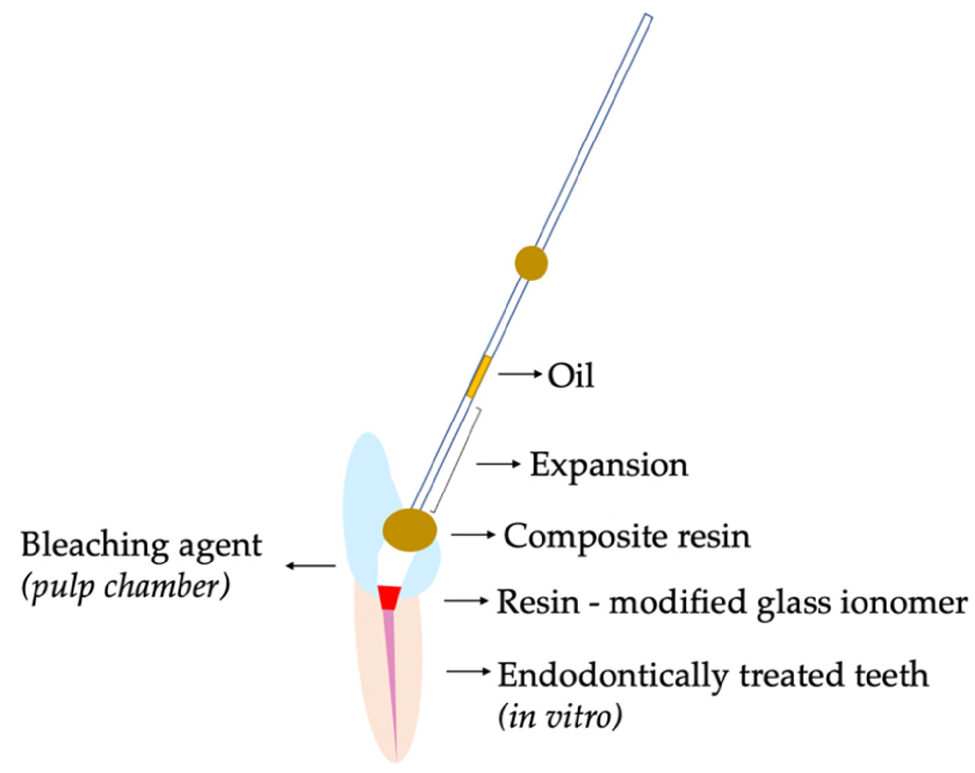
Figure 1. The experimental model, with the endodontic tooth and the assembled glass tubes.
Applsci 12 02799 g001
Figure 2. Evolution of the average expansion over time for each bleaching agent at 10 days.
Applsci 12 02799 g002
Table 1. Final average expansion after 10 days for each of the bleaching agents analyzed.
Final Average Expansion
Bleaching AgentsMeanStandard DeviationTypical Error95% Confidence IntervalMinMax
Lower LimitUpper Limit
HP 30% 335.25205.8137.58258.39412.0920725
SP (n = 30)8.404.660.856.6610.14315
HP 30% + SP (n = 30)183.07133.7124.41133.1423341435
Total (n = 90)175.57194.0820.56134.92216.223725
Table 2. Comparison of the results obtained between the different bleaching groups with the Games–Howell contrast test after 10 days.
Multiple Comparisons of Bleaching Groups
Bleaching Agent (A1)Bleaching Comparison Group (A2)Difference of Means (A1 − A2)Typical Errorp-Value95% Confidence Interval
Lower LimitUpper Limit
HP 30%SP326.8437.59<0.001234.02419.66
HP 30% + SP152.17344.810.00443.92260.42
SPHP 30%−326.8437.59<0.001419.66−234.02
HP 30% + SP−174.6724.43<0.001234.99−114.35
HP 30% + SPHP 30%−152.1744.810.004260.42−43.92
SP174.6724.43<0.001114.35234.99
Publisher’s Note: MDPI stays neutral with regard to jurisdictional claims in published maps and institutional affiliations.