Article

Antibacterial Activity and Biofilm Inhibition of New-Generation Hybrid/Fluoride-Releasing Restorative Materials

1
Department of Restorative Dentistry, School of Dentistry, Hacettepe University, 06100 Ankara, Turkey
2
Ecole de Médecine Dentaire de Marseille, Université d’Aix-Marseille, 13385 Marseille, France
3
Departments of Endodontics and Restorative Dentistry, School of Dental Medicine, University of Zagreb, 10000 Zagreb, Croatia
4
Department of Pharmaceutical Microbiology, Faculty of Pharmacy, Ankara University, 06560 Ankara, Turkey
*
Author to whom correspondence should be addressed.
Appl. Sci. 2022, 12(5), 2434; https://doi.org/10.3390/app12052434
Submission received: 9 January 2022 / Revised: 21 February 2022 / Accepted: 23 February 2022 / Published: 25 February 2022

Abstract

:
The antibacterial activity, and the effect of the application of additional topical fluoride on the bacterial activity, biofilm formation, and surface roughness of new-generation hybrid/fluoride-releasing materials were investigated. Two hundred and forty specimens were prepared in split Teflon molds (8 × 2 mm) from a resin composite (as negative control: G-aenial A’Chord/GC), Equia Forte HT Fil(GC), Equia Forte HT Fil+Equia Forte Coat, Riva Self-Cure (SDI), Riva Self-Cure+Equia Forte Coat, Zirconomer (Shofu), Beautifil II (Shofu), and Riva Silver (Shofu). Penicillin G,1U was used as positive control. The antibacterial activity was evaluated by the agar diffusion test immediately after the materials set using Streptococcus mutans (S. mutans) and Lactobacillus casei (L. casei), and repeated after application of 0.20% w/w (900 ppm) topical fluoride. The biofilm formation of S. mutans on each material was quantified by crystal violet staining. Surface roughness of the specimens was measured by a profilometer. The data were analyzed by Kruskal–Wallis, Dunn’s, one-way ANOVA, and Tukey’s HSD tests (p < 0.05). None of the tested restorative materials showed antibacterial activity and no inhibition zones were observed after treatment of the restoratives with additional topical fluoride. There were significant differences among the groups in terms of biofilm formation (p < 0.005). Equia Forte HT Fil with and without coating showed the lowest, while Riva self-cure without coating and Zirconomer showed the highest biofilm accumulation. None of the new-generation hybrid/fluoride-releasing materials demonstrated antibacterial activity and additional topical fluoride application did not make any change. Biofilm formation of the tested materials differed. All tested materials showed different surface roughness values (p < 0.005). Characteristics and compositions of the materials seemed to be more effective than the surface roughness.

1. Introduction

Tooth caries is known as one of the most common chronic oral diseases in humans [1]. The main causatives for tooth caries are bacteria, food habits, and the immune response by the host. Its etiology also includes highly complicated microecological harmony of biofilm. Streptococcus mutans (S. mutans) is considered as the primary bacterium responsible for the onset of caries while Lactobacilli are the main bacteria with respect to both caries progression and formation of secondary caries. Since acid-producing bacteria could trigger the initiation of the demineralization process, secondary caries formation might occur at the tooth–restoration interface or on the surface of the tooth [1,2,3,4,5].
With advances in dentistry, new minimal-intervention techniques and materials have been investigated in order to conserve the remaining dental hard tissues and minimize the risk of pulp damage. In recent years, comprehensive research in the field of modern dentistry has led to advancement of different new dental restorative materials with improved physical and esthetic properties and treatment modalities for tooth caries. Dental restorative materials have developed in terms of filler particles, matrix composition, and structure. Resin composites and glass ionomer cements (GICs) are the most-preferred materials as a viable alternative solution for the replacement of dental amalgam based on the concerns regarding its possible harmful effects on the environment as well as biocompatibility and esthetics. Both resin composites and GICs have distinctive features that clarify their popularity and limits [6,7,8].
Fluoride is known as an anticariogenic element. Anticariogenic potential of the fluoride is related to its unique properties, such as decreasing demineralization, increasing remineralization, and inhibition of pellicle and biofilm formation, bacterial metabolism, and microbial growth. It is assumed that fluoride released from restorative materials affects the formation of tooth caries through all mentioned mechanisms and therefore might decrease or inhibit demineralization and increase the remineralization of tooth hard tissues [9,10].
Recently, due to increasing demand to use fluoride-releasing dental materials for the restoration of cavities or build-up cores, several fluoride-containing hybrid dental restoratives have evolved in the dental market, such as resin-modified GICs, giomers, compomers, and bioactive resin composites. These hybrid restoratives were launched to handle the drawbacks of conventional GICs and resin composites without sacrificing their clinical advantages. They have shown a variety of fluoride-release potentials due to the differences in their matrices and hardening mechanisms [3,8,9,11].
Conventional GICs’ particular characteristics that make them advantageous as a restorative or an adhesive material include chemical adhesion to the tooth hard tissues without any intermediate bonding agent, anticariogenic properties caused by fluoride release, and bio and thermal compatibility with the tooth enamel. In recent years, handling properties and chemical formulations of the GICs have been considerably changed for various clinical applications [3,8]. The most recently launched materials, termed as glass hybrids (GHs), have been shown to come with adequate wear resistance (due to the resin-based coating agent after placement). GHs contain a multifunctional monomer and are reinforced with ultrafine, highly reactive fluoroaluminosilicate glass particles [8].
Some commercial GICs consist of strontium-containing glasses. In this formulation, strontium replaces calcium. It is stable and insoluble under neutral circumstances but is released from GICs when the pH shifts towards acidic challenges [3].
To improve the antibacterial properties of GICs, silver nanoparticles have also been incorporated into their formula [3]. Another improvement in GICs was the introduction of zirconia-reinforced GICs. The manufacturer of this GIC claims that this product possesses the strength and durability of amalgam as well as the protective benefits of GICs without mercury. Zirconia and/or zirconium oxide has been used as an indirect restorative material since 1998. The strength of zirconia allows it to be used for crown and bridge restoration in all areas of the mouth [12].
On the other hand, by combining the characteristics of the resin composites and GICs, hybrid products known as giomers have been obtained. In this formula, pre-reacted glass ionomer technology was employed to form the glass ionomer phase. Giomers contain fillers that are produced by the reaction of ion-leachable glasses with polyalkenoic acids. Due to the pre-reacted glass fillers, acidic resin monomers are not essential for in situ acid–base reactions. Giomers offer both protection against caries and functional and esthetic results [13,14].
From the clinical perspective, the preferred restorative materials are those that not only prevent the growth of oral bacteria, but also inhibit the colonization of bacteria on the tooth surfaces.
The antibacterial efficacy of GICs has been investigated extensively in in vitro studies [5,12,15,16,17,18,19], and some of these studies showed that conventional GICs had potential antibacterial activity and also reduced formation of biofilm by S. mutans strains [5,12,15,17,18,19,20]. On the other hand, very few studies have been published evaluating the antibacterial properties of contemporary fluoride-releasing materials [11,21]. The lack of bacterial growth inhibition of previously developed fluoride-releasing restoratives has also been shown in the literature [20,22].
Studies investigating the antibacterial efficacy and biofilm inhibition of these newly developed fluoride-releasing/hybrid restorative materials are also needed to minimize the frequency and severity of secondary caries. Therefore, the aim of this study was to investigate the antibacterial properties and biofilm formation of five different commercially available new-generation hybrid/fluoride-releasing materials: two bulk-fill GHs (Equia Forte HT Fil and Riva Self-Cure), a silver-reinforced GIC (Riva Silver), a zirconia-reinforced GIC (Zirconomer), and a giomer (Beautifil II) against S. mutans and L. casei. Additionally, surface roughness of these hybrid/fluoride-releasing materials were examined. A nanohybrid resin composite (G-aenial A’Chord) was used for comparison. The null hypothesis stated that there would be no differences: (1) in the antibacterial effect of different hybrid/fluoride-releasing materials on S. mutans and L. Casei, (2) in the biofilm formation on the surfaces, and (3) in the surface roughness of these materials.

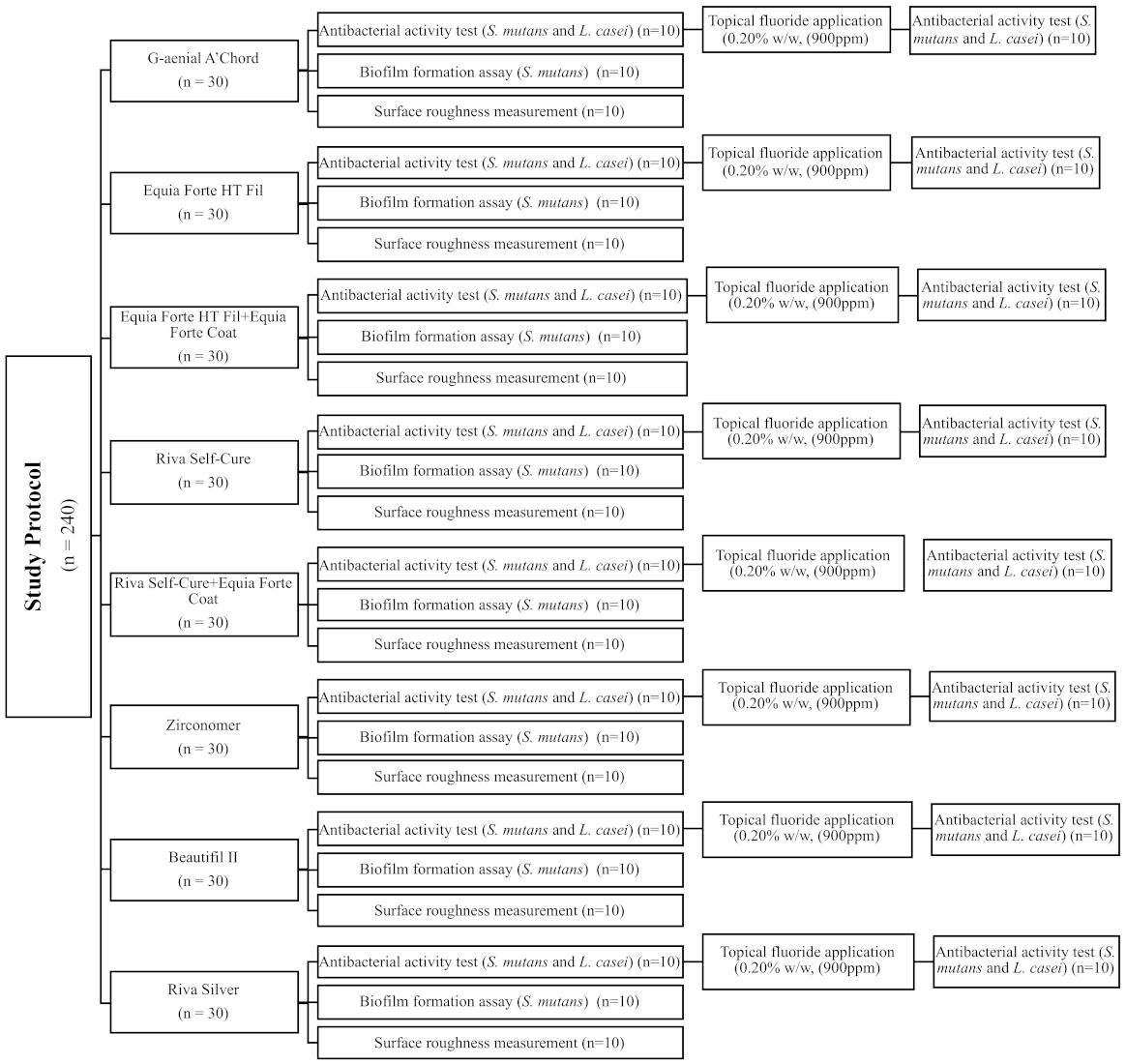
2. Materials and Methods

The tested materials are presented in Table 1 and the test protocol is illustrated in Figure 1.

2.1. Sample-Size Calculation

G*Power software (Version 3.1, Heinrich—Heine Dusseldorf University, Dusseldorf, Germany) was used to calculate the sample size with a 95% confidence interval, 85% power, and 0.50 effect size values according to one-way ANOVA-type power analysis. For each group, a minimum of 9 specimens were found to be appropriate. Therefore, 10 specimens per group was used.

2.2. Specimen Preparation

Two GHs (Equia Forte HT Fil and Riva Self-Cure), a zirconia-reinforced GIC (Zirconomer), a silver-reinforced GIC (Riva Silver), and a giomer (Beautifil II) were used in the study. Both GHs were tested either with coating as recommended by the manufacturers or without coating to test if the coating would affect their antibacterial activity. A nanohybrid resin composite was used as control.
A customized cylindrical Teflon mold (2 × 8 mm) was used for the preparation of the specimens. The Teflon mold was placed on a glass plate and the bottom surface was covered with a Mylar strip (S.S. White Limited, Middx, UK). Specimens were prepared according to the manufacturers’ instructions as follows:
Equia Forte HT Fil (n:30): Equia Forte HT capsule was activated and mixed in auto-mixer (Softly, Satelec Acteon, Merignac Cedex, France). The material was immediately inserted into the mold, covered with a Mylar strip on the top, and then condensed against a glass plate with a constant finger pressure for the duration of the setting process (2 min, 30 s).
Equia Forte HT Fil with coating (n:30): The specimens in this group were prepared as described above and, when the setting was completed, Equia Forte Coat was applied using a micro-tip applicator without air blowing, and light-cured for 20 s using an LED curing unit (440–480 nm,1500 mW/cm2, Radii plus, SDI, Victoria, Australia).
Riva Self-Cure (n:30): After the capsule was mixed for 10 s in auto-mixer, it was placed into the Teflon mold, covered with the strip on the top, and then condensed against a glass plate with a constant finger pressure for the duration of the setting process (2 min).
Riva Self-Cure with coating (n:30): The specimens were prepared and coated as described above.
Zirconomer (n:30): The specimens in this group were hand-mixed. Powder/liquid ratio was 3.6/1.0 (2 scoops:1 drop), working time was 1 min 30 s (from start of mixing), and setting time was 3 min.
Riva Silver (n:30): After the capsule was activated, it was mixed in auto-mixer for 10 s and transferred into the mold with its own applicator. Then, it was covered with a Mylar strip with slight pressure. Working time was 1 min and 30 s and setting time was 5 min.
Beautifil II (n:30): The giomer restorative was inserted into the mold with a flat-surface hand instrument, covered with strip, and polymerized using an LED curing unit for 20 s.
G-Aenial A’Chord (n:30): The specimens of this group were packed into the molds by compressing between two glass slides and Mylar strips and then polymerized for 20 s.
All specimens were polished immediately using a sequence of aluminum-oxide discs (medium, fine, and superfine) (Sof-Lex, 3M ESPE, MN, USA) for 30 s, at 20,000 rpm. Specimens were washed with distilled water and air-dried for 10 s after each step. Discs were replaced with new ones after every three specimens to obtain surfaces with homogeneous characteristics. Following polishing, specimens were sterilized in an autoclave at 121 °C for 15 min and directly subjected to antibacterial activity [23].

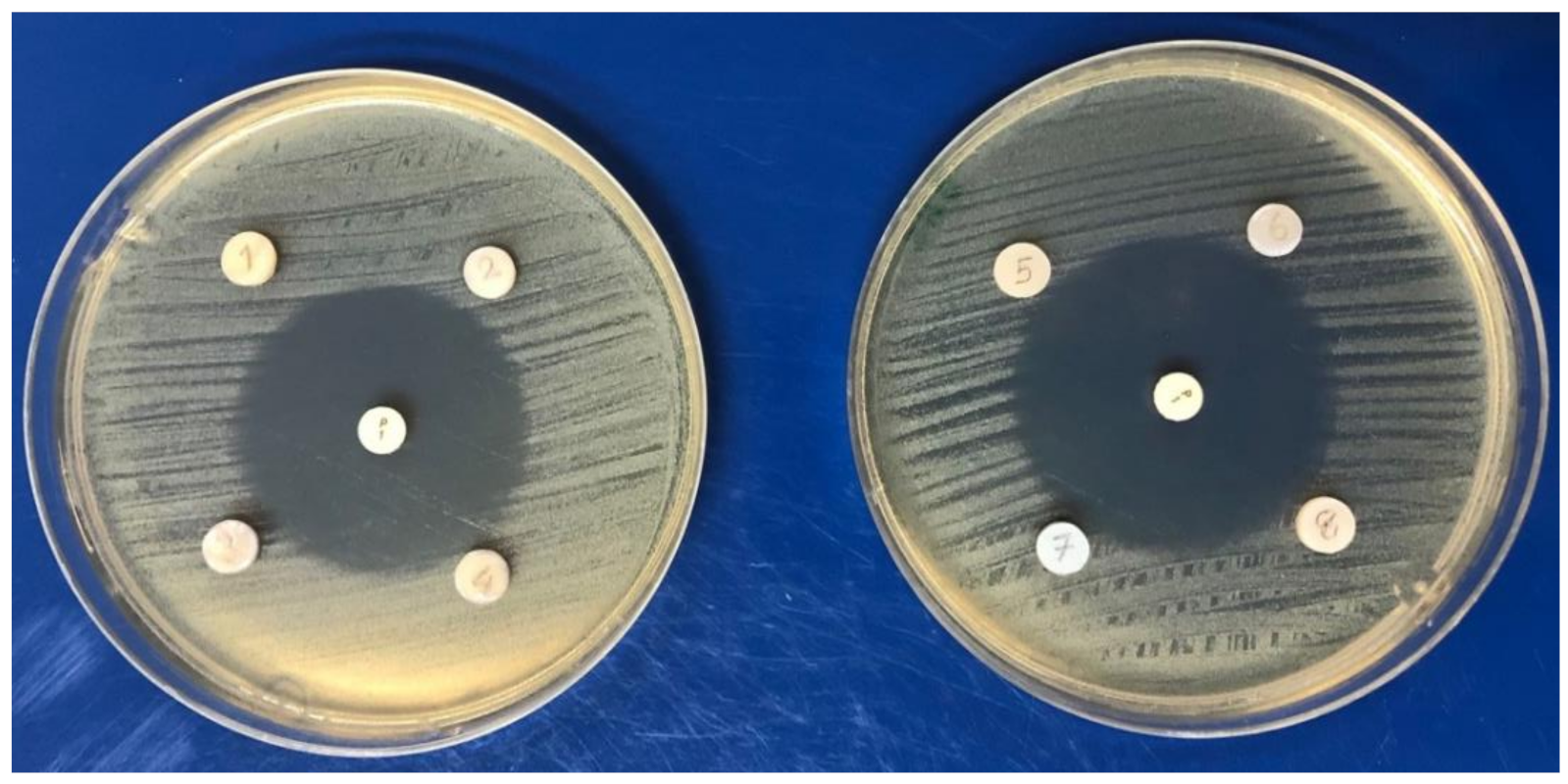
3. Antibacterial Activity Test

S. mutans ATCC 25175 and Lactobacillus casei (L. casei) ATCC 393 were used as test bacteria. Bacteria were cultured on brain heart infusion (BHI) agar (Oxoid, Hampshire, UK) and incubated at 37 °C with 5% CO2 for 48 h. Antibacterial activity of the restorative materials against test bacteria was determined by the agar diffusion method [24,25]. Suspensions of the bacteria were prepared in sterile saline (0.85% NaCl) and adjusted to McFarland 0.5 turbidity standard. The bacterial suspensions were spread on the surface of BHI agar using sterile swabs. Then, the restorative materials were placed on the surface of the inoculated agars using a sterile tweezer as follows: (1) Riva Self-cure, (2) Equia Forte HT Fil, (3) Riva Self-Cure with coating, and (4) Equia Forte HT Fil with coating were placed in one agar plate, and (5) Zirconomer, (6) G-aenial A’Chord, (7) Riva Silver, and (8) Beautifil II in another agar plate. Penicillin G,1U (Bioanalyse, Ankara/Turkey) was added to each plate as a positive control of the experiments.
The plates were incubated at 37 °C with 5% CO2 for 16–18 h. After the incubation period, the diameters of the inhibition zones were measured. The tests were performed in triplicate. As no growth inhibition was defined with all tested restorative materials, MI Paste Plus (GC, Tokyo, Japan) (The level of fluoride is 0.20% w/w, (900 ppm)) was applied to the surface of each restorative material with a cotton tip for 3 min according to the instructions of the manufacturer, to provide additional fluoride supply or to recharge. After 30 min, the specimens were sterilized again and the tests were repeated.

4. Biofilm Formation Assay

Specimens subjected to biofilm formation assay were sterilized in autoclave. S. mutans ATCC 25175 was used as test bacterium. The bacterium was cultured on BHI agar and incubated at 37 °C with 5% CO2 for 48 h. Sterile specimens were placed into 24-well plates separately. Then, each well containing sterile specimen was filled with 100 μL of filter-sterilized artificial saliva and incubated at 37 °C with 5% CO2 for 2 h. The artificial saliva prepared according to Hahnel et al.’s formula [26] allows a reproduction of the average electrolytic composition of human whole saliva. The total amount of biofilm formation (n = 10 for each group) was quantified by the crystal violet assay [27]. In the first step, the bacterial suspension was adjusted to McFarland 0.5 turbidity standard in Tryptic Soy Broth (TSB) supplemented with 4% sucrose. Then, 100 μL of bacterial suspension was added onto each specimen except for the control group. Only sterile tryptic-soy broth (TSB) supplemented with 4% sucrose was found in the wells representing the control group. All specimens were incubated at 37 °C with 5% CO2 for 24 h. After the incubation period, the specimens were removed into new 24-well plates and 100 μL 0.5% crystal violet solution was added to each well for staining the biofilm cells. After 30 min, the planktonic cells were removed by rinsing the wells three times with phosphate-buffered saline (PBS) and the specimens were removed into new 24-well plates. Then, acetone-ethanol (30:70 v/v) solution was added to the wells to dissolve bound dye within the biofilm matrix. All these steps were also applied to control groups. The optical density of the dissolved crystal violet dye was measured by a microplate reader (Thermo Scientific Multiskan GO Microplate Spectrophotometer, Vantaa, Finland) at 620 nm (OD620nm). Each optical density value of the biofilm group was subtracted from the optical density of the respective controls which were without bacteria.

5. Surface Roughness (Ra) Measurement

A contact type profilometer (Perthometer M2, Mahr GmbH, Gottingen, Germany) was used to record the surface roughness of the specimens. Five Ra readings were recorded for each specimen and the values were averaged. The profilometer was calibrated after three readings.

6. Statistical Analysis

SPSS 23 (SPSS Inc., Chicago, IL, USA) was used to perform statistical analysis. The Shapiro–Wilk test indicated the normal distribution of the surface roughness data (p > 0.05) and the Levene test indicated the homogeneity of the variances (p > 0.05). One-way analysis of variance (ANOVA) was used to compare the materials in terms of surface roughness (p < 0.001), followed by Tukey’s HSD test for multiple comparisons. The data of biofilm formation were not normally distributed (p < 0.05). Thus, the Kruskal–Wallis H test was used to compare the materials in terms of biofilm formation (p < 0.001), followed by Dunn’s test for multiple comparisons. p < 0.05 was considered as significant.

7. Results

No inhibition zones were observed in any tested restorative materials. Antibacterial activity was also not detected in any of the restorative materials tested after the application of 0.20% w/w (900 ppm) sodium fluoride. However, a 40 mm inhibition zone was observed with the Penicillin G,1U. (Figure 2).
The mean, median, and min–max of biofilm accumulated on the restorative materials are presented in Table 2. Significant differences were found among the groups (p < 0.005, Kruskal–Wallis test). Equia Forte HT Fil with and without coating showed the lowest, while Riva Self-Cure without coating and Zirconomer showed the highest biofilm accumulation. Application of coating did not result in significant difference either for Equia Forte HT or Riva Self-Cure (Dunn’s test, p = 0.999, p = 0.07, respectively). Coated Riva Self-Cure specimens showed lower biofilm accumulation than non-coat specimens.
The mean, median, and min–max of Ra values of the restorative materials are presented in Table 3. Significant differences were found among the groups (one-way ANOVA test, p < 0.005). G-aenial A’Chord showed the lowest, while Riva Silver, Zirconomer, Riva Self-Cure with and without coating showed the highest Ra values. There was not a significant difference either between coated and non-coated Equia Forte HT Fil or Riva Self-Cure with/without coating.

8. Discussion

This study determined the antibacterial efficacy of various new-generation hybrid/fluoride-releasing materials used in restorative dentistry by examining the zone of inhibition around the tested specimens in the culture plates and biofilm inhibition of these materials.
Since their introduction, antibacterial activity of GICs has attracted the interest of researchers [15,16,28,29,30]. However, the methodology of these studies showed a large variety that resulted in difficulty to compare. Most studies were carried out by agar diffusion method [16,26,28,29,30], whereas some used Direct Contact Test (DCT) [31,32,33]. There were also differences in the study periods. Short-term studies (24 and/or 48 h) were conducted predominantly, while a few studies were over a longer period of time [15,31,32].
Several factors might have an impact on the antibacterial activity of GICs, including chemical composition, fluoride release and low pH throughout the setting reaction [16,29,30,33,34]. It was concluded in the literature that each of the investigated GICs releases fluoride ions; however, the amount of release varies [35,36]. GICs have a low pH during setting reaction, lasting from several minutes to twenty-four hours [29,30,34]. Studies highlighted the importance of their antibacterial activity, resulting largely from the release of fluoride ions [33,36].
The results of the present study showed that none of the restorative materials tested inhibited the growth of either S. mutans or L. casei for 16–18 h after mixing. This might be due to the susceptibility of the tested bacteria species to fluoride ions. Therefore, the first null hypothesis that there would be no difference in the antibacterial activity of fluoride-releasing materials was not rejected.
The lack of bacterial growth inhibition by fluoride-releasing restoratives has also been shown in the literature [20,22]. DeSchepper et al. [34] and Herrera et al. [20] reported that the effect of fluoride activity on bacterial cells is dependent not only on the amount of ions but also on the pH of the material at the time of bonding, which might explain the differences in findings obtained with the materials tested. Hotwani et al. [37] compared the antibacterial potential of Fuji II LC and a giomer and reported that the antibacterial effect of the giomer against S. Mutans was superior to Fuji II LC. Conversely, Botelho [15] reported no antibacterial activity of Fuji IX against the tested bacteria.
The anticariogenic effect of a fluoride-releasing material depends on the sustainability of fluoride release and the level of reduction in fluoride release over time [20]. Previous studies emphasized that GICs are capable of recharge when treated with fluoride-containing mouth rinses or dentifrices [38,39]. To the extent of the authors’ knowledge, the effect of recharging the fluoride-releasing restorative materials with fluoride on their antibacterial activities has not yet been investigated. In the present study, as none of the tested fluoride-releasing materials showed antibacterial activity, all specimens were treated with a fluoride (900 ppm)-containing topical agent to provide additional fluoride supply or to recharge. However, for all materials, again, no inhibition zones were observed after topical fluoride application.
Biofilm on the surface of restorative material leads to the development of secondary caries that will endanger the long-term survival of the restoration. Therefore, a restorative material having a low sensitivity to bacterial adhesion is always preferred [40]. Biofilm formation can be measured quantitatively and qualitatively with different methods such as CFU counting, SEM, and CLSM after staining of the bacteria [26,41,42]. Nevertheless, all these techniques have some limitations and disadvantages. In the present study, crystal violet assay was used to evaluate biofilm formation. In vivo and in vitro studies conducted on bacterial adhesion to dental restoratives have also shown some differences [42,43]. These differences were reported to be related to the chemical structure and surface properties of the restorative materials [40].
In the present study, the fluoride-releasing materials were also tested in terms of 24 h biofilm formation. The same amount of inoculum and equal quantities were valid for all the specimens, but significant differences were detected in the biofilm formation of the materials. Equia Forte HT Fil either with or without coating showed the lowest biofilm formation. It was thought that, apart from fluoride, factors related to the materials have been involved in S. mutans biofilm formation on the tested restorative materials. Therefore, the second null hypothesis was rejected.
Although it has been well proven that the interaction of surface properties of a restorative material and biofilm formation can be influenced by the exploratory conditions of an in vitro study, it might also be assumed that the surface roughness has a significant effect on initial microbial adhesion [44,45]. Generally, rough surfaces are very attractive for bacteria with adhesion ability, as the contact area between the bacterial cell and the material is increased, as well as contribution to micromechanical retention [46]. A roughness (Ra) of 0.2 µm was reported to be the threshold for maximum reduction in bacterial adhesion on the surface of restorations [45]. However, bacterial adhesion or biofilm formation depends on not only the surface roughness of the material, but also on the shape and size of the bacterial cells and other environmental variables [45].
In the present study, the surface roughness values of all fluoride-releasing materials were higher than the 0.2 µm threshold with significant differences, leading to rejection of third null hypothesis. Therefore, it was not possible to establish a correlation between biofilm formation and surface roughness. The differences in the results may be due to different material compositions, culture conditions, and bacterium strain used in the test.
Finishing and polishing protocols can alter surface properties of the restorations, thus either promoting or inhibiting/decreasing the adhesion of oral bacteria [40]. From a clinical standpoint, it is always necessary to carry out finishing and polishing protocols. So, in the present study, a polishing technique, simulating a standard clinical procedure using aluminum-oxide discs with decreasing grain sizes was performed to prepare standardized surfaces. Although the resin composite using G-aenial A’Chord as control showed the lowest Ra value, significantly lower S. mutans biofilm formation was found on the surface of the GH Equia Forte HT Fil. Thus, the biofilm formation appears to be associated not with the surface roughness threshold, but largely dependent on the material properties.
The present study had some limitations. Firstly, different fluoride-releasing materials were tested. Results cannot be generalized to materials with different chemical structures. Secondly, even though the specimens were treated with artificial saliva to simulate the oral conditions, due to the use of only one type of bacteria and static technique used for biofilm measurement, it was not possible to entirely mimic intraoral cavity in laboratory conditions. Nevertheless, the authors are aware of the laboratory character as well as the inclusion of a limited number of bacteria. Biofilms on the teeth and restorations in the oral cavity contain clustered different bacterial strains embedded into a complex extracellular matrix. Thus, it is clear that the analysis of biofilm formation on restorative materials needs to be validated by clinical approaches that are assumed as gold standard.

9. Conclusions

Within the limitations of this in vitro study, it can be concluded:
  • The tested new-generation hybrid/fluoride-releasing dental materials show no antibacterial activity.
  • These materials are not able to develop antibacterial properties even after treatment with additional topical fluoride.
  • Biofilms accumulate to different extents on modern hybrid/fluoride-releasing restorative materials.
  • This accumulation does not depend on the surface roughness of these materials.

Author Contributions

Conceptualization: S.G., H.T. and I.M.; methodology: S.G. and S.S.G.; software: C.A.; validation: I.M.; formal analysis: U.K.V.; investigation: S.S.G.; resources: H.T.; data curation: U.K.V.; writing—review and editing: S.G.; visualization: C.A.; project administration: S.G. All authors have read and agreed to the published version of the manuscript.

Funding

This research received no external funding.

Institutional Review Board Statement

Not applicable.

Informed Consent Statement

Not applicable.

Data Availability Statement

Not applicable.

Conflicts of Interest

The authors declare no conflict of interest.

References

  1. Ismail, A.I.; Hasson, H.; Sohn, W. Dental caries in the second millennium. J. Dent. Educ. 2001, 65, 953–959. [Google Scholar] [CrossRef] [PubMed]
  2. Puy, L.; Carmen, M. Evidence concerning the medical management of caries. Med. Oral Patol. Oral Cir. Bucal 2008, 13, E325–E330. [Google Scholar]
  3. Tuzuner, T.; Dimkov, A.; Nicholson, J.W. The effect of antimicrobial additives on the properties of dental glass-ionomer cements: A review. Acta Biomater. Odontol. Scand. 2019, 5, 9–21. [Google Scholar] [CrossRef] [PubMed] [Green Version]
  4. Beighton, D. The complex oral microflora of high-risk individuals and groups and its role in the caries process. Community Dent. Oral Epidemiol. 2005, 33, 248–255. [Google Scholar] [CrossRef] [PubMed]
  5. Hoszek, A.; Ericson, D. In vitro fluoride release and the antibacterial effect of glass ionomers containing chlorhexidine gluconate. Oper. Dent. 2008, 33, 696–701. [Google Scholar] [CrossRef] [Green Version]
  6. Dasgupta, S.; Saraswathi, M.V.; Somayaji, K.; Pentapati, K.C.; Shetty, P. Comparative evaluation of fluoride release and recharge potential of novel and traditional fluoride-releasing restorative materials: An in vitro study. J. Conserv. Dent. 2018, 21, 622–626. [Google Scholar]
  7. De Castilho, A.R.; Duque, C.; Negrini Tde, C.; Sacono, N.T.; de Paula, A.B.; Sacramento, P.A.; de Souza Costa, C.A.; Spolidorio, D.M.; Puppin-Rontani, R.M. Mechanical and biological characterization of resin-modified glass-ionomer cement containing doxycycline hyclate. Arch. Oral Biol. 2012, 57, 131–138. [Google Scholar] [CrossRef]
  8. Gurgan, S.; Kutuk, Z.B.; Ergin, E.; Oztas, S.S.; Cakir, F.Y. Clinical performance of a glass ionomer restorative system: A 6-year evaluation. Clin. Oral Investig. 2017, 21, 2335–2343. [Google Scholar] [CrossRef]
  9. Asmussen, E.; Peutzfeldt, A. Long-term fluoride release from a glass ionomer cement, a compomer, and from experimental resin composites. Acta Odontol. Scand. 2002, 60, 93–97. [Google Scholar] [CrossRef]
  10. Yi, J.; Dai, Q.; Weir, M.D.; Melo, M.A.S.; Lynch, C.D.; Oates, T.W.; Zhang, K.; Zhao, Z.; Xu, H.H.K. A nano-CaF2-containing orthodontic cement with antibacterial and remineralization capabilities to combat enamel white spot lesions. J. Dent. 2019, 89, 103172. [Google Scholar] [CrossRef]
  11. Garcia-Contreras, R.; Scougall-Vilchis, R.J.; Contreras-Bulnes, R.; Sakagami, H.; Morales-Luckie, R.A.; Nakajima, H. Mechanical, antibacterial and bond strength properties of nano-titanium-enriched glass ionomer cement. J. Appl. Oral Sci. 2015, 23, 321–328. [Google Scholar] [CrossRef] [Green Version]
  12. Tiwari, S.; Kenchappa, M.; Bhayya, D.; Gupta, S.; Saxena, S.; Satyarth, S.; Singh, A.; Gupta, M. Antibacterial activity and fluoride release of glass-ionomer cement, compomer and zirconia reinforced glass-ionomer cement. J. Clin. Diagn. Res. 2016, 10, ZC90–ZC93. [Google Scholar] [CrossRef]
  13. Kimyai, S.; Bahari, M.; Naser-Alavi, F.; Behboodi, S. Effect of two different tooth bleaching techniques on microhardness of giomer. J. Clin. Exp. Dent. 2017, 9, e249–e253. [Google Scholar] [CrossRef]
  14. Kooi, T.J.; Tan, Q.Z.; Yap, A.U.; Guo, W.; Tay, K.J.; Soh, M.S. Effects of food-simulating liquids on surface properties of giomer restoratives. Oper. Dent. 2012, 37, 665–671. [Google Scholar] [CrossRef] [Green Version]
  15. Botelho, M.G. Inhibitory effects on selected oral bacteria of antibacterial agents incorporated in a glass ionomer cement. Caries Res. 2003, 37, 108–114. [Google Scholar] [CrossRef]
  16. Herrera, M.; Castillo, A.; Bravo, M.; Liebana, J.; Carrion, P. Antibacterial activity of resin adhesives, glass ionomer and resin-modified glass ionomer cements and a compomer in contact with dentin caries samples. Oper. Dent. 2000, 25, 265–269. [Google Scholar]
  17. Hosida, T.Y.; Delbem, A.C.B.; Morais, L.A.; Moraes, J.C.S.; Duque, C.; Souza, J.A.S.; Pedrini, D. Ion release, antimicrobial and physio-mechanical properties of glass ionomer cement containing micro or nanosized hexametaphosphate, and their effect on enamel demineralization. Clin. Oral Investig. 2019, 23, 2345–2354. [Google Scholar] [CrossRef]
  18. Naik, R.G.; Dodamani, A.S.; Khairnar, M.R.; Jadhav, H.C.; Deshmukh, M.A. Comparative assessment of antibacterial activity of different glass ionomer cements on cariogenic bacteria. Restor. Dent. Endod. 2016, 41, 278–282. [Google Scholar] [CrossRef]
  19. Dimkov, A.; Gjorgievska, E.; Nicholson, J.W.; Kaftandzieva, A. Antibacterial effects of conventional glass ionomer cement. Bratisl. Lek. Listy 2016, 117, 31–35. [Google Scholar] [CrossRef] [Green Version]
  20. Herrera, M.; Castillo, A.; Baca, P.; Carrion, P. Antibacterial activity of glass-ionomer restorative cements exposed to cavity-producing microorganisms. Oper. Dent. 1999, 24, 286–291. [Google Scholar]
  21. Sungurtekin-Ekci, E.; Ozdemir-Ozenen, D.; Duman, S.; Acuner, I.C.; Sandalli, N. Antibacterial surface properties of various fluoride-releasing restorative materials in vitro. J. Appl. Biomater. Funct. Mater. 2015, 13, e169–e173. [Google Scholar] [CrossRef] [PubMed]
  22. Meiers, J.C.; Miller, G.A. Antibacterial activity of dentin bonding systems, resin-modified glass ionomers, and polyacid-modified composite resins. Oper. Dent. 1996, 21, 257–264. [Google Scholar] [PubMed]
  23. Souza, J.C.; Mota, R.R.; Sordi, M.B.; Passoni, B.B.; Benfatti, C.A.; Magini, R.S. Biofilm formation on different materials used in oral rehabilitation. Braz. Dent. J. 2016, 27, 141–147. [Google Scholar] [CrossRef]
  24. Luczaj-Cepowicz, E.; Marczuk-Kolada, G.; Zalewska, A.; Pawinska, M.; Leszczynska, K. Antibacterial activity of selected glass ionomer cements. Postepy Hig. Med. Dosw. 2014, 68, 23–28. [Google Scholar] [CrossRef] [PubMed]
  25. Yesilyurt, C.; Er, K.; Tasdemir, T.; Buruk, K.; Celik, D. Antibacterial activity and physical properties of glass-ionomer cements containing antibiotics. Oper. Dent. 2009, 34, 18–23. [Google Scholar] [CrossRef] [PubMed] [Green Version]
  26. Hahnel, S.; Ionescu, A.C.; Cazzaniga, G.; Ottobelli, M.; Brambilla, E. Biofilm formation and release of fluoride from dental restorative materials in relation to their surface properties. J. Dent. 2017, 60, 14–24. [Google Scholar] [CrossRef] [PubMed]
  27. Wongpraparatana, I.; Matangkasombut, O.; Thanyasrisung, P.; Panich, M. Effect of vital tooth bleaching on surface roughness and streptococcal biofilm formation on direct tooth-colored restorative materials. Oper. Dent. 2018, 43, 51–59. [Google Scholar] [CrossRef]
  28. Duque, C.; Negrini Tde, C.; Hebling, J.; Spolidorio, D.M. Inhibitory activity of glass-ionomer cements on cariogenic bacteria. Oper. Dent. 2005, 30, 636–640. [Google Scholar]
  29. Scherer, W.; Lippman, N.; Kaim, J. Antimicrobial properties of glass-ionomer cements and other restorative materials. Oper. Dent. 1989, 14, 77–81. [Google Scholar]
  30. Vermeersch, G.; Leloup, G.; Delmee, M.; Vreven, J. Antibacterial activity of glass-ionomer cements, compomers and resin composites: Relationship between acidity and material setting phase. J. Oral Rehabil. 2005, 32, 368–374. [Google Scholar] [CrossRef]
  31. Lewinstein, I.; Matalon, S.; Slutzkey, S.; Weiss, E.I. Antibacterial properties of aged dental cements evaluated by direct-contact and agar diffusion tests. J. Prosthet. Dent. 2005, 93, 364–371. [Google Scholar] [CrossRef]
  32. Perez, C.R.; Hirata, R., Jr.; Sergio, P.P. Evaluation of antimicrobial activity of fluoride-releasing dental materials using a new in vitro method. Quintessence Int. 2003, 34, 473–477. [Google Scholar]
  33. Boeckh, C.; Schumacher, E.; Podbielski, A.; Haller, B. Antibacterial activity of restorative dental biomaterials in vitro. Caries Res. 2002, 36, 101–107. [Google Scholar] [CrossRef]
  34. DeSchepper, E.J.; White, R.R.; von der Lehr, W. Antibacterial effects of glass ionomers. Am. J. Dent. 1989, 2, 51–56. [Google Scholar]
  35. Ariffin, Z.; Ngo, H.; McIntyre, J. Enhancement of fluoride release from glass ionomer cement following a coating of silver fluoride. Aust. Dent. J. 2006, 51, 328–332. [Google Scholar] [CrossRef] [Green Version]
  36. Hsu, H.M.; Huang, G.F.; Chang, H.H.; Wang, Y.L.; Guo, M.K. A continuous flow system for assessing fluoride release/uptake of fluoride-containing restorative materials. Dent. Mater. 2004, 20, 740–749. [Google Scholar] [CrossRef]
  37. Hotwani, K.; Thosar, N.; Baliga, S.; Bundale, S.; Sharma, K. Antibacterial effects of hybrid tooth colored restorative materials against Streptococcus mutans: An in vitro analysis. J. Conserv. Dent. 2013, 16, 319–322. [Google Scholar] [CrossRef]
  38. Naoum, S.; Ellakwa, A.; Martin, F.; Swain, M. Fluoride release, recharge and mechanical property stability of various fluoride-containing resin composites. Oper. Dent. 2011, 36, 422–432. [Google Scholar] [CrossRef]
  39. Ionescu, A.; Brambilla, E.; Hahnel, S. Does recharging dental restorative materials with fluoride influence biofilm formation? Dent. Mater. 2019, 35, 1450–1463. [Google Scholar] [CrossRef]
  40. Dutra, D.; Pereira, G.; Kantorski, K.Z.; Valandro, L.F.; Zanatta, F.B. Does finishing and polishing of restorative materials affect bacterial adhesion and biofilm formation? A systematic review. Oper. Dent. 2018, 43, E37–E52. [Google Scholar] [CrossRef]
  41. Anami, L.C.; Pereira, C.A.; Guerra, E.; Assuncao e Souza, R.O.; Jorge, A.O.; Bottino, M.A. Morphology and bacterial colonisation of tooth/ceramic restoration interface after different cement excess removal techniques. J. Dent. 2012, 40, 742–749. [Google Scholar] [CrossRef]
  42. Hannig, C.; Hannig, M.; Rehmer, O.; Braun, G.; Hellwig, E.; Al-Ahmad, A. Fluorescence microscopic visualization and quantification of initial bacterial colonization on enamel in situ. Arch. Oral Biol. 2007, 52, 1048–1056. [Google Scholar] [CrossRef] [PubMed]
  43. Wang, S.P.; Ge, Y.; Zhou, X.D.; Xu, H.H.; Weir, M.D.; Zhang, K.K.; Wang, H.H.; Hannig, M.; Rupf, S.; Li, Q.; et al. Effect of anti-biofilm glass-ionomer cement on Streptococcus mutans biofilms. Int. J. Oral Sci. 2016, 8, 76–83. [Google Scholar] [CrossRef] [Green Version]
  44. Bollen, C.M.; Lambrechts, P.; Quirynen, M. Comparison of surface roughness of oral hard materials to the threshold surface roughness for bacterial plaque retention: A review of the literature. Dent. Mater. 1997, 13, 258–269. [Google Scholar] [CrossRef]
  45. Song, F.; Koo, H.; Ren, D. Effects of Material Properties on Bacterial Adhesion and Biofilm Formation. J. Dent. Res. 2015, 94, 1027–1034. [Google Scholar] [CrossRef] [PubMed]
  46. Anselme, K.; Davidson, P.; Popa, A.M.; Giazzon, M.; Liley, M.; Ploux, L. The interaction of cells and bacteria with surfaces structured at the nanometre scale. Acta Biomater. 2010, 6, 3824–3846. [Google Scholar] [CrossRef] [PubMed]
Figure 1. Study Protocol.
Applsci 12 02434 g001
Figure 2. Fluoride-releasing restorative materials and the resin composite inoculated on BHI agar plates: Penicillin G,1U at the middle and 1—Riva Self-Cure, 2—Equia Forte HT Fil, 3—Riva Self-Cure with coating, 4—Equia Forte HT Fil with coating, 5—Zirconomer, 6—G-aenial A’Chord, 7—Riva Silver, 8—Beautifil II.
Applsci 12 02434 g002
Table 1. Descriptions of the materials used in the study.
MaterialMaterial TypeCompositionManufacturer
G-aenial A’ChordNano hybrid resin compositeBis-MEPP, Filler load: 82% by weight: glass-filler (300 nm barium glass) 16 nm (fumed silica), organic filler (300 nm barium glass; 16 nm fumed silica).GC Corp., Tokyo, Japan
Equia Forte HT FilBulk-fill glass hybridFluoroaluminosilicate glass, polyacrylic acid, iron oxide
polybasic carboxylic acid, water
GC Corp., Tokyo, Japan
Riva Self-CureBulk-fill glass hybridStrontium Fluoro-aluminosilicate glass, polyacrylic acid copolymer powders, pigment, polyacrylic acid copolymer, tartaric acidSDI, Victoria, Australia
ZirconomerZirconia-reinforced glass ionomerAlumino-fluoro-silicate glass, zirconium oxide, tartaric acid, polyacrylic acid, deionized waterShofu INC, Kyoto, Japan
Beautifil IIGiomerBis-GMA 7.5%, triethylenglycol dimethacrylate 5%, aluminofluoro-borosilicate glass 7.5%, Al2O3, DL-camphorquinoneShofu Dental, Kyoto, Japan
Riva SilverSilver-reinforced glass ionomerPolyacrylic acid, tartaric acid, balancing component, alloy powderSDI, Victoria, Australia
Equia Forte coatLight-cured resin coatingUrethane methacrylate, methyl methacrylate, camphorquinone, colloidal silica, phosphoricester monomerGC Corp., Tokyo, Japan
MI Paste Plus StrawberryTopical crème with calcium, phosphate, and fluoridePure water, glycerol, CPP-ACP, D-sorbitol, CMC-Na, propylene glycol, silicon dioxide, titanium dioxide, xylitol, phosphoric acid, sodium fluoride, flavoring, sodium saccharin, ethyl p-hydroxybenzoate, propyl, p-hydroxybenzoate, butyl p-hydroxybenzoateGC Corp., Tokyo, Japan
Table 2. Amount of biofilm (OD620nm) accumulated on the fluoride-releasing restorative materials (n:10).
MaterialsMean ± SDMedianMin–Maxp
G-aenial A’Chord0.096 ± 0.020 ab0.1050.057–0.109<0.005
Equia Forte HT Fil without coating0.056 ± 0.031 a0.0700.011–0.087
Equia Forte HT Fil with coating0.019 ± 0.014 a0.0120.005–0.042
Riva Self-Cure without coating0.226 ± 0.078 b0.2170.151–0.352
Riva Self-Cure with coating0.081 ± 0.057 ab0.0810.019–0.192
Zirconomer0.202 ± 0.068 b0.2020.070–0.269
Beautifil II0.110 ± 0.038 ab0.0990.070–0.183
Riva Silver0.127 ± 0.082 ab0.1140.057–0.295
a and b imply significant differences.
Table 3. The Mean (±SD), Median, and Min–Max Ra values (µm) of the fluoride-releasing materials (n:10).
MaterialsMean ± SDMedianMin–Maxp
G-aenial A’Chord0.163 ± 0.123 a0.2400.090–0.469<0.005
Equia Forte HT Fil without coating0.591 ± 0.302 b0.4900.206–1.005
Equia Forte HT Fil with coating0.586 ± 0.305 b0.5580.300–1.201
Riva Self-Cure without coating1.353 ± 0.309 c1.3530.734–1.662
Riva Self-Cure with coating1.260 ± 0.341 c1.2600.896–1.918
Zirconomer1.534 ± 0.250 c1.6491.206–1.801
Beautifil II0.455 ± 0.182 b0.4800.174–0.653
Riva Silver1.713 ± 0.273 c1.7131.358–2.130
a, b, and c imply significant differences.
Publisher’s Note: MDPI stays neutral with regard to jurisdictional claims in published maps and institutional affiliations.

