Article

Microwave- and Ultrasound-Assisted Extraction of Cucurbita pepo Seeds: A Comparison Study of Antioxidant Activity, Phenolic Profile, and In-Vitro Cells Effects

by
Catarina Macedo
,
Ana Margarida Silva
,
Ana Sofia Ferreira
,
Manuela M. Moreira
,
Cristina Delerue-Matos
and
Francisca Rodrigues
*
REQUIMTE/LAQV-Polytechnique of Porto-School of Engineering, Rua Dr. António Bernardino de Almeida, 431, 4249-015 Porto, Portugal
*
Author to whom correspondence should be addressed.
Appl. Sci. 2022, 12(3), 1763; https://doi.org/10.3390/app12031763
Submission received: 11 January 2022 / Revised: 4 February 2022 / Accepted: 7 February 2022 / Published: 8 February 2022
(This article belongs to the Special Issue Advances in Natural Bioactive Compounds and Biological Effects)

Abstract

:
Nowadays there is a growing demand for nutraceuticals to prevent diseases related to redox imbalances, such as atherosclerosis and diabetes, being crucial to search for new matrixes rich in bioactive compounds. This work aims to characterize the value-added compounds extracted from Curcubita pepo seeds using green methodologies, namely microwave-assisted extraction (MAE) and ultrasound-assisted extraction (UAE), employing water as an extracting solvent for two ratios (condition 1: 1 mg/20 mL; condition 2: 2.5 mg/20 mL). The extract with the best antioxidant/antiradical activity in FRAP (71.09 μmol FSE/g DW) and DPPH (5.08 mg TE/g DW) assays was MAE condition 1, while MAE condition 2 exhibited the highest activity in the ABTS assay (13.29 mg AAE/g DW) and TPC (16.89 mg GAE/g DW). A remarkable scavenging capacity was observed, particularly for HOCl, with IC50 values ranging from 1.88–13.50 μg/mL. A total of 21 phenolic compounds were identified, being catechin (4.567–7.354 mg/g DW), caffeine (1.147–2.401 mg/g DW) and gallic acid (0.945–1.337 mg/g DW) predominant. No adverse effects were observed on Caco-2 viability after exposure to MAE extracts, while the other conditions led to a slight viability decrease in NSC-34. These results highlighted that the extract from MAE condition 2 is the most promising as a potential nutraceutical ingredient.

1. Introduction

Nutraceuticals are bioactive compounds extracted from their original food matrix [1] that provide medical or health benefits by preventing or treating diseases [2]. In the past years, nutraceuticals received particular attention from consumers due to their potential to improve health, delay the aging process, increase life expectancy, prevent chronic diseases, and even support the structure or function of the body [3]. Different studies reported that nutraceuticals, such as ginseng, green tea, sumac, folic acid and cod liver oil, present promising results in different pathological complications, such as atherosclerosis [4,5], cardiovascular diseases [6,7], diabetes [8], cancer [9,10] and neurological disorders [11]. Furthermore, recently published reports demonstrated the positive effects of nutraceutical plants as Zizyphus jujube [12] or Lavandula officinalis [13] on the Alzheimer’s disease, learning and memory. Extracts from coneflowers or herbs of other plants, namely Echinacea angustfolia, E. pallida and E. purpurea, revealed to improve immune function and lower susceptibility to some diseases [3]. The ability of nutraceuticals to neutralize these conditions is often related with their capacity to restore redox balance [14,15,16].
The Cucurbitaceae family, also known as cucurbits, are a large group of almost 800 species that include squashes, pumpkins, melons and gourds [17]. Cucurbita pepo, also referred to as “summer squash” or “zucchini”, originates from Central and South America, being currently cultivated worldwide in warm regions [18]. It is the most produced species of the Cucurbitaceae family, and one of the oldest known cultivated species, with Mexican archaeological evidence dating from 7000 BC [19,20]. C. pepo is an economically important crop in which immature fruits, leaves, peel, blossoms, and seeds are consumed due to their nutritional and medicinal benefits [21].
The seeds of the Cucurbitaceae family are globally used for the treatment of different diseases, particularly due to their antiviral, anti-inflammatory, anti-ulcerative, antidiabetic and antioxidant activities as well as analgesic for urinary disorders [17]. A few studies using C. pepo seed extracts were published in the past years, revealing wound healing [22], hair growth promotion [23] and anthelmintic [24] properties, besides their ability to inhibit the cell growth of hyperplastic and cancer cells [25]. Moreover, the treatment with C. pepo extract conducted to a substantial improvement of the lower urinary tract symptoms that are suggestive of benign prostatic hyperplasia [26].
According to different authors, C. pepo seeds are rich in several bioactive compounds, such as polyphenols [27], which have anti-aging and anticarcinogenic effects and may protect against vascular inflammation and cardiovascular and neurodegenerative diseases [28,29,30,31]. α- and γ-tocopherols [32] are also present, defending cells from oxidative stress and inflammation [33], while carotenoids (e.g., β-carotene and β-cryptoxanthin) may protect against different chronic diseases, including cancer [34] and cardiovascular diseases [35]. Besides that, carotenoids improve the cognitive and visual functions [36]. In addition, C. pepo seeds are rich in polyunsaturated fatty acids [37], zinc [38], and phytosterols such as β-sitosterol [32], which may reduce the blood cholesterol [39] and decrease the risk of certain types of cancer [40]. β-sitosterol is also the bioactive compound responsible for the successful use of C. pepo seeds in the treatment of benign prostatic hyperplasia [41,42,43]. Berberine and palmatine are also present in considerable amounts [24], conferring to nematocidal [24], antimalarial [44], antileishmaniasis [45], anti-schistosomiasis [46] and Toxoplasma gondii inhibitory properties [47].
Nevertheless, most of the authors employed organic solvents in the extractive step or, at least, used cold pressing methods aiming to obtain the lipidic fraction. Therefore, the use of water as an alternative solvent led to a greener extraction process and, simultaneously, avoided the extraction of a significant amount of lipids that are naturally present in C. pepo seeds. Allied to the selection of less-polluting solvents, it is imperative to select eco-friendly extraction methods. Therefore, technologies such as microwave-assisted extraction (MAE) and ultrasound-assisted extraction (UAE) arise as an alternative to the traditional ones [48]. MAE is a conventional, automated green extraction technique that allows the extraction of active components from different matrices [49,50]. By using microwave energy, MAE heats the solvents in contact with samples, disrupts the cell membrane, and releases the intracellular components into the solvents [50,51]. This is opposite to the conventional methods, such as Soxhlet extraction, that often requires 12–24 h of extraction periods and hundreds of milliliters of organic solvents [50]. When compared to such methods, MAE requires shorter extraction times, decreasing the degradation of the extracted components as well as the costs and the volume of the solvents used, and therefore improving the purity of the final extracts [52,53,54,55,56].
On the other hand, UAE is also an environmentally friendly, simple, effective, and inexpensive technique, with applications in pharmaceutical, cosmetic, and alimentary fields that have become more popular since 2007 [57,58,59]. This sustainable technique uses ultrasounds waves, which induce cavitation and thermal and mechanical effects in the extraction medium, and disrupt the cell walls of the matrix [58]. This phenomenon leads to the release of intracellular components into the solvents, without producing considerable modifications in the structure and properties of the compounds [60]. Some advantages of this technique include the use of small amounts of matrixes and solvents, short extraction times and samples throughput increment [61]. UAE also avoids the thermal decomposition of heat sensitive compounds, since it is a non-thermal process [62]. Furthermore, UAE achieves higher extraction yields in comparison with maceration and Soxhlet extractions [63]. Overall, these methodologies are less time-consuming, easy to execute, low-cost and usually lead to higher extraction yields, being reported by different authors as successfully in the recovery of high-added value compounds from plants [64,65,66].
Considering these points, the main goal of this study was the extraction of value-added compounds from C. pepo seeds using two green extraction methods, namely MAE and UAE, and water as solvent. Besides that, aiming to evaluate the influence of the ratio on the bioactive composition of the extracts, two different amounts of sample were used, namely 1 g/20 mL (condition 1) and 2.5 g/20 mL (condition 2). Afterwards, the extracts were characterized regarding antioxidant activity, radical scavenging capacity, phenolic profile, and in vitro cellular effects, aiming to determine their potential use as ingredients for nutraceutical purposes. To the best of our knowledge, this is the first study that uses these techniques and solvent to valorize C. pepo seeds. The possibility of it being used as a nutraceutical ingredient was comprehensively evaluated by analyzing its antioxidant activity, radical scavenging capacity, phenolic profile, and in vitro cellular effects.

2. Materials and Methods

2.1. Chemicals

Trolox and 2,2-diphenyl-1-picryl-hydrazyl (DPPH) were obtained from Sigma-Aldrich (Steinheim, Germany). Folin–Ciocalteu’s reagent and gallic acid were purchased from Sigma Chemical Co. (St. Louis, MO, USA). Dulbecco’s Modified Eagle Medium (DMEM), Fetal Bovine Serum (FBS), Hank’s Balanced Salt Solution (HBSS), non-essential amino acids, penicillin, streptomycin, and trypsin–EDTA were obtained from Invitrogen Corporation (Life Technologies, S.A., Madrid, Spain). Dimethyl sulfoxide (DMSO) was supplied by AppliChem (Darmstadt, Germany).
HPLC solvents were provided by Sigma-Aldrich (Milan, Italy). Phenolic compounds’ individual standards used for the identification or quantification in extracts were purchased from Sigma-Aldrich (Steinheim, Germany) and their purity was at least above 95%.
The Caco-2 cell line was obtained from American Type Culture Collection (ATCC, USA). Mouse Motor Neuron-Like Hybrid cells (NSC-34 cell line) were obtained from Cedarlane (Hornby, ON, Canada).

2.2. Samples

C. pepo seeds were obtained from local producers in September 2021, Braga, Portugal. The seeds were dehydrated (Excalibur Food Dehydrator, Sacramento, CA, USA) at 41 °C for 24 h, grinded in a miller (Moulinex A320) and stored at 4 °C in the dark until further extraction.

2.3. Preparation of C. pepo Extracts

The MAE was performed on a MARS-X 1500 W (Microwave Accelerated Reaction System for Extraction and Digestion, CEM, Mathews, NC, USA), using closed Teflon extraction vessels. Samples of grinded C. pepo seeds were extracted with 20 mL of deionized water. Two different quantities of samples (1 mg/20 mL and 2.5 mg/20 mL) were used in order to determine the best ratio. Microwave power was fixed at 300 W and the extraction was performed at 25 °C for 30 min, with constant medium stirring, according to the procedure described by Silva et al. [67].
The UAE was accomplished using a Sonic Vibracell (model VC 750, Newtown, CT, USA), at room temperature for 30 min. Sonication was performed with a probe of 13 mm diameter with 40% of amplitude. The power was fixed at 750 W and the frequency at 20 kHz, as reported by Lameirão et al. [68].
After extraction, the solutions were filtrated through a Whatman No. 1 filter paper and frozen at −80 °C for subsequent lyophilization (Telstar, model Cryodos−80, Spain). After lyophilization, extracts were stocked at room temperature (20 °C) and kept in the dark. For the further experiments, the final residue was dissolved in deionized water, except for the DPPH assay, in which the extracts were dissolved in a hydroalcoholic (1:10, v/v) solution.
In the following sections, the nomenclature used will be MAE condition 1 (1 mg/20 mL), MAE condition 2 (2.5 mg/20 mL), UAE condition 1 (1 mg/20 mL) and UAE condition 2 (2.5 mg/20 mL). These ratios were based on preliminary studies performed by the research team (data not shown).

2.4. Total Phenolic Content (TPC)

The determination of the total phenolic content (TPC) was performed following the Folin–Ciocalteu procedure designed by Singleton and Rossi [69], with slight modifications. Gallic acid was used as standard for the calibration curve (linearity range = 5–100 μg/mL; R2 > 0.997). The results were expressed as milligrams of gallic acid equivalents (GAE) per gram of extract on dry weight (DW) (mg GAE/g DW).

2.5. In Vitro Antioxidant and Antiradical Activities

2.5.1. ABTS●+ Radical Scavenging Activity Assay

The extracts ABTS●+ scavenging capacity was directly assessed in a 96-well microplate as described by Re et al. [70], with minor modifications. Ascorbic acid was prepared as standard for the calibration curve (linearity range: 5–50 μg/mL; R2 > 0.981). The results were presented as mg ascorbic acid equivalents (AAE) per gram of extract on DW (mg AAE/g DW).

2.5.2. DPPH Radical Scavenging Activity Assay

The antiradical activity by scavenging of DPPH radicals was evaluated following the procedure previously described by Pinto et al. [71]. Trolox was the standard used for the calibration curve (linearity range: 5–75 μg/mL; R2 > 0.990). Results were expressed as mg of Trolox equivalents (TE) per gram of extract on DW (mg TE/g DW).

2.5.3. Ferric Reducing Antioxidant Power Assay

Ferric reducing antioxidant power (FRAP) was determined based on the reduction of a ferric complex (Fe3+-TPTZ) to the ferrous form (Fe2+-TPTZ) by antioxidants, according to Benzie and Strain [72], with minor modifications. The reaction mixture was incubated at 37 °C for 30 min and the absorbance was measured at 595 nm. The calibration curve was prepared with a solution of ferrous sulphate 1mM as standard (linearity range: 25–500 μM; R2 > 0.999). The results were expressed in μmol of ferrous sulphate equivalents (FSE) per gram of extract on DW (μmol FSE/g DW).

2.5.4. Reactive Oxygen Species Scavenging Capacity Assays

Hypochlorous Acid Scavenging Assay (HOCl)

The hypochlorous acid (HOCl) quenching capacity of C. pepo extracts and the positive controls (catechin and gallic acid) was determined according to the protocol described by Gomes et al. [73]. The HOCl solution was prepared using NaOCl 1% (w/v) and the pH was adjusted to 6.2 using H2SO4. The fluorescence signal was monitored for 5 min at 37 °C. Results were expressed as the inhibition, in IC50 (µg/mL), of HOCl-induced oxidation of DHR to rhodamine.

Superoxide Anion Radical Scavenging Assay (O2●−)

The superoxide anion radical (O2●−) scavenging assay was performed following the procedure described by Gomes et al. [73]. O2●− was produced using a non-enzymatic system (NADH/PMS/O2) that induces the reduction of NBT into a purple-colored diformazan. The absorbance was measured at 560 nm for 5 min. The results were presented as the inhibition, in IC50 (µg/mL), of the NBT reduction to diformazan.

Peroxyl Radical Scavenging Assay (ORAC)

The peroxyl radical scavenging assay was performed to determine the capacity of C. pepo extracts to quench this reactive oxygen species (ROS), following the procedure described by Gomes et al. [73]. The positive controls used were catechin and gallic acid. GraphPad Prism 9.2.0 software (La Jolla, CA, USA) was used to plot the curves of inhibition percentage versus extract concentration and calculate the IC50 values. Results were expressed as mg TE/g DW.

2.6. Identification and Quantification of the Polyphenols Profile

The polyphenol identification and quantification were performed by HPLC with photodiode array (PDA) detection, as described in detail by Moreira et al. [64]. A Gemini C18 column (250 mm × 4.6 mm, 5 µm, Phenomenex, Alcobendas, Spain) was used for the separation at 25 °C. The results were expressed as mg of each phenolic compound per gram of extract on DW (mg/g DW).

2.7. Cell Viability Assays

The 3-(4,5-dimethylthiazol-2-yl)-2,5-diphenyltetrazolium bromide (MTT) assay was performed to evaluate the effect of the extracts on intestinal and neuronal cell lines aiming to screen the absence of toxicity for future use as a nutraceutical ingredient. Briefly, cells were incubated during 24 h with fresh medium in the absence or in the presence of the extracts (0.1, 1, 10, 100 and 1000 μg/mL) dissolved in cell culture medium. Two cell lines were employed: Caco-2 (clone type C2BBe1) and NCS-34. Passage 69–70 and 29–30 of Caco-2 and NCS-34 were, respectively, used for the MTT assay. Cells were grown according to the methodology described by Pinto et al. [74].

2.8. Statistical Analysis

Data were presented as mean ± standard deviation of at least three independent experiments. IBM SPSS Statistics 28.0.1.0 software (SPSS Inc., Chicago, IL, USA) was used to investigate statistical differences among results. After the evaluation of the normality of the data, one-way ANOVA was applied to determine the differences between samples and post hoc comparisons of the means were carried out using Tukey’s HSD test. A meaningful significance was accepted for p < 0.05.

3. Results and Discussion

3.1. Extraction Yield of C. pepo Seed Extracts

Extraction yield is one of the principal factors for the selection of an extractive technique, depending not only on the matrix and solvents used, but also on the technique and respective extraction conditions (e.g., ratio, temperature, volume, among others) [64,75]. Therefore, to optimize the extraction efficiency, different extraction conditions were used, namely the solid–liquid ratios (1 g/20 mL and 2.5 g/20 mL) and the extraction techniques (MAE and UAE). As observed in Table 1, the extraction yields of C. pepo seed extracts varied between 16.30% (MAE condition 2) and 28.41% (UAE condition 2). UAE led to higher yields (25.19% and 28.41%, respectively, for condition 1 and 2) when compared to MAE (23.62% and 16.30%, respectively, for condition 1 and 2). These differences may be due to a higher amount of compounds extracted by the UAE technique.

3.2. Antioxidant/Antiradical Activity

The extraction conditions and the experimental values of TPC, ABTS, DPPH and FRAP assays are presented in Table 1. The TPC varied from 12.17 mg GAE/g DW (UAE condition 1) to 16.89 mg GAE/g DW (MAE condition 2). These results are in line with the results obtained by HPLC–PDA analysis (Section 3.4), which confirmed that the MAE condition 2 extract has the highest content in phenolic compounds, while the UAE condition 1 extract achieved one of the lowest. The TPC values are considerably higher than the ones obtained for C. pepo seeds extracted using acetone as a solvent in an Ultra Turax mixer (8.37 mg GAE/g DW) [76]. The TPC of MAE condition 2 extract is in line with the value reported by Mondal et al. [77] for an ethanolic extract of C. pepo leaves and stems (17.49 mg GAE/g DW), using the cold extraction method. Oppositely, conventional extracts of C. pepo fresh seeds dried at 65 °C and prepared with ultrapure water displayed a substantially lower content (2.39 mg GAE/g DW) [78]. The TPC values are also considerably higher than the ones reported for C. maxima seed oil extracted using chloroform/methanol (54.41 mg GAE/Kg) as solvent [79]. Moreover, Peiretti et al. obtained a TPC of 9.82 mg of catechin equivalents (CAE)/g of C. pepo seed extract using a mixture of 80:20 methanol/water (v/v) as solvent, and a solid material to solvent ratio of 1:10 (w/v) in an ultrasonic water bath [27]. Nevertheless, the units used by the authors are not the same, making it not possible to completely compare.
Regarding the DPPH assay, the results ranged from 4.35 mg TE/g DW (UAE condition 1) to 5.08 mg TE/g DW (MAE condition 2), with no significant differences between the extracts. These values are lower than the ones reported using conventional extracts of C. pepo fresh seeds dried at 65 °C and employing water as a solvent (118.19 μmol of TE/g) [78]. As far as we know, no more studies have been performed regarding the scavenging capacity of this radical.
In regards to the antioxidant activity evaluated by the FRAP assay, the MAE condition 1 extract achieved the best result (71.09 µmol FSE/g DW), while the UAE condition 2 extract achieved the worst (45.80 µmol FSE/g DW). These values are in line with the ones reported by Peirerri et al. (54 µmol FSE/g DW) for C. pepo seeds using 80:20 methanol/water (v/v) as a solvent [27]. Nevertheless, it should be highlighted that these authors used organic solvents, while in the present study, water was the only solvent employed.
Concerning the ABTS assay, the results varied between 11.38 mg AAE/g DW (UAE condition 2) and 13.29 mg AAE/g DW (MAE condition 1), with no significant differences between extracts. These results are not in accordance with the TPC ones, which could be justified by the ABTS mechanism of assay. The ABTS assay measures the relative ability of the antiradical compounds present in the extracts to scavenge the ABTS•+ cation generated in vitro. Nevertheless, some matrixes may have interferents, which do not scavenge the cation and lead to worse results. Therefore, the antiradical assays should always combine different radical assays (such as ABTS or DPPH).
Kulczyński et al. obtained an ABTS value of 79.30 mg Trolox/100 g DW for C. maxima seed extracts using an ultrasonic water bath for 1h at 30 °C, employing water as an extraction solvent [80]. Using ethanol and methanol as an extraction solvent, Nawirska–Olszańska et al. obtained ABTS values of 7.92 and 1.95 μM Trolox/g fresh weight (FW), respectively, using C. pepo (Miranda cultivar) seed extracts and the UAE technique [81]. Nevertheless, the results obtained in the present study could not be compared with these ones since the units are different.

3.3. Reactive Oxygen Species Scavenging Capacity Assays

Reactive species are the principal cause of oxidative stress, being divided in reactive oxygen and nitrogen species (ROS and RNS, respectively). These molecules, produced during the body aerobic metabolism, may cause oxidative damage of amino acids, DNA, lipids, and proteins [82,83,84]. The radical scavenging activity results are summarized in Table 2.
Among ROS, superoxide radical (O2●−) has particular importance as it is one of the most aggressive oxygen species in the human organism, being enrolled in the development of aging and chronic diseases, such as atherosclerosis, ischemic heart disease, diabetes mellitus, cancer, neurodegenerative diseases, immunosuppression and others [85,86,87,88]. Phenolic compounds have demonstrated a strong scavenging capacity of superoxide anion [89]. Concerning this species quenching assay (Table 2), gallic acid and catechin were the best scavengers, with IC50 values of 24.55 µg/mL and 84.40 µg/mL, respectively, followed by MAE condition 2 (IC50 = 134.59 µg/mL), UAE condition 2 (IC50 = 178.68 µg/mL) and UAE condition 1 (IC50 = 221.88 µg/mL). It is important to emphasize that there are no significant differences (p > 0.05) between MAE condition 2 extract and catechin used as positive control.
Among the ROS studied, the best results were achieved by hypochlorous acid (HOCl). For this species, the UAE condition 1 (IC50 = 1.88 µg/mL) and MAE condition 1 (IC50 = 2.29 µg/mL) extracts achieved the best results, being not significantly different from gallic acid (IC50 = 3.27 µg/mL) used as positive control. These results are followed by MAE condition 2 (IC50 = 6.32 µg/mL) and UAE condition 2 (IC50 = 13.50 µg/mL) extracts.
Regarding the oxygen radical absorbance capacity, the values of C. pepo extracts varied between 0.04 µg TE/mg DW (MAE condition 2) and 1.13 µg TE/mg DW (UAE condition 1), with no significant difference between the extracts and the positive controls employed.
To the best of our knowledge, this is the first study that screened the radical scavenging activity of C. pepo extracts. Nevertheless, comparing with other seed matrixes, the results are very interesting. For example, using tea seed oil extracted by cold pressing, Liu et al. obtained a IC50 = 1.73 mg/mL for the O2●− radical [90], which indicates that this extract has a lower radical scavenging capacity than the C. pepo extracts tested. Annatto seed extracts, obtained by a conventional extraction employing distilled water as the solvent, displayed an IC50 = 1.0 µg/mL for the HOCl and an IC50 = 0.11 µmol TE/mL for the ORAC assay. However, no O2●− radical scavenging activity was detected within the assayed concentration range (15–25 µg/mL) [91].

3.4. Phenolic Compounds of C. pepo Seed Extract

Phenolic compounds are a group of non-enzymatic antioxidants that have the ability to combat free radicals, which, when in excess, may lead to increased oxidative stress and consequently to cell aging. The phenolic composition of the C. pepo extracts is summarized in Table 3.
MAE condition 2 achieved the highest concentration of phenolic compounds (20.34 mg/g DW), followed by MAE condition 1 (17.01 mg/g DW), UAE condition 1 (12.51 mg/g DW) and UAE condition 2 (9.15 mg/g DW). The lower concentration of phenolic compounds in the UAE extracts may be explained by the partial degradation of phenolic acids by ultrasonic waves and the creation of highly reactive hydroxyl radicals during the UAE process, which has been reported by several authors [92,93,94].
In all extracts, the phenolic compound present in the highest quantity was catechin. Quantitatively, in addition to catechin, the compounds identified in higher amounts were caffeine and gallic acid in the UAE condition 1 and UAE condition 2 extracts, and caffeine, phloridzin, gallic acid, protocatechuic acid, and chlorogenic acid in the MAE condition 1 and MAE condition 2 extracts. Moreover, caffeine, catechin, naringin, gallic acid, protocatechuic acid, caftaric acid, chlorogenic acid, 4-O-caffeyolquinic acid, vanillic acid, caffeic acid, syringic acid, p-coumaric acid, ferulic acid and 4,5-di-O-caffeoylquinic acid are present in all samples. Phloridzin and rutin were only detected in the UAE extracts. The phenolic compounds 3,5-di-caffeoylquinic acid, quercetin-3-O-galactoside, resveratrol, quercetin-3-O-glucopyranoside, ellagic acid, cinnamic acid, quercitrin, kaempferol-3-O-glucoside, isorhamnetin-3-O-glucoside, kaempferol-3-O-rutinoside, isorhamnetin-3-O-rutinoside, naringenin, trans-epsilon viniferin, quercetin, phloretin, tiliroside, kaempferol, apigenin and chrysin were not detected in any of the C. pepo extracts.
To the best of our knowledge, this is the first work that identified and quantified the phenolic compounds present in C. pepo seed extracts. Nevertheless, it is possible to compare the results with other Cucurbita seed extracts. The results obtained by Ennebs et al. shown that C. moschata seed extract contains less phenolic compounds than C. pepo, as only six phenolic acids (quinic acid, protocatechuic acid, caffeic acid, syringic acid, trans-ferulic acid and 4,5-Di-O-caffeoylquinic acid) were identified in C. moschata seed extracts using LC-ESI-MS, employing hexane, chloroform, ethyl acetate and methanol as the extraction solvent [95].
Employing ultrasounds as the extraction method and methanol/H2O 80:20 (v/v) as the solvent, Iswaldi et al. identified 10 phenolic acids (p-coumaric acid, ferulic acid, caftaric acid, caffeic acid, 3-O-caffeoylquinic acid, caffeic acid, 2-O-caffeoylmalic acid, dicaffeoyltartaric acid, dicaffeic acid and sinapic acid) and 16 flavonoids (including luteolin O-glucoside, quercetin 3-O-rhamnosyl-rhamnosyl-glucoside, quercetin rutinoside, glucoside, isorhamnetin 3-rutinoside-7-rhamnoside and different kaempferol glycosides) in C. pepo whole fruit extracts, using LC-DAD-Q-TOF MS [96].

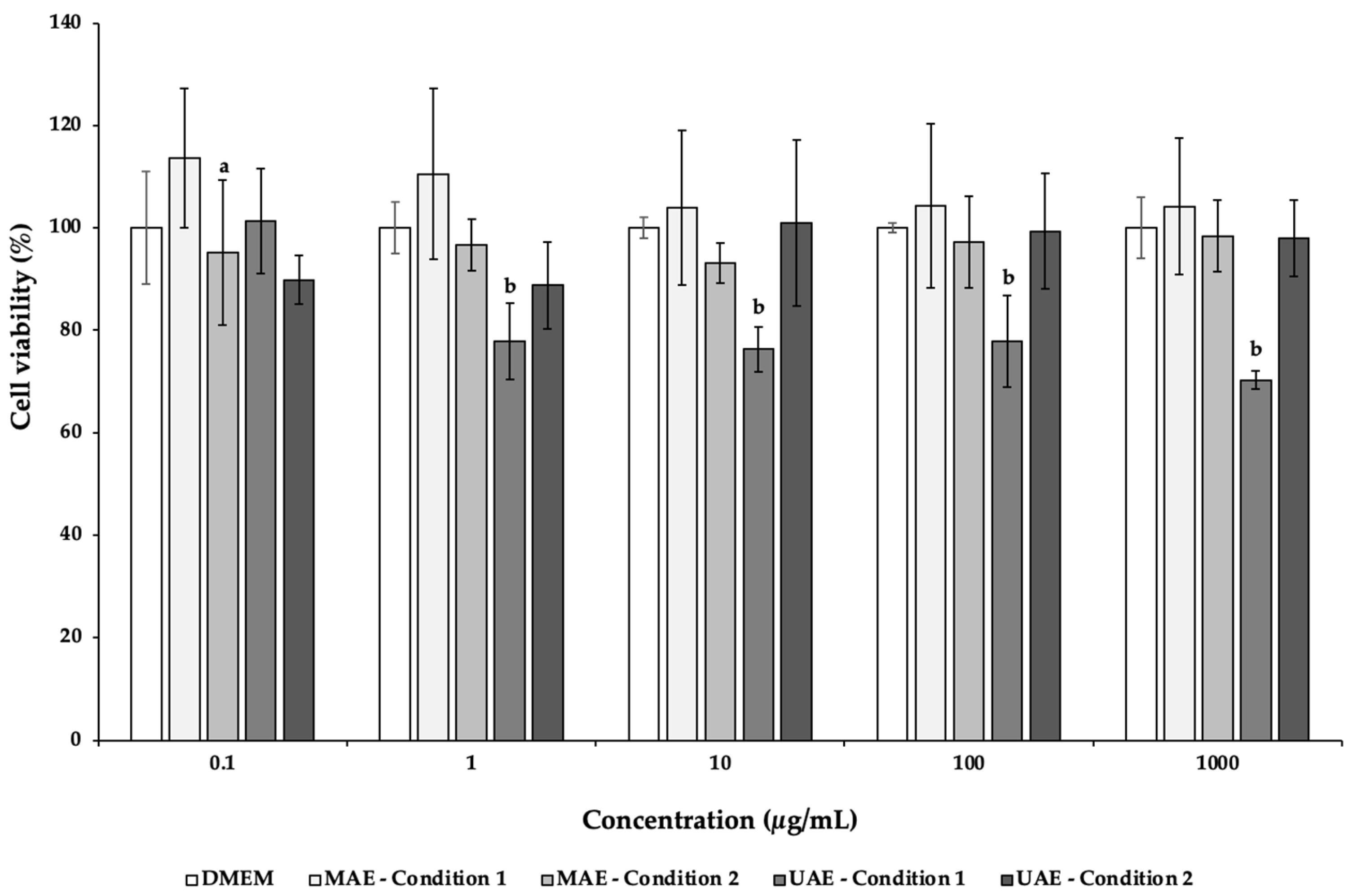
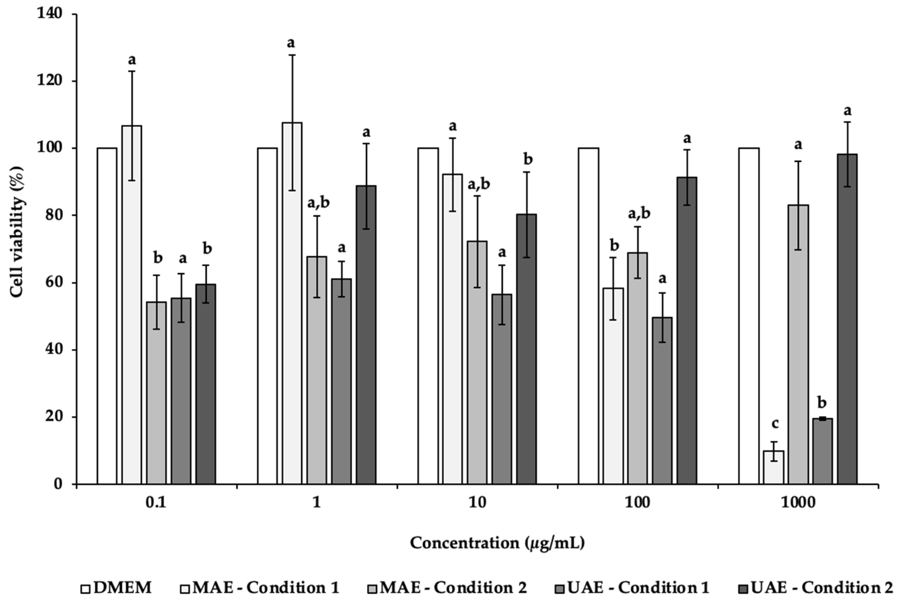
3.5. Cytotoxic Effects of C. pepo Seed Extracts towards Intestinal and Neuronal Cells

Caco-2, an intestinal cell line, was used to evaluate the cytotoxicity of the C. pepo extracts obtained by UAE and MAE. This cell line allows the study the compound absorption across the intestinal epithelium, being commonly employed to evaluate the extracts safety [97]. According to Figure 1, the exposure of this cell line to increasing concentrations (0.1–1000 µg/mL) of MAE C. pepo extracts showed a viability around 100%, without significant differences (p > 0.05). On the other hand, only the concentration of 0.1 μg/mL of the UAE condition 1 extract maintained a viability of 101.36%, being significantly different from the other concentrations (p < 0.05). At higher concentrations, the viability ranged between 70.20% (1000 μg/mL) and 77.82% (1 μg/mL). The UAE condition 2 displayed viabilities above 88.78%, being not significantly different (p > 0.05). The lower viability displayed by Caco-2 cells incubated with UAE C. pepo extracts may be explained by the higher content on neochlorogenic acid and caftaric acid present in the extracts. These compounds have antiproliferative activity and may effectively inhibit the growth of colorectal cancer cells [98,99].
In the present study, NSC-34 cell line was used to evaluate the potential neurotoxicity of C. pepo extracts obtained by UAE and MAE (Figure 2). NSC-34 is a hybrid cell line that has similar morphological and physiological properties to motor neurons [100]. This cell line in culture mimics the synthesis and storage of acetylcholine (ACh) and the expression of neurofilament proteins [100]. After 24 h of incubation in the presence of different concentrations (0.1–1000 µg/mL) of C. pepo extracts, it was possible to observe a decrease of the NSC-34 cell viability, mainly in the MAE and UAE condition 1 extracts (Figure 2). Indeed, at the highest concentration tested (1000 µg/mL), the cells’ viability was 9.92% and 19.58%, for MAE condition 1 and UAE condition 1, respectively, with significant differences (p < 0.05). In these cases, it was possible to determine the IC50 values of 253.68 (MAE condition 1) and 95.32 µg/mL (UAE condition 1).
Oppositely, MAE and UAE condition 2 showed an increasing trend with increased concentrations. For MAE condition 2, the results varied from 54.17% (0.1 μg/mL) to 82.97% (1000 μg/mL). In the case of UAE condition 2 extracts, the highest cell viability (98.21%) was found for the highest concentration tested (1000 μg/mL), following in descending order 100, 1, 10 and 0.1 μg/mL.
The inferior cells’ viability observed after exposure to MAE condition 1 and UAE condition 1 extracts may be due to the presence in higher amounts of the antiproliferative compounds neochlorogenic acid and caftaric acid, which is not observed in MAE condition 2 and UAE condition 2 extracts. Besides that, MAE condition 2 and UAE condition 2 extracts present other compounds (e.g., catechin, gallic acid or protocatechuic acid) with benefic cell effects in higher quantities, justifying a possible protective effect. Further studies are needed to justify these differences.
To the best of our knowledge, this is the first study that screens the in vitro effects of C. pepo extracts on the viability of cell lines. Li et al. [101] evaluated the cytotoxicity effect of C. moschata polysaccharides on Caco-2 through the CCK-8 assay, reporting a viability above 90% for all tested concentrations (0, 250, 500, 1000, 1500 μg/mL) [101]. These results are in line with the ones obtained in the present study for Caco-2.

4. Conclusions

In this work, value-added compounds from C. pepo seeds were extracted using green techniques—UAE and MAE—and an eco-friendly solvent—water, aiming a potential use as nutraceutical ingredient. Two different ratio conditions were employed.
FRAP scavenging activity ranged from 45.80 to 71.09 μmol FSE/g DW (UAE condition 2 and MAE condition 1, respectively), while DPPH values varied between 4.35 mg TE/g DW (UAE condition 1) and 5.08 mg TE/g DW (MAE condition 2). The extract with the best performance at ABTS assay was MAE condition 1 (13.29 mg AAE/g DW), while UAE condition 2 was the worst (11.38 mg AAE/g DW). The extract with the highest TPC value was MAE condition 2 (16.89 mg GAE/g DW), and UAE condition 1 had the lowest (12.17 mg GAE/g DW). All extracts displayed a remarkable scavenging capacity for HOCl, with IC50 values ranging from 1.88 (UAE condition 1) to 13.50 μg/mL (UAE condition 2).
HPLC–PDA analysis revealed the presence of alkaloids, chalconoids, flavanols, flavanones, flavonols, phenolic acids and stilbenoids. A total of 21 phenolic compounds were identified, with catechin, caffeine and gallic acid being present in higher quantities. No adverse effects were detected in Caco-2 viability to MAE extracts up to 1000 μg/mL, while all the other conditions tested led to a slight decrease of cell viability.
The results shown that a higher sample-to-solvent ratio led to an increase in the amount of phenolic compounds present in both MAE and UAE extracts, which led to better results in ORAC, O2●−, TPC and DPPH assays. Overall, these results highlighted that C. pepo seeds are a potential source of antioxidant/antiradical compounds and that the extract MAE condition 2 is rich in bioactive compounds with potential uses in the nutraceutical field.
Moreover, the results obtained in this study show that higher sample-to-solvent ratios using MAE technology can lead to the extraction of a greater amount of phenolic compounds. Therefore, further studies are needed to screen the effects on neuronal enzymes (such as acetylcholinesterase or butyrylcholinesterase) as well as 3D intestinal permeation studies to screen the amounts that are absorbed at intestinal level.

Author Contributions

Conceptualization, F.R.; methodology, C.M., A.M.S., A.S.F., M.M.M. and F.R.; software, A.M.S., C.M. and M.M.M.; validation, M.M.M., F.R. and C.D.-M.; formal analysis, C.M., A.M.S., M.M.M. and F.R.; investigation, C.M., A.M.S., A.S.F., M.M.M. and F.R; resources, C.D.-M. and F.R.; writing—original draft preparation, C.M. and A.M.S.; writing—review and editing, F.R., M.M.M. and C.D.-M.; supervision, F.R.; project administration, F.R.; funding acquisition, C.D.-M. and F.R. All authors have read and agreed to the published version of the manuscript.

Funding

This research was funded by UIDB/50006/2020 and UIDP/50006/2020 by the Fundação para a Ciência e a Tecnologia (FCT)/Ministério da Ciência, Tecnologia e Ensino Superior (MCTES) through national funds.

Institutional Review Board Statement

Not applicable.

Informed Consent Statement

Not applicable.

Data Availability Statement

Data available on request due to restrictions e.g., privacy or ethical.

Acknowledgments

Ana Margarida Silva (SFRH/BD/144994/2019) and Ana Sofia Ferreira (2020.07519.BD) are thankful for the Ph.D. grants financed by POPH-QREN and subsidized by the European Science Foundation and Ministério da Ciência, Tecnologia e Ensino Superior. Manuela M. Moreira (CEECIND/02702/2017) and Francisca Rodrigues (CEECIND/01886/2020) are thankful for their contracts financed by FCT/MCTES—CEEC Individual Program Contract.

Conflicts of Interest

The authors declare no conflict of interest.

References

  1. Prakash, V.; van Boekel, M.A.J.S. Chapter 19-Nutraceuticals: Possible future ingredients and food safety aspects. In Ensuring Global Food Safety; Boisrobert, C.E., Stjepanovic, A., Oh, S., Lelieveld, H.L.M., Eds.; Academic Press: San Diego, CA, USA, 2010; pp. 333–338. [Google Scholar]
  2. Dudeja, P.; Gupta, R.K. Chapter 40-Nutraceuticals. In Food Safety in the 21st Century; Gupta, R.K., Singh, M.D., Eds.; Academic Press: San Diego, CA, USA, 2017; pp. 491–496. [Google Scholar]
  3. Nasri, H.; Baradaran, A.; Shirzad, H.; Rafieian-Kopaei, M. New Concepts in Nutraceuticals as Alternative for Pharmaceuticals. Int. J. Prev. Med. 2014, 5, 1487–1499. [Google Scholar]
  4. Madihi, Y.; Merrikhi, A.; Khodai, M.; Rafieian-Kopaei, M.; Shahinfard, N.; Ansari, R.; Shirzad, H.; Mesripour, A. Impact of Sumac on Postprandial High-Fat Oxidative Stress. Pak. J. Med. Sci. 2013, 29, 340–345. [Google Scholar] [CrossRef]
  5. Setorki, M.; Rafieian-Kopaei, M.; Merikhi, A.; Heidarian, E.; Shahinfard, N.; Ansari, R.; Nasri, H.; Esmael, N.; Baradaran, A. Suppressive Impact of Anethum Graveolens Consumption on Biochemical Risk Factors of Atherosclerosis in Hypercholesterolemic Rabbits. Int. J. Prev. Med. 2013, 4, 889–895. [Google Scholar] [PubMed]
  6. Khosravi-Boroujeni, H.; Mohammadifard, N.; Sarrafzadegan, N.; Sajjadi, F.; Maghroun, M.; Khosravi, A.; Alikhasi, H.; Rafieian, M.; Azadbakht, L. Potato Consumption and Cardiovascular Disease Risk Factors among Iranian Population. Int. J. Food Sci. Nutr. 2012, 63, 913–920. [Google Scholar] [CrossRef] [PubMed]
  7. Khosravi-Boroujeni, H.; Sarrafzadegan, N.; Mohammadifard, N.; Sajjadi, F.; Maghroun, M.; Asgari, S.; Rafieian-Kopaei, M.; Azadbakht, L. White Rice Consumption and CVD Risk Factors among Iranian Population. J. Health Popul. Nutr. 2013, 31, 252–261. [Google Scholar] [CrossRef] [Green Version]
  8. Khodai, M.; Madihi, Y.; Merrikhi, A.; Rafieian-kopaei, M.; Nasri, H. Serum Lipoprotein (a) in Diabetic Patients with Various Renal Function not yet on Dialysis. Pak. J. Med. Sci. 2013, 29, 354–357. [Google Scholar]
  9. Shirzad, H.; Burton, R.C.; Smart, Y.C.; Rafieian-Kopaei, M.; Shirzad, M. Natural Cytotoxicity of nc-2+ Cells against the Growth and Metastasis of Wehi-164 Fibrosarcoma. Scand. J. Immunol. 2011, 73, 85–90. [Google Scholar] [CrossRef]
  10. Shirzad, M.; Kordyazdi, R.; Shahinfard, N.; Nikokar, M. Does Royal Jelly Affect Tumor Cells? J. Herbmed. Pharmacol. 2013, 2, 45–48. [Google Scholar]
  11. Akhlaghi, M.; Shabanian, G.; Rafieian-Kopaei, M.; Parvin, N.; Saadat, M.; Akhlaghi, M. Citrus Aurantium Blossom and Preoperative Anxiety. Rev. Bras. Anestesiol. 2011, 61, 702–712. [Google Scholar] [CrossRef] [Green Version]
  12. Rabiei, Z.; Rafieian-Kopaei, M.; Heidarian, E.; Saghaei, E.; Mokhtari, S. Effects of Zizyphus Jujube Extract on Memory and Learning Impairment Induced by Bilateral Electric Lesions of the Nucleus Basalis of Meynert in Rat. Neurochem. Res. 2014, 39, 353–360. [Google Scholar] [CrossRef]
  13. Rabiei, Z.; Rafieian-Kopaei, M.; Mokhtari, S.; Alibabaei, Z.; Shahrani, M. The Effect of Pretreatment with Different Doses of Lavandula Officinalis Ethanolic Extract on Memory, Learning and Nociception. Biomed. Aging Pathol. 2014, 4, 71–76. [Google Scholar] [CrossRef]
  14. Parsaei, P.; Karimi, M.; Asadi, S.Y.; Rafieian-Kopaei, M. Bioactive Components and Preventive Effect of Green Tea (Camellia Sinensis) Extract on Post-Laparotomy Intra-Abdominal Adhesion in Rats. Int. J. Surg. 2013, 11, 811–815. [Google Scholar] [CrossRef] [PubMed] [Green Version]
  15. Asadi-Samani, M.; Farkhad, N.; Rafieian-Kopaei, M. A Review on Phytochemistry and Pharmacological Effects of Prangos Ferulacea (L.) Lindl. Life Sci. J. 2013, 10, 360–367. [Google Scholar]
  16. Baradaran, A.; Nasri, H.; Nematbakhsh, M.; Rafieian-Kopaei, M. Antioxidant Activity and Preventive Effect of Aqueous Leaf Extract of Aloe Vera on Gentamicin-Induced Nephrotoxicity in Male Wistar Rats. Clin. Ter. 2014, 165, 7–11. [Google Scholar]
  17. Gutierrez, R. Review of Cucurbita Pepo (Pumpkin) its Phytochemistry and Pharmacology. Med. Chem. 2016, 6, 12–21. [Google Scholar] [CrossRef]
  18. Zhang, C.; Zhu, Q.; Liu, S.; Gao, P.; Zhu, Z.; Wang, X.; Luan, F. The Complete Chloroplast Genome Sequence of the Cucurbita Pepo L. (Cucurbitaceae). Mitochondrial DNA Part B 2018, 3, 717–718. [Google Scholar] [CrossRef] [Green Version]
  19. Adnan, M.; Gul, S.; Batool, S.; Bibi, F.; Rehman, A.; Yaqoob, S.; Shabir, H.; Yousaf, T.; Mussarat, S.; Ali, N.; et al. A Review on the Ethnobotany, Phytochemistry, Pharmacology and Nutritional Composition of Cucurbita Pepo L. J. Phytopharm. 2017, 6, 133–139. [Google Scholar]
  20. Smith, B.D. The Initial Domestication of Cucurbita Pepo in the Americas 10,000 Years Ago. Science 1997, 276, 932–934. [Google Scholar] [CrossRef] [Green Version]
  21. Kulczyński, B.; Sidor, A.; Gramza-Michałowska, A. Antioxidant Potential of Phytochemicals in Pumpkin Varieties Belonging to Cucurbita Moschata and Cucurbita Pepo Species. CyTA-J. Food 2020, 18, 472–484. [Google Scholar] [CrossRef]
  22. Rabrenović, B.B.; Dimić, E.B.; Novaković, M.M.; Tešević, V.V.; Basić, Z.N. The most Important Bioactive Components of Cold Pressed Oil from Different Pumpkin (Cucurbita Pepo L.) Seeds. LWT—Food Sci. Technol. 2014, 55, 521–527. [Google Scholar] [CrossRef]
  23. Hajhashemi, V.; Rajabi, P.; Mardani, M. Beneficial Effects of Pumpkin Seed Oil as a Topical Hair Growth Promoting Agent in a Mice Model. Avicenna J. Phytomed 2019, 9, 499–504. [Google Scholar] [PubMed]
  24. Grzybek, M.; Kukula-Koch, W.; Strachecka, A.; Jaworska, A.; Phiri, A.M.; Paleolog, J.; Tomczuk, K. Evaluation of Anthelmintic Activity and Composition of Pumpkin (Cucurbita Pepo L.) Seed Extracts—in vitro and in vivo Studies. Int. J. Mol. Sci. 2016, 17, 1456. [Google Scholar] [CrossRef]
  25. Medjakovic, S.; Hobiger, S.; Ardjomand-Woelkart, K.; Bucar, F.; Jungbauer, A. Pumpkin Seed Extract: Cell Growth Inhibition of Hyperplastic and Cancer Cells, Independent of Steroid Hormone Receptors. Fitoterapia 2016, 110, 150–156. [Google Scholar] [CrossRef] [PubMed] [Green Version]
  26. Vahlensieck, W.; Theurer, C.; Pfitzer, E.; Patz, B.; Banik, N.; Engelmann, U. Effects of Pumpkin Seed in Men with Lower Urinary Tract Symptoms due to Benign Prostatic Hyperplasia in the One-Year, Randomized, Placebo-Controlled GRANU Study. Urol. Int. 2014, 94, 286–295. [Google Scholar] [CrossRef] [PubMed]
  27. Peiretti, P.G.; Meineri, G.; Gai, F.; Longato, E.; Amarowicz, R. Antioxidative Activities and Phenolic Compounds of Pumpkin (Cucurbita Pepo) Seeds and Amaranth (Amaranthus Caudatus) Grain Extracts. Nat. Prod. Res. 2017, 31, 2178–2182. [Google Scholar] [CrossRef]
  28. Piccolella, S.; Crescente, G.; Candela, L.; Pacifico, S. Nutraceutical Polyphenols: New Analytical Challenges and Opportunities. J. Pharm. Biomed. Anal. 2019, 175, 112774. [Google Scholar] [CrossRef] [PubMed]
  29. Serino, A.; Salazar, G. Protective Role of Polyphenols against Vascular Inflammation, Aging and Cardiovascular Disease. Nutrients 2018, 11, 53. [Google Scholar] [CrossRef] [PubMed] [Green Version]
  30. Brglez Mojzer, E.; Knez Hrnčič, M.; Škerget, M.; Knez, Ž.; Bren, U. Polyphenols: Extraction Methods, Antioxidative Action, Bioavailability and Anticarcinogenic Effects. Molecules 2016, 21, 901. [Google Scholar] [CrossRef]
  31. Losada-Barreiro, S.; Bravo-Díaz, C. Free Radicals and Polyphenols: The Redox Chemistry of Neurodegenerative Diseases. Eur. J. Med. Chem. 2017, 133, 379–402. [Google Scholar] [CrossRef]
  32. Kim, M.Y.; Kim, E.J.; Kim, Y.-N.; Choi, C.; Lee, B.-H. Comparison of the Chemical Compositions and Nutritive Values of Various Pumpkin (Cucurbitaceae) Species and Parts. Nutr. Res. Pr. 2012, 6, 21–27. [Google Scholar] [CrossRef] [Green Version]
  33. Wallert, M.; Ziegler, M.; Wang, X.; Maluenda, A.; Xu, X.; Yap, M.L.; Witt, R.; Giles, C.; Kluge, S.; Hortmann, M.; et al. α-Tocopherol Preserves Cardiac Function by Reducing Oxidative Stress and Inflammation in Ischemia/Reperfusion Injury. Redox Biol. 2019, 26, 101292. [Google Scholar] [CrossRef]
  34. Jian, L.; Du, C.J.; Lee, A.H.; Binns, C.W. Do Dietary Lycopene and other Carotenoids Protect against Prostate Cancer? Int. J. Cancer 2005, 113, 1010–1014. [Google Scholar] [CrossRef]
  35. Sesso, H.D. Carotenoids and Cardiovascular Disease: What Research Gaps Remain? Curr. Opin. Lipidol. 2006, 17, 11–16. [Google Scholar] [CrossRef]
  36. Moran, N.E.; Mohn, E.S.; Hason, N.; Erdman, J.W., Jr.; Johnson, E.J. Intrinsic and Extrinsic Factors Impacting Absorption, Metabolism, and Health Effects of Dietary Carotenoids. Adv. Nutr. 2018, 9, 465–492. [Google Scholar] [CrossRef] [Green Version]
  37. Ryan, E.; Galvin, K.; O’Connor, T.P.; Maguire, A.; O’Brien, N.M. Phytosterol, Squalene, Tocopherol Content and Fatty Acid Profile of Selected Seeds, Grains, and Legumes. Mater. Veg. 2007, 62, 85–91. [Google Scholar] [CrossRef] [PubMed]
  38. Glew, R.; Glew, R.; Chuang, L.-T.; Huang, Y.-S.; Millson, M.; Constans, D.; VanderJagt, D. Amino Acid, Mineral and Fatty Acid Content of Pumpkin Seeds (Cucurbita Spp.) and Cyperus Esculentus Nuts in the Republic of Niger. Mater. Veg. 2006, 61, 49–54. [Google Scholar] [CrossRef]
  39. Piironen, V.; Lindsay, D.; Miettinen, T.; Toivo, J.; Lampi, A.-M. Plant Sterols: Biosynthesis, Biological Function and their Importance to Human Nutrition. J. Sci. Food Agric. 2000, 80, 939–966. [Google Scholar] [CrossRef]
  40. Raicht, R.F.; Cohen, B.I.; Fazzini, E.P.; Sarwal, A.N.; Takahashi, M. Protective Effect of Plant Sterols against Chemically Induced Colon Tumors in Rats. Cancer Res. 1980, 40, 403–405. [Google Scholar] [PubMed]
  41. Gossell-Williams, M.; Davis, A.; O’connor, N. Inhibition of Testosterone-Induced Hyperplasia of the Prostate of Sprague-Dawley Rats by Pumpkin Seed Oil. J. Med. Food 2006, 9, 284–286. [Google Scholar] [CrossRef]
  42. Zerafatjou, N.; Amirzargar, M.; Biglarkhani, M.; Shobeirian, F.; Zoghi, G. Pumpkin Seed Oil (Cucurbita Pepo) Versus Tamsulosin for Benign Prostatic Hyperplasia Symptom Relief: A Single-Blind Randomized Clinical Trial. BMC Urol. 2021, 21, 1–7. [Google Scholar] [CrossRef]
  43. Carbin, B.-E.; Larsson, B.; Lindahl, O. Treatment of Benign Prostatic Hyperplasia with Phytosterols. Br. J. Urol. 1990, 66, 639–641. [Google Scholar] [CrossRef]
  44. Leitao da-Cunha, E.V.; Fechine, I.M.; Guedes, D.N.; Barbosa-Filho, J.M.; Sobral da Silva, M. Protoberberine alkaloids. In The Alkaloids: Chemistry and Biology; Cordell, G.A., Ed.; Academic Press: Cambridge, MA, USA, 2005; Volume 62, pp. 1–75. [Google Scholar]
  45. Vennerstrom, J.L.; Lovelace, J.K.; Waits, V.B.; Hanson, W.L.; Klayman, D.L. Berberine Derivatives as Antileishmanial Drugs. Antimicrob. Agents Chemother. 1990, 34, 918–921. [Google Scholar] [CrossRef] [Green Version]
  46. Dkhil, M. Role of Berberine in Ameliorating Schistosoma Mansoni-Induced Hepatic Injury in Mice. Biol. Res. 2014, 47, 1–7. [Google Scholar] [CrossRef] [PubMed] [Green Version]
  47. Krivogorsky, B.; Pernat, J.A.; Douglas, K.A.; Czerniecki, N.J.; Grundt, P. Structure–Activity Studies of Some Berberine Analogs as Inhibitors of Toxoplasma Gondii. Bioorg. Med. Chem. Lett. 2012, 22, 2980–2982. [Google Scholar] [CrossRef] [PubMed]
  48. Panja, P. Green Extraction Methods of Food Polyphenols from Vegetable Materials. Curr. Opin. Food Sci. 2018, 23, 173–182. [Google Scholar] [CrossRef]
  49. Kataoka, H. New Trends in Sample Preparation for Analysis of Plant-Derived Medicines. Curr. Org. Chem. 2010, 14, 1698–1713. [Google Scholar] [CrossRef]
  50. Llompart, M.; Garcia-Jares, C.; Celeiro, M.; Dagnac, T. Microwave-Assisted Extraction. In Encyclopedia of Analytical Science, 3rd ed.; Worsfold, P., Poole, C., Townshend, A., Miró, M., Eds.; Academic Press: Oxford, UK, 2019; pp. 67–77. [Google Scholar]
  51. Kataoka, H. Pharmaceutical Analysis | sample preparation. In Encyclopedia of Analytical Science, 3rd ed.; Worsfold, P., Poole, C., Townshend, A., Miró, M., Eds.; Academic Press: Oxford, UK, 2019; pp. 231–255. [Google Scholar]
  52. Ahuja, S.; Diehl, D. Chapter 2 Sampling and Sample Preparation. In Comprehensive Analytical Chemistry; Elsevier: Amsterdam, The Netherlands, 2006; Volume 47, pp. 15–40. [Google Scholar] [CrossRef]
  53. Sasaki, K.; Honda, W.; Ohsawa, S.; Miyake, Y.; Kawashima, Y. A Study of Microwave Sterilizer for Injection Ampules (no.4): Application to Sterilization of Thermally Labile Drug Solutions. J. Pharm. Sci. Technol. Jpn. 1998, 58, 125–135. [Google Scholar]
  54. Beejmohun, V.; Fliniaux, O.; Grand, É.; Lamblin, F.; Bensaddek, L.; Christen, P.; Kovensky, J.; Fliniaux, M.-A.; Mesnard, F. Microwave-Assisted Extraction of the Main Phenolic Compounds in Flaxseed. Phytochem. Anal. 2007, 18, 275–282. [Google Scholar] [CrossRef]
  55. Proestos, C.; Komaitis, M. Application of Microwave-Assisted Extraction to the Fast Extraction of Plant Phenolic Compounds. LWT 2008, 41, 652–659. [Google Scholar] [CrossRef]
  56. Gallo, M.; Ferracane, R.; Graziani, G.; Ritieni, A.; Fogliano, V. Microwave Assisted Extraction of Phenolic Compounds from Four Different Spices. Molecules 2010, 15, 6365–6374. [Google Scholar] [CrossRef]
  57. Rostagno, M.A.; Prado, J.M. Natural Product Extraction: Principles and Applications; RSC Green Chemistry: Cambridge, UK, 2013. [Google Scholar]
  58. Reddy, A.V.B.; Moniruzzaman, M.; Madhavi, V.; Jaafar, J. Recent Improvements in the Extraction, Cleanup and Quantification of Bioactive Flavonoids; Elsevier: Amsterdam, The Netherlands, 2020; Volume 66, pp. 197–223. [Google Scholar] [CrossRef]
  59. Roohinejad, S.; Nikmaram, N.; Brahim, M.; Koubaa, M.; Khelfa, A.; Greiner, R. Chapter 16-potential of novel technologies for aqueous extraction of plant bioactives. In Water Extraction of Bioactive Compounds; Dominguez González, H., González Muñoz, M.J., Eds.; Elsevier: Amsterdam, The Netherlands, 2017; pp. 399–419. [Google Scholar]
  60. Moldoveanu, S.; David, V. Chapter 6-solvent extraction. In Modern Sample Preparation for Chromatography, 2nd ed.; Moldoveanu, S., David, V., Eds.; Elsevier: Amsterdam, The Netherlands, 2021; pp. 191–279. [Google Scholar]
  61. Ishtiaq, F.; Farooq, R.; Farooq, U.; Farooq, A.; Siddique, M.; Shah, S.H.; Shaheen, M. Application of Ultrasound in Pharmaceutics. World Appl. Sci. J. 2009, 6, 886–893. [Google Scholar]
  62. Ma, Y.-Q.; Ye, X.-Q.; Fang, Z.-X.; Chen, J.-C.; Xu, G.-H.; Liu, D.-H. Phenolic Compounds and Antioxidant Activity of Extracts from Ultrasonic Treatment of Satsuma Mandarin (Citrus Unshiu Marc.) Peels. J. Agric. Food Chem. 2008, 56, 5682–5690. [Google Scholar] [CrossRef] [PubMed]
  63. Sun, Y.; Liu, Z.; Wang, J. Ultrasound-Assisted Extraction of Five Isoflavones from Iris Tectorum Maxim. Sep. Purif. Technol. 2011, 78, 49–54. [Google Scholar] [CrossRef]
  64. Moreira, M.M.; Barroso, M.F.; Boeykens, A.; Withouck, H.; Morais, S.; Delerue-Matos, C. Valorization of Apple Tree Wood Residues by Polyphenols Extraction: Comparison between Conventional and Microwave-Assisted Extraction. Ind. Crop. Prod. 2017, 104, 210–220. [Google Scholar] [CrossRef] [Green Version]
  65. Pimentel-Moral, S.; Borrás-Linares, I.; Lozano-Sánchez, J.; Arráez-Román, D.; Martínez-Férez, A.; Segura-Carretero, A. Microwave-Assisted Extraction for Hibiscus Sabdariffa Bioactive Compounds. J. Pharm. Biomed. Anal. 2018, 156, 313–322. [Google Scholar] [CrossRef]
  66. Chemat, F.; Rombaut, N.; Sicaire, A.-G.; Meullemiestre, A.; Fabiano-Tixier, A.-S.; Abert-Vian, M. Ultrasound Assisted Extraction of Food and Natural Products. Mechanisms, Techniques, Combinations, Protocols and Applications. A Review. Ultrason. Sonochem. 2017, 34, 540–560. [Google Scholar] [CrossRef] [PubMed]
  67. Silva, A.; Pinto, D.; Fernandes, I.; Freitas, V.; Cádiz-Gurrea, M.; Costa, P.; Delerue-Matos, C.; Rodrigues, F. An Insight into Kiwiberry Leaf Valorization: Phenolic Composition, Bioactivity and Health Benefits. Molecules 2021, 26, 2314. [Google Scholar] [CrossRef] [PubMed]
  68. Lameirão, F.; Pinto, D.; Vieira, E.F.; Peixoto, A.F.; Freire, C.; Sut, S.; Dall’Acqua, S.; Costa, P.; Delerue-Matos, C.; Rodrigues, F. Green-Sustainable Recovery of Phenolic and Antioxidant Compounds from Industrial Chestnut Shells Using Ultrasound-Assisted Extraction: Optimization and Evaluation of Biological Activities in Vitro. Antioxidants 2020, 9, 267. [Google Scholar] [CrossRef] [Green Version]
  69. Singleton, V.L.; Rossi, J.A. Colorimetry of Total Phenolics with Phosphomolybdic-Phosphotungstic Acid Reagents. Am. J. Enol. Vitic. 1965, 16, 144. [Google Scholar]
  70. Re, R.; Pellegrini, N.; Proteggente, A.; Pannala, A.; Yang, M.; Rice-Evans, C. Antioxidant Activity Applying an Improved ABTS Radical Cation Decolorization Assay. Free Radic. Biol. Med. 1999, 26, 1231–1237. [Google Scholar] [CrossRef]
  71. Pinto, D.; Cádiz-Gurrea, M.D.L.L.; Vallverdú-Queralt, A.; Delerue-Matos, C.; Rodrigues, F. Castanea Sativa Shells: A Review on Phytochemical Composition, Bioactivity and Waste Management Approaches for Industrial Valorization. Food Res. Int. 2021, 144, 110364. [Google Scholar] [CrossRef]
  72. Benzie, I.F.F.; Strain, J.J. Ferric Reducing/Antioxidant Power Assay: Direct Measure of Total Antioxidant Activity of Biological Fluids and Modified Version for Simultaneous Measurement of Total Antioxidant Power and Ascorbic Acid Concentration. Methods Enzymol. 1999, 299, 15–27. [Google Scholar] [PubMed]
  73. Gomes, A.; Fernandes, E.; Silva, A.M.; Santos, C.M.; Pinto, D.C.; Cavaleiro, J.A.; Lima, J.L. 2-Styrylchromones: Novel Strong Scavengers of Reactive Oxygen and Nitrogen Species. BioOrganic Med. Chem. 2007, 15, 6027–6036. [Google Scholar] [CrossRef] [PubMed]
  74. Pinto, D.; Vieira, E.F.; Peixoto, A.F.; Freire, C.; Freitas, V.; Costa, P.; Delerue-Matos, C.; Rodrigues, F. Optimizing the Extraction of Phenolic Antioxidants from Chestnut Shells by Subcritical Water Extraction Using Response Surface Methodology. Food Chem. 2020, 334, 127521. [Google Scholar] [CrossRef] [PubMed]
  75. Galan, A.-M.; Calinescu, I.; Trifan, A.; Winkworth-Smith, C.; Calvo-Carrascal, M.; Dodds, C.; Binner, E. New Insights into the Role of Selective and Volumetric Heating during Microwave Extraction: Investigation of the Extraction of Polyphenolic Compounds from Sea Buckthorn Leaves Using Microwave-Assisted Extraction and Conventional Solvent Extraction. Chem. Eng. Process Intensif. 2017, 116, 29–39. [Google Scholar] [CrossRef]
  76. Ethiraj, S.; Balasundaram, J. Phytochemical and Biological Activity of Cucurbita Seed Extract. J. Adv. Biotechnol. 2016, 6, 813–821. [Google Scholar] [CrossRef]
  77. Mondal, S.; Hossain, I.; Islam, M. Determination of Antioxidant Potential of Cucurbita Pepo Linn. (An Edible Herbs of Bangladesh). J. Pharmacogn. Phytochem. 2017, 6, 1016–1019. [Google Scholar]
  78. Saavedra, M.J.; Aires, A.; Dias, C.; Almeida, J.A.; De Vasconcelos, M.C.B.M.; Santos, P.; Rosa, E.A. Evaluation of the Potential of Squash Pumpkin by-Products (Seeds and Shell) as Sources of Antioxidant and Bioactive Compounds. J. Food Sci. Technol. 2013, 52, 1008–1015. [Google Scholar] [CrossRef] [Green Version]
  79. Rezig, L.; Chouaibi, M.; Ojeda-Amador, R.M.; Gomez-Alonso, S.; Salvador, M.D.; Fregapane, G.; Hamdi, S. Cucurbita Maxima Pumpkin Seed Oil: From the Chemical Properties to the Different Extracting Techniques. Not. Bot. Horti Agrobot. Cluj-Napoca 2018, 46, 663–669. [Google Scholar] [CrossRef] [Green Version]
  80. Kulczyński, B.; Gramza-Michałowska, A.; Królczyk, J.B. Optimization of Extraction Conditions for the Antioxidant Potential of Different Pumpkin Varieties (Cucurbita Maxima). Sustainability 2020, 12, 1305. [Google Scholar] [CrossRef] [Green Version]
  81. Nawirska-Olszańska, A.; Kita, A.; Biesiada, A.; Sokół-Łętowska, A.; Kucharska, A.Z. Characteristics of Antioxidant Activity and Composition of Pumpkin Seed Oils in 12 Cultivars. Food Chem. 2013, 139, 155–161. [Google Scholar] [CrossRef] [PubMed]
  82. Gutteridge, J.M. Lipid Peroxidation and Antioxidants as Biomarkers of Tissue Damage. Clin. Chem. 1995, 41, 1819–1828. [Google Scholar] [CrossRef] [PubMed]
  83. Halliwell, B. Antioxidant Characterization: Methodology and Mechanism. Biochem. Pharmacol. 1995, 49, 1341–1348. [Google Scholar] [CrossRef]
  84. Kehrer, J.P. Free Radicals as Mediators of Tissue Injury and Disease. Crit. Rev. Toxicol. 1993, 23, 21–48. [Google Scholar] [CrossRef]
  85. Sánchez-Moreno, C.; Larrauri, J.A.; Saura-Calixto, F. Free Radical Scavenging Capacity and Inhibition of Lipid Oxidation of Wines, Grape Juices and Related Polyphenolic Constituents. Food Res. Int. 1999, 32, 407–412. [Google Scholar] [CrossRef]
  86. Malencić, D.; Gasic, O.; Popović, M.; Boza, P. Screening for Antioxidant Properties of Salvia Reflexa Hornem. Phytother Res. 2000, 14, 546–548. [Google Scholar] [CrossRef]
  87. Gülçin, I.; Oktay, M.; Küfrevioğlu, O.I.; Aslan, A. Determination of Antioxidant Activity of Lichen Cetraria Islandica (L) Ach. J. Ethnopharmacol. 2001, 79, 325–329. [Google Scholar] [CrossRef]
  88. Devasagayam, T.P.A.; Tilak, J.C.; Boloor, K.K.; Sane, K.S.; Ghaskadbi, S.S.; Lele, R.D. Free Radicals and Antioxidants in Human Health: Current Status and Future Prospects. J. Assoc. Physicians India 2004, 52, 794–804. [Google Scholar]
  89. Shahidi, F.; Janitha, P.K.; Wanasundara, P.D. Phenolic Antioxidants. Crit. Rev. Food Sci. Nutr. 1992, 32, 67–103. [Google Scholar] [CrossRef]
  90. Liu, G.; Zhu, W.; Zhang, J.; Song, D.; Zhuang, L.; Ma, Q.; Yang, X.; Liu, X.; Zhang, J.; Zhang, H.; et al. Antioxidant Capacity of Phenolic Compounds Separated from Tea Seed Oil in Vitro and in Vivo. Food Chem. 2021, 371, 131122. [Google Scholar] [CrossRef]
  91. Chisté, R.C.; Mercadante, A.Z.; Gomes, A.; Fernandes, E.; Lima, J.; Bragagnolo, N. In Vitro Scavenging Capacity of Annatto Seed Extracts against Reactive Oxygen and Nitrogen Species. Food Chem. 2011, 127, 419–426. [Google Scholar] [CrossRef] [PubMed]
  92. Karabegović, I.T.; Stojičević, S.S.; Veličković, D.T.; Todorovic, Z.; Nikolić, N.; Lazić, M.L. The Effect of Different Extraction Techniques on the Composition and Antioxidant Activity of Cherry Laurel (Prunus Laurocerasus) Leaf and Fruit Extracts. Ind. Crop. Prod. 2014, 54, 142–148. [Google Scholar] [CrossRef]
  93. Bubalo, M.C.; Ćurko, N.; Tomašević, M.; Ganić, K.K.; Redovniković, I.R. Green Extraction of Grape Skin Phenolics by Using Deep Eutectic Solvents. Food Chem. 2016, 200, 159–166. [Google Scholar] [CrossRef] [PubMed]
  94. Sharmila, G.; Nikitha, V.; Ilaiyarasi, S.; Dhivya, K.; Rajasekar, V.; Kumar, N.; Muthukumaran, K. Ultrasound Assisted Extraction of Total Phenolics from Cassia Auriculata Leaves and Evaluation of its Antioxidant Activities. Ind. Crop. Prod. 2016, 84, 13–21. [Google Scholar] [CrossRef]
  95. Enneb, S.; Drine, S.; Bagues, M.; Triki, T.; Boussora, F.; Guasmi, F.; Nagaz, K.; Ferchichi, A. Phytochemical Profiles and Nutritional Composition of Squash (Cucurbita Moschata D.) from Tunisia. S. Afr. J. Bot. 2020, 130, 165–171. [Google Scholar] [CrossRef]
  96. Iswaldi, I.; Gómez-Caravaca, A.M.; Lozano-Sánchez, J.; Arraez-Roman, D.; Carretero, A.S.; Gutierrez, A.F. Profiling of Phenolic and other Polar Compounds in Zucchini (Cucurbita Pepo L.) by Reverse-Phase High-Performance Liquid Chromatography Coupled to Quadrupole Time-of-Flight Mass Spectrometry. Food Res. Int. 2013, 50, 77–84. [Google Scholar] [CrossRef]
  97. Artursson, P.; Karlsson, J. Correlation between Oral Drug Absorption in Humans and Apparent Drug Permeability Coefficients in Human Intestinal Epithelial (Caco-2) Cells. Biochem. Biophys. Res. Commun. 1991, 175, 880–885. [Google Scholar] [CrossRef]
  98. Szwajgier, D.; Paduch, R.; Kukuła-Koch, W.; Polak-Berecka, M.; Waśko, A. Study on Biological Activity of Bread Enriched with Natural Polyphenols in Terms of Growth Inhibition of Tumor Intestine Cells. J. Med. Food 2020, 23, 181–190. [Google Scholar] [CrossRef]
  99. Elansary, H.O.; Mahmoud, E.A. In Vitro Antioxidant and Antiproliferative Activities of Six International Basil Cultivars. Nat. Prod. Res. 2015, 22, 2149–2154. [Google Scholar] [CrossRef]
  100. Cashman, N.R.; Durham, H.D.; Blusztajn, J.K.; Oda, K.; Tabira, T.; Shaw, I.T.; Dahrouge, S.; Antel, J.P. Neuroblastoma × Spinal Cord (NSC) Hybrid Cell Lines Resemble Developing Motor Neurons. Dev. Dyn. 1992, 194, 209–221. [Google Scholar] [CrossRef]
  101. Li, F.; Wei, Y.; Zhao, J.; Yu, G.; Huang, L.; Li, Q. Transport Mechanism and Subcellular Localization of a Polysaccharide from Cucurbia Moschata across Caco-2 Cells Model. Int. J. Biol. Macromol. 2021, 182, 1003–1014. [Google Scholar] [CrossRef] [PubMed]
Figure 1. Effects of C. pepo extracts obtained by UAE and MAE exposure on the viability of Caco-2 cells at different concentrations (0.1–1000 µg/mL), measured by MTT assay. Values are expressed as mean ± standard deviation (n = 3). Different letters represent significant differences between concentrations of the same sample (p < 0.05), according to Tukey’s HSD test.
Applsci 12 01763 g001
Figure 2. Effects of C. pepo extracts obtained by UAE and MAE exposure on the viability of NSC-34 cells at different concentrations (0.1–1000 µg/mL), as measured by the MTT assay. Values are expressed as mean ± standard deviation (n = 3). Different letters indicate significant differences between concentrations of the same sample (p < 0.05), according to Tukey’s HSD test.
Applsci 12 01763 g002
Table 1. Extraction yield, total phenolic content (TPC), in vitro antioxidant/antiradical activities (evaluated by ABTS, FRAP and DPPH assays) of C. pepo extracts prepared by MAE and UAE. Values are expressed as mean ± standard deviation (n = 3).
Extraction TechniquesRatio (g/mL)Extraction Yield (%)TPC
(mg GAE/g DW)
FRAP
(µmol FSE/g DW)
ABTS
(mg AAE/g DW)
DPPH
(mg TE/g DW)
MAE1/20 (condition 1)23.62 ± 0.0212.63 ± 1.01 b71.09 ± 1.07 a13.29 ± 0.694.93 ± 0.93
2.5/20 (condition 2)16.30 ± 0.0716.89 ± 1.06 a59.75 ± 1.09 b12.42 ± 0.855.08 ± 0.84
UAE1/20 (condition 1)25.19 ± 0.0412.17 ± 0.72 b52.32 ± 1.95 c12.87 ± 1.764.35 ± 0.73
2.5/20 (condition 2)28.41 ± 0.1012.40 ± 0.91 b45.80 ± 1.37 d11.38 ± 3.544.54 ± 1.40
Different letters (a, b, c, d) in the same column indicate significant differences between extracts (p < 0.05). TPC: total phenolic content; FRAP: ferric reducing antioxidant power; FSE: ferrous sulphate equivalents; AAE: ascorbic acid equivalents; TE: trolox equivalents; DW: dry weight.
Table 2. Reactive oxygen species scavenging capacity (evaluated by O2●−, HOCl and ORAC assays) of C. pepo extracts prepared by microwave-assisted extraction and ultrasound-assisted extraction (MAE and UAE, respectively). Values are expressed as mean ± standard deviation (n = 3).
Reactive Oxygen Species
Extraction TechniqueConditionsO2●−
IC50 (µg/mL)
HOCl
IC50 (µg/mL)
ORAC
µg TE/mg DW
MAECondition 1-2.29 ± 0.11 c0.28 ± 0.025
Condition 2134.59 ± 29.31 b,c6.32 ± 0.10 b0.04 ± 0.01
UAECondition 1221.88 ± 0.00 a1.88 ± 0.23 c1.13 ± 0.28
Condition 2178.68 ± 42.60 a,b13.50 ± 0.75 a0.67 ± 0.06
Positive controls
Catechin84.40 ± 10.33 c,d0.31 ± 0.05 d6.60 ± 9.29
Gallic acid24.55 ± 3.49 d3.27 ± 0.29 c7.31 ± 2.63
Different letters (a, b, c, d) in the same column indicate significant differences between extracts (p < 0.05). IC50 = In vitro concentration required to decrease in 50% the reactivity of the studied reactive species in the tested media. ORAC: Peroxyl radical scavenging assay; TE: trolox equivalents; DW: dry weight.
Table 3. Identification and quantification (mg/g DW) of the phenolic compounds present in C. pepo seed extracts obtained by UAE and MAE at different extraction conditions through HPLC–PDA analysis. Results are expressed as mean ± standard deviations (n = 3).
CompoundsUAE (mg/g DW)MAE (mg/g DW)
Condition 1Condition 2Condition 1Condition 2
Alkaloids
Caffeine2.401 ± 0.1201.147 ± 0.0571.483 ± 0.0742.373 ± 0.119
Chalconoids
PhloridzinNDND1.502 ± 0.0752.062 ± 0.103
Flavanols
Catechin5.634 ± 0.2824.567 ± 0.2285.753 ± 0.2887.354 ± 0.368
Epicatechin0.278 ± 0.014<LOD0.549 ± 0.0270.654 ± 0.033
Flavanones
Naringin0.016 ± 0.0010.072 ± 0.0040.102 ± 0.0050.116 ± 0.006
Flavonols
RutinNDND0.045 ± 0.0020.054 ± 0.003
Myricetin0.299 ± 0.015ND0.392 ± 0.020<LOQ
Phenolic acids
Gallic acid1.337 ± 0.0670.945 ± 0.0471.061 ± 0.0531.236 ± 0.062
Protocatechuic acid0.378 ± 0.0190.712 ± 0.0361.989 ± 0.0991.970 ± 0.098
Neochlorogenic acid0.224 ± 0.011<LOD0.106 ± 0.005<LOQ
Caftaric acid0.174 ± 0.0090.176 ± 0.0090.090 ± 0.0050.081 ± 0.004
Chlorogenic acid0.079 ± 0.0040.111 ± 0.0061.490 ± 0.0741.419 ± 0.071
4-O-caffeyolquinic acid0.396 ± 0.0180.369 ± 0.0180.399 ± 0.0200.533 ± 0.0267
Vanillic acid0.224 ± 0.0110.269 ± 0.0130.531 ± 0.0270.835 ± 0.042
Caffeic acid0.050 ± 0.0020.032 ± 0.0020.152 ± 0.0080.184 ± 0.009
Syringic acid0.047 ± 0.0020.083 ± 0.0040.183 ± 0.0090.197 ± 0.010
p-Coumaric acid0.123 ± 0.0060.142 ± 0.0070.257 ± 0.0130.303 ± 0.015
trans-Ferulic acid0.061 ± 0.0030.044 ± 0.0020.080 ± 0.0040.132 ± 0.007
Sinapic acid0.155 ± 0.0800.093 ± 0.0050.062 ± 0.003ND
4,5-di-O-caffeoylquinic acid0.555 ± 0.0280.336 ± 0.0170.693 ± 0.0350.840 ± 0.042
Stilbenoids
trans-polydatin0.072 ± 0.0040.055 ± 0.0030.090 ± 0.004<LOQ
Total12.5119.15417.00620.340
ND: not detected; LOD: limit of detection; LOQ: limit of quantitation; DW: dry weight.
Publisher’s Note: MDPI stays neutral with regard to jurisdictional claims in published maps and institutional affiliations.

