Article

Turning Waste into A Resource: Isolation and Characterization of High-Quality Collagen and Oils from Atlantic Bluefin Tuna Discards

1
Centre of Molecular Medicine and Biobanking, Department of Physiology and Biochemistry, Faculty of Medicine and Surgery, University of Malta, MSD 2080 Msida, Malta
2
Institute of Applied Sciences, Malta College for Science Arts and Technology, PLA 9032 Paola, Malta
3
Department of Geosciences, Faculty of Sciences, University of Malta, MSD 2080 Msida, Malta
4
Scienze e Tecnologie Biologiche Chimiche e Farmaceutiche, Universita degli Studi di Palermo, 90128 Palermo, Italy
*
Authors to whom correspondence should be addressed.
Appl. Sci. 2022, 12(3), 1542; https://doi.org/10.3390/app12031542
Submission received: 3 December 2021 / Revised: 22 January 2022 / Accepted: 26 January 2022 / Published: 31 January 2022

Abstract

:
At the behest of the Green Deal, circular economy concepts are currently being widely promoted, not least within the aquaculture sector. The current study aims to demonstrate the technical feasibility of extracting collagen and fish oils from waste Atlantic bluefin tuna biomass originating from the Maltese aquaculture industry. For collagen, a three-stage methodology, consisting of pre-treatment, extraction, and retrieval, was applied to biomass originating from bone, skin, muscle, and internal organs (offal) in order to extract both acid-soluble collagen (ASC) and pepsin-soluble collagen (PSC). The chemical identity of the extracted collagen was confirmed through the conduction of hydroxyproline and SDS-PAGE tests as well as through FTIR, whilst the extracted collagen was also tested for its microbiological and heavy metal profiles. The collagen yield was found to be highest for skin tissue and for PSC-based protocols and is comparable to the yield cited in the literature for other tuna species. Oils were extracted through low temperature, high temperature, and enzymatic means. The fatty acid profile of the extracted oils was assessed using GC-FID; this indicated high proportions of EPA and DHA. Yield indicated that the enzymatic extraction of oil is most effective. High heat and the presence of iron-containing muscle starting material promote oxidation and rancidity. Further effort into the optimization of both collagen and lipid extraction protocols must be invested, with a special focus on the production of high-value fractions that are much closer to the quality required for human use/consumption.

1. Introduction

Worldwide, the volume of fish hailing from the aquaculture industry (80 million tons) is rivaling that hailing from wild-capture fisheries (98 million tons), with global fish demand increasing annually [1]. The aquaculture sector in the Maltese Islands (central Mediterranean) revolves mainly around the fattening of wild Atlantic bluefin tuna (Thunnus thynnus) individuals, with up to 17,000 metric tons of tuna being sold yearly, representing 92% of total local aquaculture output [2]. The processing of large volumes of fish generates large volumes of waste; it is, in fact, estimated that upwards of 35% of the entire fish biomass is discarded, with some filleting and canning operations discarding up to 70% of the original fish mass [2,3].
The sanctioned disposal at sea within Maltese territorial waters of vast volumes (estimated at an annual figure of 3000–5000 metric tons) of non-commercialized Atlantic bluefin tuna biomass (e.g., fish heads, tails, fins, and offal) represents a missed opportunity in terms of an under-exploited resource as well as an environmental hazard (mainly in terms of degraded water quality), given the importance to the local economy represented by the tourism industry. These aforementioned problems and their persistence indicate that the Atlantic bluefin tuna capture-fisheries industry is not entirely sustainable; however, one must note how the ongoing initiative to increase the sustainability of this industry by ICCAT in the last 20 years has led to the species no longer being considered endangered [4]. There is a significant economic contribution from this industry to the local GDP, with EUR 198 million in sales in 2020 in Malta [2] and a value exceeding EUR 875 million in the Mediterranean as of 2018 [5]. This means that the termination of such operations would be resisted; instead, better management is required. The treatment and valorization of the wastes created from such an industry could, therefore, lessen these environmental burdens. Moreover, the EU waste policy, which is part of the European Green Deal’s priorities, aims to contribute to the circular economy by extracting high-quality resources from waste whenever possible. The main elements of this proposal relating to bio-waste include the recycling and re-use of municipal waste (including bio-waste), to be increased to 70% by 2030, as well as the reduction of food waste generation by 30% by 2025 [6].
These considerable amounts of waste comprise various bioactive materials of considerable economic value that can be produced from this discarded biomass, consistent with circular economy tenets. Various biologically active molecules (BAMs) are being extracted from waste fish biomass worldwide, including oils, proteins, peptides, and hydroxyapatite, amongst others [7].
The creation of new value chains through the extraction of valuable BAMs from a currently discarded waste is consistent with the spirit of several contemporary, relevant policies, including the new Circular Economy Action Plan (CEAP) “less waste, more value” priority and the “Towards a Zero Pollution for Air, Water and Soil” Action Plan, respectively [8]. The objectives of this reported research are compatible with the goals of the same Action Plans, which, in turn, are key foundations of the European Green Deal. In a communication sent on the 17 May 2021 to the European Parliament and Council, the European Economic and Social Committee, and the Committee of the Regions, the European Commission stressed the need to align the aims of the Blue Growth Strategy with those of a Sustainable Blue Economy by, for example, promoting low-impact aquaculture. In addition, the implementation plan of the EU Commission’s Mission Starfish, approved in June 2021 and launched in September 2021, which aims to promote the restoration of marine and inland waters across Europe by 2030, advocates the delivery of zero-carbon and low-impact aquaculture as a way to promote the circular, low-carbon, and multi-purpose use of marine space.
Collagen is an extremely sought-after structural protein, representing up to 30% of total protein content in the skin and bones of all animal species. Due to the many applications in the food, cosmetics, and pharmaceutical industries, the demand for collagen and gelatin has increased globally. Up to 300,000 metric tons of collagen and gelatin are estimated to be produced yearly, with the vast majority coming from bovine and porcine sources. As discussed elsewhere, the cultural and religious implications, as well as the possibility of infectious outbreaks related to these terrestrial sources, may limit their use for collagen production [3,9,10,11]. Thus, reliable alternative sources of collagen would be of great economic potential. Apart from the untapped commercial significance, marine collagen is also of interest due to its unique characteristics. These collagens are easy to extract and exhibit high biocompatibility and negligible biological contaminants and toxins. One must, however, consider the lower denaturation temperatures when compared to mammalian collagens [12].
Currently, collagen is extracted and retrieved from marine sources through a series of common steps, including pre-treatment, extraction, purification, and retrieval. The pre-treatment step entails the removal of non-collagenous proteins, lipid, and mineralized material in the case of bones and scales through chemical treatment [12,13,14]. Due to its triple helical structure, collagen is not readily soluble in water and must be solubilized prior to its extraction. Acidic solubilization (ASC) via the use of acetic acid or citric acid is the most common extraction method [12]. Enzymatically solubilized collagen extraction is also commonly utilized, with pepsin being the agent most frequently used. Such proteases aid in the cleavage of the non-helical regions of the collagen structure (telopeptides), increasing solubility and extraction efficiency greatly [15,16].
Recovery of the solubilized collagen is accomplished via salting out, where the precipitate formed is retrieved via centrifugation or filtration. This is then redissolved in a minimum amount of acetic or citric acid and dialyzed. Further purification steps may be employed. Once a sufficiently clean collagen sample is obtained, this is freeze-dried to obtain the dry collagen sample [15,17,18].
Marine-derived oils are some of the most widely used dietary supplements, owing to their high concentrations of n-3 PUFAs, namely, eicosapentaenoic acid (EPA) and docosahexaenoic acid (DHA). These are reported to have cardio-protective functions and to reduce thrombosis risk [19], as well as having anti-aging benefits on brain health and development as well as eye health. In vitro studies show that they may be useful in combating chronic diseases such as diabetes and Alzheimer’s [19]. The market value for EPA and DHA is expected to exceed EUR 5.14 billion by 2026 [20,21]. Depleting marine fisheries resources necessitate alternative ways to produce fish oils that do not include the capture of whole fish for its extraction; therefore, interest in the production of marine oils from the waste material of the fisheries and aquaculture industries has greatly increased. The extraction and concentration of ω-3 PUFAs can be achieved in multiple ways [22]. The wet pressing method is the traditional procedure that is employed together with fish meal processing. This process involves the cooking, pressing, and separation (decantation and centrifugation) of the fish tissue. The high temperature and pressure employed may affect the condition of PUFAs in the oil due to hydrolysis and oxidation reactions, and, therefore, this method may not be suitable to produce high-quality EPA and DHA fatty acids destined for human use [23].
Stringent regulations for the production of fish oils in North America and Europe are also pushing for newer technologies that will lead to fish oils richer in uncompromised fatty acids, such as the omega-3 fatty acids mentioned here [20]. Newer methods include the extraction of fish lipids via enzymatic hydrolysis. Enzymes may be obtained from the endogenous enzymes of the fish itself (from proteolytic enzymes of the fish gut) or from pure proteases such as pepsin and Alcalase® [24]. The use of supercritical fluids such as CO2 for the extraction of marine oils has also attracted major attention due to the low temperatures and oxygen-free conditions that prevent the oxidation and degradation of unsaturated fatty acids [25].
Various purification processes may be employed to enhance the quality of the oil and render it acceptable for human and/or animal consumption. Processes such as degumming for the removal of phospholipids and bleaching for the removal of oxidation products, organic pollutants, pigments, and deodorization are normally employed in order to obtain a suitable end-product [26].
Considering the large annual catches of Maltese Atlantic bluefin tuna (Thunnus thynnus), the waste generated from these operations could prove to be a lucrative source material for collagen and EPA- and DHA-rich oil. Although the study of collagen extraction methods from fish species has received significant attention globally, studies focusing primarily on the development of methods that are conducive to industrial scale-up are lacking. Furthermore, no studies regarding the isolation and characterization of collagen and fish oils from locally farmed Atlantic bluefin tuna exist, and, indeed, basic questions remain unanswered regarding feasibility. Thus, the aims of this study are to optimize collagen extraction methods from the non-marketable fish waste of Thunnus thynnus from the Maltese capture-fisheries industry, with the intent of suggesting such methods for industrial scale-up, characterizing the extracted collagen, and comparing different tissue types and extraction methods. We extract lipids from the same waste material and characterize and analyze the quality and fatty acid profile of oils extracted from tissues using different extraction methods.

2. Materials and Methods

2.1. Procurement of Raw Material

Fresh fish waste, consisting of cuttings from the head, frame, and fins was separated using an electric band saw and handheld knife from the by-products of farmed Atlantic bluefin tuna shortly after processing. The material was provided by MFF Ltd., which operates its tuna pens in the southeast of Malta with the help of the Aquaculture Directorate within the Department of Fisheries and Aquaculture. The cuttings were then divided according to tissue type. Material containing muscle and viscera, bone, and skin was separated and transported within 30 min of harvesting in a polystyrene box and stored at −20 °C to avoid degradation. Samples were thawed at room temperature under running water prior to extraction procedures.

2.2. Collagen Analysis and Extraction

2.2.1. Raw Material Pre-Treatment

The preparation of acid-soluble collagen was based on the method developed by Nagai et al. [9]. All procedures for the preparation of acid-soluble collagen (ASC) and pepsin-soluble collagen (PSC) were carried out at ambient temperature except for overnight stirring and centrifugation at 4 °C. For deproteinization, the tissue was cut into approx. 1 cm3 portions, suspended in 5 volumes of 0.1 M NaOH for 72 h, and stirred, changing the NaOH solution every 24 h. For larger extractions exceeding 600 mL, 72 h might not be sufficient; this would be indicated by the supernatant still being murky. In that case, NaOH treatment was repeated until the supernatant was clear. The insoluble components were repeatedly washed with distilled water until a neutral pH was obtained, then filtered through a cheesecloth. In the case of mineralized tissue, such as bone and scales, the tissue was treated with 3 volumes of 0.5 M EDTA solution at pH 7.5 for 3 days, replacing the solution every 24 h. This was then washed with distilled water and filtered through a cheesecloth. The alkali-insoluble components were de-fatted in 5 volumes of 10% butyl alcohol for 2 days. Since tuna is considerably fat-rich, this step was of utmost importance as oil and fat will significantly alter the odor and color of the final product. Prior to filtration of the material using cheesecloth, the material was left to reach room temperature. This was done to ensure that fat is not retained as a solid on the sides of the container and instead is washed out with the butyl alcohol, as intended. Similar to the NaOH step, this pre-treatment stage should be repeated if the starting material was large or especially fatty.

2.2.2. Extraction

The pre-treated tissue was washed with cold distilled water and extracted in 5 volumes of 0.5 M acetic acid; for PSC extractions, 20 units/g residue of porcine pepsin was also added; the extraction was then carried out for 3 days with constant stirring at 4 °C. The extract was filtered through multiple nylon filters using a vacuum pump and Buchner funnel and flask to remove any small solid fragments. The residue retained by the filtered extract was resuspended in 0.5 M acetic acid (and pepsin in the case of PSC) for another 3 days and was then also filtered. Both filtrates were combined and centrifuged at 12,000 rpm for 5 min to remove any solid fragments that were not removed during vacuum filtration. Salt precipitation using 2.6 M NaCl was carried out on the supernatant and left overnight at 4 °C; the salted-out crude collagen was collected via centrifugation at 12,000 rpm for 20 min, and clear supernatant was discarded.

2.2.3. Cleaning and Retrieval

For ASC, the collagen collected was resolubilized in a minimum amount of citric acid, filtered through multiple layers of nylon cloth filters, and dialyzed against 0.1 M acetic acid for 48 h and against distilled water for another 48 h, with changes in solution every 24 h. In the case of the PSC, an additional cleaning step was employed due to the presence of fish skin chromophores, which imparted a brownish color and fishy odor that persisted up until this step. The crude collagen collected was redissolved in 0.5 M citric acid at pH 2.2 containing 0.3% hydrogen peroxide in a 4:1 v/v. This was then filtered through multiple layers of nylon cloth filter and centrifuged at 12,000 rpm to remove any remaining debris which did not dissolve in the citric acid. The solution was left overnight for further decolorization. The solution was then reprecipitated in 2.5 M NaCl under vigorous stirring to prevent any co-precipitation of any non-collagenous contaminants. This was followed by centrifugation at 12,000 rpm and the clean supernatant was discarded. The cleaned-out collagen was then dialyzed and lyophilized as previously described.

2.2.4. Quantification of Total Protein Content and Hydroxyproline

The dialysate was mixed to ensure homogeneity and various samples were lyophilized; these were used to approximate the total mass of lyophilized collagen obtained. The value obtained was then compared to the initial mass of tissue used, and a percentage yield was calculated.
%   P e r c e n t a g e   y i e l d = M a s s   o f   l y o p h i l i z e d   c o l l a g e n I n i t u a l   t i s s u e   m a s s × 100
For total protein determination, a BSA calibration curve was prepared. A stock solution of 1.5 mg/mL of collagen standard in 0.5 M acetic acid was prepared. The stock solution was subjected to a twofold serial dilution inside a 96-well plate using 0.5 M acetic acid as a diluent and 10 µL of stock; 250 µL 10× diluted dye reagent was added to each well and mixed thoroughly using a microplate mixer. The microplate was incubated at room temperature for 5–60 min, after which the absorbance was measured at 595 nm. The results obtained are expressed in terms of collagen equivalents.
In the case of collagen, a known amount of lyophilized material was subjected to salt/alkaline extraction, as described by Maehre et al. [27], with minor modifications, whereby, 0.5 g of raw material was homogenized with 30 mL of 0.1 M sodium hydroxide (NaOH) in 3.5% sodium chloride (NaCl) using a homogenizer. The homogenates were incubated at 60 °C for 90 min before centrifugation at 4000× g for 30 min at 4 °C. The supernatants were frozen and kept at −20 °C until analyzed. From the resulting supernatant, 10 µL was placed in a microplate, and the value obtained was compared to the calibration curve to determine protein concentration.
Hydroxyproline analysis, which can be used to quantify collagen, was carried out using the Hydroxyproline Assay kit from Sigma Aldrich (MAK008) [28], which uses chloroamine oxidation to pyrrol, followed by a nucleophilic addition to 4-(dimethylamino) benzaldehyde (DMAB). This results in a colored Schiff base product that was measured at 560 nm; 10 μL of 0.2, 0.4, 0.6, 0.8, and 1.0 μg/well of hydroxyproline standards were pipetted in each well, followed by 100 μL of chloramine T and oxidation buffer, followed by 50 μL DMAB reagent and 50 μL of perchloric acid. The collagen samples were preliminary hydrolyzed using 12 M HCl at 120 °C for 3 h. The resulting solution was diluted and treated as the standard solution.
%   H y d r o x y p r o l i n e = H y d r o x y p r o l i n e   c o n c e n t r a t i o n T o t a l   p r o t e i n   c o n c e n t r a t i o n × 100

2.2.5. SDS-PAGE

SDS-PAGE was carried out as described by Maniatis, Fritsch and Sambrook [29] using Tris-HCl/glycine buffer with a 7% resolving gel and 5% stacking gel. The sample containing 50 µg of lyophilized material was mixed in an SDS gel-loading buffer containing bromophenol blue as a staining agent; the collagen sample was dissolved in 0.5 moldm−3 acetic acid prior to being added to the SDS gel-loading buffer to prevent the precipitation of protein; this was then boiled for 3 min. Denatured samples were loaded onto the gel, together with a high molecular weight protein marker (ThermoFisher, 10–250 kDa). The gel was then run at 8 Vcm−1; once the dye front moved into the resolving gel, the voltage was increased to 15 Vcm−1. Once the migration was complete, the gel was removed from the electrophoresis apparatus and fixed and stained using 0.1% (w/v) Coomassie Blue R-250 in 10% (v/v) acetic acid and 40% methanol, followed by destaining in 10% acetic acid and 10% methanol.

2.2.6. FTIR

For sample preparation, approximately 2 mg of lyophilized material was mixed with oven-dry KBr in an agate pestle and mortar; the mixture was transferred to a manual pellet die and pressed, and the disc formed was then analyzed using a Shimazdu FTIR; the spectrum was run at a wavenumber from 500–4000 cm−1.

2.2.7. Microbiological Assessment

For this assessment, 100 mg of sample was transferred into a sterile Eppendorf tube, followed by 1000 µL of sterile PBS containing 0.01% Tween 80 to aid in the dispersion of the collagen. The resultant solution was mixed for 5 min and allowed to settle; 50 µL of resultant suspension was plated directly onto previously prepared plates of total count agar for bacteria and Sabouraud-dextrose agar (SDA Oxoid™ Basingstoke, United Kingdom) for yeast and fungi. The plates were incubated upside down for 24–48 h at 35 °C. The resultant colonies were counted. To quantify the initial number of bacteria present in the collagen, 50 µL of the resultant dilution was plated onto previously prepared Mac Conkey agar without salt. This allowed the quantification and detection of the following bacteria: Escherichia coli, Enterobacter spp., Klebsiella spp., Aerobacter aerogenes, and Staphylococcus species. Detection and quantification of Bacilli species were carried out using Brilliance™ Bacillus Cereus Agar Base, which was supplemented with Polymixin B in order to inhibit the most Gram-positive bacteria and Gram-negative bacteria other than Bacillus cereus. For the detection, quantification, and differentiation of Salmonella and Shigella, XLD agar was used. Each sample plate was incubated with a negative control containing only 0.01% Tween 80 in PBS and a positive control containing each agar. Staphylococcus epidermidis ATCC® 12228™ and Staphylococcus aureus ATCC ® 12600™ were used as a positive control for mannitol salt agar, Escherichia coli ATCC® 8739™, Pseudomonas aeruginosa ATCC® 9027™ was used for Chromogenic coliform agar, Bacillus cereus ATCC® 11778™ was used for Brilliance™ Bacillus Cereus Agar, Salmonella enterica subsp. enterica serovar typhimurium ATCC™ 14028™ was used as a positive control for XLD agar, and Aspergillus brasiliensis (niger) ATCC® 9642™ was used as a positive control for Sabouraud-dextrose agar.

2.2.8. Heavy Metal Analysis

Before analysis for heavy metals, 1 g of collagen was subjected to wet acid digestion. This was carried out in 10 mL thermal glass reactor vials to which 10 mL of a solution composed of 1:3 85% perchloric acid and 79% fuming nitric acid was added. The vials were placed in a thermal block and heated to 120 °C for 2 h, at which point there were no visible residues of undissolved collagen. The vials were left to cool at room temperature and volumetrically transferred to 25 mL volumetric flasks and topped up with ultra-purified water. The heavy metal (Pb, Cd, Cr, Cu, Co, Zn, Ni, and Fe) concentrations in all samples were determined by atomic absorption spectrophotometer (AAS) (Model AA-7000, Shimadzu Corporation, Japan) using an air-acetylene flame with digital read-out system [30,31,32]. Samples were aspirated through the nebulizer, and the absorbance was measured against a method blank as a reference. The calibration curve was obtained using standard samples containing 5, 10, 15, 20, and 25 µg/L for all the different metals used. The correlation coefficient was found for Cd 0.981, Cr 0.879, Cu 0.969, Pb 0.949, Zn 0.979, Co 0.849, and Ni 0.934. For comparative purposes, the heavy metals in an equivalent quantity of collagen from bovine achilleas tendon (Sigma C9879) were also studied. Mercury analysis was outsourced to Inspectorate International Limited, Dubai, United Arab Emirates, using inductively coupled plasma–optical emission spectrometry (ICP-OES).

2.3. Fish Oil Extraction and Analysis

2.3.1. Extraction Techniques

The fish waste material collected was treated using three different methods of extraction: cold extraction (CT), warm extraction (WT), and (enzymatic) pepsin extraction (PE). The protocols used for the lipid extractions are as outlined below. Cold extraction was applied to the different tuna waste tissues: muscle, skin, and mixed tissue. A subsample of each selected fish tissue (100 g) was macerated thoroughly in 200 mL of distilled water for 5 min using a kitchen blender. The mixture obtained was then stirred for 30 min without heating. After stirring, the mixture was filtered through a fine nylon sieve under pressure. Filtrate was collected, and solid residue was discarded. The filtrate containing the oil was transferred to 50 mL plastic centrifuge tubes. The tubes were then centrifuged at 12,000 rpm for 20 min; the top oil layer was then collected using a plastic pipette, and the total volume was measured. To perform a warm extraction, the same procedure was followed; however, after blending, the mixture was heated at 75 °C on a heating mantle for 30 min whilst constantly stirring, prior to the filtration step. To perform the enzymatic extraction, a 100 mL solution of 0.5 Mdm−3 acetic acid solution was prepared and was added to the selected minced sample, followed by 0.5 g (0.01 g/g of tissue) of pepsin enzyme. The mixture was heated to 40 °C, and the enzyme was allowed to act for 2 h on the blended fish tissue. The rest of the extraction procedure was carried out as mentioned above.

2.3.2. GC-FID Fatty Acid Profile Analysis

Each of the oil samples collected from muscle, skin, and mixed tissue through cold extraction, warm extraction, and enzyme extraction were analyzed by GC-FID equipment (Inspectorate International Limited, Dubai, UAE) to determine the quantity of EPA, DHA, and other fatty acids present in the oil. The method involved methylation, followed by quantification by GC-FID, AOAC 19th Edn 969.33. (LOD: 50 ppm, LOQ: 100 ppm).

2.3.3. Determination of Oxidation Parameters

To determine the peroxide value, 2 g of oil sample was dissolved in 5 mL of chloroform and 7 mL of acetic acid; 0.5 mL of potassium iodide was then added. This was allowed to react for 5 min and then quenched with 30 mL of distilled water to halt the reaction. The iodine liberated was then titrated against 0.002 M sodium thiosulfate solution. Peroxide value was calculated using the following equation.
P V = V   ×   T   ×   100 m
The p-anisidine value was determined by dissolving 1 g (w) of oil in 10 mL of iso-octane; 5 mL of the prepared solution was then added to 1 mL of 0.25% p-anisidine v/v in acetic acid solution. The solution was then mixed and left to stand in the dark for 10 min. The spectrophotometer was blanked (Eb) using 5 mL of iso-octane with 1 mL of p-anisidine solution. The absorbances (Ea) were measured at 350 nm. The p-anisidine value was calculated using the following equation.
A V = ( 25 1.2 E b E a w
Total oxidation value was determined via the following calculation; this combines both oxidation parameters into one value.
T O T O X = 2 P V + A V

2.3.4. Determination of Free Fatty Acid Content and Degree of Unsaturation

To determine acidity due to free fatty acids, 1 g of oil was dissolved in 10 mL of diethyl ether:ethanol solution; this was mixed thoroughly and titrated against a previously standardized 0.01 M ethanolic KOH solution using phenolphthalein as an indicator. For the determination of the degree of unsaturation, 1 g of oil sample was dissolved in 20 mL of cyclohexane:acetic acid; 25 mL of Wilj’s reagent was then added, and the mixture was placed inside a dark cupboard for an hour; 20 mL of saturated potassium iodide (KI) solution and 150 mL distilled water were then added. This was then titrated against a previously standardized 0.1 M thiosulfate solution.

3. Results

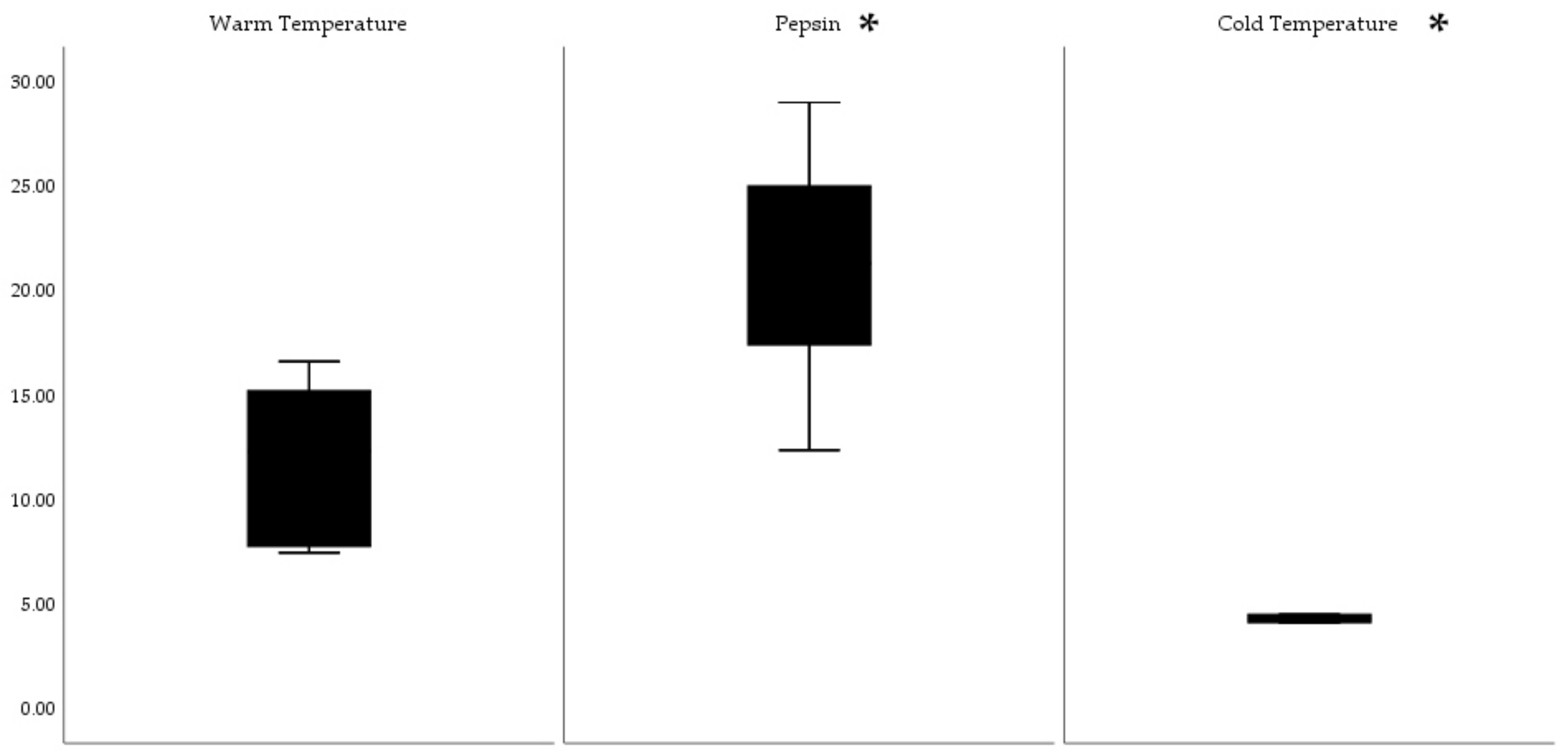
3.1. Isolation of Collagen

In this study, collagen was extracted from bone, skin, muscle, and general waste (a mixture of bone, muscle, and viscera in approximately equal proportions) obtained from the bluefin tuna farming industry. For each waste type, the yields for the ASC and PSC components were compared. The collagen yield was found to be lower for all ASC-tissue extracted, ranging from 1.2 g/100 g of collagen derived from general waste to 1.64 g/100 g from bone to 4.9 g/100 g from skin, compared to 3.3, 2.01, and 16.6 g/100 g, respectively, for the PSC components. The increased yield noted for PSC extraction occurred as the proteases used to cleave the telopeptide cross-linking regions of the collagen structure that represent the main sites of intra- and inter-molecular cross-linkage in the collagen structure, effectively exposing further collagen for extraction. The values reported in this study were consistent with those reported for other tuna, where the value obtained here for ASC skin (4.9%) was similar to that obtained for pacific bluefin tuna at 5.4% [33]. Similarly, for the PSC skin, the reported value is consistent with the 16.7% obtained by Ahmed et al. [34] for bigeye tuna (Thunnus obesus).
The sensorial qualities (appearance and odor) of the extracted PSC were found to be initially poor due to the presence of chromophores and as well as the use of acetic acid that left a residual odor. However, the introduction of a lengthier second salting-out step and slow addition of the crude collagen solution to the sodium chloride residue allowed for an improved separation of collagen from the inclusions present. Visual quality was further improved by a decolorization and deodorization step implemented during the second salting-out step using a concurrent treatment with 4% hydrogen peroxide, commonly used in the food and pharmaceutical industry [35]. Furthermore, after filtration of the salted-out material, the dissolution of collagen was carried out in citric acid, which eliminated residual acetic acid odor in the final product (see the flow diagram in Figure 1).
Microbiological analysis of six common microbial contaminates (Escherichia coli Enterobacter spp., Klebsiella spp., Aerobacter aerogenes, and Staphylococcus epidermidis and aureus) was also carried out; all tests were negative, and no evidence of these bacteria was found; furthermore, the total count for bacteria and fungi proved to be negative.

3.2. Characterization of Extracted Collagen

The molecular weight of the different collagen samples obtained in the preceding steps was determined using denaturing SDS-PAGE. The banding patterns observed are presented in Figure 2.
All bluefin tuna collagen samples were found to consist of two α tropocollagen chains (α1 and α2) with an estimated molecular weight of approximately 120–130 kDa, a dimer of two alpha chains that made up the β component (approximately 250 kDa), and a trimer referred to as the gamma component (γ). This is very similar to that reported for other fish and tuna species [36]. The absence of any other bands further down the gel in most lanes indicated that the collagen structure was intact and confirmed the absence of extraneous non-specific proteins. The pattern discussed is indicative of the type I collagen found primarily in skin and bone [37].
To further confirm the presence of collagen present in the different samples, the quantity of hydroxproline as a percentage of the total protein was assessed. Uniquely, hydroxyproline is found at high levels in collagen, where it stabilizes the triple helix and may, therefore, be used to quantify collagen. Hyroxyproline (HYP) was found to range from 7.44% in general waste up to 12.14% in bone and 14.40% in skin. The HYP value recorded in this study for general waste was comparable to the +6.41% to 7.8% previously described for other tuna species [34,38]. In the case of bone and skin, no direct comparison was possible with existing literature; however, the % HYP in bone is comparable to the one reported by Atma et al. [39] for catfish. Skin possessed a slightly higher % of hydroyproline when compared to the reported 9.86% value by Nguyen et al. [40].

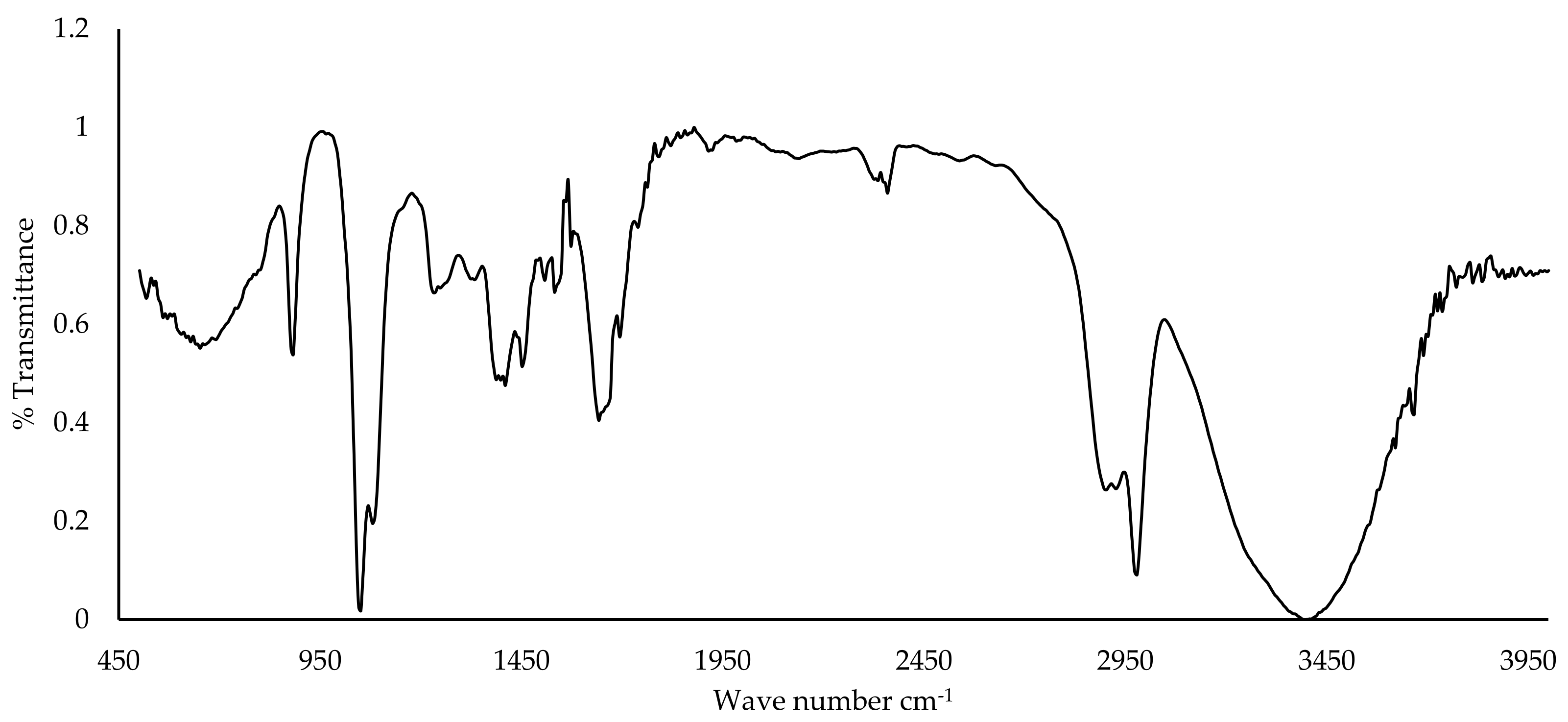
3.3. FTIR Spectra of Collagen

FTIR spectroscopy was used to study the functional groups present in the collagen from general waste and bone samples, as seen in Figure 3.
All the spectra obtained for the collagen extracted from different source tissues were similar to known FTIR spectra for tuna collagen [32], where the characteristic amide III, II, and I bands, as well as amide A and B, were observed. The amide I band is associated with protein secondary structures and was observed at around 1650 cm−1. This is in agreement with Muyonga et al. [41] and Jeong et al. [42], who noted that this band may be observed between 1600 and 1700 cm−1 The amide II band reflects the N-H bending vibrations coupled to C-N stretching, while the two amide A and B bands that were observed over 3450 and 2950 cm−1 represent N-H stretching associated with hydrogen bonding and CH2 asymmetric stretching.

3.4. Heavy Metal Assessment

An assessment of heavy metals in the collagen samples from different tissues was carried out and the results are shown in Table 1. Copper and nickel were not detected in any of the tissues studied, while zinc, cobalt, and iron were found at levels that were lower than that found in the equivalent amount of control tissue and within the limits set by Regulation (EC) No. 1223/2009 for heavy metal contents in cosmetics [43]. Mercury was found at levels below 0.1 mg/kg (0.1 ppm), whilst copper and nickel were below the LOD.

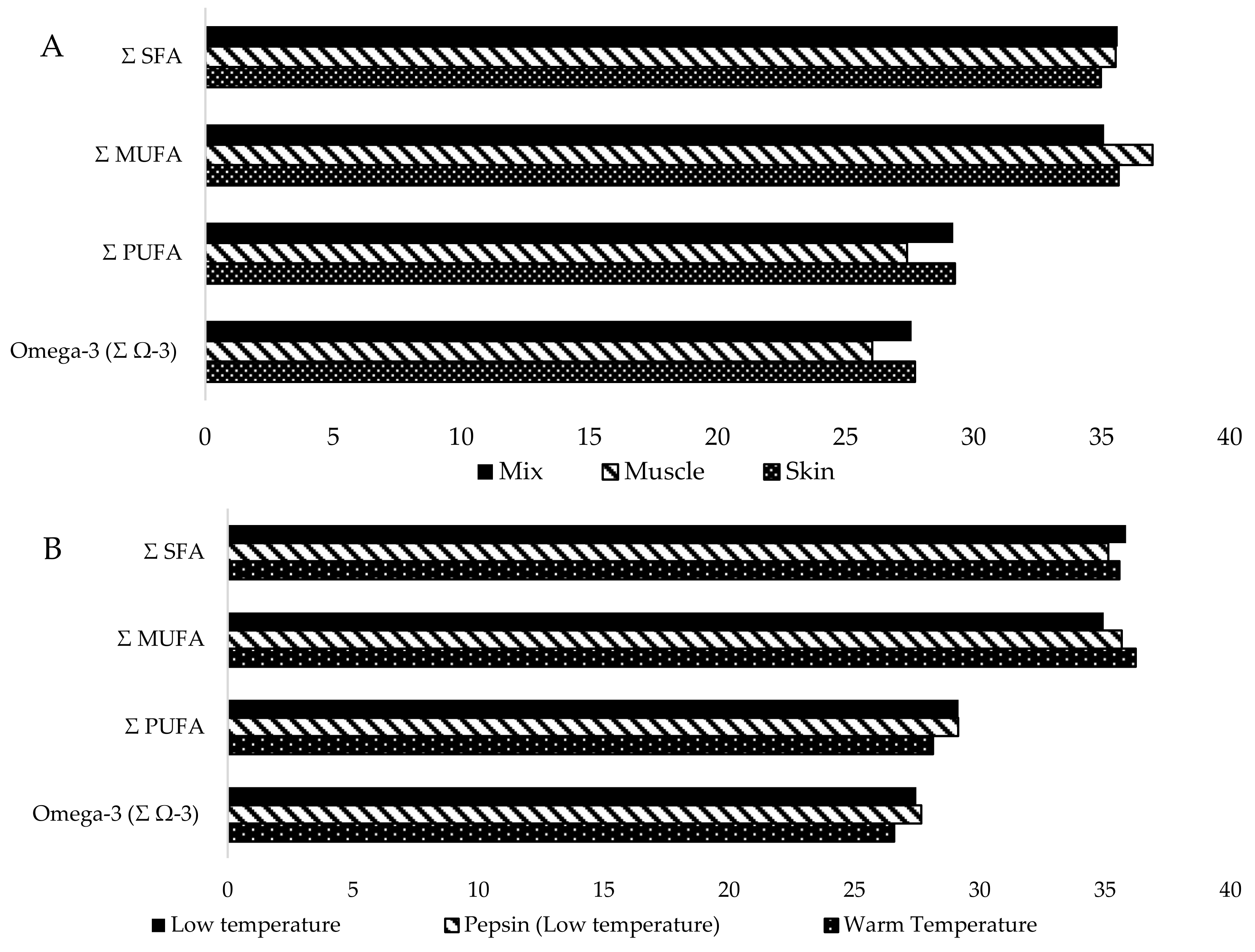
3.5. Tuna Oil Extraction Yields

The oil yields obtained in Figure 4 and Figure 5 showed considerable differences between both the tissues and the extraction methods. Enzymatically aided extraction exhibited the highest yields (12.5–29.0 mL/100 g) for all three tissue types when compared to the cold-extraction and high-temperature extraction methods (Figure 4). Skin tissue treated with pepsin produced the highest percentage yield at 29%, closely followed by mixed tissue treated with pepsin at 25%. An ANOVA test was then carried out to assess whether there were any significant differences between the extraction methods. When testing grouped treatments (Warm Temp, Pepsin, Cold Temp), the ANOVA null hypothesis was rejected (p > 0.05, Sig: 0.002), indicating a significant difference between groups. Post-hoc Bonferroni analysis revealed that a significant difference was observed between cold temperature and pepsin treatments (Sig: 0.003, 95% confidence interval). ANOVA testing between the three tissue types tested (assorted muscle, skin, and mixed tissue) indicated no significant difference (Sig: 0.379, <0.05).

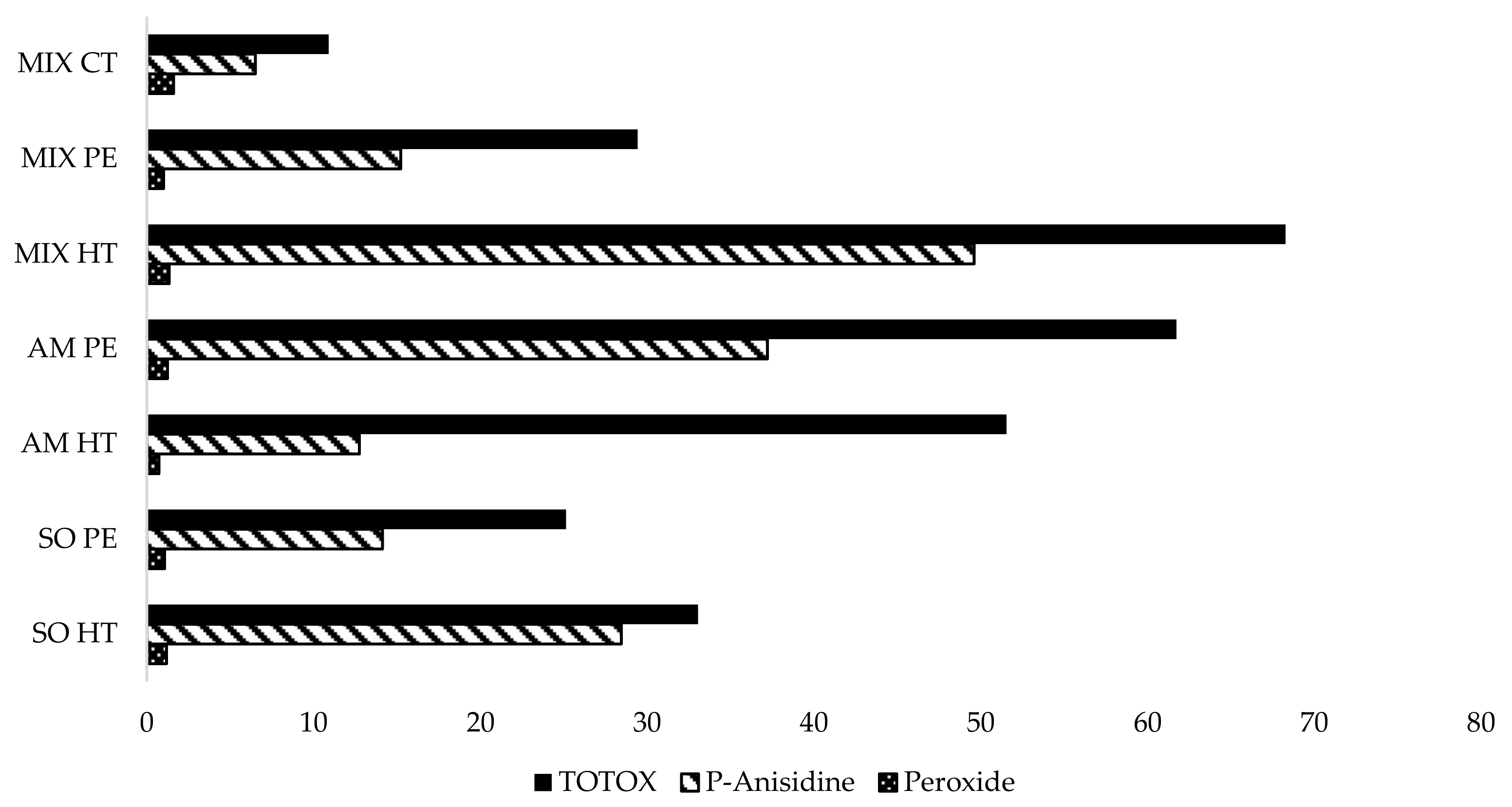
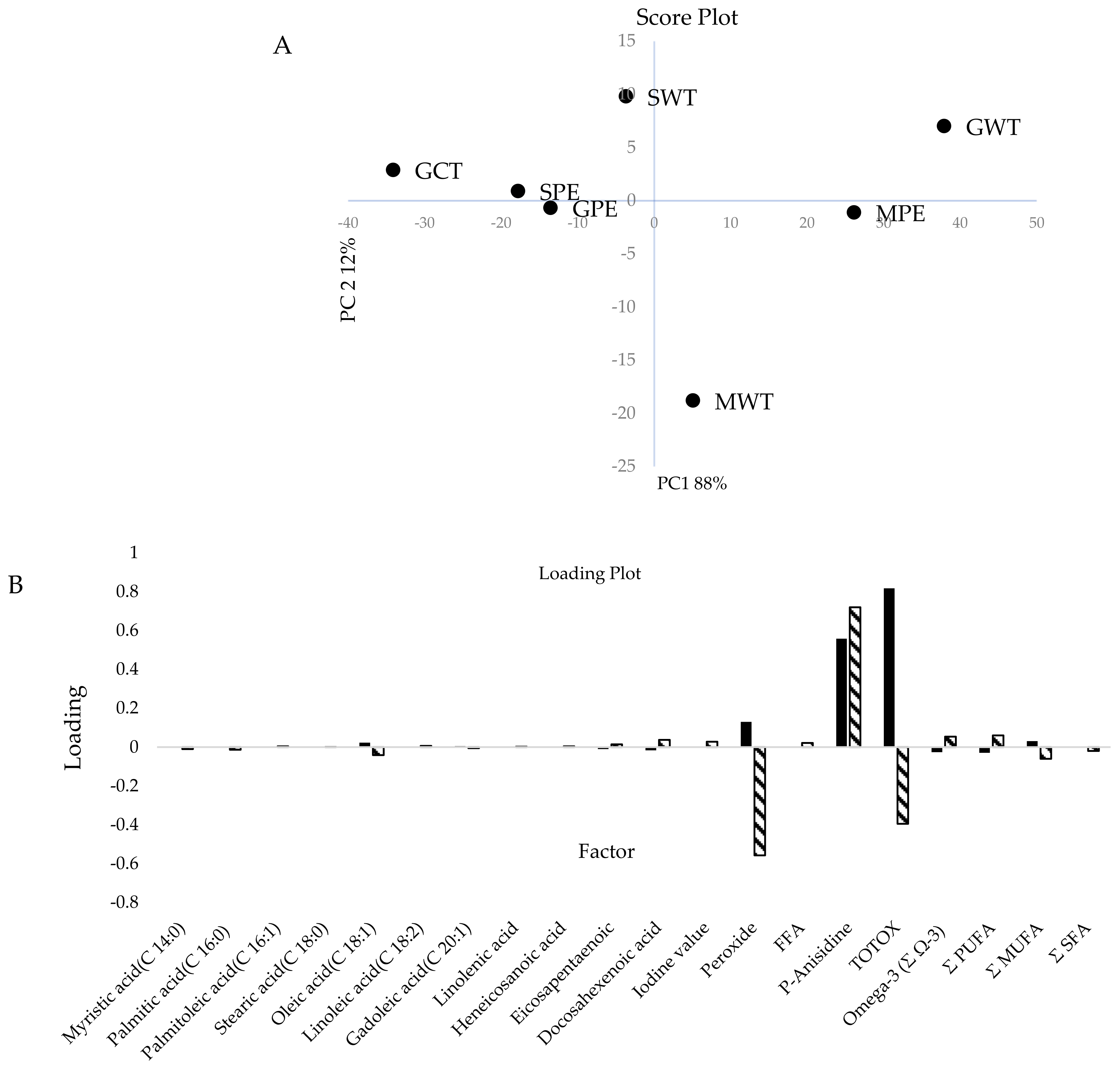
3.6. GC-FID Fatty Acid Profile Analysis and Oxidation Parameters

In general, individual fatty acid component concentration, according to gas chromatography, was consistent across extraction methods and tissue types. Figure 6 shows the major fatty acids obtained from the extracted oils, whilst Figure 7A shows that warm extracted oils, as well as muscle-derived oils (shown in Figure 7B), have higher overall levels of monounsaturated fatty acids (MUFAs) as well as lower levels of polyunsaturated fatty acids (PUFAs) when compared to other tissues and treatment types.
TOTOX values for the oil samples tested indicated higher values for oils extracted using higher temperatures. Figure 8 shows that samples treated with high-temperature extraction exhibited total oxidation values that were considerably higher than those at lower temperatures (CT and PE). Samples extracted from assorted muscles also exhibited higher TOTOX values.
Application of principal component analysis carried out on the data obtained from the fatty acid profile and oxidation testing, shown in Figure 9, indicated that by the second principal component, 100% of the variability observed within the oils could be explained. From the analysis of the loading plots obtained, it was found that the oxidation parameters, namely, the amount of peroxides and secondary oxidation products, were responsible for the observed variation in the data. Analysis of the score plot showed the existence of two major clusters. The first group was found to contain oils derived using the thermal process, whilst the second group was found to contain oils that were obtained under cold extraction. The only exception was for oils obtained using muscle tissue, both thermal and non-thermally extracted.

4. Discussion

4.1. Collagen Isolation

In this study, clear differences in collagen yield were noted between tissue types, with the PSC from skin yielding significantly higher yields (16.6% of dry collagen/wet initial mass) than those from bone and general waste. The lower yield from bone was unexpected as it contains a higher protein percentage (10.2–21.7% protein) according to proximate composition analysis in tuna species [44]. This, however, may be attributed to the much greater effort needed for the maceration and demineralization of the rigid structure. The low yields from general waste may be ascribed to the presence of muscle and viscera, which, on average, contain higher moisture and lipid levels and lower protein. These results may justify the removal of muscles and viscera from the carcass at the outset since expenditure on chemical treatment achieves only a slight increase in collagen yield.
The average protein content in the skin from various tuna species is known to range between 20.54–36.09%, varying with species, age, size, and environmental conditions [44,45]. Considering that around 80% of total fish skin protein is comprised of collagen, the 16.6% yield obtained from the PSC skin extraction represents a high percentage of the maximum potential yield [46]. Certainly, the intense feeding program to which farmed tuna are subjected within a restricted environment probably contributed to the overall high isolation values reported in this study.
To improve the sensorial qualities of the collagen product, different methods were tested and various improvements were made. In particular, residual odors of fish and acetic acid, as well as pigmentation and occlusion of insoluble material, were found to persist in the earlier extraction methods. These are not considered acceptable in products designed for cosmetic preparations. The undesirable odor attributed to the presence of fish oil contaminants in the final product was addressed by the addition of a 48 h defatting step using 10% butanol, which effectively removed most contaminating fish oils. The acetic acid smell, pigmentation, and occlusions of insoluble material were removed by introducing an extra cleaning step and redissolving the salted out “contaminated” collagen in 0.5 M 4:1 v/v, citric acid pH 2.2, 0.3% hydrogen peroxide. This removed the traces of acetic acid and allowed for the occluded insoluble material to be separated from the solution and removed through centrifugation. The use of 0.3% hydrogen peroxide was effective in oxidizing the residual chromophores and hemoglobin present with the collagen. The dissolved collagen was resalted out using a drop-wise addition of the dissolved collagen in twice the final amount of 2.2 M sodium chloride containing 4% of hydrogen peroxide under vigorous stirring. The use of larger volumes and vigorous stirring effectively controlled the rate of collagen precipitation. The slower the rate of precipitation, the purer the final product obtained. Controlling the rate of collagen precipitation effectively prevented the occlusion of any remaining insoluble material. This method takes advantage of the Von Weymarn equation and theory, whereby the particle size of precipitates is inversely proportional to the relative supersaturation of the solution [47].
By keeping the concentration of solutions low and the solubility of the compound of interest high through pH adjustment, larger precipitate particles were formed; the larger the particles, the smaller the surface area to volume ratio for any contaminants to get occluded and absorbed into the precipitated collagen. The solution obtained was refrigerated at 4 °C for 12 h; this, in turn, further reduced the solubility of collagen and resulted in a solid pallet that floated on top of the mother liquor. The further addition of 4% hydrogen peroxide in contact with the precipitated-out collagen was found to effectively reduce the coloration of the final product.

4.2. Characterization of Collagen

FTIR spectrum gives a good indication of the presence and general structure of the collagen; however, this data cannot be used as an exclusive test for the presence and purity of collagen. All bluefin tuna collagen samples were found to consist of two α tropocollagen chains (α1 and α2), with an estimated molecular weight of approximately 120–130 kDa, a dimer of two alpha chains that make up the β component (approximately 250 kDa), and a trimer that is the gamma component, representing a trimer of α chains. This is similar to that reported for other fish and tuna species [36]. The absence of any other bands further down the gel in most lanes indicated that the collagen structure was intact and confirmed the absence of extraneous non-specific proteins. The pattern discussed is indicative of the type I collagen found primarily in skin and bone [37].
To further confirm the presence of collagen in different samples, the quantity of hydroxyproline as a percentage of the total protein was assessed. Uniquely, hydroxyproline is found at high levels in collagen, where it stabilizes the triple helix and thus may be used to quantify collagen. Hyroxyproline (HYP) was found to range from 7.44% in general waste up to 12.14% in bone and 14.40% in skin. The HYP value recorded in this study for general waste was comparable to the +6.41% to 7.8% previously described for other tuna species [33,38]. These low values may be due to the presence of muscle and viscera content, which have lower values of collagenous protein and, hence, hydroxyproline. The purified collagen obtained from skin and bone seems to be in agreement with the 12.5% of hydroxyproline content specified by Edwards and O’Brien [48].
Assessment of the heavy metals (Zn, Cr, Co, Cu, Ni, Fe, and Pb) in the collagen samples from skin, bone, and muscle, with the exception of cadmium in bone, was either negative or present at levels that did not exceed the limits of the regulations such as Regulation (EC) No. 853/2004 [43]. Likewise, the outsourced assessment of mercury was also within the permitted limits of the same regulation. The general absence or low levels of these heavy metals may be linked to the farmed origin of the fish and the nature of the (collagen) source material. Lares et al. suggested that farmed tuna tend to have lower mercury and cadmium levels as the fed diet contains fewer heavy metals [49]. In general, it was found that collagen obtained from bone had a higher amount of heavy metal content when compared to the general waste. These results are in agreement with the study carried out by Asgedom et al., where it was shown that a higher concentration of heavy metals tends to accumulate in the fish bones compared to the fleshy parts [50]. Although these results are promising, further testing on heavy metal deposition from other organs and tissues in Atlantic bluefin tuna, such as the liver, where most heavy metals would be concentrated, would be recommended if the product is to be considered for pharmaceutical or nutraceutical purposes. Despite this, it should be considered that most nutraceutical products and cosmetics containing collagen are very unlikely to be made primarily of collagen as they would be a part of formulations, mixtures, and creams and would therefore contain a considerable number of additives, reducing the concentration of potentially harmful heavy metals per serving/application.
Microbiological analysis of the extracted collagen indicated the complete absence of the six common microbial species tested. This is to be expected as the acetic acid and, later, citric acid used in the extraction process are effective antimicrobial and bactericidal agents even at the low concentrations used in this study. Furthermore, the high NaCl concentrations utilized in the separation process would further reduce the risk of microbial contamination [51].

4.3. Oil Extraction

The high-temperature extraction is the most analogous to the traditional extraction method. This was even more effective when compared to the cold extraction method. This is due to the higher temperature, allowing for the mechanical separation of the solid and liquid fractions, the rupture of adipocytic tissue, and the subsequent release of the oils in the liquid phase. The use of enzymes such as pepsin allows for the breakdown of the tissues at lower temperatures; this method seems to be more effective in the liberation of the oil globules in the adipocytic tissue [52,53].
Much like the collagen extraction process, limitations regarding the maceration and filtration of the material are also a very considerable bottleneck in the retrieval of this material. Time-related issues may be more problematic for lipid extraction when compared to collagen extraction as processes occurring at room temperature or higher for a prolonged period may influence the quality of the final product considerably. This means that for larger-scale procedures, more efficient and temperature-controlled procedures may need to be implemented in order to produce products of the highest quality. TOTOX values indicated that samples treated with high temperature exhibited total oxidation values that were considerably higher than those at lower temperatures (CT and PE). Samples extracted from assorted muscle also exhibited higher TOTOX values; this may be indicative of accelerated oxidation of the lipids due to the presence of natural components present in muscle and viscera, especially iron, myoglobin, hydrogen peroxide, and ascorbic acid, which are known to accelerate lipid oxidation. These act as catalysts that induce the formation of reactive oxygen species [54].
The Global Organization for EPA and DHA Omega-3s (GOED) gives a global target for the TOTOX value that unflavored fish oils must have. It indicates that a TOTOX value exceeding 26 is unsuitable. From the analysis of the oils obtained, only Mix-CT and SO-PE fell within these parameters, whilst the others exceeded it. Mix HT and AM PE/WT were by far the highest in TOTOX value; this indicates that muscle and viscera content, coupled with high-temperature extraction, considerably affected the quality of the oil [54].
Oils obtained using muscle tissue (both thermally and non-thermally extracted) have been found to have a high concentration of both primary oxidation and secondary oxidation products. This is mainly attributed to the presence of a higher concentration of iron (as hemoglobin) present within the muscle tissue. The results obtained are consistent with the literature, given that the major factors affecting the rate of oxidation are the degree of unsaturation, the amount of oxygen, temperature and light, and the presence of metals (mainly transition metals such as Fe and Cu) [55,56,57]. Benedet and Shibamoto demonstrated that trace amounts of Fe, Cu, Cr, Pb, and Cd contribute oxidative effects to lipid peroxidation [58].
The presence of these trace metals enhanced the rate of oxidation of edible oils by increasing the generation of free radicals from fatty acids and hydroperoxides. On the other hand, it was found that thermal extracted oils derived from skin had a lower degree of oxidation; this was mainly attributed to the lack of iron and other hydrolytic enzymes that might be present in the viscera used in the general waste. This could be due to several reasons; however, a number of studies have shown that fish skin contains a significant antioxidant effect, derived from the presence of proteins shown in studies carried out on Alaska pollack skin gelatin hydrolysate [59], yellowfin (Thunnus albacares) fish protein [60], and snakehead fish skin [61].

4.4. Circularity

There is an ever-growing need to make more efficient use of already available biomaterials through circularity. This reduces pressure on the natural environment and primary streams [62]. Additionally, the sustainable consumption of resources was recently listed as one of the priority areas of the European Green Deal, as the European Union (EU) has also recognized the importance of reducing the mining of natural resources [63].
In the light of the results demonstrated in this study, it may be worthwhile for local Atlantic bluefin tuna ranching operators to invest in waste valorization technologies that will not only contribute to the CEAP “less waste, more value” priority and lessen the environmental burden of their industry but also potentially introduce an additional revenue stream. Waste re-utilization and valorization could also contribute and form part of the already existing strategies put forward by the Aquaculture directorate in the “Aquaculture strategy for the Maltese islands 2014–2025” and therefore ensure responsible waste management, forming part of the permit and license conditions for the operation of tuna penning activities in Maltese waters. The same obligations would also be relevant to the rest of the Mediterranean ranching industry [64].

5. Conclusions

This study is concerned with the valorization of waste generated from the local ranched Atlantic bluefin tuna industry. Significantly higher yields of collagen were found to be obtained from the pepsin-extracted component derived from tuna skin (16.6%) when compared to other components. Furthermore, improvements to the quality and quantity of collagen, using a lengthier salting-out step, defatting through butanol, a peroxide treatment, and the use of citric acid instead of acetic acid to reduce the chromophore persistence and residual odor observed in previously preliminary extraction procedures, are described. Collagen extracted from the by-products of the Atlantic bluefin tuna industry may, therefore, be of considerable value to the nutraceutical and pharmaceutical industries. This is of particular relevance, given that the high cost of raw materials during marine-based collagen formulation has been identified as one of the major constraints hindering market growth. Satisfactory lipid retrieval, with a substantial presence of ω-3 poly-unsaturated fatty acids, was also achieved, with skin and general waste samples yielding over 20% lipid composition. It was shown that the tissue type and extraction method greatly affected the quality and quantity of oils obtained.
Further effort into the optimization of both collagen and lipid extraction protocols is recommended, focusing on the production of high-value fractions that are much closer to the quality required for human use/consumption. The latter objective is challenging at the laboratory scale, where scaling-up infrastructure is limited and not tailored to produce such fractions. The implementation of better maceration, agitation (including ultrasonication), filtration, and centrifugation methods are expected to increase collagen and oil yield and quality considerably. Investigating alternative green methods of extraction that could reduce the amount of toxic waste by-products generated during the extraction procedures would also be of great interest, given the current emphasis within the EU’s Green Deal of the DNSH (Do No Significant Harm) principle.
It is also suggested that the impact of changes to policies, such as waste management becoming an integral part of the license agreement, be considered. Finally, this study is an important step to make better use of already-available high-value biomaterials through circularity. These results indicate that it may be possible to reduce the vast volumes of biomass currently being disposed of into the sea. Such endeavors would also generate new revenue streams while contributing to a more sustainable industry.

Author Contributions

Conceptualization, N.C., F.L., M.Z.M., V.A. and A.D.; methodology, N.C., F.L. and M.Z.M.; investigation, N.C. and F.L.; resources, M.Z.M., V.A. and A.D.; data curation, N.C. and F.L.; writing—original draft preparation, N.C., F.L., and M.Z.M.; writing—review and editing, N.C., F.L., M.Z.M. and A.D.; visualization, N.C. and F.L.; supervision, M.Z.M.; project administration, M.Z.M., V.A., J.G. and A.D.; funding acquisition, M.Z.M., V.A. and A.D. All authors have read and agreed to the published version of the manuscript.

Funding

This work has been financially supported by the Bythos Interreg V-A Italia-Malta 2014–2020 cooperation project, approved for funding in 2018 (Project code C1-1.1-9).

Institutional Review Board Statement

Not applicable.

Data Availability Statement

The data presented in this study is available on request from the corresponding author Neil Cutajar.

Acknowledgments

We thank Pamela Cuschieri for her initial preliminary work in the extraction of collagen.

Conflicts of Interest

The authors declare no conflict of interest.

References

  1. FAO. The State of World Fisheries and Aquaculture 2018—Meeting the Sustainable Development Goals. Rome. Licence: CC BY-NC-SA 3.0 IGO 2020. Available online: https://www.fao.org/3/ca9229en/ca9229en.pdf (accessed on 18 October 2021).
  2. NSO. Aquaculture: 2019 (Rep.). Malta: National Statistics Office 2020. Available online: https://nso.gov.mt/en/News_Releases/Documents/2021/11/News2021_210.pdf (accessed on 10 November 2021).
  3. Ferraro, V.; Cruz, I.B.; Jorge, R.F.; Malcata, F.X.; Pintado, M.E.; Castro, P.M.L. Valorisation of natural extracts from marine source focused on marine by-products: A review. Food Res. Int. 2010, 43, 2221–2233. [Google Scholar] [CrossRef]
  4. WWF. Bluefin Symposium Celebrates Conservation Gains While Urging Continued Action; WWF: Morges, Switzerland, 2021; Available online: https://www.wwfmmi.org/?2090941%2FBluefin-Symposium-Celebrates-Conservation-Gains--While-Urging-Continued-Action (accessed on 15 January 2022).
  5. McKinney, R. A Global Tuna Valuation: Netting Billions 2020. A Global Tuna Valuation: Netting Billions 2020|The Pew Charitable Trusts. 2020. Available online: https://www.pewtrusts.org/en/research-and-analysis/reports/2020/10/netting-billions-2020-a-global-tuna-valuation (accessed on 15 January 2022).
  6. European Commision. Communication from the Commission to the European Parliament, the Council, the European Eco-nomic and Social Committee and the Committee of the Regions. Eur-Lex. Available online: https://eur-lex.europa.eu/legal-content/EN/TXT/?qid=1583933814386&uri=COM%3A2020%3A98%3AFIN (accessed on 14 January 2022).
  7. Caruso, G. Fishery Wastes and By-products: A Resource to Be Valorised. J. Fish. Sci. 2015, 9, 80–83. [Google Scholar]
  8. Johnson, C.; Ruiz Sierra, A.; Dettmer, J.; Sidiropoulou, K.; Zicmane, E.; Canalis, A.; Llorente, P.; Paiano, P.; Mengal, P.; Puzzolo, V. The bio-based industries joint undertaking as a catalyst for a green transition in Europe under the European Green Deal. EFB Bioecon. J. 2021, 1, 100014. [Google Scholar] [CrossRef]
  9. Nagai, T. Isolation of collagen from fish waste material—Skin, bone and fins. Food Chem. 2000, 68, 277–281. [Google Scholar] [CrossRef]
  10. Jongjareonrak, A.; Benjakul, S.; Visessanguan, W.; Tanaka, M. Isolation and characterization of collagen from bigeye snapper (Priacanthus macracanthus) skin. J. Sci. Food Agric. 2005, 85, 1203–1210. [Google Scholar] [CrossRef]
  11. Raman, M.; Gopakumar, K. Fish Collagen and its Applications in Food and Pharmaceutical Industry: A Review 2018. Available online: https://www.ecronicon.com/ecnu/pdf/ECNU-13-00529.pdf (accessed on 12 October 2020).
  12. Jafari, H.; Lista, A.; Siekapen, M.M.; Ghaffari-Bohlouli, P.; Nie, L.; Alimoradi, H.; Shavandi, A. Fish Collagen: Extraction, Characterization, and Applications for Biomaterials Engineering. Polymers 2020, 12, 2230. [Google Scholar] [CrossRef]
  13. Nagai, T.; Suzuki, N. Preparation and characterization of several fish bone collagens. J. Food Biochem. 2000, 24, 427–436. [Google Scholar] [CrossRef]
  14. Karayannakidis, P.D.; Zotos, A. Fish Processing By-Products as a Potential Source of Gelatin: A Review. J. Aquat. Food Prod. Technol. 2014, 25, 65–92. [Google Scholar] [CrossRef]
  15. Silva, T.; Moreira-Silva, J.; Marques, A.; Domingues, A.; Bayon, Y.; Reis, R. Marine Origin Collagens and Its Potential Applications. Mar. Drugs 2014, 12, 5881–5901. [Google Scholar] [CrossRef] [Green Version]
  16. Skierka, E.; Sadowska, M. The influence of different acids and pepsin on the extractability of collagen from the skin of Baltic cod (Gadus morhua). Food Chem. 2007, 105, 1302–1306. [Google Scholar] [CrossRef]
  17. Pal, G.K.; Suresh, P.V. Sustainable valorisation of seafood by-products: Recovery of collagen and development of collagen-based novel functional food ingredients. Innov. Food Sci. Emerg. Technol. 2016, 37, 201–215. [Google Scholar] [CrossRef]
  18. Benjakul, S.; Thiansilakul, Y.; Visessanguan, W.; Roytrakul, S.; Kishimura, H.; Prodpran, T.; Meesane, J. Extraction and characterisation of pepsinsolubilised collagens from the skin of bigeye snapper (Priacanthus tayenus and Priacanthus macracanthus). J. Sci. Food Agric. 2010, 90, 132–138. [Google Scholar] [CrossRef]
  19. Sahena, F.; Zaidul, I.S.M.; Jinap, S.; Saari, N.; Jahurul, H.A.; Abbas, K.A.; Norulaini, N.A. PUFAs in Fish: Extraction, Fractionation, Importance in Health. Compr. Rev. Food Sci. Food Saf. 2009, 8, 59–74. [Google Scholar] [CrossRef]
  20. Hedge, A. EPA/DHA (Omega 3) Ingredients Market to Hit $5.7 Billion. 2019. Available online: https://www.prnewswire.com/news-releases/epadha-omega-3-ingredients-market-to-hit-5-7-billion-by-2026-global-market-insights-inc-300953616.html (accessed on 17 October 2020).
  21. Hilleman, D.E.; Wiggins, B.S.; Bottorff, M.B. Critical differences between dietary supplement and prescription omega-3 fatty acids: A narrative review. Adv. Ther. 2020, 37, 656–670. [Google Scholar] [CrossRef] [PubMed] [Green Version]
  22. Ghaly, A.E.; Ramakrishnan, V.V.; Brooks, M.S.; Budge, S.M.; Dave, D. Fish Processing Wastes as a Potential Source of Proteins, Amino Acids and Oils: A Critical Review. J. Microb. Biochem. Technol. 2013, 5, 107–129. [Google Scholar] [CrossRef] [Green Version]
  23. FAO. The Production of Fish Meal and Oil. Rome: Food and Agriculture Organization of the United Nations. 1986. Available online: https://www.fao.org/3/X6899E/X6899E02.html (accessed on 17 October 2020).
  24. Arruda, L.F.; Borghesi, R.; Portz, L.; Cyrino, J.E.; Oetterer, M. Fish silage in black bass (Micropterus salmoides) feed as an alternative to fish meal. Braz. Arch. Biol. Technol. 2009, 52, 1261–1266. [Google Scholar] [CrossRef] [Green Version]
  25. Rubio-Rodríguez, N.; de Diego, S.M.; Beltrán, S.; Jaime, I.; Sanz, M.T.; Rovira, J. Supercritical fluid extraction of fish oil from fish by-products: A comparison with other extraction methods. J. Food Eng. 2012, 109, 238–248. [Google Scholar] [CrossRef] [Green Version]
  26. Bonilla, J.R.; Concha, J.L.H. Methods of extraction refiningand concentration of fish oil as a source of omega-3 fatty acids. Transform. Agro-Ind. 2018, 19, 645–668. [Google Scholar]
  27. Mæhre, H.; Jensen, I.-J.; Eilertsen, K.-E. Enzymatic pre-treatment increases the protein Bioaccessibility and extractability in Dulse (Palmaria palmata). Mar. Drugs 2006, 14, 196. [Google Scholar] [CrossRef] [Green Version]
  28. Sigma-Aldrich. MAK008 Technical Bulletin—Sigma-Aldrich. MAK008 Hydroxyproline Assay Kit Technical Bulletin. Available online: https://www.sigmaaldrich.com/deepweb/assets/sigmaaldrich/product/documents/418/543/mak008bul.pdf (accessed on 8 November 2021).
  29. Maniatis, T.; Fritsch, E.F.; Sambrook, J. Molecular Cloning: A Laboratory Manual; Cold Spring Harbor Laboratory: Cold Spring Harbor, NY, USA, 1982. [Google Scholar]
  30. Kenawy, I.M.M.; Hafez, M.A.H.; Akl, M.A.; Lashein, R.R. Determination by AAS of some trace heavy metal ions in some natural and biological samples after their preconcentration using newly chemically modified chloromethylated polystyrene-PAN ion exchanger. Analyt. Sci. 2000, 16, 493–500. [Google Scholar] [CrossRef] [Green Version]
  31. Loska, K.; Wiechuła, D. Application of principal component analysis for the estimation of source of heavy metal contamination in surface sediments from the Rybnik Reservoir. Chemosphere 2003, 51, 723–733. [Google Scholar] [CrossRef]
  32. Tapia, J.; Vargas-Chacoff, L.; Bertrán, C.; Pena-Cortes, F.; Hauenstein, E.; Schlatter, R.; Jimenez, C.; Tapia, C. Heavy metals in the liver and muscle of Micropogonias manni fish from Budi Lake, Araucania Region, Chile: Potential risk for humans. Environ. Monit. Assess. 2012, 184, 3141–3151. [Google Scholar] [CrossRef] [PubMed] [Green Version]
  33. Tanaka, T.; Takahashi, K.; Tsubaki, K.; Hirata, M.; Yamamoto, K.; Biswas, A.; Moriyama, T.; Kawamura, Y. Isolation and characterization of acid-soluble bluefin Tuna (Thunnus orientalis) Skin collagen. Fish. Aquat. Sci. 2018, 21, 1–8. [Google Scholar] [CrossRef]
  34. Ahmed, R.; Haq, M.; Chun, B.S. Characterization of Marine Derived Collagen Extracted from the by-Products of Bigeye Tuna (Thunnus Obesus). Int. J. Biol. Macromol. 2019, 135, 668–676. [Google Scholar] [CrossRef] [PubMed]
  35. Kersten, K.M.; Breen, M.E.; Mapp, A.K.; Matzger, A.J. Pharmaceutical solvate formation for the incorporation of the antimicrobial agent hydrogen peroxide. Chem. Commun. 2018, 54, 9286–9289. [Google Scholar] [CrossRef] [PubMed]
  36. Gómez-Guillén, M.C.; Turnay, J.; Fernández-Dıaz, M.D.; Ulmo, N.; Lizarbe, M.A.; Montero, P. Structural and physical properties of gelatin extracted from different marine species: A comparative study. Food Hydrocoll. 2002, 16, 25–34. [Google Scholar] [CrossRef] [Green Version]
  37. Rabotyagova, O.S.; Cebe, P.; Kaplan, D.L. Collagen structural hierarchy and susceptibility to degradation by ultraviolet radiation. Mater. Sci. Eng. C 2008, 28, 1420–1429. [Google Scholar] [CrossRef] [PubMed] [Green Version]
  38. Alemán, A.; Giménez, B.; Montero, P.; Gómez-Guillén, M.C. Antioxidant activity of several marine skin gelatins. LWT-Food Sci. Technol. 2011, 44, 407–413. [Google Scholar] [CrossRef]
  39. Sanaei, A.V.; Mahmoodani, F.; See, S.F.; Yusop, S.M.; Babji, A.S. Optimization of gelatin extraction and physico-chemical properties of catfish (Clarias gariepinus) bone gelatin. Int. Food Res. J. 2013, 20, 423–430. [Google Scholar]
  40. Nguyen, B.C.; Kha, T.C.; Nguyen, K.H.N.; Nguyen, H.M.X. Optimization of Enzymatic Hydrolysis of Collagen from Yellowfin Tuna Skin (Thunnus Albacares) by Response Surface Methodology and Properties of Hydrolyzed Collagen. J. Food Processing Preserv. 2021, 45, e15319. [Google Scholar] [CrossRef]
  41. Muyonga, J.H.; Cole, C.G.B.; Duodu, K.G. Characterisation of acid soluble collagen from skins of young and adult Nile Perch (lates niloticus). Food Chem. 2004, 85, 81–89. [Google Scholar] [CrossRef]
  42. Jeong, H.-S.; Venkatesan, J.; Kim, S.-K. Isolation and characterization of collagen from marine fish (Thunnus obesus). Biotechnol. Bioprocess Eng. 2013, 18, 1185–1191. [Google Scholar] [CrossRef]
  43. Regulation (EC) No 1223/2009 of the European Parliament and of the Council of 30 November 2009 on Cosmetic Products OJ L 342, 22.12. 2009, p. 59–209. Available online: https://eur-lex.europa.eu/legal-content/EN/ALL/?uri=celex%3A32009R1223 (accessed on 19 October 2021).
  44. Murthy, L.N.; Rao, B.M.; Prasad, M.M. Biochemical and Microbiological Evaluation of Tuna Loin Processing Waste. Fish. Technol. 2012, 49, 45–49. [Google Scholar]
  45. Karunarathna, U. Nutritional evaluation in five species of tuna. Nutr. Physiol. 2019. [Google Scholar] [CrossRef]
  46. Kołodziejska, I.; Skierka, E.; Sadowska, M.; Kołodziejski, W.; Niecikowska, C. Effect of extracting time and temperature on yield of gelatin from different fish offal. Food Chem. 2008, 107, 700–706. [Google Scholar] [CrossRef]
  47. Barlow, D.A.; Baird, J.K.; Su, C.-H. Theory of the von weimarn rules governing the average size of crystals precipitated from a supersaturated solution. J. Cryst. Growth 2004, 264, 417–423. [Google Scholar] [CrossRef]
  48. Edwards, C.A.; O’Brien, W.D., Jr. Modified assay for determination of hydroxyproline in a tissue hydrolyzate. Clin. Chim. Acta 1980, 104, 161–167. [Google Scholar] [CrossRef] [Green Version]
  49. Lares, M.L.; Huerta-Diaz, M.A.; Marinone, S.G.; Valdez-Márquez, M. Mercury and cadmium concentrations in farmed bluefin tuna (Thunnus orientalis) and the suitability of using the caudal peduncle muscle tissue as a monitoring tool. J. Food Prot. 2012, 75, 725–730. [Google Scholar] [CrossRef]
  50. Asgedom, A.G.; Besta, M.B.; Gebremedhin, Y.W. Bioaccumulation of Heavy Metals in Fishes of Hashenge Lake, Tigray, Northern Highlands of Ethiopia. Am. J. Chem. 2012, 2, 326–334. [Google Scholar] [CrossRef]
  51. Ryssel, H.; Kloeters, O.; Germann, G.; Schäfer, T.; Wiedemann, G.; Oehlbauer, M. The antimicrobial effect of acetic acid—an alternative to common local antiseptics? Burns 2009, 35, 695–700. [Google Scholar] [CrossRef]
  52. Bimbo, A.P. Fish Meal and Oil. In The Seafood Industry; Martin, R.E., Flick, G.J., Eds.; Springer: Boston, MA, USA, 1990. [Google Scholar]
  53. Taati, M.M.; Shabanpour, B.; Ojagh, S.M. Investigation on fish oil extraction by enzyme extraction and wet reduction methods and quality analysis. AACL Bioflux 2018, 11, 83–90. [Google Scholar]
  54. Amaral, A.B.; Silva, M.V.; Lannes, S.C. Lipid oxidation in meat: Mechanisms and protective factors—A review. Food Sci. Technol. 2018, 38 (Suppl. S1), 1–15. [Google Scholar] [CrossRef] [Green Version]
  55. GOED, Technical Guidance Documents. 2020. Available online: https://scioninstruments.com/wp-content/uploads/2020/11/TGD-2020-06-29.pdf (accessed on 24 October 2021).
  56. Meira, M.; Quintella, C.M.; dos Santos Tanajura, A.; da Silva, H.G.R.; Fernando, J.D.S.; da Costa Neto, P.R.; Pepe, J.M.; Santos, M.A.; Nascimento, L.L. Determination of the Oxidation Stability of Biodiesel and Oils by Spectrofluorimetry and Multivariate Calibration. Talanta 2011, 85, 430–434. [Google Scholar] [CrossRef] [PubMed] [Green Version]
  57. Sikwese, F.E.; Duodu, K.G. Antioxidant Effect of a Crude Phenolic Extract from Sorghum Bran in Sunflower Oil in the Presence of Ferric Ions. Food Chem. 2007, 104, 324–331. [Google Scholar] [CrossRef] [Green Version]
  58. Benedet, J.A.; Umeda, H.; Shibamoto, T. Antioxidant Activity of Flavonoids Isolated from Young Green Barley Leaves toward Biological Lipid Samples. J. Agric. Food Chem. 2007, 55, 5499–5504. [Google Scholar] [CrossRef] [PubMed]
  59. Kim, S.-K.; Kim, Y.-T.; Byun, H.-G.; Nam, K.-S.; Joo, D.-S.; Shahidi, F. Isolation and characterization of antioxidative peptides from gelatin hydrolysate of Alaska pollack skin. J. Agric. Food Chem. 2001, 49, 1984–1989. [Google Scholar] [CrossRef]
  60. Jun, S.-Y.; Park, P.-J.; Jung, W.-K.; Kim, S.-K. Purification and characterization of an antioxidative peptide from enzymatic hydrolysate of yellowfin sole (Limanda aspera) frame protein. Eur. Food Res. Technol. 2004, 219, 20–26. [Google Scholar] [CrossRef]
  61. Baehaki, A.; Widiastuti, I.; Nainggolan, C.; Gofar, N. Antioxidant activities of snakehead (Channa Striat A) fish skin: Peptides hydrolysis using protease tp2 isolate from swamp plant silage. Potravinarstvo Slovak J. Food Sci. 2020, 14, 379–384. [Google Scholar] [CrossRef]
  62. Andersen, S.; Allen, B.; Domingo, G.C. Biomass in the EU Green Deal: Towards Consensus on the Use of Biomass for EU Bioenergy’ Policy Report, 2021. Available online: https://ieep.eu/uploads/articles/attachments/a14e272d-c8a7-48ab-89bc-31141693c4f6/Biomass%20in%20the%20EU%20Green%20Deal.pdf?v=63804370211 (accessed on 29 December 2021).
  63. Camilleri, M.A. European environment policy for the circular economy: Implications for business and industry stakeholders. Sustain. Dev. 2020, 28, 1804–1812. [Google Scholar] [CrossRef]
  64. Borg, M. Aquaculture-from Conflict to Blue Growth: An Example of Aquaculture Cages Re-Allocation. The European Maritime Spatial Planning Platform. 2018. Available online: https://maritime-spatial-planning.ec.europa.eu/practices/aquaculture-conflict-blue-growth-example-aquaculture-cages-re-allocation (accessed on 14 January 2022).
Figure 1. Flow diagram indicating steps involved in the pre-treatment, extraction, retrieval, and purification of Atlantic bluefin tuna collagen.
Applsci 12 01542 g001
Figure 2. SDS-PAGE pattern showing various collagen extracts from left to right: Protein ladder, PSC-Skin 1, PSC-General Waste, PSC-Bone, ASC-Skin, ASC-General waste. Gamma, beta, and alpha bands are clearly visible.
Applsci 12 01542 g002
Figure 3. FTIR spectra for collagen extracted from different bluefin tuna waste tissues.
Applsci 12 01542 g003
Figure 4. Yield (mL per 100 g) of oils from different tissues and treatments (MIXWT—mixed tissue warm temperature; MIXPE—mixed pepsin extraction; MIXCT—mixed tissue cold temperature; AMWT—assorted muscle warm temperature; AMPE—assorted muscle pepsin extraction; SOWT—skin only warm temperature; SOPE—skin only pepsin extraction).
Applsci 12 01542 g004
Figure 5. Average yield (mL per 100 g) of oils from different treatments, grouping together tissue types. Significant difference indicated between pepsin and cold temperature (*).
Applsci 12 01542 g005
Figure 6. % amount of different fatty acids determined by GC-FID, obtained using different extraction temperatures.
Applsci 12 01542 g006
Figure 7. % amount of different saturated fatty acids (SFAs), monounsaturated fatty acids (MUFAs), polyunsaturated fatty acids (PUFAs), and omega-3 fatty acids, obtained using different parts (A) and different extraction temperatures (B).
Applsci 12 01542 g007
Figure 8. Fish oil oxidation parameters obtained from mix fish waste (MIX), muscle (AM), and skin (SO) using different extraction temperatures: CT (cold temperature), HT (high temperature), and pepsin-assisted extraction (PE).
Applsci 12 01542 g008
Figure 9. Principal component analysis on the various fish oils obtained using fatty acid profile data and oxidation parameters showing two clusters in the score plot (A) and the loadings of each factor contributing to the observed variation (B).
Applsci 12 01542 g009
Table 1. Summary of heavy metals (mg/kg) present in different tissues.
Heavy MetalBone PSCBone ASCGeneral Waste 1General Waste 2Bovine Collagen
Cd0.081.560.08<0.01<0.01
Zn5.128.050.580.420.42
Cr3.080.380.150.090.09
Cu----<0.01
Co5.962.190.08<0.01-
Ni-----
Pb1.09-0.02<0.01<0.01
Fe0.201.460.200.240.24
Publisher’s Note: MDPI stays neutral with regard to jurisdictional claims in published maps and institutional affiliations.

