
Nanodevices for Biological and Medical Applications: Development of Single-Molecule Electrical Measurement Method


Abstract
:Featured Application
Abstract
1. Introduction
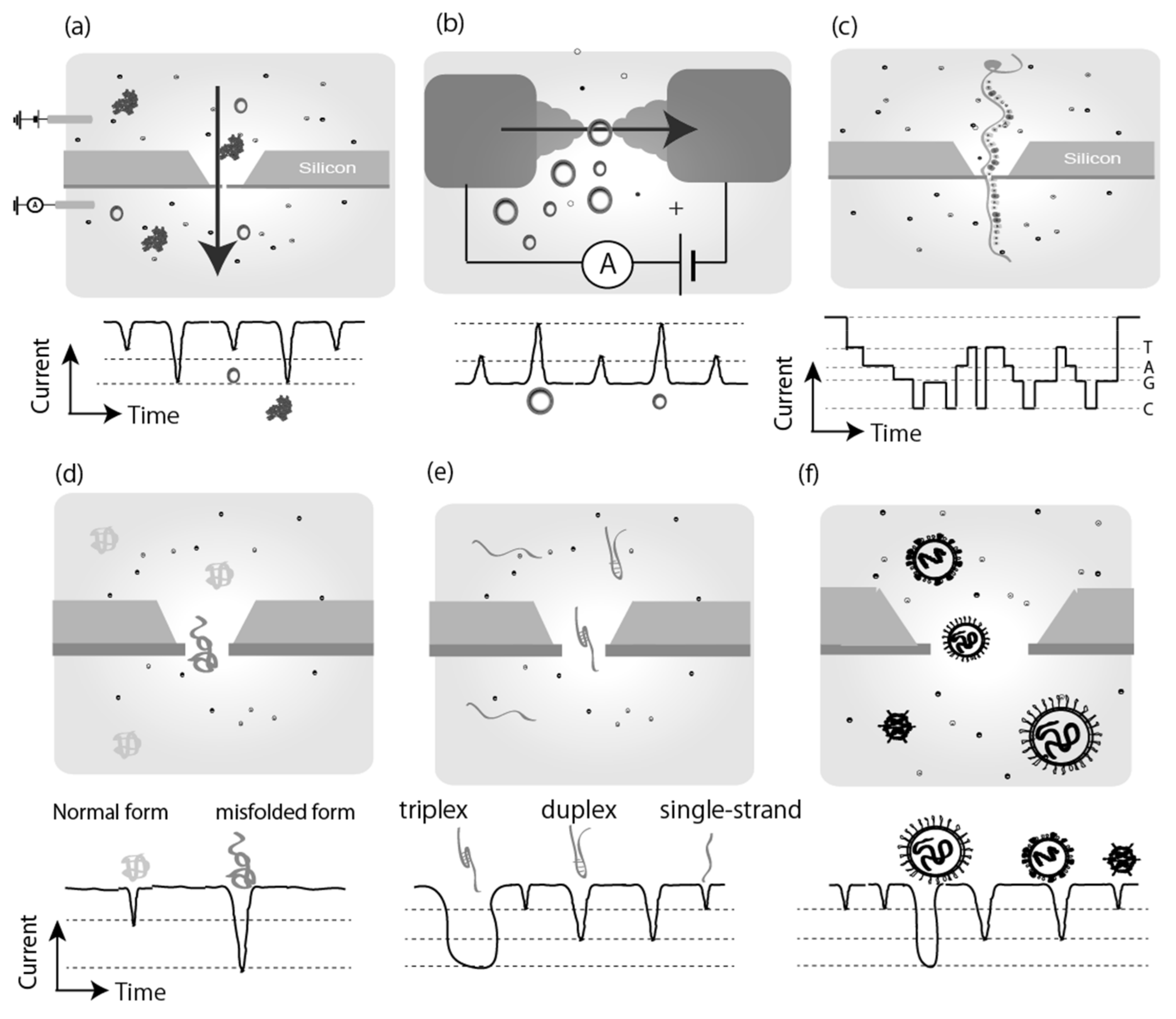
2. Single Molecule Electrical Measurements: Biomolecule Detection
2.1. Direct Sensing by Single Molecule Electrical Measurements
2.2. Indirect Sensing by Single Molecule Electrical Measurements
2.3. Nucleotide Sensing
2.4. Amino Acids, Peptides and Proteins Sensing
2.5. Glycans, and Biocompatible Polymer (PEG) Sensing
2.6. Second Messengers, Ion Sensing
2.7. Single-Molecule Electrical Detection Based Sequencing
3. Applications of Single Molecule Measurement: Disease Diagnosis, Monitoring
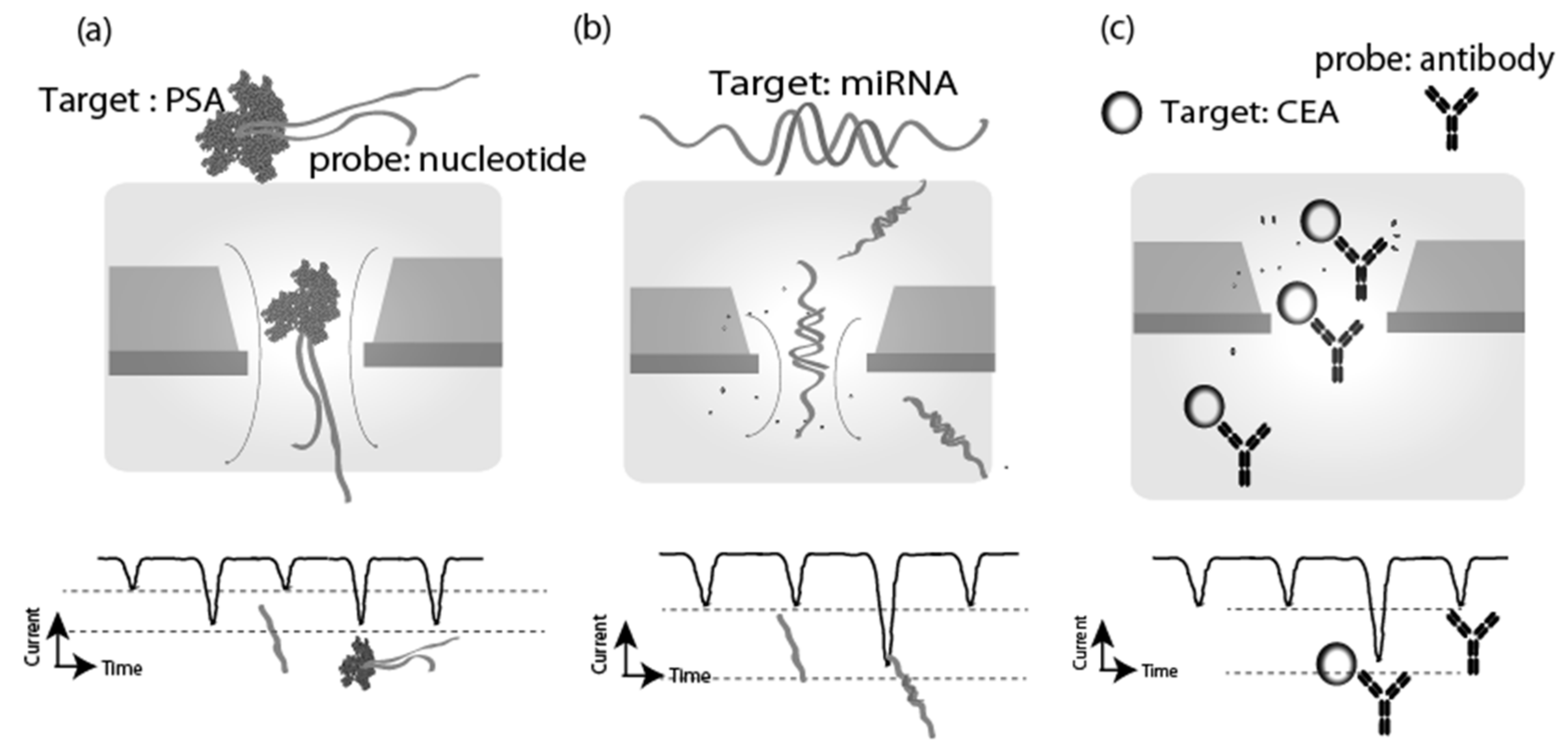
3.1. Cancer Diagnosis
3.2. Alzheimer’s Disease, Huntington’s Disease, and Prion Diseases Detection
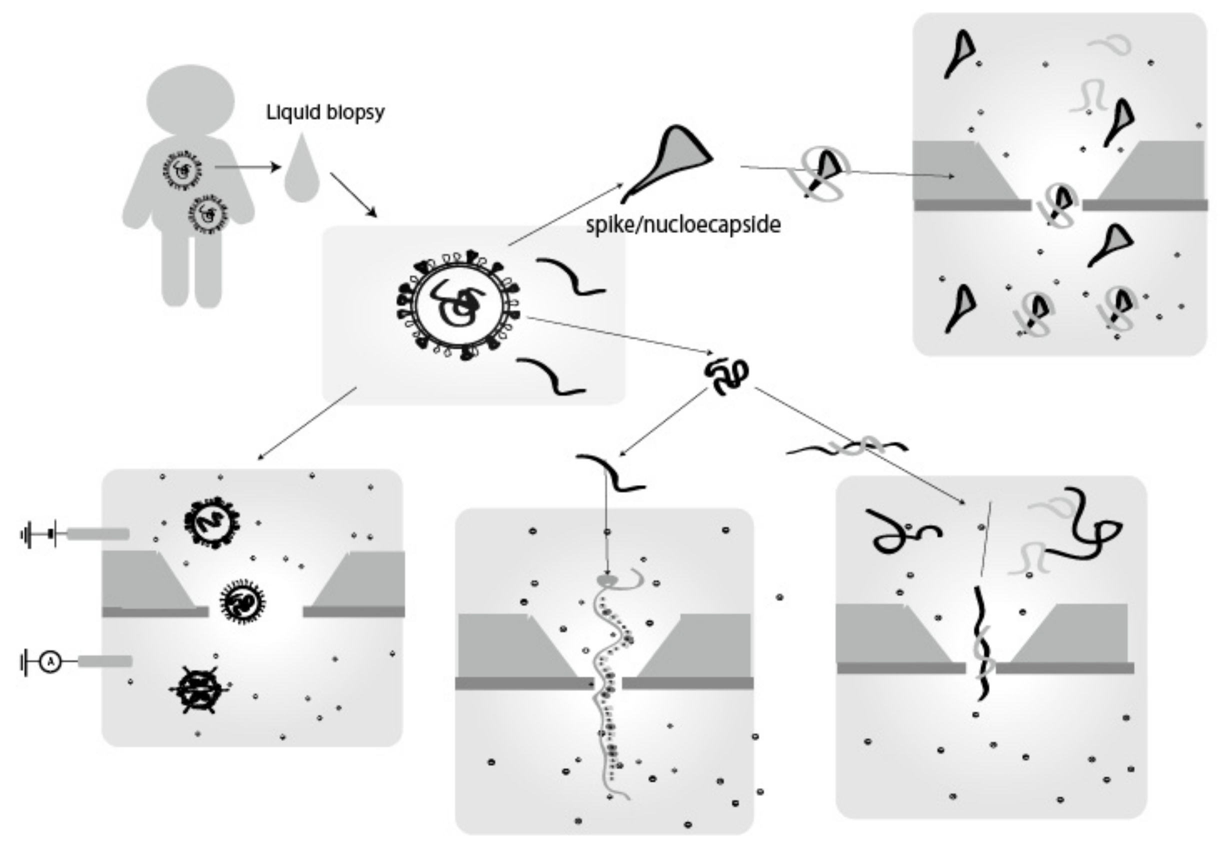
3.3. Virus Detection
3.4. Drug Screening and Environmental Monitoring
4. Discussion and Future Prospects
Funding
Institutional Review Board Statement
Data Availability Statement
Conflicts of Interest
References
- Wang, G.; Wang, L.; Han, Y.; Zhou, S.; Guan, X. Nanopore Stochastic Detection: Diversity, Sensitivity, and Beyond. Acc. Chem. Res. 2013, 46, 2867–2877. [Google Scholar] [CrossRef] [PubMed]
- Howorka, S.; Siwy, Z. Nanopore analytics: Sensing of single molecules. Chem. Soc. Rev. 2009, 38, 2360–2384. [Google Scholar] [CrossRef] [PubMed]
- Bulbul, G.; Chaves, G.; Olivier, J.; Ozel, R.E.; Pourmand, N. Nanopipettes as Monitoring Probes for the Single Living Cell: State of the art and future directions in molecular biology. Cells 2018, 7, 55. [Google Scholar] [CrossRef] [PubMed] [Green Version]
- Stanley, J.; Pourmand, N. Nanopipettes—The past and the present, N Pourmand. APL Mater. 2020, 8, 100902. [Google Scholar] [CrossRef]
- Actis, P.; Mak, A.C.; Pourmand, N. Functionalized nanopipettes: Toward label-free, single cell biosensors. Bioanal. Rev. 2010, 1, 177–185. [Google Scholar] [CrossRef] [Green Version]
- Zwolak, M.; Di Ventra, M. Colloquium: Physical approaches to DNA sequencing and detection. Rev. Mod. Phys. 2008, 80, 141–165. [Google Scholar] [CrossRef] [Green Version]
- Venkatesan, B.M.; Bashir, R. Nanopore sensors for nucleic acid analysis. Nat. Nanotech. 2011, 6, 615–624. [Google Scholar] [CrossRef]
- Deamer, D.W.; Branton, D. Characterization of Nucleic Acids by Nanopore Analysis. Acc. Chem. Res. 2002, 35, 817–825. [Google Scholar] [CrossRef] [Green Version]
- Ying, Y.L.; Cao, C.; Long, Y.T. Single molecule analysis by biological nanopore sensors. Analyst 2014, 139, 3826–3835. [Google Scholar] [CrossRef]
- Kono, N.; Arakawa, K. Nanopore sequencing: Review of potential applications in functional genomics. Dev. Growth Differ. 2019, 61, 316–326. [Google Scholar] [CrossRef] [Green Version]
- Wang, Y.; Zhao, Y.; Bollas, A.; Wang, Y.; Au, K.F. Nanopore sequencing technology, bioinformatics and applications. Nat. Biotech. 2021, 39, 1348–1365. [Google Scholar] [CrossRef]
- Benner, S.; Chen, R.J.A.; Wilson, N.A.; Abu-Shumays, R.; Hurt, N.; Lieberman, K.R.; Deamer, D.W.; Dunbar, W.B.; Akeson, M. Sequence-specific detection of individual DNA polymerase complexes in real time using a nanopore. Nat. Nanotechnol. 2007, 2, 718–724. [Google Scholar] [CrossRef] [PubMed] [Green Version]
- Schatz, M.C. Nanopore sequencing meets epigenetics. Nat. Methods 2017, 14, 347–348. [Google Scholar] [CrossRef] [PubMed]
- Wallace, E.V.B.; Stoddart, D.; Heron, A.J.; Mikhailova, E.; Maglia, G.; Donohoe, T.J.; Bayley, H. Identification of epigenetic DNA modifications with a protein nanopore. Chem. Commun. 2010, 46, 8195–8197. [Google Scholar] [CrossRef]
- Schreiber, J.; Wescoe, Z.L.; Abu-Shumays, R.; Vivian, J.T.; Baatar, B.; Karplus, K.; Akeson, M. Error rates for nanopore discrimination among cytosine, methylcytosine, and hydroxymethylcytosine along individual DNA strands. Proc. Natl. Acad. Sci. USA 2013, 110, 18910–18915. [Google Scholar] [CrossRef] [PubMed] [Green Version]
- Tsutsui, M.; Taniguchi, M.; Yokota, K.; Kawai, T. Identifying single nucleotides by tunnelling current. Nat. Nanotechnol. 2010, 5, 286–290. [Google Scholar] [CrossRef] [PubMed]
- Chang, S.A.; Huang, S.; He, J.; Liang, F.; Zhang, P.M.; Li, S.Q.; Chen, X.; Sankey, O.; Lindsay, S. Electronic Signatures of all Four DNA Nucleosides in a Tunneling Gap. Nano Lett. 2010, 10, 1070–1075. [Google Scholar] [CrossRef] [PubMed]
- Prasongkit, J.; Grigoriev, A.; Pathak, B.; Ahuja, R.; Scheicher, R.H. Transverse Conductance of DNA Nucleotides in a Graphene Nanogap from First Principles. Nano Lett. 2011, 11, 1941–1945. [Google Scholar] [CrossRef] [PubMed] [Green Version]
- Huang, S.; He, J.; Chang, S.A.; Zhang, P.M.; Liang, F.; Li, S.Q.; Tuchband, M.; Fuhrmann, A.; Ros, R.; Lindsay, S. Identifying single bases in a DNA oligomer with electron tunnelling. Nat. Nanotechnol. 2010, 5, 868–873. [Google Scholar] [CrossRef] [PubMed]
- Im, J.; Sen, S.; Lindsay, S.; Zhang, P.M. Recognition Tunneling of Canonical and Modified RNA Nucleotides for Their Identification with the Aid of Machine Learning. ACS Nano 2018, 12, 7067–7075. [Google Scholar] [CrossRef]
- Tsutsui, M.; Matsubara, K.; Ohshiro, T.; Furuhashi, M.; Taniguchi, M.; Kawai, T. Electrical detection of single methylcytosines in a DNA oligomer. J. Am. Chem Soc. 2011, 133, 9124–9128. [Google Scholar] [CrossRef]
- Ohshiro, T.; Konno, M.; Asai, A.; Komoto, Y.; Yamagata, A.; Doki, Y.; Eguchi, H.; Ofusa, K.; Taniguchi, M.; Ishii, H. Single-Molecule RNA Sequencing for Simultaneous Detection of m6A and 5mC. Sci. Rep. 2021, 11, 19304. [Google Scholar] [CrossRef]
- Komoto, Y.; Ohshiro, T.; Taniguchi, M. Detection of an alcohol-associated cancer marker by single-molecule quantum sequencing. Chem. Comm. 2020, 56, 14299–14302. [Google Scholar] [CrossRef] [PubMed]
- Karhanek, M.; Kemp, J.T.; Pourmand, N.; Davis, R.W.; Webb, C.D. Single DNA molecule detection using nanopipettes and nanoparticles. Nano Lett. 2005, 5, 403–407. [Google Scholar] [CrossRef] [PubMed]
- Ouldali, H.; Sarthak, K.; Ensslen, T.; Piguet, F.; Manivet, P.; Pelta, J.; Behrends, J.C.; Aksimentiev, A.; Oukhaled, A. Electrical recognition of the twenty proteinogenic amino acids using an aerolysin nanopore. Nat. Biotechnol. 2019, 38, 176–181. [Google Scholar] [CrossRef] [PubMed]
- Rodriguez-Larrea, D. Single-amino acid discrimination in proteins with homogeneous nanopore sensors and neural networks. Biosens. Bioelectron. 2021, 180, 7. [Google Scholar] [CrossRef]
- Zhao, Y.A.; Ashcroft, B.; Zhang, P.M.; Liu, H.; Sen, S.M.; Song, W.; Im, J.; Gyarfas, B.; Manna, S.; Biswas, S.; et al. Single-molecule spectroscopy of amino acids and peptides by recognition tunnelling. Nat. Nanotechnol. 2014, 9, 466–473. [Google Scholar] [CrossRef] [PubMed]
- Ohshiro, T.; Tsutsui, M.; Yokota, K.; Furuhashi, M.; Taniguchi, M.; Kawai, T. Detection of post-translational modifications in single peptides using electron tunnelling currents. Nat. Nanotechnol. 2014, 9, 835–840. [Google Scholar] [CrossRef]
- Piguet, F.; Ouldali, H.; Pastoriza-Gallego, M.; Manivet, P.; Pelta, J.; Oukhaled, A. Identification of single amino acid differences in uniformly charged homopolymeric peptides with aerolysin nanopore. Nat. Commun. 2018, 9, 966. [Google Scholar] [CrossRef]
- Oukhaled, A.; Bacri, L.; Pastoriza-Gallego, M.; Betton, J.M.; Pelta, J. Sensing Proteins through Nanopores: Fundamental to Applications. ACS Chem. Biol. 2012, 7, 1935–1949. [Google Scholar] [CrossRef] [PubMed]
- Movileanu, L.; Howorka, S.; Braha, O.; Bayley, H. Detecting protein analytes that modulate transmembrane movement of a polymer chain within a single protein pore. Nat. Biotechnol. 2000, 18, 1091–1095. [Google Scholar] [CrossRef] [PubMed]
- Oukhaled, G.; Mathe, J.; Biance, A.L.; Bacri, L.; Betton, J.M.; Lairez, D.; Pelta, J.; Auvray, L. Unfolding of Proteins and Long Transient Conformations Detected by Single Nanopore Recording. Phys. Rev. Lett. 2007, 98, 158101. [Google Scholar] [CrossRef] [PubMed] [Green Version]
- Chang, T.L.; Tsai, C.Y.; Sun, C.C.; Chen, C.C.; Kuo, L.S.; Chen, P.H. Ultrasensitive electrical detection of protein using nanogap electrodes and nanoparticle-based DNA amplification. Biosens. Bioelectron. 2007, 22, 3139–3145. [Google Scholar] [CrossRef]
- Fennouri, A.; Przybylski, C.; Pastoriza-Gallego, M.; Bacri, L.; Auvray, L.; Daniel, R. Single Molecule Detection of Glycosaminoglycan Hyaluronic Acid Oligosaccharides and Depolymerization Enzyme Activity Using a Protein Nanopore. ACS Nano 2012, 6, 9672–9678. [Google Scholar] [CrossRef] [PubMed]
- Kullman, L.; Winterhalter, M.; Bezrukov, S.M. Transport of Maltodextrins through Maltoporin: A Single-Channel Study. Biophys. J. 2002, 82, 803–812. [Google Scholar] [CrossRef] [Green Version]
- Bacri, L.; Oukhaled, A.; Hemon, E.; Bassafoula, F.B.; Auvray, L.; Daniel, R. Discrimination of Neutral Oligosaccharides through a Nanopore. Biochem. Biophys. Res. Commun. 2011, 412, 561–564. [Google Scholar] [CrossRef] [PubMed]
- Robertson, J.W.F.; Rodrigues, C.G.; Stanford, V.M.; Rubinson, K.A.; Krasilnikov, O.V.; Kasianowicz, J.J. Single-molecule mass spectrometry in solution using a solitary nanopore. Proc. Natl. Acad. Sci. USA 2007, 104, 8207–8211. [Google Scholar] [CrossRef] [Green Version]
- Movileanu, L.; Cheley, S.; Bayley, H. Partitioning of Individual Flexible Polymers into a Nanoscopic Protein Pore. Biophys. J. 2003, 8, 897–910. [Google Scholar] [CrossRef] [Green Version]
- Oukhaled, A.G.; Biance, A.L.; Pelta, J.; Auvray, L.; Bacri, L. Transport of Long Neutral Polymers in the Semidilute Regime through a Protein Nanopore. Phys. Rev. Lett. 2012, 108, 088104. [Google Scholar] [CrossRef]
- Umehara, S.; Karhanek, M.; Davis, R.W.; Pourmand, N. Label-free biosensing with functionalized nanopipette probes. Proc. Natl. Acad. Sci. USA 2009, 106, 4611–4616. [Google Scholar] [CrossRef] [Green Version]
- Ying, Y.L.; Wang, H.Y.; Sutherland, T.C.; Long, Y.T. Monitoring of an ATP-Binding Aptamer and its Conformational Changes Using an α-Hemolysin Nanopore. Small 2011, 7, 87–94. [Google Scholar] [CrossRef] [PubMed]
- Komoto, Y.; Ohshiro, T.; Taniguchi, M. Development of Single-Molecule Electrical Identification Method for Cyclic Adenosine Monophosphate Signaling Pathway. Nanomaterials 2021, 11, 784. [Google Scholar] [CrossRef] [PubMed]
- Komoto, Y.; Ohshiro, T.; Yoshida, T.; Tarusawa, E.; Yagi, T.; Washio, T.; Taniguchi, M. Time-resolved neurotransmitter detection in mouse brain tissue using an artificial intelligence nanogap. Sci. Rep. 2020, 10, 11244. [Google Scholar] [CrossRef] [PubMed]
- Cheley, S.; Gu, L.Q.; Bayley, H. Stochastic sensing of nanomolar inositol 1,4,5-trisphosphate with an engineered pore. Chem. Biol. 2002, 9, 829–838. [Google Scholar] [CrossRef] [Green Version]
- Gu, L.Q.; Braha, O.; Conlan, S.; Cheley, S.; Bayley, H. Stochastic sensing of organic analytes by a pore-forming protein containing a molecular adapter. Nature 1999, 398, 686–690. [Google Scholar] [CrossRef] [PubMed]
- Braha, O.; Gu, L.Q.; Zhou, L.; Lu, X.F.; Cheley, S.; Bayley, H. Simultaneous stochastic sensing of divalent metal ions. Nat. Biotechnol. 2000, 18, 1005–1007. [Google Scholar] [CrossRef]
- Zhu, Z.P.; Wang, D.Y.; Tian, Y.; Jiang, L. Ion/Molecule Transportation in Nanopores and Nanochannels: From Critical Principles to Diverse Functions. J. Am. Chem. Soc. 2019, 141, 8658–8669. [Google Scholar] [CrossRef]
- Liu, L.; You, Y.; Zhou, K.; Guo, B.Y.; Cao, Z.; Zhao, Y.L.; Wu, H.C. A Dual-Response DNA Probe for Simultaneously Monitoring Enzymatic Activity and Environmental pH Using a Nanopore. Angew. Chem. Int. Ed. 2019, 131, 15071–15076. [Google Scholar] [CrossRef]
- Roozbahani, G.M.; Chen, X.H.; Zhang, Y.W.; Xie, R.Q.; Ma, R.; Li, D.E.; Li, H.Z.; Guan, X.Y. Peptide-Mediated Nanopore Detection of Uranyl Ions in Aqueous Media. ACS Sens. 2017, 2, 703–709. [Google Scholar] [CrossRef] [Green Version]
- Wen, S.; Zeng, T.; Liu, L.; Zhao, K.; Zhao, Y.L.; Liu, X.J.; Wu, H.C. Highly Sensitive and Selective DNA-Based Detection of Mercury(II) with α-Hemolysin Nanopore. J. Am. Chem. Soc. 2011, 133, 18312–18317. [Google Scholar] [CrossRef]
- Umehara, S.; Pourmand, N.; Webb, C.D.; Davis, R.W.; Yasuda, K.; Karhanek, M. Current rectification with poly-L-lysine-coated quartz nanopipettes. Nano Lett. 2006, 6, 2486–2492. [Google Scholar] [CrossRef] [PubMed] [Green Version]
- Ozel, R.E.; Bulbul, G.; Perez, J.; Pourmand, N. Functionalized Quartz Nanopipette for Intracellular Superoxide Sensing: A Tool for Monitoring Reactive Oxygen Species Levels in Single Living Cell. ACS Sens. 2018, 3, 1316–1321. [Google Scholar] [CrossRef] [PubMed]
- Tsutsui, M.; Yokota, K.; Yoshida, T.; Hotehama, C.; Kowada, H.; Esaki, Y.; Taniguchi, M.; Washio, T.; Kawai, T. Identifying Single Particles in Air Using a 3D-Integrated Solid-State Pore. ACS Sens. 2019, 4, 748–755. [Google Scholar] [CrossRef] [PubMed]
- Arima, A.; Tsutsui, M.; Washio, T.; Baba, Y.; Kawai, T. Solid-State Nanopore Platform Integrated with Machine Learning for Digital Diagnosis of Virus Infection. Anal. Chem. 2021, 93, 215–227. [Google Scholar] [CrossRef] [PubMed]
- Taniguchi, M.; Minami, S.; Ono, C.; Hamajima, R.; Morimura, A.; Hamaguchi, S.; Akeda, Y.; Kanai, Y.; Kobayashi, T.; Kamitani, W.; et al. Combining machine learning and nanopore construction creates an artificial intelligence nanopore for coronavirus detection. Nat. Commun. 2021, 12, 3726. [Google Scholar] [CrossRef] [PubMed]
- Branton, D.; Deamer, D.W.; Marziali, A.; Bayley, H.; Benner, S.A.; Butler, T.; Di Ventra, M.; Garaj, S.; Hibbs, A.; Huang, X.; et al. The potential and challenges of nanopore sequencing. Nat. Biotechnol. 2008, 26, 1146–1153. [Google Scholar] [CrossRef] [PubMed]
- Garalde, D.R.; Snell, E.A.; Jachimowicz, D.; Sipos, B.; Lloyd, J.H.; Bruce, M.; Pantic, N.; Admassu, T.; James, P.; Warland, A.; et al. Highly parallel direct RNA sequencing on an array of nanopores. Nat. Method. 2018, 15, 201. [Google Scholar] [CrossRef]
- Di Ventra, M.; Taniguchi, M. Decoding DNA, RNA and peptides with quantum tunnelling. Nat. Nanotechnol. 2016, 11, 117–126. [Google Scholar] [CrossRef]
- Ohshiro, T.; Matsubara, K.; Tsutsui, M.; Furuhashi, M.; Taniguchi, M.; Kawai, T. Single-Molecule Electrical Random Resequencing of DNA and RNA. Sci. Rep. 2012, 2, 501–507. [Google Scholar] [CrossRef]
- Ohshiro, T.; Tsutsui, M.; Yokota, K.; Taniguchi, M. Quantitative analysis of DNA with single-molecule sequencing. Sci. Rep. 2018, 8, 8517. [Google Scholar] [CrossRef] [Green Version]
- Restrepo-Perez, L.; Joo, C.; Dekker, C. Paving the way to single-molecule protein sequencing. Nat. Nanotechnol. 2018, 13, 786–796. [Google Scholar] [CrossRef] [PubMed] [Green Version]
- Hu, Z.L.; Huo, M.Z.; Ying, Y.L.; Long, Y.T. Biological Nanopore Approach for Single-Molecule Protein Sequencing. Angew. Chem. Int. Ed. 2020, 133, 14862–14873. [Google Scholar] [CrossRef]
- Norris, A.L.; Workman, R.E.; Fan, Y.; Eshleman, J.R.; Timp, W. Nanopore sequencing detects structural variants in cancer. Cancer Biol. Ther. 2016, 17, 246–253. [Google Scholar] [CrossRef]
- Yang, W.J.; Liu, Y.; Dong, R.Y.; Liu, J.; Lang, J.D.; Yang, J.L.; Wang, W.W.; Li, J.J.; Meng, B.; Tian, G. Accurate Detection of HPV Integration Sites in Cervical Cancer Samples Using the Nanopore MinION Sequencer Without Error Correction. Front. Genet. 2020, 11, 660. [Google Scholar] [CrossRef] [PubMed]
- Valle-Inclan, J.E.; Stangl, C.; de Jong, A.C.; van Dessel, L.F.; van Roosmalen, M.J.; Helmijr, J.C.; Renkens, I.; Janssen, R.; de Blank, S.; de Witte, C.J.; et al. Optimizing Nanopore sequencing-based detection of structural variants enables individualized circulating tumor DNA-based disease monitoring in cancer patients. Genome Med. 2021, 13, 86. [Google Scholar] [CrossRef]
- Quan, L.L.; Dong, R.Y.; Yang, W.J.; Chen, L.Y.; Lang, J.D.; Liu, J.; Song, Y.; Ma, S.Q.; Yang, J.L.; Wang, W.W.; et al. Simultaneous detection and comprehensive analysis of HPV and microbiome status of a cervical liquid-based cytology sample using Nanopore MinION sequencing. Sci. Rep. 2019, 9, 19337. [Google Scholar] [CrossRef]
- Minervini, C.F.; Cumbo, C.; Orsini, P.; Brunetti, C.; Anelli, L.; Zagaria, A.; Minervini, A.; Casieri, P.; Coccaro, N.; Tota, G.; et al. TP53 gene mutation analysis in chronic lymphocytic leukemia by nanopore MinION sequencing. Diagn. Pathol. 2016, 11, 96. [Google Scholar] [CrossRef] [Green Version]
- Wang, Y.; Zheng, D.L.; Tan, Q.L.; Wang, M.X.; Gu, L.Q. Nanopore-based detection of circulating microRNAs in lung cancer patients. Nat. Nanotechnol. 2011, 6, 668–674. [Google Scholar] [CrossRef]
- Zhang, J.H.; Liu, X.L.; Hu, Z.L.; Ying, Y.L.; Long, Y.T. Intelligent identification of multi-level nanopore signatures for accurate detection of cancer biomarkers. Chem. Commun. 2017, 53, 10176–10179. [Google Scholar] [CrossRef]
- Zhang, X.Y.; Wang, Y.; Fricke, B.L.; Gu, L.Q. Programming Nanopore Ion Flow for Encoded Multiplex MicroRNA Detection. ACS Nano 2014, 8, 3444–3450. [Google Scholar] [CrossRef]
- Tian, K.; He, Z.J.; Wang, Y.; Chen, S.J.; Gu, L.Q. Designing a Polycationic Probe for Simultaneous Enrichment and Detection of MicroRNAs in a Nanopore. ACS Nano 2013, 7, 3962–3969. [Google Scholar] [CrossRef] [PubMed] [Green Version]
- Ying, Y.L.; Li, D.W.; Li, Y.; Lee, J.S.; Long, Y.T. Enhanced translocation of poly(dt)45 through an α-hemolysin nanopore by binding with antibody. Chem. Commun. 2011, 47, 5690–5692. [Google Scholar] [CrossRef] [PubMed]
- Shim, J.; Humphreys, G.I.; Venkatesan, B.M.; Munz, J.M.; Zou, X.Q.; Sathe, C.; Schulten, K.; Kosari, F.; Nardulli, A.M.; Vasmatzis, G.; et al. Detection and Quantification of Methylation in DNA using Solid-State Nanopores. Sci. Rep. 2013, 3, 1389. [Google Scholar] [CrossRef] [Green Version]
- Xi, D.M.; Li, Z.; Liu, L.P.; Ai, S.Y.; Zhang, S.S. Ultrasensitive Detection of Cancer Cells Combining Enzymatic Signal Amplification with an Aerolysin Nanopore. Anal. Chem. 2018, 90, 1029–1034. [Google Scholar] [CrossRef]
- Li, X.; Zhang, P.; Dou, L.; Wang, J.; Sun, K.; Zhang, X.; Song, G.; Zhao, C.; Li, K.; Bai, Y.; et al. Detection of Circulating Tumor Cells in Breast Cancer Patients by Nanopore Sensing with Aptamer-Mediated Amplification. ACS Sens. 2020, 5, 2359–2366. [Google Scholar] [CrossRef]
- Duan, L.; Yobas, L. Label-Free Multiplexed Electrical Detection of Cancer Markers on a Microchip Featuring an Integrated Fluidic Diode Nanopore Array. ACS Nano 2018, 12, 7892–7900. [Google Scholar] [CrossRef]
- Wang, S.Y.; Haque, F.; Rychahou, P.G.; Evers, B.M.; Guo, P.X. Engineered Nanopore of Phi29 DNA-Packaging Motor for Real-Time Detection of Single Colon Cancer Specific Antibody in Serum. ACS Nano 2013, 7, 9814–9822. [Google Scholar] [CrossRef] [PubMed] [Green Version]
- Shim, J.; Kim, Y.; Humphreys, G.I.; Nardulli, A.M.; Kosari, F.; Vasmatzis, G.; Taylor, W.R.; Ahlquist, D.A.; Myong, S.; Bashir, R. Nanopore-Based Assay for Detection of Methylation in Double-Stranded DNA Fragments. ACS Nano 2015, 9, 290–300. [Google Scholar] [CrossRef] [PubMed]
- Lin, Y.; Ying, Y.L.; Shi, X.; Liu, S.C.; Long, Y.T. Direct sensing of cancer biomarkers in clinical samples with a designed nanopore. Chem. Commun. 2017, 53, 11564–11567. [Google Scholar] [CrossRef] [PubMed] [Green Version]
- Guo, B.Y.; Song, P.; Zhou, K.; Liu, L.; Wu, H.C. Simultaneous Sensing of Multiple Cancer Biomarkers by a Single DNA Nanoprobe in a Nanopore. Anal. Chem. 2020, 92, 9405–9411. [Google Scholar] [CrossRef] [PubMed]
- Tang, H.R.; Wang, H.; Yang, C.; Zhao, D.D.; Qian, Y.Y.; Li, Y.X. Nanopore-based Strategy for Selective Detection of Single Carcinoembryonic Antigen (CEA) Molecules. Anal. Chem. 2020, 92, 3042–3049. [Google Scholar] [CrossRef] [PubMed]
- Liu, L.; Li, T.; Zhang, S.W.; Song, P.; Guo, B.Y.; Zhao, Y.L.; Wu, H.C. Simultaneous Quantification of Multiple Cancer Biomarkers in Blood Samples through DNA-Assisted Nanopore Sensing. Angew. Chem. Int. Ed. 2018, 57, 11882–11887. [Google Scholar] [CrossRef] [PubMed]
- Kang, I.; Wang, Y.; Reagan, C.; Fu, Y.M.; Wang, M.X.; Gu, L.Q. Designing DNA interstrand lock for locus-specific methylation detection in a nanopore. Sci. Rep. 2013, 3, 2381. [Google Scholar] [CrossRef] [PubMed] [Green Version]
- Chen, X.H.; Zhang, Y.W.; Roozbahani, G.M.; Guan, X.Y. Salt-Mediated Nanopore Detection of ADAM-17. ACS Appl. Bio Mater. 2019, 2, 504–509. [Google Scholar] [CrossRef]
- Wang, Y.; Tian, K.; Shi, R.C.; Gu, A.; Pennella, M.; Alberts, L.; Gates, K.S.; Li, G.F.; Fan, H.X.; Wang, M.X.; et al. Nanolock-Nanopore Facilitated Digital Diagnostics of Cancer Driver Mutation in Tumor Tissue. ACS Sens. 2017, 2, 975–981. [Google Scholar] [CrossRef] [PubMed]
- Sun, K.; Chen, P.; Yan, S.; Yuan, W.; Wang, Y.; Li, X.; Dou, L.; Zhao, C.; Zhang, J.; Wang, Q.; et al. Ultrasensitive Nanopore Sensing of Mucin 1 and Circulating Tumor Cells in Whole Blood of Breast Cancer Patients by Analyte-Triggered Triplex-DNA Release. ACS Appl. Mater. Interfaces 2021, 13, 21030–21039. [Google Scholar] [CrossRef] [PubMed]
- Ilyas, A.; Asghar, W.; Allen, P.B.; Duhon, H.; Ellington, A.D.; Iqbal, S.M. Electrical detection of cancer biomarker using aptamers with nanogap break-junctions. Nanotechnology 2012, 23, 275502. [Google Scholar] [CrossRef] [Green Version]
- Arima, A.; Tsutsui, M.; Yoshida, T.; Tatematsu, K.; Yamazaki, T.; Yokota, K.; Kuroda, S.; Washio, T.; Baba, Y.; Kawai, T. Digital Pathology Platform for Respiratory Tract Infection Diagnosis via Multiplex Single-Particle Detections. ACS Sens. 2020, 5, 3398–3403. [Google Scholar] [CrossRef] [PubMed]
- Batovska, J.; Lynch, S.E.; Rodoni, B.C.; Sawbridge, T.I.; Cogan, N.O.I. MinION nanopore sequencing of an influenza genome. Front. Microbiol. 2015, 6, 766. [Google Scholar]
- Batovska, J.; Lynch, S.E.; Rodoni, B.C.; Sawbridge, T.I.; Cogan, N.O. Metagenomic arbovirus detection using MinION nanopore sequencing. J. Virol. Methods 2017, 249, 79–84. [Google Scholar] [CrossRef]
- Greninger, A.L.; Naccache, S.N.; Federman, S.; Yu, G.; Mbala, P.; Bres, V.; Stryke, D.; Bouquet, J.; Somasekar, S.; Linnen, J.M.; et al. Rapid metagenomic identification of viral pathogens in clinical samples by real-time nanopore sequencing analysis. Genome Med. 2015, 7, 99. [Google Scholar] [CrossRef] [PubMed] [Green Version]
- Niedzwiecki, D.J.; Iyer, R.; Borer, P.N.; Movileanu, L. Sampling a Biomarker of the Human Immunodeficiency Virus across a Synthetic Nanopore. ACS Nano 2013, 7, 3341–3350. [Google Scholar] [CrossRef] [PubMed] [Green Version]
- Wanunu, M.; Dadosh, T.; Ray, V.; Jin, J.M.; McReynolds, L.; Drndic, M. Rapid electronic detection of probe-specific microRNAs using thin nanopore sensors. Nat. Nanotechnol. 2010, 5, 807–814. [Google Scholar] [CrossRef] [PubMed]
- Kwak, D.K.; Kim, J.S.; Lee, M.K.; Ryu, K.S.; Chi, S.W. Probing the Neuraminidase Activity of Influenza Virus Using a Cytolysin a Protein Nanopore. Anal. Chem. 2020, 92, 14303–14308. [Google Scholar] [CrossRef] [PubMed]
- Zhao, Q.T.; de Zoysa, R.S.S.; Wang, D.Q.; Jayawardhana, D.A.; Guan, X.Y. Real-Time Monitoring of Peptide Cleavage Using a Nanopore Probe. J. Am. Chem. Soc. 2009, 131, 6324–6325. [Google Scholar] [CrossRef]
- Ding, T.L.; Yang, J.; Pan, V.; Zhao, N.; Lu, Z.H.; Ke, Y.G.; Zhang, C. DNA nanotechnology assisted nanopore-based analysis. Nucleic Acids Res. 2020, 48, 2791–2806. [Google Scholar] [CrossRef] [Green Version]
- Wanunu, M.; Sutin, J.; Meller, A. DNA Profiling Using Solid-State Nanopores: Detection of DNA-Binding Molecules. Nano Lett. 2009, 9, 3498–3502. [Google Scholar] [CrossRef] [Green Version]
- Lee, D.H.; Oh, S.; Lim, K.; Lee, B.; Yi, G.S.; Kim, Y.R.; Kim, K.B.; Lee, C.K.; Chi, S.W.; Lee, M.K. Tertiary RNA Folding-Targeted Drug Screening Strategy Using a Protein Nanopore. Anal. Chem. 2021, 93, 2811–2819. [Google Scholar] [CrossRef]
- Yao, F.J.; Duan, J.; Wang, Y.; Zhang, Y.; Guo, Y.L.; Guo, H.L.; Kang, X.F. Nanopore Single-Molecule Analysis of DNA–Doxorubicin Interactions. Anal. Chem. 2015, 87, 338–342. [Google Scholar] [CrossRef]
- Ohshiro, T.; Komoto, Y.; Konno, M.; Koseki, J.; Asai, A.; Ishii, H.; Taniguch, M. Direct Analysis of Incorporation of an Anticancer Drug into DNA at Single-Molecule Resolution. Sci. Rep. 2019, 9, 3886. [Google Scholar] [CrossRef] [Green Version]
- De Coster, W.; Van Broeckhoven, C. Newest methods for detecting structural variations. Trends Biotechnol. 2019, 37, 973–982. [Google Scholar] [CrossRef] [PubMed] [Green Version]
- Zou, Z.; Yang, H.; Yan, Q.; Qi, P.; Qing, Z.H.; Zheng, J.; Xu, X.; Zhang, L.H.; Feng, F.; Yang, R.H. Synchronous screening of multiplexed biomarkers of Alzheimer’s disease by a length-encoded aerolysin nanopore-integrated triple-helix molecular switch. Chem. Commun. 2019, 55, 6433. [Google Scholar] [CrossRef] [PubMed]
- Lenhart, B.; Wei, X.J.; Watson, B.; Wang, X.Q.; Zhang, Z.H.; Li, C.Z.; Moss, M.; Liu, C. In Vitro Biosensing of β-Amyloid Peptide Aggregation Dynamics using a Biological Nanopore. Sens. Actuators B 2021, 338, 129863. [Google Scholar] [CrossRef] [PubMed]
- Houghtaling, J.; List, J.; Mayer, M. Nanopore-Based, Rapid Characterization of Individual Amyloid Particles in Solution: Concepts, Challenges, and Prospects. Small 2018, 14, 1802412. [Google Scholar] [CrossRef] [PubMed]
- Madampage, C.A.; Andrievskaia, O.; Lee, J.S. Nanopore detection of antibody prion interactions. Anal. Biochem. 2010, 396, 36–41. [Google Scholar] [CrossRef]
- Madampage, C.; Tavassoly, O.; Christensen, C.; Kumari, M.; Lee, J.S. Nanopore analysis: An emerging technique for studying the folding and misfolding of proteins. Prion 2012, 6, 116–123. [Google Scholar] [CrossRef] [PubMed] [Green Version]
- Charalampous, T.; Kay, G.L.; Richardson, H.; Aydin, A.; Baldan, R.; Jeanes, C.; Rae, D.; Grundy, S.; Turner, D.J.; Wain, J.; et al. Nanopore metagenomics enables rapid clinical diagnosis of bacterial lower respiratory infection. Nat. Biotechnol. 2019, 37, 783–792. [Google Scholar] [CrossRef]
- Mitsuhashi, S.; Frith, M.C.; Mizuguchi, T.; Miyatake, S.; Toyota, T.; Adachi, H.; Oma, Y.; Kino, Y.; Mitsuhashi, H.; Matsumoto, N. Tandem-genotypes: Robust detection of tandem repeat expansions from long DNA reads. Genome Biol. 2019, 20, 58. [Google Scholar] [CrossRef] [Green Version]
- Sanchis-Juan, A.; Stephens, J.; French, C.E.; Gleadall, N.; Mégy, K.; Penkett, C.; Shamardina, O.; Stirrups, K.; Delon, I.; Dewhurst, E.; et al. Complex structural variants in Mendelian disorders: Identification and breakpoint resolution using short- and long-read genome sequencing. Genome Med. 2018, 10, 95. [Google Scholar] [CrossRef] [Green Version]
- Kafetzopoulou, L.E.; Pullan, S.T.; Lemey, P.; Suchard, M.A.; Ehichioya, D.U.; Pahlmann, M.; Thielebein, A.; Hinzmann, J.; Oestereich, L.; Wozniak, D.M.; et al. Metagenomic sequencing at the epicenter of the Nigeria 2018 Lassa fever outbreak. Science 2019, 363, 74–77. [Google Scholar] [CrossRef] [Green Version]
- Minh, Q.N.; Tong, H.D.; Kuijk, A.; van de Bent, F.; Beekman, P.; van Rijn, C.J.M. Gas sensing performance at room temperature of nanogap interdigitated electrodes for detection of acetone at low concentration. RSC Adv. 2017, 7, 50279–50286. [Google Scholar] [CrossRef] [Green Version]
- Guan, X.Y.; Gu, L.Q.; Cheley, S.; Braha, O.; Bayley, H. Stochastic sensing of TNT with a genetically engineered pore. ChemBioChem 2005, 6, 1875–1881. [Google Scholar] [CrossRef] [PubMed]
- Taniguchi, M.; Ohshiro, T. Applications of Microfluidic Systems in Biology and Medicine: Nanopore Device for Single-Molecule Sensing Method and Its Application; Springer: Berlin/Heidelberg, Germany, 2019; ISBN 978-981-13-6229-3. [Google Scholar] [CrossRef]
- Hou, X.; Guo, W.; Jiang, L. Biomimetic smart nanopores and nanochannels. Chem. Soc. Rev. 2011, 40, 2385–2401. [Google Scholar] [CrossRef] [PubMed]
- Theberge, A.B. Microdroplets in Microfluidics: An Evolving Platform for Discoveries in Chemistry and Biology. Angew. Chem. Int. Ed. 2010, 49, 5846–5868. [Google Scholar] [CrossRef] [PubMed]
- Kaji, N.; Okamoto, Y.; Tokeshi, M.; Baba, Y. Nanopillar, nanoball, and nanofibers for highly efficient analysis of biomolecules. Chem. Soc. Rev. 2010, 39, 948–956. [Google Scholar] [CrossRef] [PubMed]
- Ohshiro, T.; Komoto, Y.; Taniguchi, M. Single-Molecule Counting of Nucleotide by Electrophoresis with Nanochannel-Integrated Nano-Gap Devices. Micromachines 2020, 11, 982. [Google Scholar] [CrossRef] [PubMed]
- Luo, S.H.; Hoff, B.H.; Maier, S.A.; de Mello, J.C. Scalable Fabrication of Metallic Nanogaps at the Sub-10 nm Level. Adv. Sci. 2021, 8, 2102756. [Google Scholar] [CrossRef] [PubMed]
- Wang, J.J.; Ying, Y.L.; Zhong, C.B.; Zhang, L.M.; Yan, F.; Long, Y.T. Instrumentational implementation for parallelized nanopore electrochemical measurements. Analyst 2021, 146, 4111. [Google Scholar] [CrossRef] [PubMed]
- Korlach, J.; Bjornson, K.P.; Chaudhuri, B.P.; Cicero, R.L.; Flusberg, B.A.; Gray, J.J.; Holden, D.; Saxena, R.; Wegener, J.; Turner, S.W. Real-time DNA sequencing from single polymerase molecules. Science 2009, 323, 133–138. [Google Scholar]
- Mashaghi, S.; Abbaspourrad, A.; Weitz, D.A.; van Oijen, A.M. Droplet microfluidics: A tool for biology, chemistry and nanotechnology. Trends Anal. Chem 2016, 82, 118–125. [Google Scholar] [CrossRef] [Green Version]
- Dombi, P.; Papa, Z.; Vogelsang, J.; Yalunin, S.V.; Sivis, M.; Herink, G.; Schafer, S.; Gross, P.; Ropers, C.; Lienau, C. Strong-field nano-optics. Rev. Mod. Phys. 2020, 92, 025003. [Google Scholar] [CrossRef]
- Reisner, W.; Larsen, N.B.; Silahtaroglu, A.; Kristensen, A.; Tommerup, N.; Tegenfeldt, J.O.; Flyvbjerg, H. Single-molecule denaturation mapping of DNA in nanofluidic channels. Proc. Natl. Acad. Sci. USA 2010, 107, 13294–13299. [Google Scholar] [CrossRef] [PubMed] [Green Version]
- Jiang, J.; Bosnick, K.; Maillard, M.; Brus, L. Single Molecule Raman Spectroscopy at the Junctions of Large Ag Nanocrystals. J. Phys. Chem. B 2003, 107, 9964–9972. [Google Scholar] [CrossRef]
- Zrimsek, A.B.; Chiang, N.H.; Mattei, M.; Zaleski, S.; McAnally, M.O.; Chapman, C.T.; Henry, A.I.; Schatz, G.C.; Van Duyne, R.P. Single-Molecule Chemistry with Surface- and Tip-Enhanced Raman Spectroscopy. Chem. Rev. 2017, 117, 7583–7613. [Google Scholar] [CrossRef] [PubMed]
- Konishi, T.; Kiguchi, M.; Takase, M.; Nagasawa, F.; Nabika, H.; Ikeda, K.; Uosaki, K.; Ueno, K.; Misawa, H.; Murakoshi, K. Single Molecule Dynamics at a Mechanically Controllable Break Junction in Solution at Room Temperature. J. Am. Chem. Soc. 2013, 135, 1009–1014. [Google Scholar] [CrossRef] [PubMed]
- Kaneko, S.; Yasuraoka, K.; Kiguchi, M. Bias Voltage Induced Surface-Enhanced Raman Scattering Enhancement on the Single-Molecule Junction. J. Phys. Chem. C 2019, 123, 6502–6507. [Google Scholar] [CrossRef]
- Yang, W.; Lim, D.K. Recent Advances in the Synthesis of Intra-Nanogap Au Plasmonic Nanostructures for Bioanalytical Applications. Adv. Mater. 2020, 32, e2002219. [Google Scholar] [CrossRef]
- Taniguchi, M.; Ohshiro, T.; Komoto, Y.; Takaai, T.; Yoshida, T.; Washio, T. High-Precision Single-Molecule Identification Based on Single-Molecule Information within a Noisy Matrix. J. Phys. Chem. C 2019, 123, 15867–15873. [Google Scholar] [CrossRef]
- Taniguchi, M. Combination of Single-Molecule Electrical Measurements and Machine Learning for the Identification of Single Biomolecules. ACS Omega 2020, 5, 959–964. [Google Scholar] [CrossRef] [Green Version]
- Komoto, Y.; Ohshiro, T.; Taniguchi, M. Length discrimination of homo-oligomeric nucleic acids with single-molecule measurement. Anal. Sci. 2021, 37, 513–518. [Google Scholar] [CrossRef]
- Luh, F.; Yen, Y. FDA guidance for next generation sequencing-based testing: Balancing regulation and innovation in precision medicine. NPJ Genom. Med. 2018, 3, 28. [Google Scholar] [CrossRef] [PubMed]
- Allegretti, M.; Fabi, A.; Buglioni, S.; Martayan, A.; Conti, L.; Pescarmona, E.; Ciliberto, G.; Giacomini, P. Tearing down the walls: FDA approves next generation sequencing (NGS) assays for actionable cancer genomic aberrations. J. Exp. Clin. Cancer Res. 2018, 37, 47. [Google Scholar] [CrossRef] [PubMed] [Green Version]
- Yuan, M.; Huang, L.L.; Chen, J.H.; Wu, J.; Xu, Q. The emerging treatment landscape of targeted therapy in non-small-cell lung cancer. Signal Transduct. Target. Ther. 2019, 4, 61. [Google Scholar] [CrossRef] [PubMed] [Green Version]


| Target Sample | Device | Detection | Sample | Reference |
|---|---|---|---|---|
| Nucleotide (DNA) | Nanopore (Bionanopore) | Ion current signal characterization | DNA | Review: [7,8,9,10,11] |
| [12] | ||||
| Nucleotide (epi DNA, 5 mC, 5 hmC) | Nanopore (Bionanopore) | Ion current signal characterization | DNA | [13,14,15] |
| Nucleotide (DNA) | Nanogap | Tunnel current signal characterization | DNA | [16,17,18,19] |
| Nucleotide (epiDNA, /5 mC, m6A, N2-Et-dG) | Nanogap | Ion current signal characterization | DNA | [20,21,22,23] |
| Nucleotide (DNA) | Nanopipette | Ion current signal characterization | DNA | [24] |
| Amino acid | Nanopore (Aerolysin bionanopore) | Ion current signal characterization | 13 of the 20 natural amino acids | [25] |
| Amino acid, Peptide | Nanopore (Alpha-haemolysin bionanopore) | Ion current signal characterization | 9 mutants in peptide | [26] |
| Amino acid, Peptide | Nanogap | Tunneling current signal characterization | Amino acids, peptide | [27,28] |
| Peptide | Nanopore (Aerolysin bionanopore) | Ion current signal characterization | Peptide | [29] |
| Proteins | Nanopore | Ion current signal characterization | Amino acids, Peptide, Protein | Review: [30] |
| Proteins | Nanopore (Bioenginered Bionanopore) | Ion current signal characterization | Streptavidin | [31] |
| Proteins | Nanopore (Alpha-haemolysin bionanopore) | Ion current signal characterization | Maltose binding protein of Escherichia coli (MBP) | [32] |
| Proteins | Nanogap | Ion current-voltage response | Antigen-DNA/Gold nanoparticle | [33] |
| Polymer (Carbohydrates) | Nanopore (Aerolysin bionanopore) | Ion current signal characterization | Glycosaminoglycans | [34] |
| Polymer (Carbohydrates) | Nanopore (Maltoporin bionanopore) | Ion current signal characterization | Maltoheptaose (m7), Maltohexaose (m6), Maltopentaose (m5), Maltotetraose (m4), Maltotriose (m3) | [35] |
| Polymer (Carbohydrates) | Nanopore (Alpha-haemolysin bionanopore) | Ion current signal characterization | Oligosaccharides | [36] |
| Polymer (Neutral polymer) | Nanopore (Alpha-haemolysin bionanopore) | Ion current signal characterization | Ethylene glycole | [37,38,39] |
| Polymer (Polyelectrolytes) | Nanopipette | Ion current signal characterization | Poly-L-lysine (PLL) | [40] |
| Second messenger | Nanopore (Alpha-haemolysin bionanopore) with ATP aptamer ligand probe | Ion current signal characterization | ATP | [41] |
| Second messenger | Nanogap | Tunnel current signal characterization | Cyclic AMP, AMP, ADP, ATP | [42] |
| Second messenger | Nanogap | Tunnel current signal characterization | Dopamine, Norepinephrine, Serotonine | [43] |
| Organic molecule | Nanopore (Alpha-haemolysin bionanopore) | Ion current signal characterization | Inositol 1,4,5-trisphosphate (IP3) | [44] |
| Organic molecule | Nanopore (β-CD adapted alpha-haemolysin bionanopore) | Ion current signal characterization | Adamantan amine | [45] |
| Ion | Nanopore (Bionanopore) | Ion current signal characterization | Divalent ions (Zn2+, Co2+, Cd2+) | [46] |
| Ion | Nanopore (Polymer modified Solid state nanopore) | Ion current-voltage response | Proton (H+) | [47] |
| Ion | Nanopore (Alpha-haemolysin bionanopore) with DNA ligand probe | Ion current-voltage response | Proton (H+) | [48] |
| Ion | Nanopore (Bionanopore) with 14-amino-acid peptide ligand probe | Ion current-voltage response | UO22+ ions | [49] |
| Ion | Nanopore (Bionanopore) | Ion current signal characterization | Hg2+ | [50] |
| Ion | Nanopiptte | Ion current-voltage response | Proton (H+) | [51] |
| Ion | Nanopiptte | Ion current-voltage response | Reactive Oxygen Species (ROS: O2·−) | [52] |
| Nanopaticle | Nanopore (Solid state nanopore) | Ion current signal characterization | Particulates (Atmospheric particulate matter) | [53] |
| Virus (Influenza A, B, Coronavirus, Adenovirus, Respiratory Syncytial virus) | Nanopore (Solid state nanopore) | Ion current signal characterization by machine learning | Virus sample | Review: [54] |
| Virus (Coronavirus) | Nanopore (Solid state nanopore) | Ion current signal characterization by machine learning | Virus sample | [55] |
| Sequencing (Nucleotide/Peptide) | Nanopore/Nanogap | Ion /Tunnel current signal characterization | DNA | Review: [56] |
| Sequencing (Nucleotide) | Nanopore (Bionanopore) | Ion current signal characterization | RNA | [57] |
| Sequencing (Nucleotide/Peptide) | Nanogap | Tunnel current signal characterization | DNA | Review: [58], [6,59] |
| Sequencing (miRNA) | Nanogap | Tunnel current signal characterization | RNA/DNA | [60] |
| Sequencing (Peptide) | Nanopore/Nanogap | Ion current/Tunnel signal characterization | Peptide | Review: [61] |
| Sequencing (Peptide) | Nanopore | Ion current signal characterization | Peptide | Review: [62] |
| Detection Purpose | Target Marker | Detection Method | Detection Device | Sample Source | Reference |
|---|---|---|---|---|---|
| Cancer (pancreatic cancer) | DNA: Tumor suppressor genes (CDKN2A/p16 and SMAD4/DPC4) | Sequencing | bionanopore (Minion) | pancreatic cancer cell lines | [63] |
| Cancer (cervical cancer) | DNA: human papillomavirus (HPV), which is integrated into the human genome, | Sequencing | bionanopore (MinION) | patient tissue | [64] |
| Cancer (prostate cancer) | DNA: tumor-specific genomic structural variants | Sequencing | bionanopore (MinION) | blood (circulating tumor DNA) | [65] |
| Cancer (cervical cancer) | DNA: human papillomavirus (HPV) | Sequencing | bionanopore (MinION) | exfoliated cervical epithelial cell | [66] |
| Cancer (chronic lymphocytic leukemia) | DNA: TP53 gene mutation | Sequencing | bionanopore (MinION) | peripheral blood | [67] |
| Cancer (lung cancer) | miRNA (miR-155) | Binding by complementary probe | bionanopore (alpha-haemolysin) | blood | [68] |
| Cancer (colorectal cancer) | miRNA (miR-21) | Binding by complementary probe | bionanopore | serum | [69] |
| Cancer (lung cancer) | miRNA (miR-155, miR182-5p, miR-210, miR-21) | Binding by complementary probe | bionanopore | synthesized nucleotide | [70] |
| Cancer/HIV | miRNA (let7b, miR155, miR21), protein (HIV-TAT) | Binding by complementary probe | bionanopore | synthesized nucleotide/peptide | [71] |
| Cancer | IgG antibody (HED10) | Binding by poly (dT)45 DNA ligand | bionanopore (alpha-haemolysin) | - | [72] |
| Cancer | DNA:5-methylcytosine (5 mC) in DLX1 | Binding by MBD1 (MBD-1x) proteins ligand | solid state nanopore | - | [73] |
| Cancer | DNA: Target DNA extracted from cell | Binding by complementary probe hybridization | bionanopore (Aerolysin) | cell culture (Ramons, A549, Jurkat, MCF-7, Hela) | [74] |
| Cancer (breast cancer) | Circulating tumor cells (CTC) | Binding by breast cancer cell-specific aptamer probe | bionanopore (MspA) | blood: tumor cells (CTC) from blood, cell (MCF-7, HpeG2, U87) | [75] |
| Cancer | Cancer marker proteins (carcinoembryonic antigen, α-fetoprotein antigen, and human epidermal growth factor receptor-2) | Binding by target proteins corresponding monoclonal antibody ligand | solid-state nanopore (multiplex pore) | - | [76] |
| Cancer (colon cancer) | EpCAM antibody | Binding by/Colon Cancer EpCAM Antigen peptide ligand | bionanopore (engineered EpCAM phi29 connector protein) | antibody from mice blood serum | [77] |
| Cancer (prostate, colon, lung, liver, breast cancer) | Mehtylated CpG DNA | Binding by methyl-binding protein ligand | solid-state nanopore (SiN) | synthesized DNA | [78] |
| Cancer (prostate cancer) | Cancer biomarkers: Prostate-specific antigen (PSA) | Binding by DNA aptamer probe | solid-state nanopore (SiN) | PSA in serum | [79] |
| Cancer | Vascular endothelial growth factor (VEGF), matrix metallopeptidase-9 (MMP-9) | Binding by DNA aptamer probe | bionanopore (α-haemolysin) | - | [80] |
| Cancer | CarcinoembryonicAntigen (CEA) | Binding by DNA aptamer probe | solid-state nanopore (Nanopipettes) | - | [81] |
| Cancer | Cancer biomarkers: PSA, CEA, AFP, NSE, CA19-9 | Binding (Multiplex assay) by antigen/Barcode probe DNA | bionanopore (α-haemolysin) | bovine blood serum | [82] |
| Cancer (CpG related disease) | Target/probe mismatch DNA | Chemical Reaction (difference between U-T mismatch, and mC-T mismatch in Hg2+) | bionanopore | synthesized DNA | [83] |
| Cancer (lung cancer, liver cancer, and prostate cancer) | ADAM-17 | Chemical Reaction (reacted target:ADAM-17/ no-reacted molecules: ADAM-9, 12) | bionanopore (α-haemolysin) | synthesized peptide | [84] |
| Cancer (thyroid cancer) | BRAF V600E mutation | Chemical Reaction (The mutant allele/probe duplex can form a mutation sequence-specific nanolock with Hg2+) | bionanopore | tumor tissues of thyroid cancer patients | [85] |
| Cancer (breast Cancer) | mucin 1 protein (MUC1) | Chemical Reaction Mediated signal Conversion (DNA hydrogel Breakdown by biomarker) | bionanopore | CTC | [86] |
| Cancer (reast, lung, cervical, bladder, esophageal and ovarian cancer) | Cancer biomarker: EGFR protein | Binding by RNA aptamer probe | nanogap (RNA aptamer modified) | EGFR protein | [87] |
| Virus infection | Influenza A, B, Coronavirus, Adenovirus, Respiratory Syncytial virus | Ion current signal characterization by machine learning | solid state nanopore | virus sample | [88] |
| Virus infection | HCoV-229E, SARS-CoV, MERS-CoV, and SARS-CoV-2. Detection of SARS-CoV-2 | Ion current signal characterization by machine learning | solid state nanopore | virus sample | [53] |
| Virus infection | Influenza A | Sequencing | bionanopore (MinION) | virus sample | [89] |
| Virus infection | Ross River virus | Sequencing | bionanopore (MinION) | virus sample | [90] |
| Virus infection | chikungunya virus (CHIKV), Ebola virus (EBOV), and hepatitis C virus (HCV) | Sequencing | bionanopore (MinION) | virus sample | [91] |
| Virus infection | HPV | Sequencing | bionanopore (MinION) | virus sample | [64] |
| Virus infection | NCp7, a protein biomarker of the HIV-1 virus, | Binding | solid state nanopore | virus sample | [92] |
| Virus infection (miR122) | Hepatitis C virus replication | Binding (miRNA) | solid state nanopore | rat liver | [93] |
| Virus infection | Neuraminidase (NA),biomarker for influenza A virus (IAV) detection | Binding | bionanopore (cytosine A bionanopore) | neuraminidase (NA), biomarker | [94] |
| Drug detection | R-ibuprofen, S-ibuporofen/ (M113F/K147N)73βCD complex | Binding | bionanopore | R-ibuprofen, S-ibuporofen | [95] |
| Drug detecti | Cocaine | Binding by Complementary DNA in DNA oligami | solid state nanopore | synthesized DNA | [96] |
| Drug detection | DNA Intercalator molecules | Binding by DNA probe | solid state nanopore | ethidium, propidium, ethidium homodimer | [97] |
| Drug detection | Adenine | Binding by adenine-sensing riboswitch DNA aptamer probe | bionanopore (alpha-haemolysin) | adenine | [98] |
| Drug detection | Doxorubicin | Binding by DNA probe | bionanopore (alpha-haemolysin) | doxorubicin | [99] |
| Drug detection | Target DNA analog drug | Sequencing | nanogap | FTD | [100] |
| Alzheimer’s disease (AD)/Parkinson’s disease (PD) | APP and SNCA, genes | Sequencing | bionanopore (MinION) | blood | [101] |
| Alzheimer’s disease | Tau 381, AAT, BACE1 | Binding By DNA aptamer probe | bionanopore (aerolysin) | blood: serum | [102] |
| Alzheimer’s disease | β-Amyloid peptide | Self-aggregating | bionanopore | - | [103], Review: [104] |
| Prion Diseases, Transmissible spongiform encephalopathies (TSEs) | Prion peptide (PrP(143-169)) | Binding by monoclonal antibody M2188 | bionanopore (alpha-haemolysin) | synthesized peptides | [105] |
| Alzheimer disease, Parkinson disease, Prion Diseases, Huntington disease | β-Amyloid, α-synuclein, Bovine/Human PrP | Self-aggregating, self-folding | bionanopore (alpha -haemolysin) | - | Review: [106] |
| Bacterial lower respiratory infections (LRIs) | MecA blaTEM, sul1 and dfrA17,gene, E.coil, MRSA | Sequencing (metagenome) | bionanopore (MinION) | respiratory samples from patients | [107] |
| Polyglutamine diseases (e.g., spinal and bulbar muscular atrophy, Huntington’s disease) | Tandem repeat regions (CAG, CAA, GGGGCC and iCCTG) | Sequencing (Tandem genotyping) | bionanopore (MinION) | - | [108] |
| Rare Mendelian disorders | Structural variants of genes DNA | Sequencing | bionanopore (Minion) | - | [109] |
| Lassa fever (Lassa virus) | Lassa virus | Sequencing | bionanopore (Minion) | [110] | |
| Volatile organic compounds (VOCs) | Acetone | capacitance signal characterization | nanogap | - | [111] |
| TNT | Nitroaromatics | Binding | bionanopore (bio engineered nanopore) | TNT | [112] |
Publisher’s Note: MDPI stays neutral with regard to jurisdictional claims in published maps and institutional affiliations. |
© 2022 by the author. Licensee MDPI, Basel, Switzerland. This article is an open access article distributed under the terms and conditions of the Creative Commons Attribution (CC BY) license (https://creativecommons.org/licenses/by/4.0/).
Share and Cite
Ohshiro, T. Nanodevices for Biological and Medical Applications: Development of Single-Molecule Electrical Measurement Method. Appl. Sci. 2022, 12, 1539. https://doi.org/10.3390/app12031539
Ohshiro T. Nanodevices for Biological and Medical Applications: Development of Single-Molecule Electrical Measurement Method. Applied Sciences. 2022; 12(3):1539. https://doi.org/10.3390/app12031539
Chicago/Turabian StyleOhshiro, Takahito. 2022. "Nanodevices for Biological and Medical Applications: Development of Single-Molecule Electrical Measurement Method" Applied Sciences 12, no. 3: 1539. https://doi.org/10.3390/app12031539
APA StyleOhshiro, T. (2022). Nanodevices for Biological and Medical Applications: Development of Single-Molecule Electrical Measurement Method. Applied Sciences, 12(3), 1539. https://doi.org/10.3390/app12031539