Article

Evaluation of Skeletal, Dentoalveolar, and Sagittal Airway Changes Induced by the Mandibular Anterior Repositioning Appliance (MARA) in Class II Malocclusion: A Retrospective Controlled Study on Lateral Cephalograms

1
Department of Experimental and Clinical Medicine, University of Florence, 50121 Florence, Italy
2
Independent Researcher, 50100 Florence, Italy
3
Orthodontic Practice Popovic & Colleagues, Kronberger Straße 10, 65812 Bad Soden, Germany
4
Radiodiagnostic Unit n. 2, Department of Experimental and Clinical Biomedical Sciences, University of Florence, 50134 Florence, Italy
*
Author to whom correspondence should be addressed.
Appl. Sci. 2022, 12(3), 1484; https://doi.org/10.3390/app12031484
Submission received: 15 December 2021 / Revised: 20 January 2022 / Accepted: 27 January 2022 / Published: 29 January 2022
(This article belongs to the Special Issue Imaging Techniques for Oral and Dental Applications)

Abstract

:
The aim of the study was to evaluate the changes in skeletal, dentoalveolar, and airway dimensions induced by the mandibular anterior repositioning appliance (MARA) in the treatment of Class II malocclusion. A group of 24 Class II patients treated with MARA was evaluated with lateral cephalograms at the start (13.8 ± 2 years) and at the end of treatment (15.7 ± 1.9 years) and compared with a matched untreated Class II group of 20 subjects selected from the archive of the AOOF Legacy. Statistical comparisons were performed with the Student’s t-test. No significant differences were observed for the sagittal airway dimensions. The MARA group showed a significant improvement of Wits (−1.4 mm; p = 0.009) and ANB angle (−0.6°; p = 0.038), a reduction in the overjet (−1.9 mm; p = 0.001), of the overbite (−1.5 mm; p = 0.005), an improvement of molar relationship (1.8 mm; p = 0.000) and a proclination of the lower incisors (2.7°; p = 0.012). During active treatment, MARA did not produce significant favorable changes in the sagittal airway dimensions in Class II subjects when compared with untreated controls. Clinically, these findings highlighted that MARA was able to provide an effective correction of Class II malocclusion with favorable dentoskeletal changes.

1. Introduction

Class II malocclusions represent a prevalent orthodontic discrepancy resulting from a complex interaction between multiple factors that influence growth and are generally associated with mandibular retrusion rather than maxillary protrusion [1]. In recent years, literature has shown that the sagittal skeletal pattern, in addition to craniofacial growth, is also involved in changes in airway size. In this regard, the factors that seem to contribute to the onset of respiratory disorders, in addition to neuro-muscular tone and inflammatory conditions, include the presence of specific craniofacial features [2]. In fact, literature has demonstrated that patients with Class II skeletal imbalance associated with mandibular retrusion have smaller airways and a higher risk of future respiratory problems than children with Class I malocclusion [3,4,5,6]. Recently, interest in these disorders has grown significantly because of the serious implications of this condition on multiple levels if not treated early, such as nocturnal snoring [7], but also more significant health conditions such as obstructive sleep apnea (OSAS) [8]. Since functional appliances represent one of the treatment protocols used for the correction of Class II malocclusions, the literature has demonstrated that these devices could increase the airway dimensions and decrease the risk of respiratory disorders [9]. In this context, it has been demonstrated that the advancement of the mandible with functional appliances influences the position of the hyoid bone including tongue, and it improves the morphology of upper airways [10,11]. In this panorama, the orthodontist will have a crucial role not only in the treatment of craniofacial anomalies but also in the early interception and reduction in incidence of respiratory disorders. Several studies have evaluated the effects of various functional devices on upper airway modification, but only one examined the mandibular anterior repositioning appliance (MARA) [12]. Specifically, except for that study, only skeletal and dentoalveolar changes induced by the MARA have been evaluated in the literature, without considering airway dimensions. The aim of this retrospective controlled clinical study was to evaluate the changes in skeletal, dentoalveolar, and airway dimensions on lateral cephalograms induced by the MARA in the treatment of Class II malocclusion.

2. Materials and Methods

A group of 24 patients (14 females and 10 males) with Class II malocclusion treated consecutively with the MARA was selected from the archives of the Private Practice of Drs Popovic and Colleagues in Bad Soden, Germany, from April 2017 through April 2019.
The sample inclusion criteria were:
- Patients with Class II malocclusion where treatment with MARA was indicated;
- Patients with age at T1 less than 18 years;
- Duration of MARA therapy not longer than 2 years;
- Interval between T1 and T2 not longer than 2.5 years;
- Radiographic documentation of suitable quality.
The mean duration of active therapy with MARA was 11.3 ± 5.6 months. Lateral cephalograms taken at T1 (start of treatment) and T2 (after MARA therapy) were available for all patients. The radiographic documentation allowed evaluation of the stages of individual skeletal maturity as related to the cervical vertebral maturation (CVM) method [13].
The treated group was compared with an untreated Class II control group of 20 subjects (11 females, 9 males). The control group was selected from the records of the American Association of Orthodontists Foundation (AAOF) Craniofacial Growth Legacy Collection web archive (http://www.aaoflegacycollection.org, accessed on 27 March 2021, Oregon Growth Study, Burlington Growth Study, Forsyth Twin Growth Study, Michigan Growth Study, Denver Growth Study, and Iowa Growth Study).
The control group was selected in order to match the treated group as to the type of dentoskeletal disharmony, chronologic age at T1 and T2, skeletal maturation at T1 and T2, and sex distribution. Demographic data of the samples are reported in Table 1.
Following the selection of patients in the control group, high-definition lateral cephalograms were requested from the AAOF Legacy website.

2.1. Cephalometric Analysis

Lateral cephalograms of patients belonging to both treated and control groups were traced and digitalized by one investigator (V.B.) using the Viewbox4 program (dHAL software, Kifissia, Greece). The accuracy of the landmark placement was verified by a second investigator (L.F.), resolving any discrepancies.
The cephalometric measurements used were (airway variables were obtained from Cretella, Lombardo et al. 2020) [14]:
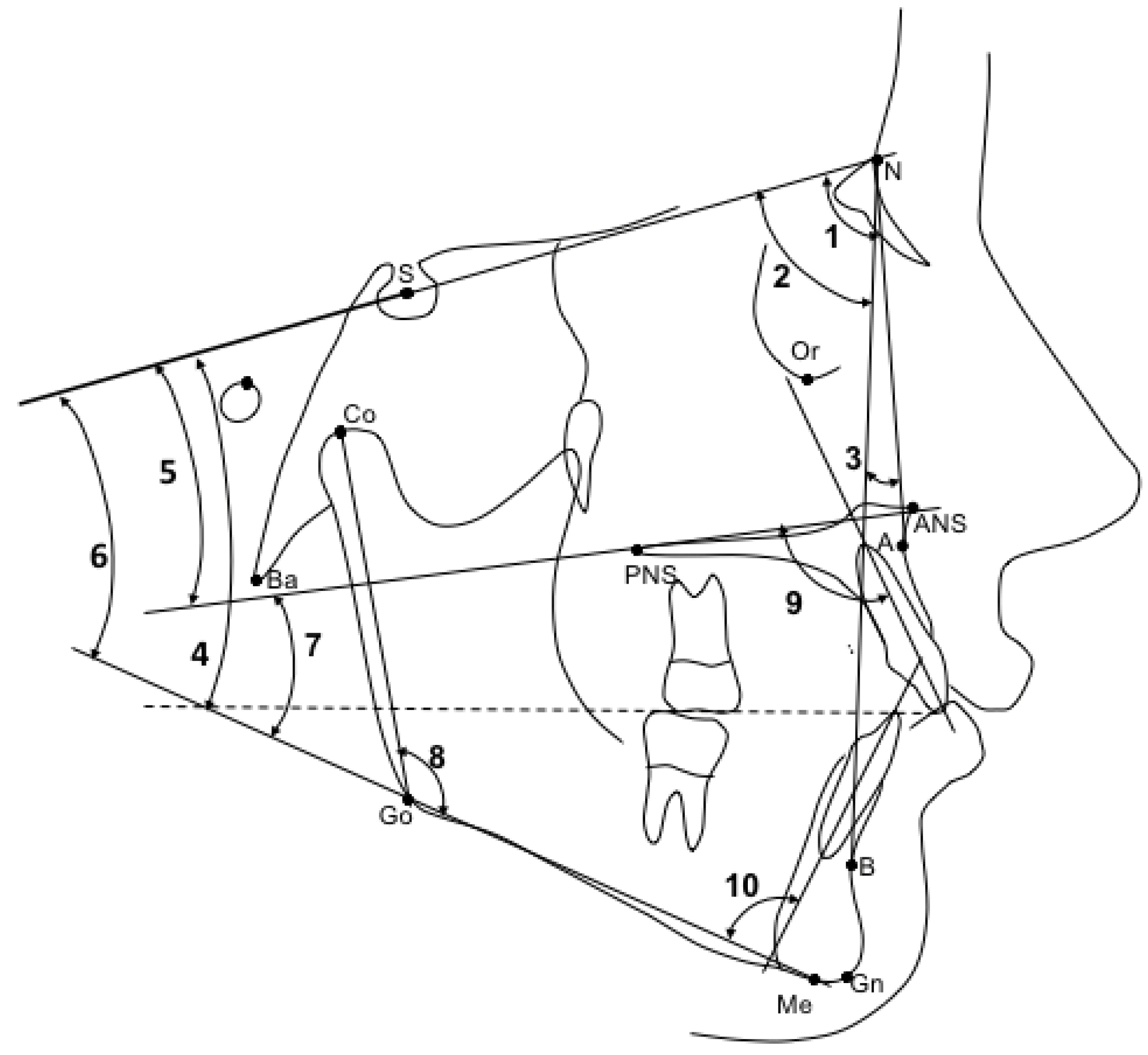
  • SNA (°): angle between sella, nasion, and subspinale point A. It indicates the antero-posterior position of the maxilla relative to anterior cranial base (Figure 1, 1);
  • SNB (°): angle between sella, nasion, and supramentale point B. It indicates the antero-posterior position of the mandible to anterior cranial base (Figure 1, 2);
  • ANB (°): angle resulting from the difference between the SNA and SNB angles, which provide an indication of the sagittal skeletal relationships (Figure 1, 3);
  • Wits (mm): is the measure of the segment Ao-Bo where Ao and Bo indicate respectively the projection of point A and point B on the occlusal plane (Figure 2, a);
  • SN-Occlusal Plane (°): angle between the cranial base (SN) and the occlusal plane (Figure 1, 4);
  • SN-Palatal Plane (°): angle between the cranial base (SN) and the palatal plane (Figure 1, 5);
  • SN-Mandibular Plane (°): angle between the cranial base (SN) and the mandibular plane (Figure 1, 6);
  • Palatal Plane-Mandibular Plane (°): angle between the bispinal plane and the mandibular plane, expressing intermaxillary divergence (Figure 1, 7);
  • Co-Gn (mm): distance from condylion to anatomic gnathion, expressing total mandibular length (Figure 2, b);
  • Co-Go (mm): distance from condylion to gonion, expressing the height of the mandibular ramus (Figure 2, c);
  • Co-Go-Me (°): mandibular angle (Figure 1, 8);
  • Overjet (mm): parameter indicating the measurement of the distance in the sagittal plane between the incisal margin of the upper incisors and the vestibular surface of the lower incisors (Figure 2, d);
  • Overbite (mm): distance in the vertical plane between the incisal margin of the upper and lower central incisors (Figure 2, e);
  • Molar relationship (mm): distance between the projections in the occlusal plane of the point of contact between the first permanent molars and the adjacent mesial tooth (second deciduous molar or second premolar) (Figure 2, f);
  • Inc. Sup.-Pal. Pl. (°): angle expressing the inclination of the axis of the upper central incisor relative to the palatal plane (Figure 1, 9);
  • Inc. Inf.-Mand. Pl. (°): angle expressing the inclination of the axis of the lower central incisor relative to the mandibular plane (Figure 1, 10);
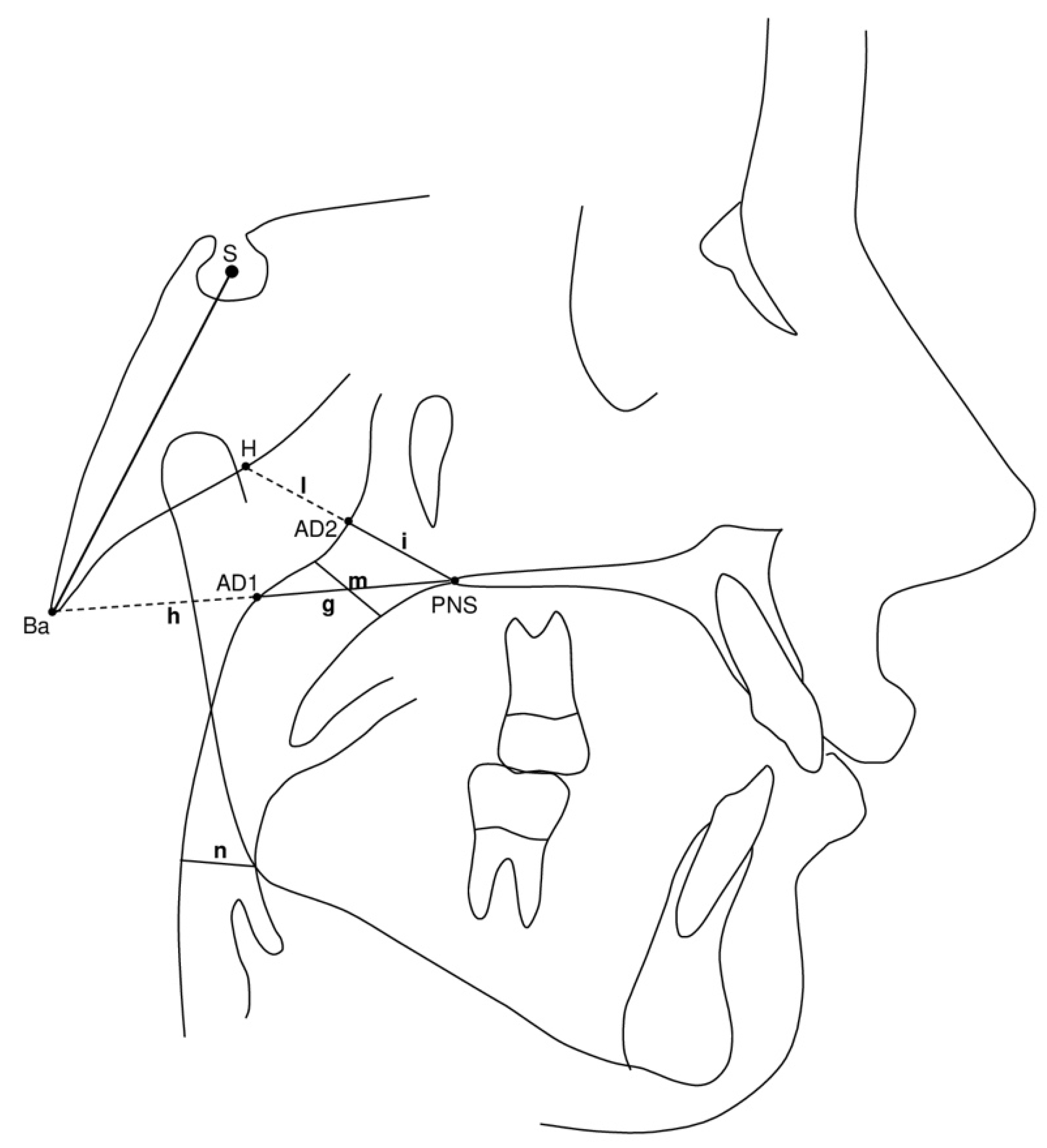
  • PNS-AD1 (mm): lower airway dimension; distance between the PNS and the nearest adenoid tissue measured through the PNS-Ba line (AD1) (Figure 3, g);
  • AD1-Ba (mm): lower adenoid size; defined as the soft tissue thickness at the posterior nasopharynx wall through the PNS-Ba line (Figure 3, h);
  • PNS-AD2 (mm): upper airway dimension; distance between the PNS and the nearest adenoid tissue measured through a perpendicular line to S-Ba from PNS (AD2) (Figure 3, i);
  • AD2-H (mm): upper adenoid size; defined as the soft tissue thickness at the posterior nasopharynx wall through the PNS-H line (H, Hormion, point located at the intersection between the perpendicular line to S-Ba from PNS and the cranial base) (Figure 3, I);
  • McNamara’s upper pharynx dimension (mm): the minimum distance between the upper soft palate and the nearest point on the posterior pharynx wall (Figure 3, m);
  • McNamara’s lower pharynx dimension (mm): the minimum distance between the point where the posterior tongue contour crosses the mandible and the nearest point on the posterior pharynx wall (Figure 3, n).

2.2. Method Error

As for intra-operator method error, 15 lateral cephalograms randomly selected were initially digitalized. After 15 days, all films were retraced and digitized by the same operator (V.B.). Intra-operator repeatability was calculated with the interclass correlation coefficients (ICCs). The random error was calculated by means of method of moments estimator (MME) [15].

2.3. Statistical Analyses

Sample size was calculated considering as a primary outcome variable ANB angle. For a difference of 1.5° for ANB, a standard deviation of 1.4° [16], an alfa of 0.05, and a power of 80%, a sample of at least 15 subjects per group was necessary. Inclusion of all consecutively treated patients should be considered for the retrospective design of the study; consequently, a greater number of patients were involved in order to ensure at least 15 patients per group.
Descriptive statistics for continuous variables are reported with mean and standard deviation. Statistical comparisons in the distribution of cervical vertebral maturation stages at T1 and T2 were performed with the chi-square test. In the presence of normally distributed data (Shapiro–Wilk test), statistical comparisons between cephalometric variables at T1 and on changes in the T1-T2 interval were performed with independent sample t-tests. If data were not normally distributed (Shapiro–Wilk test), between-group comparisons were carried out with the Mann–Whitney test.

3. Results

The ICCs values were all “excellent“ [17] as they ranged between 0.902 (Co-Go) and 0.992 (Co-Gn) for linear measurements and between 0.954 (ANB) and 0.995 (Co-Go-Me) for angular measurements. Random error ranged between 0.27 mm (OVB) and 1.45 mm (Co-Go) for linear measurements and between 0.32° (SNB) and 1.48° (Inf.-P.Mand) for angular measurements.
No statistically significant differences were observed between treated and control samples’ demographic data reported in Table 1.
From the comparison between the treated and the control groups, no statistically significant differences were observed for any of the cephalometric variables at baseline (Table 2), including skeletal, dentoalveolar, and airway dimensions.
Descriptive statistics and comparison of changes in the T1–T2 interval between the treated group and the control group are reported in Table 3.
From the comparison between the MARA group and the control group (Table 3), no statistically significant differences were observed for any of the variables analyzed to evaluate the sagittal dimensions of the airways.
On the contrary, considering the variables expressing sagittal skeletal relationships, during active treatment, the MARA group showed a significant improvement of Wits and ANB angle (−1.0 mm vs. 0.4 mm and −0.7° vs. −0.1°, p = 0.009 and p = 0.038) compared to the control group. Analyzing the dentoalveolar changes, the MARA group showed a statistically significant reduction in the overjet (−2.2 mm vs. −0.3 mm p = 0.001), of the overbite (−1.4 mm vs. 0.1 mm p = 0.005), an improvement of molar relationship (1.6 mm vs. −0.2 mm p = 0.000), and a greater proclination of the lower incisors compared to the control group (3.2° vs. 0.5° p = 0.012).

4. Discussion

Class II malocclusions represent a prevalent orthodontic problem generally associated with mandibular retrusion rather than maxillary protrusion [1]. Over the years, the literature has shown that functional appliances could be used to treat this malocclusion orthopedically, not only by determining skeletal and dentoalveolar effects but also causing a modification of the upper airway dimensions, highlighting a possible relationship between the upper airway, skeletal structures, soft tissues, and the muscular component [18].
The present study evaluated the changes in skeletal, dentoalveolar, and sagittal dimensions of the oropharyngeal airways induced by treatment with a fixed functional appliance (MARA) in patients with Class II malocclusion.
For ethical reasons, we used a control group belonging to the AAOF Legacy collections that, while it might show some bias [19], proves to be a suitable representation of what occurs to growing subjects with untreated Class II malocclusion.
During the active phase of treatment (T1-T2), the results of the present study showed no favorable effects on upper airway dimensions in the treated group compared to the control group.
In the literature, there is only one study performed by CBCT evaluating volumetric changes in the upper airway in patients with Class II malocclusions treated with MARA followed by a fixed appliance and compared with an untreated group [12]. In disagreement with what was observed in our study, Rizk et al. showed that treatment with MARA determines a significant increase in the upper airway (p = 0.005 in reference to air volume) and a repositioning of the hyoid bone in treated patients compared to the control group (A-P distance 4.3 mm vs. 1.5 mm, p = 0.000). The different conclusions reported by this study could be interpreted by considering that the observation period represents an important role in the development of the changes in the maxillofacial and oropharyngeal complex over time. In this regard, Vogler et al. [20] reported by means of MRI examinations an increase in adenoid thickness during the first decade of life, reaching the maximum values between 7 and 10 years of age, and then recording a continuous regression up to the age of 60 years. In Rizk et al.’s study [12], the mean age of the treated group with MARA is 11.7 ± 1.75 years at T1 period and 14.5 ± 1.2 years at T2, while the mean age of the subjects treated in our study is 13.8 years at T1 and 15.7 years at T2. Since the treated and the control groups in Rizk et al.’s study [12] were selected by matching the stage of skeletal maturity without considering the chronologic age of the patients, the airway volume augmentation observed in the treated group could be associated mainly with adenoids and Waldeyer lymphatic ring regression occurring from the age of 11 years, rather than an airway development. On the contrary, in our study, the modification of airway dimensions following MARA treatment was not registered in the results, as the mean ages of the patients in the treated and control groups are higher than in the study by Rizk et al. [12]. Vogler et al.’s conclusions [20] were additionally confirmed by the study of Songu et al. [21], which emphasized the fact that the nasopharyngeal dimension increases simultaneously and linearly with the adenoids until the age of 10 years, reaching a peak between 10 and 11 years. From that point, the adenoids and palatine tonsils regress rapidly, reaching a growth plateau at 11 years. After this age, their size shows a slight reduction, and then it stabilizes. In addition to these considerations, as reported by Cozza et al. [22], airway changes in relation to skeletal modifications. In relation to this, Arens et al. [23] by MRI analysis concluded that the middle and lower thirds of the face continue to grow linearly along the sagittal and axial planes during infancy and, simultaneously, the soft tissues that delineate the upper airways, including adenoids and tonsils, evolve in constant proportion to bone structures in order to maintain airway patency.
In addition, the literature has demonstrated that fixed functional appliances can stimulate mandibular growth in Class II patients with mandibular retrusion if the device is applied at the ideal timing [24]. In relation to this, Huanca Ghislanzoni et al. [25] showed that pubertal growth spurt represents the most favorable timing for treatment with MARA in order to achieve a greater amount of mandibular skeletal effects, such as a statistically significant augmentation of total mandibular length (2.6 mm), and a minimal amount of dental compensation at the lower arch level. In our study, as reported in Table 3, not all patients were treated with MARA at the pubertal growth spurt (12.5%), whereas, based on the individual skeletal maturity, most subjects were pre-pubertal (25%) or post-pubertal (66.7%) at T1. In the present study, MARA did not induce statistically significant skeletal changes in mandibular growth or in mandibular sagittal position. Based on these findings, the lack of changes in upper airway dimensions may be related to the absence of skeletal changes at the mandibular level since both develop consistently and simultaneously.
In disagreement with what was observed in Rizk et al.’s study [12], Cortese et al. [26] evaluated changes in airway dimensions on lateral cephalograms in 10 patients treated with mandibular activators, such as the Andresen monoblock and the Twin Block, according to Clark, compared to a control group of 10 untreated subjects. In line with the results of our study, Cortese et al. [26] found no significant differences in upper, middle, and lower airway changes between the two groups during the observation period (superior-posterior airway space (SPAS) 1.3 mm vs. −0.5 mm, p = 0.1157; middle airway space (MAS) 1 mm vs. 0.2 mm, p = 0.2954; inferior airway space (IAS) 1.1 mm vs. −0.3 mm, p = 0.4682).
Our study not only focused on the analysis of airway dimensions but also evaluated the skeletal and dentoalveolar effects induced by MARA. On skeletal level, the results of the present study showed that Wits and ANB angle were significantly reduced in the treated group, demonstrating the achievement of a better relationship between the mandible and the maxilla (Wits −1.0 mm and ANB −0.7°, p = 0.009 and p = 0.038). These results are in line with the study of Pangrazio-Kulbersh et al. [18], and they are also supported by the study by Ghislanzoni et al. [16], who showed that MARA determines favorable long-term skeletal changes, although of low entity, including the reduction in ANB angle and Wits in the treated group compared to the controls (ANB −1.7° vs. −0.4°, p = 0.000; Wits −1.1 mm vs. 0.7 mm, p = 0.040). In disagreement with the results of these two studies, our study did not show a modification of mandibular growth. As previously reported, this is presumably attributable to the fact that the MARA appliance was not applied in all treated subjects at the pubertal growth spurt, which represents the ideal treatment timing for stimulating mandibular growth using orthopedic appliances [25].
Since MARA was not applied in all patients at the pubertal growth spurt, most of the effects produced by the treatment were observed at the dentoalveolar level. In fact, our study showed an improvement in molar relationship, overjet, and overbite, and a proclination of the lower incisors, in agreement with the results of the studies of Gonner et al. [18], Chiqueto et al. [27], and Ghislanzoni et al. [16].
These findings can be supported clinically by the following Figures that refer to a patient treated with MARA at the pubertal growth spurt. In particular, Figure 4a shows a patient with Class II malocclusion before treatment, while Figure 4b represents the correction of the malocclusion after treatment with the functional device.

Limitations

The limitations of the present study were its retrospective and short-term nature and the comparison of the treated groups with a historical control sample of subjects with untreated Class II malocclusion. Another limitation was that not all patients were treated with MARA device at the pubertal growth spurt, which represents the most favorable period of treatment. In fact, as previously mentioned, the lack of changes in upper airway dimensions could be related to the absence of skeletal changes at the mandibular level since both develop consistently and simultaneously. Finally, the changes in airway dimensions were assessed in 2D. Ideally, these changes should have been described in 3D. The use of CBCTs in growing patients, however, raises ethical concerns in terms of radiation exposure.

5. Conclusions

During active treatment, MARA did not produce significant favorable changes in the sagittal airway dimensions Class II subjects when compared with untreated controls.
The functional appliance analyzed in this study improved both the sagittal skeletal (significant improvement in Wits and ANB angle) and the dentoalveolar relationships (significant decrease in overjet and overbite, improvement of the molar relationship). However, the device induced a significant proclination of the lower incisors and no significant changes in mandibular growth. Clinically, these findings highlight that MARA was able to provide an effective correction of Class II malocclusion with favorable dentoskeletal changes. Future studies should include only Class II patients treated at the pubertal growth spurt, with the use of skeletal anchorage to control the proclination of the lower incisors, and with a long-term follow-up.

Author Contributions

Conceptualization, V.G. and L.F.; Methodology, V.G. and L.F.; Formal Analysis, M.D.S. and V.B.; Investigation, M.D.S. and V.B.; Resources, M.D.S., V.B. and N.P.; Data Curation, V.G., L.F. and N.P.; Writing—Original Draft Preparation, M.D.S.; Writing—Review and Editing, M.D.S., L.F., V.B. and N.P.; Visualization, V.G., L.F., D.F., F.P., N.P. and C.N.; Supervision, D.F., F.P., N.P. and C.N.; Project Administration, V.G. and L.F. All authors have read and agreed to the published version of the manuscript.

Funding

This research received no external funding.

Institutional Review Board Statement

The study was conducted according to the guidelines of the Declaration of Helsinki and approved by the Landeszahnärztekammer Hessen Körperschaft des öffentlichen Rechts (Ethic 03/2021).

Informed Consent Statement

Informed consent was obtained from all subjects involved in the study.

Data Availability Statement

The data will be available on request.

Conflicts of Interest

The authors declare no conflict of interest.

Abbreviations

MARA = mandibular anterior repositioning appliance; OSAS = obstructive sleep apnea; T1 = start of treatment; T2 = after MARA therapy; CVM = cervical vertebral maturation method; AAOF Legacy = Association of Orthodontists Foundation Craniofacial Growth Legacy Collection; ICCs = interclass correlation coefficients; MME = method of moments estimator; CBCT = cone beam computed tomography; MRI = magnetic resonance imaging; SPAS = superior-posterior airway space; MAS = middle airway space; IAS = inferior airway space; SD = standard deviation; Diff. = difference; CI = confidence interval; Occ. = occlusal; Pal. = palatal; Pl. = plane; Mand. = mandibular; Inc. = incisor; McNa = McNamara; deg. = degrees; mm = millimeters.

References

  1. McNamara, J.A. Components of class II malocclusion in children 8–10 years of age. Angle Orthod. 1981, 51, 177–202. [Google Scholar] [CrossRef] [PubMed]
  2. Zhong, Z.; Tang, Z.; Gao, X.; Zeng, X.L. A comparison study of upper airway among different skeletal craniofacial patterns in nonsnoring Chinese children. Angle Orthod. 2010, 80, 267–274. [Google Scholar] [CrossRef] [PubMed]
  3. Baccetti, T.; Franchi, L.; Stahl, F. Comparison of 2 comprehensive Class II treatment protocols including the bonded Herbst and headgear appliances: A double-blind study of consecutively treated patients at puberty. Am. J. Orthod. Dentofac. Orthop. 2009, 135, 698.e1–698.e10. [Google Scholar] [CrossRef] [PubMed]
  4. Katyal, V.; Pamula, Y.; Martin, A.J.; Daynes, C.N.; Kennedy, J.D.; Sampson, W.J. Craniofacial and upper airway morphology in pediatric sleep-disordered breathing: Systematic review and meta-analysis. Am. J. Orthod. Dentofac. Orthop. 2013, 143, 20–30. [Google Scholar] [CrossRef] [PubMed]
  5. El, H.; Palomo, J.M. Airway volume for different dentofacial skeletal patterns. Am. J. Orthod. Dentofac. Orthop. 2011, 139, e511–e521. [Google Scholar] [CrossRef] [PubMed]
  6. Banabilh, S.M.; Samsudin, A.R.; Suzina, A.H.; Dinsuhaimi, S. Facial profile shape, malocclusion and palatal morphology in Malay obstructive sleep apnea patients. Angle Orthod. 2010, 80, 37–42. [Google Scholar] [CrossRef]
  7. Carroll, J.L. Obstructive sleep-disordered breathing in children: New controversies, new directions. Clin. Chest Med. 2003, 24, 261–282. [Google Scholar] [CrossRef]
  8. Kaditis, A.G.; Alonso Alvarez, M.L.; Boudewyns, A.; Alexopoulos, E.I.; Ersu, R.; Joosten, K.; Larramona, H.; Miano, S.; Narang, I.; Trang, H.; et al. Obstructive sleep disordered breathing in 2- to 18-year-old children: Diagnosis and management. Eur. Respir. J. 2016, 47, 69–94. [Google Scholar] [CrossRef]
  9. Li, Y. Early orthodontic treatment of skeletal Class II malocclusion may be effective to prevent the potential for OSAHS and snoring. Med. Hypotheses 2009, 73, 594–595. [Google Scholar] [CrossRef]
  10. Xiang, M.L.; Hu, B.; Liu, Y.; Sun, J.; Song, J. Changes in airway dimensions following functional appliances in growing patients with skeletal class II malocclusion: A systematic review and meta-analysis. Int. J. Pediatr. Otorhinolaryngol. 2017, 97, 170–180. [Google Scholar] [CrossRef]
  11. Mohamed, R.N.; Basha, S.; Al-Thomali, Y. Changes in upper airway dimensions following orthodontic treatment of skeletal Class II malocclusion with twin block appliance: A systematic review. Turk. J. Orthod. 2020, 33, 59–64. [Google Scholar] [CrossRef] [PubMed]
  12. Rizk, S.; Kulbersh, V.P.; Al-Qawasmi, R. Changes in the oropharyngeal airway of Class II patients treated with the mandibular anterior repositioning appliance. Angle Orthod. 2016, 86, 955–961. [Google Scholar] [CrossRef] [PubMed] [Green Version]
  13. Baccetti, T.; Franchi, L.; McNamara, J.A. The Cervical Vertebral Maturation (CVM) method for the assessment of optimal treatment timing in dentofacial orthopedics. Semin. Orthod. 2005, 11, 119–129. [Google Scholar] [CrossRef]
  14. Cretella Lombardo, E.; Franchi, L.; Lione, R.; Chiavaroli, A.; Cozza, P.; Pavoni, C. Evaluation of sagittal airway dimensions after face mask therapy with rapid maxillary expansion in Class III growing patients. Int. J. Pediatr. Otorhinolaryngol. 2020, 130, 109794. [Google Scholar] [CrossRef]
  15. Springate, S. The effect of sample size and bias on the reliability of estimates of error: A comparative study of Dahlberg’s formula. Eur. J. Orthod. 2012, 34, 158–163. [Google Scholar] [CrossRef] [Green Version]
  16. Ghislanzoni, L.T.H.; Toll, D.E.; Defraia, E.; Baccetti, T.; Franchi, L. Treatment and posttreatment outcomes induced by the mandibular advancement repositioning appliance; A controlled clinical study. Angle Orthod. 2011, 81, 684–691. [Google Scholar] [CrossRef] [Green Version]
  17. Cicchetti, D.V.; Sparrow, S.A. Developing criteria for establishing interrater reliability of specific items: Applications to assessment of adaptive behavior. Am. J. Ment. Defic. 1981, 86, 127–137. [Google Scholar]
  18. Gönner, U.; Özkan, V.; Jahn, E.; Toll, D.E. Effect of the MARA appliance on the position of the lower anteriors in children, adolescents and adults with Class II malocclusion. J. Orofac. Orthop. 2007, 68, 397–412. [Google Scholar] [CrossRef]
  19. Pandis, N. Use of controls in clinical trials. Am. J. Orthod. Dentofac. Orthop. 2012, 141, 250–251. [Google Scholar] [CrossRef]
  20. Vogler, R.C.; Wippold, F.J.; Pilgram, T.K. Age-specific size of the normal adenoid pad on magnetic resonance imaging. Clin. Otolaryngol. Allied Sci. 2000, 25, 392–395. [Google Scholar] [CrossRef]
  21. Songu, M.; Adibelli, Z.H.; Tuncyurek, O.; Adibelli, H. Age-specific size of the upper airway structures in children during development. Ann. Otol. Rhinol. Laryngol. 2010, 119, 541–546. [Google Scholar] [CrossRef] [PubMed]
  22. Cozza, P.; Baccetti, T.; Franchi, L.; De Toffol, L.; McNamara, J.A. Mandibular changes produced by functional appliances in Class II malocclusion: A systematic review. Am. J. Orthod. Dentofac. Orthop. 2006, 129, 599.e1–599.e12. [Google Scholar] [CrossRef] [PubMed] [Green Version]
  23. Arens, R.; McDonough, J.M.; Corbin, A.M.; Hernandez, M.E.; Maislin, G.; Schwab, R.J.; Pack, A.I. Linear dimensions of the upper airway structure during development: Assessment by magnetic resonance imaging. Am. J. Respir. Crit. Care Med. 2002, 165, 117–122. [Google Scholar] [CrossRef] [PubMed]
  24. Perinetti, G.; Primožič, J.; Furlani, G.; Franchi, L.; Contardo, L. Treatment effects of fixed functional appliances alone or in combination with multibracket appliances: A systematic review and meta-analysis. Angle Orthod. 2015, 85, 480–492. [Google Scholar] [CrossRef] [PubMed]
  25. Huanca Ghislanzoni, L.T.; Baccetti, T.; Toll, D.; Defraia, E.; McNamara, J.A.; Franchi, L. Treatment timing of MARA and fixed appliance therapy of Class II malocclusion. Eur. J. Orthod. 2013, 35, 394–400. [Google Scholar] [CrossRef] [PubMed]
  26. Cortese, M.; Pigato, G.; Casiraghi, G.; Ferrari, M.; Bianco, E.; Maddalone, M. Evaluation of the oropharyngeal airway space in class II malocclusion treated with mandibular activator: A retrospective study. J. Contemp. Dent. Pract. 2020, 21, 666–672. [Google Scholar] [CrossRef] [PubMed]
  27. Chiqueto, K.; Henriques, J.F.C.; Barros, S.E.C.; Janson, G. Angle Class II correction with MARA appliance. Dent. Press J. Orthod. 2013, 18, 35–44. [Google Scholar] [CrossRef] [PubMed] [Green Version]
Figure 1. Angular variables considered in the study.
Applsci 12 01484 g001
Figure 2. Linear variables considered in the study.
Applsci 12 01484 g002
Figure 3. Cephalometric measurements for the analysis of airway dimensions.
Applsci 12 01484 g003
Figure 4. (a) Before treatment with MARA device. (b) After treatment with MARA appliance.
Applsci 12 01484 g004
Table 1. Demographics with statistical comparisons (independent-samples t-tests or Mann–Whitney U test or chi-square test).
VariablesMARA Group
(n = 24)
Control Group
(n = 20)
Diff.p-Value
MeanSDMeanSD
Age at T1 (years)13.82.013.81.80.00.902
Age at T2 (years)15.71.915.41.60.30.517
T1-T2 interval (years)1.90.41.60.50.30.071
CVM at T1CS1 = 4; CS2 = 3; CS3 = 2; CS4 = 6; CS5 = 6; CS6 = 2CS1 = 3; CS2 = 3; CS3 = 1; CS4 = 4; CS5 = 7; CS6 = 2 0.980
CVM at T2CS2 = 2; CS3 = 4; CS4 = 7; CS5 = 5; CS6 = 5CS2 = 1; CS3 = 4; CS4 = 5; CS5 = 7; CS6 = 3 0.862
Table 2. Descriptive statistics and statistical comparisons (independent-samples t-tests or Mann–Whitney U test) of the starting forms (cephalometric values at T1).
VariablesMARA Group (n = 24)Control Group (n = 20)Diff.p-Value95% CI of the Difference
MeanSDMeanSDLowerUpper
Age (years)13.82.013.81.80.00.902−1.11.2
SNA (deg)80.34.281.13.2−0.80.517−3.01.5
SNB (deg)75.43.275.92.8−0.50.632−2.31.4
ANB (deg)4.92.45.21.6−0.30.638−1.61.0
Wits (mm)2.72.52.33.10.40.591−1.32.2
SN-Occ. Pl. (deg)17.23.617.83.8−0.60.551−2.91.6
SN-Pal. Pl. (deg)7.82.97.82.90.00.974−1.71.8
SN-Mand. Pl. (deg)31.86.130.54.71.30.424−2.04.7
Pal. Pl. to Mand. Pl. (deg)24.05.722.75.01.30.431−2.04.6
Co-Gn (mm)103.512.8102.75.90.80.175
Co-Go (mm)52.67.652.24.50.40.465
Co-Go-Me (deg)121.66.2121.55.40.10.948−3.53.7
Overjet (mm)4.92.04.91.30.00.936−1.11.0
Overbite (mm)2.91.84.12.2−1.20.062−2.40.1
Molar Relationship (mm)−0.11.5−0.61.70.50.338−0.51.5
Upper Inc. to Pal. Pl. (deg)111.27.3107.47.63.80.099−0.78.3
Lower Inc. to Mand. Pl. (deg)102.06.5103.57.8−1.50.495−5.82.9
PNS-AD1 (mm)22.94.823.73.3−0.80.536−3.31.8
AD1-Ba (mm)20.64.318.54.32.10.115−0.54.7
PNS-AD2 (mm)18.13.918.74.1−0.60.649−3.01.9
AD2-H (mm)11.33.710.63.80.70.529−1.63.0
McNa Upper Pharinx (mm)12.43.912.63.4−0.20.809−2.52.0
McNa Lower Pharinx (mm)9.42.610.22.8−0.80.297−2.50.8
SD = standard deviation; Diff. = difference; CI = confidence interval; Occ. = occlusal; Pal. = palatal; Pl.= plane; Mand. = mandibular; Inc. = incisor; McNa = McNamara; deg. = degrees; mm = millimeters.
Table 3. Descriptive statistics and statistical comparisons (independent-samples t-tests or Mann–Whitney U test) of the T2-T1 changes.
VariablesMARA Group
(n = 24)
Control Group
(n = 20)
Diff.p-Value95% CI of the Difference
MeanSDMeanSDLowerUpper
SNA (deg)−0.31.10.01.2−0.30.301−1.10.3
SNB (deg)0.41.00.11.30.30.476−0.40.9
ANB (deg)−0.71.0−0.10.9−0.60.038−1.20.0
Wits (mm)−1.01.70.41.6−1.40.009−2.4−0.4
SN-Occ. Pl. (deg)0.32.9−0.72.61.00.251−0.72.7
SN-Pal. Pl. (deg)0.40.90.81.4−0.40.252−1.20.3
SN-Mand. Pl. (deg)−1.33.3−0.51.6−0.80.532
Pal. Pl. to Mand. Pl. (deg)−1.73.0−1.31.9−0.40.887
Co-Gn (mm)3.22.43.02.30.20.732
Co-Go (mm)2.31.72.21.70.10.785−0.91.2
Co-Go-Me (deg)−1.12.4−1.51.70.40.570−0.91.6
Overjet (mm)−2.22.2−0.30.9−1.90.001−3.0−0.8
Overbite (mm)−1.41.90.11.0−1.50.005
Molar Relationship (mm)1.62.0−0.20.91.80.0000.92.8
Upper Inc. to Pal. Pl. (deg)−1.47.20.62.9−2.00.249−5.51.5
Lower Inc. to Mand. Pl. (deg)3.24.30.52.12.70.0120.64.9
PNS-AD1 (mm)0.62.20.43.50.20.855−1.61.9
AD1-Ba (mm)−0.12.30.33.0−0.40.211
PNS-AD2 (mm)1.52.40.42.71.10.162−0.52.7
AD2-H (mm)−1.22.50.13.1−1.30.130−3.00.4
McNa Upper Pharinx (mm)1.62.50.92.40.70.342−0.82.2
McNa Lower Pharinx (mm)1.90.41.60.50.30.641−1.71.1
SD = standard deviation; Diff. = difference; CI = confidence interval; Occ. = occlusal; Pal. = palatal; Pl. = plane; Mand. = mandibular; Inc. = incisor; McNa = McNamara; deg. = degrees; mm = millimeters. Bold character indicates a statistically significant p value.
Publisher’s Note: MDPI stays neutral with regard to jurisdictional claims in published maps and institutional affiliations.

