Article

Skeletal and Dentoalveolar Effects Induced by the Paolone-Kaitsas Appliance in the Treatment of Class II Malocclusion: A Controlled Retrospective Study on Lateral Cephalograms

by
Camilla Gavazzi
1,
Debora Franceschi
1,
Felicita Pierleoni
1,
Valeria Barone
2,
Francesco Kaitsas
3,
Maria Giacinta Paolone
4,
Lorenzo Franchi
1,* and
Veronica Giuntini
1
1
Department of Experimental and Clinical Medicine, University of Florence, 50121 Florence, Italy
2
Independent Researcher, 50100 Florence, Italy
3
Student of the Faculty of Medicine and Surgery, Università Cattolica del Sacro Cuore, 00168 Rome, Italy
4
Independent Researcher, 00152 Rome, Italy
*
Author to whom correspondence should be addressed.
Appl. Sci. 2022, 12(3), 1165; https://doi.org/10.3390/app12031165
Submission received: 17 December 2021 / Revised: 15 January 2022 / Accepted: 19 January 2022 / Published: 23 January 2022
(This article belongs to the Special Issue Imaging Techniques for Oral and Dental Applications)

Abstract

:
The aim of this study was to assess the skeletal and dentoalveolar effects induced by the Paolone-Kaitsas functional appliance (PK appliance) in the treatment of growing patients affected by Class II malocclusion. A group of 25 Class II patients, treated with the PK appliance followed by fixed appliances, was evaluated with lateral cephalograms at the start (9.6 ± 1.6 years) and at the end of treatment (13.0 ± 1.5 years), and was compared with a matched untreated Class II control group of 23 subjects selected from the web archive of the American Association of Orthodontists Foundation Craniofacial Growth Legacy. Statistical comparisons were performed with the Student’s t-tests. The treated group showed a significant decrease in SNA (−2.2°), ANB (−2.2°), and Wits appraisal (−3.4 mm), a significant increase in the SN-palatal plane angle (1.1°), and a significant improvement in overjet (−2.9 mm), overbite (−2.5 mm), and molar relationship (3.6 mm). The PK appliance produced favorable dentoalveolar and skeletal effects: it inhibited maxillary growth without effects on the mandible and it also induced a downward inclination of the palatal plane.

1. Introduction

Class II malocclusion affects about a third of the population [1], representing one of the most common problems in orthodontics. Among the numerous dental and skeletal factors that can lead to Class II malocclusion, mandibular skeletal retrusion is the main cause [2,3]. Data in the literature have shown that the characteristics of this type of malocclusion stabilize from early childhood and, in the absence of treatment or modification of the etiological factors, it never undergoes spontaneous self-correction [4]. Therefore, treatment is necessary.
As Class II malocclusion is more frequently characterized by mandibular retrusion, a therapy capable of improving mandibular growth and position is recommended in these patients [2]. This type of dental and skeletal disharmony, in fact, can be corrected by the use of functional devices that stimulate mandibular growth with anterior repositioning of the mandible.
Proffit et al. [5] divided the main etiological factors of malocclusions into three categories: specific causes, such as alterations during the embryonic, fetal, and neonatal periods, hereditary factors, and environmental factors. The latter are represented by the forces and pressures that the muscles exert physiologically on the skeletal and dental structures during development. The pressure generated by the muscles of mastication during chewing is a factor potentially significant in the development of malocclusion. As stated by McNamara [2], mandibular retrusion has an embryological origin. Furthermore, the position of the dental elements in the arch is dictated by the balance between centripetal and centrifugal muscular forces that cancel each other out. The alteration of the pressure balance generated by the early onset of a bad habit, such as finger sucking, can generate the onset of Class II malocclusion. Factors leading to mandibular retrusion, such as transverse discrepancy due to superior transverse minus [6], are considered “Class II interferences” and may cause the onset of a functional Class II malocclusion. If those interferences are not intercepted and removed, over time the malocclusion can turn into an anatomical alteration.
The Paolone-Kaitsas appliance (PK appliance) [7] is a removable functional appliance that was described for the first time in 2017. It consists of two acrylic plates, one upper and one lower, joined by two lateral three-loop springs. The plates are characterized by the presence of occlusal bite planes with complete occlusal coverage to control the occlusal plane. Lateral springs enable the progressive activation of the device in order to control the posterior facial height/anterior facial height (PFH/AFH) Index due to the larger activation in the posterior than in the anterior area. Moreover, it is possible to incorporate an expansion screw in the upper plate to correct possible maxillary contraction, which is frequently associated with Class II malocclusion. No studies evaluating the skeletal and dentoalveolar effects of the device have been published so far.
The aim of this study was to analyze the skeletal and dentoalveolar effects induced by the PK appliance in the treatment of growing patients with Class II malocclusion. This evaluation was based on the analysis of pre- and post-treatment lateral cephalograms compared with the cephalometric data of a control group of growing patients with untreated Class II malocclusion.

2. Materials and Methods

The treatment group consisted of 25 patients, 13 females and 12 males, treated consecutively with a PK appliance followed by fixed appliances. All patients were treated by the same operator (Dr. Maria Giacinta Paolone).
The sample inclusion criteria were:
Skeletal Class II malocclusion;
Dental Class II malocclusion (division 1 and division 2);
Presence of complete and good quality initial and final lateral cephalograms;
Treatment of the Class II dentoskeletal imbalance with a PK appliance followed by fixed appliances.
The sample exclusion criteria were:
Patients with cleft lip/palate abnormalities;
Patients with craniofacial syndromes.
All treated patients underwent pre-treatment (T1) and post-treatment (T2) lateral cephalograms performed using the same radiographic machine. The original cephalograms were acquired through a scanner to be analyzed. The scans were all performed using the same device (Epson Perfection V850 Pro) at the same resolution (150 DPI).
For each patient undergoing treatment, the stage of mandibular skeletal maturity was detected on the lateral cephalograms by means of the cervical vertebrae maturation method [8].
The control group consisted of 23 subjects (12 females and 11 males) with untreated Class II malocclusion selected from the web archive of the American Association of Orthodontists Foundation (AAOF) Craniofacial Growth Legacy Collection (https://www.aaoflegacycollection.org/, accessed on 22 December 2020), within the Denver Growth, Iowa Growth, Michigan Growth, and Oregon Growth collections. The patients belonging to the control group were individually matched to the treated patients in compliance with the following criteria:
Presence of Class II malocclusion;
Gender;
Chronologic age at the beginning and at end of treatment, with a maximum difference of six months;
Stage of maturation of the cervical vertebrae at the beginning and end of treatment.
For each of the patients belonging to the control group, lateral cephalograms with high definition were requested.

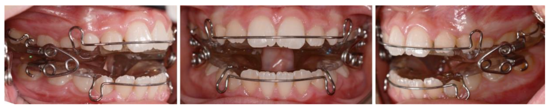
2.1. Treatment Protocol

Patients belonging to the treated group underwent a treatment protocol consisting of two phases. In the first treatment phase, the PK appliance [7] (Figure 1) was used for about 15 months, with the aim of maintaining the position of the upper jaw and promoting mandibular growth, controlling its growth in the vertical plane, in order to establish a Class I molar relationship and a correct relationship between the incisors. In the permanent dentition, the second treatment phase was performed with fixed upper and lower appliances with the aim of aligning and leveling the arches to achieve a stable occlusion. Treatment with fixed appliances lasted for about 12 months on average and it was performed with standard Edgewise brackets with 0.022 × 0.028 inch slots. As for the arch sequence, it comprised 0.016 inch CuNiTi and beta-titanium archwires followed by 0.017 × 0.025 inch and 0.019 × 0.025 inch stainless steel archwires. Finishing was achieved with 0.016 and 0.017 × 0.025 inch beta-titanium archwires. Retention after the fixed appliances was performed with upper and lower Hawley retainers.

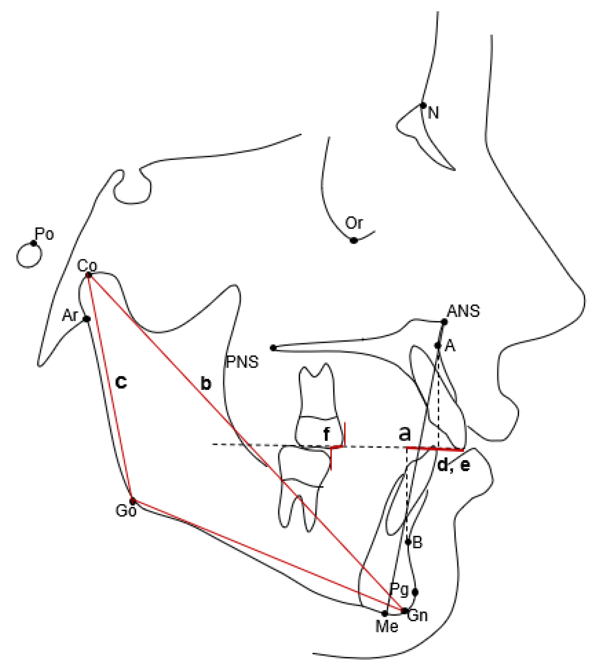
2.2. Cephalometric Analysis

Lateral cephalograms were digitized by one investigator (V.B.). A customized digitization regimen (Viewbox, version 4.0, dHAL Software, Kifissia, Greece) was created and used for cephalometric evaluation. Sixteen variables (6 linear and 10 angular) were generated (Figure 2 and Figure 3). Then, landmark locations and the accuracy of the anatomical outlines were verified by a second investigator (L.F.), and any discrepancies as to landmark placement were resolved by mutual agreement.

2.3. Method Error

In order to evaluate the intra-operator reproducibility and method error, 15 lateral cephalograms randomly selected from the two groups were re-digitized after 10 days by the same operator. The intra-observer reproducibility was assessed with the intraclass correlation coefficients (ICCs), while the method error was calculated by means of the method of moments estimator (MME) [9].

2.4. Statistical Analysis

The sample size was calculated considering as a primary outcome variable ANB angle. For an effect size of 1, an alfa of 0.05, and a power of 80%, a sample of at least 17 subjects per group was necessary. The inclusion of all consecutively treated patients should be considered for the retrospective design of the study; consequently, a greater number of patients were involved in order to ensure at least 17 patients per group.
Descriptive statistics for continuous variables are reported with their means and standard deviations. If the continuous variables were not normally distributed, descriptive statistics were reported with their medians and interquartile ranges. The statistical comparison of the distribution of the stages of cervical vertebrae maturation at T1 and T2 was performed with the Chi-square test. In the presence of normally distributed data (Shapiro–Wilk test), statistical between-group comparisons were performed with independent-samples Student’s t-tests. If the data were not normally distributed, statistical between-group comparisons were carried out with the Mann–Whitney test. A sensitivity analysis was performed by constructing statistical models with gender, Class II division, and treatment as explanatory variables. For all statistical tests, the level of significance was set at p < 0.05.

3. Results

The intraclass correlation coefficients were all “excellent” [10]. The ICC values for linear measurements ranged from a minimum of 0.902 for Co-Go to a maximum of 0.992 for Co-Gn. The ICC values for angular measurements ranged from a minimum of 0.954 for the ANB angle to a maximum of 0.995 for the Co-Go-Me angle. The random error for the linear measurements ranged from a minimum of 0.3 mm for OVB to a maximum of 1.4 mm for Co-Go, while, for angular measurements, it ranged from a minimum of 0.3 degrees for SNB to a maximum of 1.5 degrees for the lower incisor to mandibular plane.
The mean ages, stages of maturation of the cervical vertebrae at T1 and T2, and mean durations of the T1–T2 intervals for both the treated and control groups are reported in Table 1. No statistically significant differences between the two groups were observed.
Descriptive data and statistical comparisons between the treated and control groups for the starting forms and cephalometric changes from T1 to T2 are reported in Table 2 and Table 3, respectively.
The comparison of starting forms (Table 2) showed no significant differences between the treated group and the control group, except for the inclination of the palatal plane to SN (SN-Pal. Pl), which was significantly greater in the treated group compared with that in the control group (8.5° vs. 6.7°, p = 0.014).
As for the T1–T2 changes (Table 3), the PK appliance produced a statistically significant restraint in the sagittal position of the maxilla (SNA) compared with the control group (−1.7° vs. 0.5°. p = 0.000). The treated group also showed a significant decrease in the ANB angle and in the Wits appraisal with respect to the controls (−2.4° and −2.7 mm vs. −0.2° and 0.7 mm. p = 0.000 and p = 0.000). As for the changes in the vertical skeletal relationships, no statistically significant differences were found among the two groups for any of the angular measurements, except for the SN to palatal plane (SN-Pal. Pl.), which showed a significantly greater increase in the treated group compared with that in the control group (1.0° vs. −0.1°. p = 0.020). As for the dentoalveolar changes, the PK appliance group showed significantly greater decreases in overjet and overbite (−2.8 mm and −2.0 mm vs. 0.1 and 0.5 mm. p = 0.000 and p = 0.000) and a significantly greater increase in the intermolar relationship (3.8 mm vs. 0.2 mm. p = 0.000) compared with the control group.
The sensitivity analysis showed no significant differences between males and females, and the results were comparable to those of the main analysis. Class II division was not included in the statistical model because the vast majority of the cases showed Class II division 1 malocclusion (only two cases in the treated group and only one case in the control group showed Class II division 2 malocclusion).

4. Discussion

Class II malocclusion is the most common orthodontic problem in the population [1]. As stated by Cozza et al. [11], it can be expressed in multiple forms, both skeletal and dental, and their differential diagnoses can help in the selection of the most appropriate treatment approach. Among these factors, mandibular retrognathism shows a prevailing frequency [2,3]. In these cases, the use of functional appliances capable of stimulating growth and anterior repositioning of the mandible is particularly indicated. The aim of this study was to analyze the skeletal and dentoalveolar effects induced by the treatment of Class II malocclusion with a removable functional appliance followed by fixed appliances. Over the decades, numerous types of functional devices—both fixed and removable—have been proposed and analyzed in the literature. According to information reported by recent systematic reviews of the literature [11,12,13], they have been proven effective in the treatment of Class II malocclusion, resulting in a correction of the latter through both skeletal and dentoalveolar effects. The skeletal correction of Class II malocclusion is the result of the combined action of effects on the maxilla and mandible.
The PK appliance was designed with the aim of achieving stimulation of mandibular growth, both in its horizontal and vertical components, and control of vertical skeletal relationships. The latter is obtained through careful management of the occlusal plane and the facial height index; the goal, in fact, is to maintain the anterior facial height and to increase the posterior facial height. The patients belonging to the treated group underwent a therapy consisting of two phases: the first treatment phase was performed with the PK appliance for 15 months, with the aim of controlling the sagittal and vertical positions of the maxilla, promoting mandibular growth, and establishing a Class I molar relationship and a correct relationship between the incisors. Once a Class I molar relationship was reached, the second phase of treatment was performed with fixed appliances with the aim of aligning and leveling the arches to obtain a stable occlusion.
This study has highlighted the ability of the PK appliance to act on the sagittal growth of the maxilla, resulting, at the end of treatment, in a statistically significant reduction (−2.2°, p = 0.0000) of the value of the SNA angle. This result is in line with the results reported by Nucera and collaborators [13], who underlined the ability of functional removable devices to exert a restraining action on the growth of the maxilla. In contrast, the PK appliance did not demonstrate any modifying effects on the size and antero-posterior position of the mandible. Therefore, the statistically significant reduction in the ANB angle and in the Wits appraisal (−2.2°, p = 0.0000 and −3.4 mm, p = 0.0000) appears to be due to the action of the device mainly on the maxilla.
As previously stated, one of the goals of the PK appliance is to control the vertical dimension. According to Björk [14], anterior rotation of the mandible occurs when the posterior facial height shows greater development than the anterior facial height. Mandibular rotation appears to be closely related to changes in the occlusal plane during growth [15]. The posterior portion of the dentition, moreover, represents one of the main factors influencing the functional positioning of the mandible [16,17]. Longitudinal growth studies conducted by Tanaka and Sato [18] revealed that changes in the occlusal plane can change the growth pattern of Class II malocclusions and, specifically, that the continued leveling of the posterior occlusal plane is accompanied by a simultaneous decrease in the angle of the mandibular plane. Assuming that the posterior portion of the occlusal plane is the key to changing the position of the mandible, it is plausible to infer that the leveling of this plane may result in an increase in the height of the mandibular ramus and, consequently, a flattening of the mandibular plane. An excessive curve of Spee in the lower arch, in fact, can be leveled by freeing the posterior teeth from the resin of the appliance, inducing their extrusion. The analysis of the results obtained in the vertical plane from the present study confirmed a statistically significant increase in the angular value of SN-palatal plane (1.1°, p = 0.020) at the end of treatment in the treated group compared with that in the control group. The increase in this angle is indicative of a downward inclination of the palatal plane. This result, therefore, is in line with the findings of Paolone and Kaitsas [7] and reflects one of the desired effects induced by the device. The latter, in fact, was designed to exert control of verticality through the presence of lateral thickness planes and three-lobed springs. Specifically, the latter are able to progressively activate the device in order to obtain a greater activation in the posterior region than in the anterior one. On the contrary, no effects of the device on the occlusal plane were found, since, in the treated group, there were no statistically significant changes in the values of SN-occlusal plane and SN-mandibular plane.
As for dentoalveolar changes, the group treated with the PK appliance showed significant decreases in the values of the overjet and overbite, and a statistically significant improvement in the molar relation compared with the control group (respectively −2.9 mm, p = 0.0000; −2.5°, p = 0.0000; 3.6 mm, p = 0.0000). In contrast, there were no significant changes in the inclinations of the upper incisor with respect to the palatal plane and of the lower incisor with respect to the mandibular plane. The improvement in the values of the overjet and overbite, therefore, can be related to the results obtained at the skeletal level, specifically with the reduction in the SNA angle and the downward inclination of the palatal plane.
The skeletal and dentoalveolar results listed above must be analyzed by taking into account the timing for the treatment with the PK appliance in patients with Class II malocclusion. The literature is unanimous in establishing as the ideal timing of treatment with functional appliances the period coinciding with the peak of pubertal growth [19,20], corresponding to the CS3–CS4 stage of maturation of the cervical vertebrae [21]. The group undergoing treatment with the PK appliance, however, was mostly composed of prepubertal-stage subjects (mainly cervical stages CS1 and CS2) at the beginning of active therapy (92%). Moreover, at the end of the observation, most of the treated group was still in the prepubertal stage (stages CS1–2, 16%) or in the circumpubertal stage CS3 (52%), when the beginning of the pubertal growth peak was identified. By virtue of this observation, it is plausible to infer that the lack of skeletal effects induced by the device on the mandible, both on its size and its sagittal position, may be related to inappropriate treatment timing. This hypothesis is in line with the conclusions of the study conducted on the Twin-block device by O’Brien and collaborators [22]. According to this study, in fact, early treatment with the Twin-block appliance can correct Class II malocclusion mainly through dentoalveolar modifications, such as the reduction of the overjet and the improvement of the molar relationship. These effects lead to an overall reduction in the severity of the malocclusion, while the skeletal effects of early treatment are mild and probably of low clinical significance. In addition, according to Baccetti et al. [23], treatment with Twin-block performed at or immediately after the pubertal growth peak involves multiple advantages over pre-peak treatment, such as a greater skeletal contribution to molar correction, and a greater increase in mandibular length and mandibular ramus height associated with greater growth in the posterior direction of the mandibular condyle. Moreover, the use of removable functional devices followed by fixed appliances at the pubertal peak has a number of advantages: removable functional appliances are effective, as they allow about 3 mm more mandibular growth to be obtained; they are efficient in reducing the duration of active therapy [24] and treatment with fixed appliances [25], allowing the achievement of good stability at the end of therapy, which typically ends in a post-pubertal stage of development, where Class II subjects have a residual growth very similar to that of Class I subjects [4].

Limitations

The limitations of this study were its retrospective and short-term nature and the comparison of the treated groups with a historical control sample of subjects with untreated Class II malocclusion. In addition, most patients were treated with the PK appliance at a prepubertal stage, which is not the most favorable period of treatment, represented by the pubertal growth spurt. For this reason, the lack of mandibular skeletal changes induced by the appliance could be related to the inappropriate treatment timing. Future studies should investigate the role of treatment timing (prepubertal vs. pubertal stage of development) on the treatment effects produced by the PK appliance, possibly with a long-term follow up.

5. Conclusions

The PK appliance was proven to be effective in improving the maxillomandibular relationship in the sagittal plane, resulting in a significant reduction in the ANB angle and Wits appraisal in the treated group compared with those in the control group with untreated Class II malocclusion. It exerted a greater restraining effect on the maxilla growth while showing no growth stimulation or anterior repositioning of the mandible.
In the vertical plane, the PK appliance caused a downward inclination of the palatal plane, while it was not able to modify the inclination of the occlusal plane or the mandibular plane.
As for dentoalveolar changes, the device produced a reduction in overbite and overjet, along with a significant improvement in the molar relationship.

Author Contributions

Conceptualization, V.G. and L.F.; Methodology, V.G. and L.F.; Formal analysis, C.G. and V.B.; Investigation, C.G. and V.B.; Resources, M.G.P., C.G. and V.B.; Data curation, V.G. and L.F.; Writing—Original draft preparation, C.G.; Writing—review and editing, C.G., V.B., and L.F.; Visualization, V.G., L.F., D.F., F.P., F.K., and M.G.P.; Supervision, D.F., F.P., F.K., and M.G.P.; Project administration, V.G. and L.F. All authors have read and agreed to the published version of the manuscript.

Funding

This research received no external funding.

Institutional Review Board Statement

The study was conducted according to the guidelines of the Declaration of Helsinki and approved by the Lazio 1 Ethics Committee of the Region of Lazio, Italy (1631/CELazio1 Oss-R-259).

Informed Consent Statement

Informed consent was obtained from all subjects involved in the study.

Data Availability Statement

The data will be available on request.

Conflicts of Interest

The authors declare no conflict of interest.

References

  1. Proffit, W.R.; Fields, H.W.J.; Moray, L.J. Prevalence of malocclusion and orthodontic treatment need in the United States: Estimates from the NHANES III survey. Int. J. Adult Orthod. Orthognath. Surg. 1998, 13, 97–106. [Google Scholar]
  2. McNamara, J.A.J. Components of class II malocclusion in children 8-10 years of age. Angle Orthod. 1981, 51, 177–202. [Google Scholar] [CrossRef] [PubMed]
  3. Pancherz, H.; Zieber, K.; Hoyer, B. Cephalometric characteristics of Class II division 1 and Class II division 2 malocclusions: A comparative study in children. Angle Orthod. 1997, 67, 111–120. [Google Scholar] [CrossRef] [PubMed]
  4. Stahl, F.; Baccetti, T.; Franchi, L.; McNamara, J.A.J. Longitudinal growth changes in untreated subjects with Class II Division 1 malocclusion. Am. J. Orthod. Dentofac. Orthop. 2008, 134, 125–137. [Google Scholar] [CrossRef] [PubMed]
  5. Proffit, W.R.; Fields, H.W.; Larson, B.E.; Sarver, D.M. Contemporary Orthodontics 6th Ed; Elsevier: Philadelphia, PA, United States, 2019; pp. 107–139. [Google Scholar]
  6. Tollaro, I.; Baccetti, T.; Franchi, L.; Tanasescu, C.D. Role of posterior transverse interarch discrepancy in Class II, Division 1 malocclusion during the mixed dentition phase. Am. J. Orthod. Dentofac. Orthop. 1996, 110, 417–422. [Google Scholar] [CrossRef]
  7. Paolone, M.G.; Kaitsas, R. Mandibular response and directional forces with a new functional orthopedic appliance—A clinical case treated with a “Paolone-Kaitsas (PK) appliance”. Int. Orthod. 2017, 15, 708–727. [Google Scholar] [CrossRef] [PubMed]
  8. McNamara, J.A.J.; Franchi, L. The cervical vertebral maturation method: A user’s guide. Angle Orthod. 2018, 88, 133–143. [Google Scholar] [CrossRef] [PubMed] [Green Version]
  9. Springate, S.D. The effect of sample size and bias on the reliability of estimates of error: A comparative study of Dahlberg’s formula. Eur. J. Orthod. 2012, 34, 158–163. [Google Scholar] [CrossRef] [PubMed] [Green Version]
  10. Cicchetti, D.V.; Sparrow, S.A. Developing criteria for establishing interrater reliability of specific items: Applications to assessment of adaptive behavior. Am. J. Ment. Defic. 1981, 86, 127–137. [Google Scholar]
  11. Cozza, P.; Baccetti, T.; Franchi, L.; De Toffol, L.; McNamara, J.A. Mandibular changes produced by functional appliances in Class II malocclusion: A systematic review. Am. J. Orthod. Dentofac. Orthop. 2006, 129, 599.e1–599.e12. [Google Scholar] [CrossRef] [PubMed] [Green Version]
  12. D’Antò, V.; Bucci, R.; Franchi, L.; Rongo, R.; Michelotti, A.; Martina, R. Class II functional orthopaedic treatment: A systematic review of systematic reviews. J. Oral Rehabil. 2015, 42, 624–642. [Google Scholar] [CrossRef] [Green Version]
  13. Nucera, R.; Lo Giudice, A.; Rustico, L.; Matarese, G.; Papadopoulos, M.A.; Cordasco, G. Effectiveness of orthodontic treatment with functional appliances on maxillary growth in the short term: A systematic review and meta-analysis. Am. J. Orthod. Dentofac. Orthop. 2016, 149, 600–611.e3. [Google Scholar] [CrossRef]
  14. Björk, A. Prediction of mandibular growth rotation. Am. J. Orthod. 1969, 55, 585–599. [Google Scholar] [CrossRef]
  15. Aju, I.G.; Ardani, W.; Dinata, F.C.; Triwardhani, A.; Aju, G. The Importance of the Occlusal Plane in Predicting Better Facial Soft Tissue in Class II Malocclusion in Ethnic Javanese. Eur. J. Dent. 2020, 14, 429–434. [Google Scholar] [CrossRef]
  16. Fushima, K.; Kitamura, Y.; Mita, H.; Sato, S.; Suzuki, Y.; Kim, Y.H. Significance of the cant of the posterior occlusal plane in class II division 1 malocclusions. Eur. J. Orthod. 1996, 18, 27–40. [Google Scholar] [CrossRef] [PubMed] [Green Version]
  17. Sato, S.; Suzuki, Y. Relationship between the development of skeletal mesio-occlusion and posterior tooth-to-denture base discrepancy--its significance in the orthodontic reconstruction of skeletal Class III malocclusion. Nihon Kyosei Shika Gakkai Zasshi 1988, 47, 796–810. [Google Scholar] [PubMed]
  18. Tanaka, E.M.; Sato, S. Longitudinal alteration of the occlusal plane and development of different dentoskeletal frames during growth. Am. J. Orthod. Dentofac. Orthop. 2008, 134, 602.e1–e11. [Google Scholar] [CrossRef]
  19. Perinetti, G.; Primožič, J.; Furlani, G.; Franchi, L.; Contardo, L. Treatment effects of fixed functional appliances alone or in combination with multibracket appliances: A systematic review and meta-analysis. Angle Orthod. 2015, 85, 480–492. [Google Scholar]
  20. Perinetti, G.; Primožič, J.; Franchi, L.; Contardo, L. Treatment Effects of Removable Functional Appliances in Pre-Pubertal and Pubertal Class II Patients: A Systematic Review and Meta-Analysis of Controlled Studies. PLoS ONE 2015, 10, e0141198. [Google Scholar] [CrossRef] [PubMed]
  21. Baccetti, T.; Franchi, L.; McNamara, J.A. The Cervical Vertebral Maturation (CVM) method for the assessment of optimal treatment timing in dentofacial orthopedics. Semin. Orthod. 2005, 11, 119–129. [Google Scholar] [CrossRef]
  22. O’Brien, K.; Wright, J.; Conboy, F.; Sanjie, Y.W.; Mandall, N.; Chadwick, S.; Connolly, I.; Cook, P.; Birnie, D.; Hammond, M.; et al. Effectiveness of early orthodontic treatment with the Twin-block appliance: A multicenter, randomized, controlled trial. Part 1: Dental and skeletal effects. Am. J. Orthod. Dentofac. Orthop. 2003, 124, 234–243. [Google Scholar] [CrossRef]
  23. Baccetti, T.; Franchi, L.; Toth, L.R.; McNamara, J.A. Treatment timing for Twin-block therapy. Am. J. Orthod. Dentofac. Orthop. 2000, 118, 159–170. [Google Scholar] [CrossRef] [PubMed] [Green Version]
  24. Tulloch, J.F.C.; Proffit, W.R.; Phillips, C. Outcomes in a 2-phase randomized clinical trial of early class II treatment. Am. J. Orthod. Dentofac. Orthop. 2004, 125, 657–667. [Google Scholar] [CrossRef] [PubMed]
  25. Giuntini, V.; Vangelisti, A.; Masucci, C.; Defraia, E.; McNamara, J.A.J.; Franchi, L. Treatment effects produced by the Twin-block appliance vs the Forsus Fatigue Resistant Device in growing Class II patients. Angle Orthod. 2015, 85, 784–789. [Google Scholar] [CrossRef] [PubMed] [Green Version]
Figure 1. PK appliance.
Applsci 12 01165 g001
Figure 2. Cephalometric landmarks and angular measurements. Cephalometric landmarks: S, Sella; N, Nasion; ANS, Anterior Nasal Spine; PNS, Posterior Nasal Spine; A, A point; B, B point; Gn, Gnathion; Me, Menton; Go, Gonion; Co, Condylion. Cephalometric angular measurements: (1) SNA; (2) SNB; (3) ANB; (4) SN to occlusal plane; (5) SN to palatal plane; (6) SN to mandibular plane; (7) palatal plane to mandibular plane; (8) Co-Go-Me mandibular angle; (9) upper incisor to palatal plane; (10) lower incisor to mandibular plane.
Applsci 12 01165 g002
Figure 3. Cephalometric landmarks and linear measurements. Cephalometric landmarks: A, A point; B, B point; Gn, Gnathion; Me, Menton; Go, Gonion; Co, Condylion. Cephalometric linear measurements: (a) Wits appraisal; (b) Co-Gn total mandibular length; (c) Co-Go ramus height; (d) OVJ overjet; (e) OB overbite; (f) molar relationship.
Applsci 12 01165 g003
Table 1. Demographics with statistical comparisons (independent-samples t-tests, Mann–Whitney U tests, or Chi-square tests).
VariablesPK Group
(n = 25)
Control Group
(n = 23)
Diff.p
MeanSDMeanSD
Age at T1 (years)9.61.69.61.60.00.885
Age at T2 (years)13.01.512.91.40.10.722
T1-T2 interval (years)3.41.33.21.30.20.580
CVM at T1CS1 = 15; CS2 = 8; CS3 = 2CS1 = 12; CS2 = 10; CS3 = 1 0.668
CVM at T2CS1 = 2; CS2 = 2; CS3 = 13
CS4 = 3; CS5 = 5
CS1 = 1; CS2 = 2; CS3 = 11
CS4 = 5; CS5 = 4
0.905
Table 2. Descriptive statistics and statistical comparisons (independent-samples t-test) for the starting forms (cephalometric values at T1).
VariablesPK Group
(n = 25)
Control Group
(n = 23)
Diff.p Value95% CI of the Difference
Mean
Median
SD
IQR
Mean
Median
SD
IQR
LowerUpper
SNA (deg)80.22.480.23.20.00.980−1.61.7
SNB (deg)74.52.274.82.7−0.30.740−1.71.2
ANB (deg)5.71.85.41.40.30.554−0.71.2
Wits (mm)0.63.21.51.6−0.90.307
SN-Occ. Pl. (deg)21.33.819.83.41.50.148−0.63.6
SN-Pal. Pl. (deg)8.53.66.73.31.80.014
SN-Mand. Pl. (deg)33.93.932.64.51.30.293−1.13.7
Pal. Pl. to Mand. Pl. (deg)25.24.625.65.0−0.40.793−3.12.4
Co-Gn (mm)99.75.397.15.22.60.094−0.55.6
Co-Go (mm)49.43.948.54.10.90.412−1.43.3
Co-Go-Me (deg)123.35.3123.15.00.20.914−2.83.1
Overjet (mm)5.04.44.71.90.30.445
Overbite (mm)4.63.83.03.21.60.248
Molar Relationship (mm)−1.71.5−1.01.1−0.70.083−1.50.1
Upper Inc. to Pal. Pl. (deg)110.08.4107.74.12.30.246−1.66.2
Lower Inc. to Mand. Pl. (deg)98.34.799.17.1−0.80.621−4.32.6
deg.: degrees; Pal.: palatal; Pl.: plane; Mand.: mandibular; Mol.: molar; Rel.: relationship; Diff.: difference; C.I.: confidence interval; IQR: interquartile range. Bold character indicates a statistically significant p value. Italics indicates either Median or IRQ in the corresponding columns.
Table 3. Descriptive statistics and statistical comparisons (independent-samples t-test or Mann–Whitney U test) for the T1-T2 changes.
VariablesPK Group
(n = 25)
Control Group
(n = 23)
Diff.p Value95% CI of the Difference
Mean
Median
SD
IQR
Mean
Median
SD
IQR
LowerUpper
SNA (deg)−1.72.20.51.3−2.20.000−3.2−1.1
SNB (deg)0.82.40.71.90.10.741
ANB (deg)−2.42.0−0.21.0−2.20.000−3.1−1.2
Wits (mm)−2.72.60.72.4−3.40.000−4.8−1.9
SN-Occ. Pl. (deg)0.13.9−1.73.41.80.091−0.34.0
SN-Pal. Pl. (deg)1.01.8−0.11.41.10.0200.22.0
SN-Mand. Pl. (deg)0.02.1−0.91.60.90.131−0.32.0
Pal. Pl. to Mand. Pl. (deg)−1.01.8−0.82.1−0.20.629−1.40.9
Co-Gn (mm)8.45.56.44.72.00.201
Co-Go (mm)5.24.03.83.11.40.183−0.73.5
Co-Go-Me (deg)−1.12.4−1.61.90.50.459−0.81.7
Overjet (mm)−2.83.60.11.4−2.90.000
Overbite (mm)−2.02.50.51.2−2.50.000−3.7−1.4
Molar Relationship (mm)3.81.70.21.33.60.0002.74.5
Upper Inc. to Pal. Pl. (deg)0.59.00.73.2−0.20.918−4.23.8
Lower Inc. to Mand. Pl. (deg)3.54.91.63.51.90.120−0.54.4
deg.: degrees; Pal.: palatal; Pl.: plane; Mand.: mandibular; Mol.: molar; Rel.: relationship; Diff.: difference; C.I.: confidence interval; IQR: interquartile range. Bold character indicates a statistically significant p value. Italics indicates either Median or IRQ in the corresponding columns.
Publisher’s Note: MDPI stays neutral with regard to jurisdictional claims in published maps and institutional affiliations.

