Article

Ultrasonography, Microcomputed Tomography, and Macroscopic Preparation in an Anatomical Study of the Thoracic Limb of the Golden-Headed Lion Tamarin (Leontopithecus chrysomelas)

by
Maciej Zdun
1,*,
Katarzyna Szczepańska
1,
Arkadiusz Grzeczka
1,
Hieronim Frąckowiak
1,
Bartosz Gapiński
2 and
Michał Wieczorowski
2
1
Department of Basic and Preclinical Sciences, Institute of Veterinary Medicine, Nicolaus Copernicus University, 87-100 Torun, Poland
2
Department of Metrology and Measurement Systems, Institute of Mechanical Technology, Faculty of Mechanical Engineering, Poznan University of Technology, Piotrowo Str, 3, 60-965 Poznan, Poland
*
Author to whom correspondence should be addressed.
Appl. Sci. 2022, 12(3), 1031; https://doi.org/10.3390/app12031031
Submission received: 4 December 2021 / Revised: 12 January 2022 / Accepted: 18 January 2022 / Published: 19 January 2022
(This article belongs to the Special Issue Applications of Image Processing in Anatomy)

Abstract

:

Featured Application

The results of our study can be used by veterinarians dealing with exotic species. However, the comparison of results between the three methods confirms the usefulness of the imaging tech-niques and also indicates the high precision of the traditional examination.

Abstract

The aim of this study was to evaluate the normal anatomy of the forearm of the golden-headed lion tamarin (Leontopithecus chrysomelas) using microcomputed tomography (micro-CT) and ultrasonography (US) and then compare the results with the results of a gross anatomy dissection of the forearm. The results of the US examination of the musculoskeletal system of the tamarin forearm were not satisfactory. US imaging enabled observation of the shape of the soft tissue and the size of muscle groups; however, we distinguished more muscles by traditional methods. In addition, in the dissection study, the assessment of the muscles was easier. Examination of the forearm bones using micro-CT provided a complete picture of the bones in this part of the body and was less time-consuming than traditional methods. Imaging allows the anatomy to be represented as a 3D image. However, some methods are not accurate; as in our study, US did not allow a complete assessment of the forearm musculature.

1. Introduction

Since the discovery of radiology over a century ago, advances in this field have enabled better diagnosis and treatment of diseases. The development of radiology has led to more precise examination methods such as computed tomography (CT), magnetic resonance imaging (MRI), nuclear medicine, and ultrasonography (US) [1,2,3,4,5,6,7,8,9,10]. These techniques soon began to be used in other scientific fields such as anatomy. This enabled the presentation of anatomical findings in a more interesting way for the reader than figures alone. Three-dimensional computer models, simulations of biomechanics of movement, and series of images from different cross-sections make it possible to present anatomy in a wide range of non-invasive ways. CT is often used to analyse the skeletal system [11,12], whereas ultrasonography is used for soft tissues such as muscles [13,14,15,16]. However, many authors of anatomical studies still use traditional methods, which involve the preparation of dead tissues [17,18]. This may suggest that, despite the great advantages of computer-assisted imaging, research based on the preparation of cadavers is still needed. As in the cited study, based on the anatomy of the forearm of the Callitrichidae and Lemuridae [18], the authors of this research also used macroscopic anatomy to study the Leontopithecus chrysomelas and, additionally, analysed the prepared structures by means of microcomputed tomography (micro-CT) and US. The main focus of the study was the skeletomuscular system of the forearm of Leontopithecus chrysomelas. It is noteworthy that this species is endemic to the area between the de Contas and Pardo rivers in Brazil. Every case report on anatomical research or diagnostics imaging gives valuable hints, but further investigation is necessary for clinical use to better protect the golden-headed lion tamarin. Due to the rarity of this species (there may be only 1000 or fewer animals living in the wild), research on the golden-headed lion tamarin may be useful for specialised veterinarians and technicians. If the treatment of fractures or injuries affects an animal capable of breeding, the cure of the disease will result in a return to breeding and, thus, increase the chance of preserving this endangered species. The golden-headed tamarin is an arboreal animal, so the thoracic limb is crucial. It may also be helpful for scientists responsible for animal welfare in zoos and for wildlife rehabilitation centres as this species has already become an animal used in scientific research [19,20,21,22,23]. Anatomy is the basic science, providing the basis for further research. Perhaps studying the anatomy of this body area will contribute to exploring the skeletal system of these animals.
We decided to analyse the musculoskeletal system using the modern imaging diagnostic methods available to us (micro-CT and US). In this paper’s imaging methods, it is impossible to visualise the joint capsule or ligaments, the description of which is necessary when describing the joints. Visualisation of such small structures is impossible in the US (only above 0.1 mm). Vascular examinations using the US Doppler are possible only during life. Due to the availability of only post-mortem material, it was not possible to perform such an examination. Not able to perform a complete analysis with different techniques, we abandoned the CT angiography.

2. Materials and Methods

2.1. Animals

Two golden-headed lion tamarin cadavers with no history of lameness or front limbs injury were acquired from the zoo in Toruń, Poland. The monkeys were recently deposited in the anatomical collection of the Institute of Veterinary Medicine, where they were used for investigations in the anatomy laboratory. Four forelimbs were analysed.

2.2. Preparation of the Limb for Ultrasound Imaging

The whole area to be examined was shaved with a #50 clipper blade and treated with alcohol-gel slurry. The ultrasound examination was performed with a 16 MHz linear probe (SonoScape S22) by an experienced ultrasonographer in longitudinal and transverse ultrasonograms at different levels (the probe was turned 90°). The US analysis made it possible to define the boundaries between the muscles, draw attention to other muscles in the examined limb’s area, or indicate the location of attachments.

2.3. Microcomputed Tomography

Forelimbs were used for a non-medical micro-CT scan (GE Phoenix v|tome|x s240) performed in a laboratory at the Poznań University of Technology, Poland. It was prepared for the study according to guidelines [24]. The transverse plane was perpendicular to the antebrachial bones. There were 1200 X-ray images (each 200 ms over time) in a single 360° rotation of the object. The geometrical arrangement of cone-beam X-ray tubes, referred to as nanofocus, was the source of X-ray with 100 kV voltage and 180 µA. The digital detector array system containing 1000 × 1000 detectors was an important component which provided a high-resolution volumetric representation of the research object, allowing to obtain a 3D image after filtered back projection (FBP). It generated 2D slices transverse to the axis of rotation. The micro-CT measurement data were reconstructed using the dedicated Datos software provided with the micro-CT by GE Phoenix. The 1200 captures were reconstructed by combining them into a single 3D set with respect to a common axis of rotation in line with the axis of rotation of the measuring table on which the limb was mounted. The Feldkamp–Davis–Kress algorithms (FDK algorithms) were used for reconstruction [25,26,27]. The HU window known in medical tomography is not used in technical tomography. The isosurface principle based on the greyscale of the tomographic image has been used to determine the boundary depicting the boundary of a material. All DICOM data were analysed in a laboratory at the Institute of Veterinary Medicine, Nicolaus Copernicus University in Toruń. Two-dimensional images obtained during the research and the reconstructed three-dimensional image were analysed. Attention was paid to the shape of the bones and their structures and the position of the bones in relation to each other.

2.4. Traditional Preparation of Muscle and Bones of the Forearm

The research material, in the form of four forearms of the test species, was fixed in a solution of 10% formaldehyde for 14 days. In order to analyse the muscular system, the skin was removed from all forearms using surgical instruments in such a way as to not damage the muscles. The individual forearm muscles were then separated using tweezers. After analysing the number of tendons, the number of heads, and the shape of muscles, the forearm was prepared for analysis of the bone architecture of this part of the body. The muscles were removed in their place of attachment together with the vessels and nerves occurring in this place, exposing the surfaces of both bones of the forearm. The prepared structures were compared with the results obtained in US and micro-CT.

3. Results

3.1. Anatomical Examination

As a result of the preparation work the muscles were divided into four functional groups:

3.1.1. Extensors

The extensor carpi radialis longus (ECRL) underlying the brachioradialis was closely related to two muscles with similar attachments. The ECRL was attached on the initial part of the lateral epicondyle and was fused with the extensor carpi radialis brevis (ECRB) and extensor digitorum communis (EDC). The latter had a wide initial attachment, which ranged from the lateral epicondyle of the radius up to the ulna at the proximal radioulnar joint. The ECRL had a final attachment on the metacarpal bone II and the ECRB on the metacarpal bone III. The EDC was divided into a part for digit II and a part for digits III-V. The muscle underlying the extensor digitorum communis was the extensor digiti minimi (EDM). It moved digit V and was also linked to the EDC. The deepest located muscle was the extensor pollicis longus (EPL), attached below the abductor pollicis longus (APL) and the distal phalanx of the first digit. The last one was the extensor carpi ulnaris (ECU), attached on the lateral epicondyle, together with the previous muscles, and on the posterior surface of the ulnar bone. With its thin tendon, it reached the dorsolateral side of the wrist.

3.1.2. Flexors

The largest of these was the deep digital flexor (DDF) emerging from the humerus, elbow, and radius. It formed a single tendon which divided into tendons for all digits at the wrist level. The single-headed superficial digital flexor (SDF), lying on the DDF, shared a common initial attachment with the DDF but ended in a stitch for muscles II-V. Next to it, was the palmaris longus (PL). Attached to the medial epicondyle, it fused with the adjacent muscles. Its tendon ran along the medial line of the forearm on the palm side, ending at the wrist. The flexor carpi radialis (FCR) was attached on the medial side of the proximal epicondyle and ended on the metacarpal bone II. The flexor carpi ulnaris (FCU) was attached on the medial side of the forearm on the ulnar bone, had one head, and ended on the palmarolateral side of the wrist.

3.1.3. Rotators

The brachioradialis (BR) had a proximal attachment below the middle of the humerus up to its lateral epicondyle. Passing to the forearm, it tapered off and became the largest part of the radial muscle group. Progressive narrowing of the muscle meant that a flat tendon appeared as early as the mid-forearm and ended with an attachment above the styloid process.
The pronator teres (PT), attached by two heads on the humerus and ulna, ended in the middle of the radius bone. The musculus pronator quadratus (PQ) was located between the ulnar and radial bones in the distal part of the upper arm. The supinator (SU) was located on the lateral side of the arm adjacent to the ulna and also attached to the lateral epicondyle of the humerus.

3.1.4. Abductor

In the lower third of the forearm, the APL emerged from under the extensors described above without dividing into the abductor pollicis brevis (APB).
Analyses of the skeleton of the thoracic limb in the forearm section gave a picture of the bony characteristics. There were two separate bones—the ulna and the radius (Figure 1)—connected by two joints at the level of the proximal and distal epiphyses. The antebrachial interosseous space was present along the entire length of the forearm. The radius was flattened anteroposteriorly. The proximal epiphysis of the radius bone was aligned with the articular surface of the humerus, while the proximal epiphysis of the ulna was developed as an olecranon. The distal epiphysis of the radius bone had the styloid process. The distal epiphysis of the ulna extended to the well-developed ulnar head.

3.2. Ultrasonography Examination

There were five groups of muscles observed in the US:
  • The radial group included the following muscles: BR, ECRL, and ECRB, which were located at the anterolateral portion of the forearm. US imaging revealed the flat shape and location of the BR muscle belly, which was visible only in a cross-section. The ECRL and ECRB were located at the posterolateral portion of the forearm. They were more cylindrical and elongated. The supinator muscle was not visible in the figure;
  • The superficial dorsal group included the EDC and EDM, located at the lateral portion of the forearm;
  • The ulnar group was shown with the main visible muscle—the ECU—located at the caudal surface of the forearm from the lateral epicondyle of the humerus. The belly was well visible because of its cylindrical shape. The SDF located at the caudomedial portion of the forearm was the last muscle in this group;
  • The deep medial group included the APL, which originated from the interosseous membrane and passed through the wrist as a small tendon. The EPB was not detected. The main mass of the group was represented by the DDF;
  • The pronator group included the PT and PQ muscles.
The ultrasound imaging of the L. chrysomelas forearm resulted in transverse and longitudinal ultrasonograms of the hypoechoic muscles in the same anatomical planes (Figure 1). In the sagittal plane, there were muscle bellies and a linear stippled structure, which exhibited the greatest echogenicity when the fibres were under tension and perpendicular to the incident sound waves. In the cross-section of the transverse plane, the forearm muscles were not clearly visible because of their small size and location close to the bone. The ulnar and radial bone surfaces, which are curvilinear, illustrated a typical acoustic shadow artefact. Acoustic shadows are regions of decreased echogenicity distal to the bone surface, which exhibit high reflectivity to sound beams. The diagram in Figure 1 shows three transverse sonographic scans performed in lines shown in this figure. The longitudinal image of the muscles showed hypoechoic structures with small hyperechoic fibres, enabling distinction of each tendon seat, extension, and muscle thickness, as well as the linear hyperechoic margins of the radial bone.

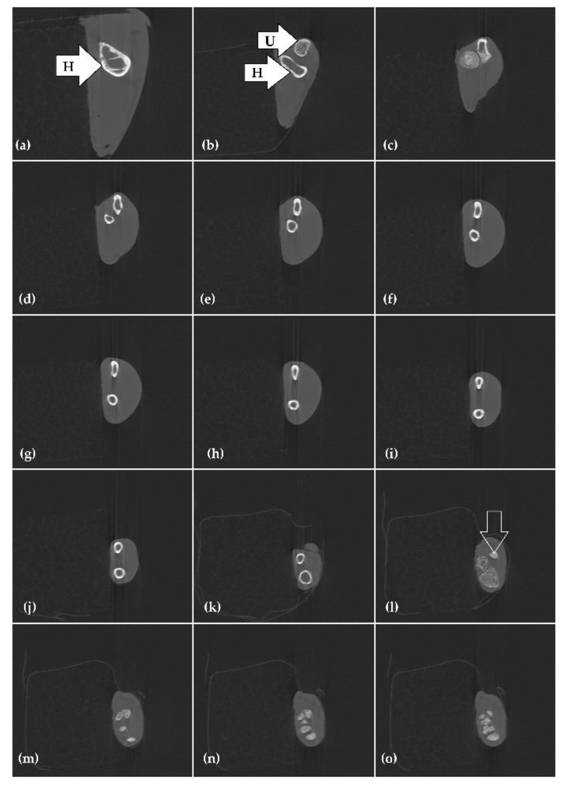
3.3. Microcomputed Tomography Examination (Micro-CT)

There were two bones in the marmoset’s forearm: the radius (R), located cranially, and the ulna (U), located medially (Figure 2). The examination showed not only all the bony elements mentioned above, but it also enabled spatial imaging. Individual images were reconstructed to create a 3D volume representation of the object (Figure 1) according to the normal bone tissue. The minimum voxel size in micro-CT scanners is 1 micrometre. A large length of the object results in an image enlarged 2.91× and 68.7 µm in voxel. Images of the bone and soft tissue of the normal lion tamarin’s forearm are shown in Figure 2. Water (HU = 0) is used in standard radiodensity calibrators. The HU of the air and bones are −1000 and 1000, respectively. Bone mineral density is a well-recognised microscopic and compositional measure of bone loss used for predicting fracture risk. Due to the increasing access to 3D modalities, the obvious benefit of volumetric imaging is avoidance of anatomical overlap in medical studies. Micro-CT scans also enable assessment of bone geometry and architecture. Three anatomical planes—A, B, and C—divided the right forearm of the L. chrysomelas body into transverse slices. The micro-CT image shows the effects of gently moving the measured object, resulting in slight blurring and blurred edges. This was due to the inability to fix the object under examination “rigidly”. If this were done, the fixation would make the measurement difficult and, more importantly, the object itself could have been damaged/distorted.

4. Discussion

The anatomical terminology for quadrupeds in Nomina Anatomica Veterinaria [28] lists the EDC, extensor digiti IV proprius and extensor digiti V proprius, as two independent muscles [29]. The extensor digiti IV proprius muscle and extensor digiti V proprius muscle are between the EDC and the ECU. The EDM is often termed as the extensor digiti quinti proprius [30]. The comparison of the results of our research with the findings of other authors’ studies shows that the golden-headed lion tamarin has muscular forearm characteristics typical of animals moving in tree crowns [31]. The analysis of the musculature of the gibbon forearm shows that the PQ may be divided into two parts, which was not demonstrated in our study [32]. In the second pronator, it is possible that there are two heads in the gibbon—the elbow and the humerus [32]. In contrast, the humeral head alone is found in the macaque [32]. As researchers point out, the muscles responsible for limb rotation are some of the distinguishing elements of the primate group, so analysis of this muscle group is important [33]. Assigned to the rotator group in this study, the brachioradialis muscle in humans acts as a strong flexor of the elbow joint [34]. Another characteristic of this group of arboreal animals is the development of the PL muscle, in contrast to the Gorilla sp., where it is often reduced [35]. The absence of the EPB muscle is common to golden-headed lion tamarin and macaca mulatta [32]. However, it is present in humans [36]. Golden-headed lion tamarins and humans also have similarities in the musculature of the FCU. In humans, this muscle has two heads and attaches to the ulnar and radial bones, whereas, in apes, this muscle is double-headed and attaches to the ulnar and humeral bone [32,37]. The general description shows the consistency of primate forearm musculature. In addition, according to the review, the musculature of the forearm of the goldfinch shows the greatest similarity to monkeys, rather than to apes or humans. The usefulness of US in the study and teaching of anatomy is supported by the fact that research is being conducted into the influence of using US as a supplementary method in the teaching of human anatomy [38]. Ultrasonography is also the most relevant technique of diagnosing muscle diseases in small animals and is widely used in current veterinary practice [4,6,39,40]. At first, the assessment of bones and fractures did not seem feasible because the bony surface reflects mostly incident acoustic waves (Figure 1). Unlike other waves (light, radio waves), ultrasound waves require a medium to travel through [4,6]. Wave echoes are used to create images. They transmit energy by alternating regions of high and low pressure. Frequency (the number of times a wave is repeated per second), wavelength (the distance travelled by a sound wave in one cycle), and velocity (the rate at which sound travels through an acoustic medium) are used to describe sound waves. Echoes contain information about structures being imaged. Reflection of the sound beam causes attenuation on return to the transducer, while the amount of attenuation is determined by the distance travelled by the sound wave and its frequency. Hyperechoic structures are white or grey, which results in their relative brightness. The structures which have no echoes within are anechoic and appear black. Focal changes in echogenicity are easier to detect in muscles because the adjacent, normal parenchyma can be compared [4,6,40]. Longitudinal and transverse US images of the muscles show hypoechoic structures with small hyperechoic fibres, which allow distinction of each muscle group or even the demonstration of sexual dimorphism, e.g., when analysing the scapula level muscle [17]. Ultrasound imaging enables measurement of the muscle thickness and diameter of the muscle from the outer borders. This technique has improved, resulting in the display of the muscle tissue with resolution of up to 0.1 mm. The intravital possibility of examining these parameters enables the assessment of physiological and pathological dimensions of structures and simultaneous contact with the patient, as in the case of human and animal elbow joint analysis [41,42]. The diameter was clearly measured between the peripheral margins of the hypoechoic muscle structures, which were distant from the bone surface. The graded compression technique was applied to move the transducer slowly over the muscles. The authors of other studies also used this method for the identification of individual muscles [43,44] as this technique visualises muscles better than others. In addition, US also enables the analysis of fascial structures [45]. Nevertheless, errors in the form of artefacts may occur during the ultrasound examination, and extensive experience with ultrasound is required to corectly interpret artefacts [46]. Even with the use of US in our study, the results only partly overlapped with traditional examination methods, and, despite the high similarity of results, these facts call into question the usefulness of US for anatomical descriptions of new species, as differences between species can be missed or the results of the study might be falsified.
The results of the micro-CT examination were more satisfactory. Although computed tomography is more commonly used to diagnose musculoskeletal problems, it can also be used to assess the anatomy of the front limb. Micro-CT has previously been used to analyse the skeletal system of the thoracic limb in mammals, reptiles, and birds [47,48,49,50,51]. In normal anatomy, CT seems to be sufficient, but micro-CT can be very useful when assessing pathology or changes in normal anatomy. In clinical practice, micro-CT is applied to assess the bone condition and changes, where measurements are expressed as Hounsfield units (HU) [1,4,6,52]. The aim of our study was to evaluate the normal appearance of the forearm bone in the sagittal plane and in 3D reconstruction and to compare it with the traditional anatomical preparation. The micro-CT enabled a comprehensive examination of the skeletal structure of the thoracic limb; the bone elements shown in the micro-CT examination were comparable to those from the preparation. The figures obtained from the micro-CT examination realistically depicted the bones, which was a satisfactory effect. The possibility of modelling the images, i.e., adding colour to the bones, as well as the possibility of the presentation of 3D images of the bones, added even more cognitive and educational value. The traditional preparation procedure was time-consuming, and the thoracic limb of this individual could not be re-examined from another angle, e.g., in terms of vascularisation or innervation. In contrast to the non-invasive specificity of micro-CT, the traditional method was less effective in this respect. In addition, micro-CT offers the possibility of analysing the cross-section of each part of the radial and ulnar bones. This is a great advantage of tomography, which also makes it possible to examine the structure of the marrow cavity, which has been a frequent research topic in recent years [53,54,55] (Figure 2). In conventional radiography, the composite of attenuation sums makes an actual, two-dimensional image. In contrast to conventional CT, in our micro-CT scan, the sum of linear attenuation coefficients along the path of an X-ray beam passing through multiple voxels was used instead to calculate individual contributions from each voxel (68.7 µm in size). Substances that can be discriminated in a CT image include fat, water, gas, and normal or abnormal tissues [4,6]. Unlike conventional scanners, high-resolution quantitative computed tomography can directly detect larger cortical pores. Nowadays, CT has become an important investigative tool and is a primary imaging technique for detecting acute traumas in human medicine. The physical principles of computed tomography (CT) imaging are complex. Both a CT and a micro-CT scanner employ an X-ray beam. In a CT device, the X-ray source rotates along a circular pattern with detectors rotating on the opposite side, whereas, in micro-CT, the X-ray tube and detector are fixed while a sample rotates on a table [56,57]. A micro-CT device has a microfocus X-ray tube, thanks to which power can be increased to 320 W. The other nanofocus X-ray tube enables the detection of elements as small as 0.2 µm [58,59]. Although CT may aid the diagnosis of an almost endless list of diseases, the possible manifestations of these diseases in images are limited [6]. A complex computer system utilises algorithms and mathematical formulae to process data into a 3D image or to generate a large number of two-dimensional slices. The computer system analyses the data on the basis of geometrical plots and assigns Hounsfield units (HU) to reconstruct the image. X-rays are absorbed by different materials to a varying degree [1,4,56,57,58,59]. If the density of a particular tissue, organ, or lesion increases, so does the absorption, which results in increased attenuation of the X-ray beam. The tissue which appears less opaque than normal is hypoattenuating. It may contain fresh blood (haemorrhage), cystic, necrotic, fatty, or oedematous infiltration [8,60]. The tissue which is more opaque than normal is hyperattenuating. It may contain fibrin and clot retraction or mineral or iodinated contrast medium. The cells which are densely fibrotic have a high nuclear/cytoplasmic ratio [56]. The attenuation of muscles is similar and yields the same HU value. Bone X-ray is the most widely available bone structure visualisation technique in veterinary practice. It provides qualitative information on bone density, fractures, injuries, and joint abnormalities. Another advantage of a traditional X-ray radiograph is its use of a very small dose of ionising radiation to produce pictures. Therefore, the main limitation of CT is radiation exposure [6].
As in small animal practice, CT and US are the techniques that provide sectional images of anatomic structures of the object [1,4,5,6,7,8,9,10,39,56,57,61]. Both of them are highly accurate and sensitive in diagnosing bone and muscle lesions. Magnetic resonance imaging can be applied as a complementary technique because this imaging modality proved to be useful for the evaluation of joint morphology in other animals [16,18]. In order to interpret CT and MRI and compare them with ultrasound and gross anatomical dissection, a particularly rare species was used to verify the results—the sagittal planes of the right forearm of the marmoset. The results of other tests should be collected as a point of reference for the L. chrysomelas species. Most authors recommend using multiple diagnostic techniques to plan the treatment of animals. Therefore, various techniques have been developed to teach students at veterinarian schools and in professional training. All cases are important to comparative anatomy and must be taken into account in clinical, surgical, and orthopaedic approaches to each part of the forearm.

5. Conclusions

Comprehensive understanding of possible arrangements of the forearm structures is of great clinical importance. CT evaluation of affected forearm and hand bones is associated with parameters of the bone and muscle status. Gross dissection and imaging modalities, such as US and micro-CT, are useful procedures for the anatomical location of these structures and for teaching. The combined and concurrent use of micro-CT and US provides the most useful and relevant information, which is seldom redundant. However, the separate use of US does not enable a reliable study of anatomy. On the other hand, it is possible with micro-CT, because the image obtained with this technique corresponds to reality and the technique itself has numerous advantages.

Author Contributions

Conceptualisation, M.Z. and K.S.; methodology, M.Z., K.S., B.G. and M.W.; formal analysis, M.Z., K.S., A.G., B.G. and M.W.; investigation, M.Z., K.S., A.G., B.G. and M.W.; writing—original draft preparation, M.Z., K.S., A.G., B.G. and M.W.; writing—review and editing, M.Z., K.S., A.G., H.F., B.G. and M.W.; visualisation, K.S., B.G. and M.W.; supervision, K.S. and H.F. All authors have read and agreed to the published version of the manuscript.

Funding

This research received no external funding.

Institutional Review Board Statement

Ethical review and approval were waived for this study because the experimental work was conducted on cadavers. All procedures involving cadavers in accordance with the law of 15 January 2015 on the protection of animals used for scientific or educational purposes do not require the approval of the local ethics committee.

Informed Consent Statement

Not applicable.

Conflicts of Interest

The authors declare no conflict of interest.

References

  1. Ohsang, K.; Jeong, B.K.; Jihyeung, K.; Hyun, B.G. Computed Tomography Evaluation of Forearm and Hand Muscules in Patients with Distal Radius Fracture. J. Clin. Densitom. 2021, 24, 88–93. [Google Scholar] [CrossRef]
  2. De Rycke, L.M.; Gielen, I.M.; Van Bree, H.; Simoens, H. Computed Tomography of the elbow joint in clinically normal dogs. Am. J. Vet. Res. 2002, 63, 1400–1407. [Google Scholar] [CrossRef] [PubMed]
  3. Istrate, A.; Peteoaca, A.; Constantinescu, R.; Angeli, G.; Tanase, A. Radiographic and Computed Tomography findings in dogs with fragmented medial coronoid process. Sci. Works Ser. C. Vet. Med. 2019, 65, 60–65. [Google Scholar]
  4. Kirberger, R.M.; McEvoy, F.J. Manual of Canine and Feline Musculoskeletal Imaging, 2nd ed.; BSAVA: Quedgeley, UK, 2016; pp. 1–118. [Google Scholar]
  5. Solis-Chavez, S.; Castillo-Rivera, M.A.; Arteaga-Silva, M.; Ibanez-Contreras, A.; Hernandez-Godinez, B.; Moron-Mendoza, A.; Mendoza-Cuevas, G.; Morales-Guadarrama, A.; Sacristan-Rock, E. Computed tomography is a feasible method for quantifying bone density in Macaca mullata. Vet. Radiol. Ultrasound. 2018, 59, 545–550. [Google Scholar] [CrossRef]
  6. Thrall, D.E. Textbook of Veterinary Diagnostic Radiology, 5th ed.; Saunders: Raleigh, NC, USA, 2007; pp. 2–805. [Google Scholar]
  7. Woodruf, W.W. Fundamentals of Neuroimaging; Saunders: Raleigh, NC, USA, 1993; pp. 33–70. [Google Scholar]
  8. Garvey, C.J. Computed tomography in clinical practice. Br. Med. J. 2002, 324, 1077–1080. [Google Scholar] [CrossRef] [Green Version]
  9. Samei, E.; Pelc, N.J. Computed Tomography: Approaches, Applications, and Operations; Springer Nature: Cham, Switzerland, 2020; pp. 3–458. [Google Scholar]
  10. Brant, W.E.; Helms, C.A. Fundamental of Diagnostic Radiology, 4th ed.; Lippincott Williams & Wilkins: Philadelphia, PA, USA, 2007; pp. 927–1183. [Google Scholar]
  11. Spoormakers, T.J.P.; Veraa, S.; Graat, E.A.M.; van Weeren, P.R.; Brommer, H. A comparative study of breed differences in the anatomical configuration of the equine vertebral column. J. Anat. 2021, 239, 829–838. [Google Scholar] [CrossRef] [PubMed]
  12. Darlim, G.; Montefeltro, F.C.; Langer, M.C. 3D skull modelling and description of a new baurusuchid (Crocodyliformes, Mesoeucrocodylia) from the Late Cretaceous (Bauru Basin) of Brazil. J. Anat. 2021, 239, 622–662. [Google Scholar] [CrossRef]
  13. El-Ansary, D.; Marshall, C.J.; Farragher, J.; Annoni, R.; Schwank, A.; McFarlane, J.; Bryant, A.; Han, J.; Webster, M.; Zito, G.; et al. Architectural anatomy of the quadriceps and the relationship with muscle strength: An observational study utilising real-time ultrasound in healthy adults. J. Anat. 2021, 239, 847–855. [Google Scholar] [CrossRef]
  14. Graham, K.M.; Kouba, A.J.; Langhorne, C.J.; Marcec, R.M.; Willard, S.T. Biological sex identification in the endangered dusky gopher frog (Lithobates sevosa): A comparison of body size measurements, secondary sex characteristics, ultrasound imaging, and urinary hormone analysis methods. Reprod. Biol. Endocrinol. 2016, 14, 1–14. [Google Scholar] [CrossRef] [PubMed] [Green Version]
  15. Tokunaga, T.; Jomane, F.N.; Mandai, S.; Ishida, T.; Hirooka, H. Estimation of the marbling development pattern in Japanese Black cattle by using serial ultrasound measurement data. Anim. Sci. J. 2021, 92, e13533. [Google Scholar] [CrossRef] [PubMed]
  16. Canoso, J.J.; Naredo, E.; Martínez-Estupiñán, L.; Mérida-Velasco, J.R.; Pascual-Ramos, V.; Murillo-González, J. Palpation of the lateral bands of the extensor apparatus of the fingers. Anatomy of a neglected clinical finding. J. Anat. 2021, 239, 663–668. [Google Scholar] [CrossRef]
  17. Keidan, L.; Barash, A.; Lenzner, Z.; Pick, C.G.; Been, E. Sexual dimorphism of the posterior cervical spine muscle attachments. J. Anat. 2021, 239, 589–601. [Google Scholar] [CrossRef]
  18. Dickinson, E.; Boettcher, M.L.; Smith, M.R.; Worden, N.A.; Swindell, S.R.; Seelye, J.S.; Pastor, F.; Hartstone-Rose, A. Myological variation in the forearm anatomy of Callitrichidae and Lemuridae. J. Anat. 2021, 239, 669–681. [Google Scholar] [CrossRef]
  19. Weatherall, D. The Use of Non-Human Primates in Research, 1st ed.; Welcome Trust: London, UK, 2006; pp. 11–141. [Google Scholar]
  20. Diogo, R.; Richmond, B.G.; Wood, B. Evolution and homologies of primate and modern human hand and forearm muscles, with notes on thumb movements and tool use. J. Hum. Evol. 2012, 63, 64–78. [Google Scholar] [CrossRef] [PubMed]
  21. Diogo, R.; Wood, B.A. Phylogenetic analyses of primates based on the muscles of the head, neck, pectoral region and upper limb. In Comprarative Anatomy and Phylogeny of Primate Muscles and Human Evolution, 1st ed.; CRC Press: Boca Raton, FL, USA, 2019; pp. 313–357. [Google Scholar]
  22. Diogo, R.; Wood, B.A. In Origin, Development, and evolution o primate muscles, with notes on human anatomical variations and anomalies. In Developmental Approaches to Human Evolution, 1st ed.; Boughner, J.C., Campbell, R., Eds.; John Wiley & Sons: Hoboken, NJ, USA, 2016; pp. 167–204. [Google Scholar]
  23. Cheng, E.C.; Scott, S.H. Morphometry of Macaca mulatta forelimb. I. shoulder and elbow muscles and segment inertial parameters. J Morphol. 2000, 245, 206–224. [Google Scholar] [CrossRef]
  24. Hb Du Sert, N.P.; Ahluwalia, A.; Alam, S.; Avey, M.T.; Baker, M.; Browne, W.J.; Clark, A.; Cuthill, I.C.; Dirnagl, U.; Emerson, M.; et al. Reporting animal research: Explanation and elaboration for the ARRIVE guidelines 2.0. PLoS Biol. 2020, 18, e3000411. [Google Scholar] [CrossRef]
  25. Illemann, J.; Bartscher, M. X-ray spectrum dependence of the magnification of cone-beam CT. In Proceedings of the 7th Conference on Industrial Computed Tomography (iCT2017), Leuven, Belgia, 7–9 February 2017; Available online: www.ndt.net/?id=20827 (accessed on 2 December 2021).
  26. Grangeat, P. Mathematical framework of cone beam 3D reconstruction via the first derivative of the radon transform. In Mathematical Methods in Tomography. Lecture Notes in Mathematics; Herman, G.T., Louis, A.K., Natterer, F., Eds.; Springer: Berlin, Germany, 2006; pp. 66–97. [Google Scholar]
  27. Willemink, M.J.; de Jong, P.A.; Leiner, T.; de Heer, L.M.; Nievelstein, R.A.J.; Budde, R.P.J.; Schilham, A.M.R. Iterative reconstruction techniques for computed tomography Part 1: Technical principles. Eur. Radiol. 2013, 23, 1623–1631. [Google Scholar] [CrossRef]
  28. Nomina Anatomica Veterinaria, 6th ed.; WAVA: Hanover, Germany, 2017.
  29. Garcia, J.F.V.; Parra, J.E.D.; Rios, J.B. Descriptive Anatomy of Lateral Digital Extensor Muscles of the Hand of the White-Footed Tamarin (Sanguinus leucopus, Gunter, 1876). Int. J. Morphol. 2016, 34, 1123–1127. [Google Scholar] [CrossRef] [Green Version]
  30. Aversi-Ferreira, T.A.; Aversi-Ferreira, R.A.G.M.F.; Bretas, R.V.; Nishimaru, H.; Nishijo, H. Comparative anatomy of the arm muscules of the Japanese Monkey (Macaca Fuscata) with some comments on locomotor mechanics and behavior. J. Med. Primatol. 2016, 10, 165–179. [Google Scholar] [CrossRef]
  31. Aversi-Ferreira, R.A.; Bretas, R.V.; Maior, R.S.; Davaasuren, M.; Paraguassú-Chaves, C.A.; Nishijo, H.; Aversi-Ferreira, T.A. Morphometric and statistical analysis of the palmaris longus muscle in human and non-human primates. BioMed Res. Int. 2014, 2014, 1–6. [Google Scholar] [CrossRef]
  32. Vanhoof, M.J.; van Leeuwen, T.; Vereecke, E.E. The forearm and hand musculature of semi-terrestrial rhesus macaques (Macaca mulatta) and arboreal gibbons (Fam. Hylobatidae). Part I. Description and comparison of the muscle configuration. J. Anat. 2020, 237, 774–790. [Google Scholar] [CrossRef]
  33. Kikuchi, Y. Comparative analysis of muscle architecture in primate arm and forearm. Ant. Histol. Embryol. 2010, 39, 93–106. [Google Scholar] [CrossRef]
  34. Boland, M.R.; Spigelman, T.; Uhl, T.L. The Function of Brachioradialis. Journal of Hand Surgery. Am. Soc. Surg. Hand. 2008, 33, 1853–1859. [Google Scholar] [CrossRef] [PubMed]
  35. Diogo, R.; Patou, J.M.; Pastor, J.F.; de Paz, F.J.; Ferrero, E.M.; Bello, G.; Barbosa, M.; Wood, B.A. Photographic and Descriptive Musculoskeletal Atlas of Gorilla: With Notes on the Attachments, Variations, Innervation, Synonymy and Weight of the Muscles; CRC Press: Boca Raton, FL, USA, 2010; pp. 26–50. [Google Scholar]
  36. Nayak, S.R.; Krishnamurthy, A.; Pai, M.M.; Prabhu, L.V.; Ramanathan, L.A.; Kumar, C.G.; Thomas, M.M. Multiple variations of the extensor tendons of the forearm. Rom. J. Morphol. Embryol. 2008, 49, 97–100. [Google Scholar]
  37. Murray, W.M.; Delp, S.L.; Buchanan, T.S. Variation of muscle moment arms with elbow and forearm position. J. Biomech. 1995, 28, 513–525. [Google Scholar] [CrossRef]
  38. Sweetman, G.M.; Crawford, G.; Hird, K.; Fear, M.W. The benefits and limitations of using ultrasonography to supplement anatomical understanding. Anat. Sci. Educ. 2013, 6, 141–148. [Google Scholar] [CrossRef] [PubMed]
  39. Beggs, I. Shoulder. In Musculoskeletal Ultrasound, 1st ed.; Lippincott Williams & Wilkins: Philadelphia, PA, USA, 2014. [Google Scholar]
  40. Kramer, M.; Gerwing, M.; Hach, V.; Schimke, E. Sonography of the musculoskeletal system in dogs and cats. Vet. Radiol. Ultrasound. 1997, 38, 139–149. [Google Scholar] [CrossRef]
  41. Konin, G.P.; Nazarian, L.N.; Walz, D.M. US of the elbow: Indications, technique, normal anatomy, and pathologic conditions. Radiographics 2013, 33, 125–147. [Google Scholar] [CrossRef] [PubMed]
  42. Bellegard, G.; Lopes, É.R.; Bisetto, S.P.; Hage, M.C.F. Musculoskeletal ultrasonography of the elbow joint in dogs: Applicability and evaluation protocol. Pesqui. Vet. Bras. 2019, 39, 419–428. [Google Scholar] [CrossRef] [Green Version]
  43. Konschake, M.; Stofferin, H.; Moriggl, B. Ultrasound visualization of an underestimated structure: The bicipital aponeurosis. Surg. Radiol. Anat. 2017, 39, 1317–1322. [Google Scholar] [CrossRef] [Green Version]
  44. Van Alfen, N.; Mah, J.K. Neuromuscular ultrasound: A new tool in your toolbox. Can. J. Neurol. Sci. 2018, 45, 504–515. [Google Scholar] [CrossRef]
  45. Duarte, M.L.; Iared, W.; Oliveira, A.S.B.; Santos, L.R.D.; Peccin, M.S. Ultrasound versus electromyography for the detection of fasciculation in amyotrophic lateral sclerosis: Systematic review and meta-analysis. Radiol. Bras. 2020, 53, 116–121. [Google Scholar] [CrossRef] [PubMed]
  46. Kirberger, R.M. Imaging artifacts in diagnostic ultrasound—A review. Vet. Radiol. Ultrasound. 1995, 36, 297–306. [Google Scholar] [CrossRef]
  47. Hanson, N.A.; Bagi, C.M. Alternative approach to assessment of bone quality using micro-computed tomography. Bone 2004, 35, 326–333. [Google Scholar] [CrossRef] [PubMed]
  48. Bribiesca-Contreras, F.; Sellers, W.I. Three-dimensional visualisation of the internal anatomy of the sparrowhawk (Accipiter nisus) forelimb using contrast-enhanced micro-computed tomography. PeerJ 2017, 5, e3039. [Google Scholar] [CrossRef] [Green Version]
  49. Chen, Y.; Lin, G.; Chen, Y.; Fok, A.; Slack, J.M. Micro-computed tomography for visualizing limb skeletal regeneration in young Xenopus frogs. Anat. Rec. 2012, 295, 1562–1565. [Google Scholar] [CrossRef] [PubMed] [Green Version]
  50. Kamal, B.M. Sequential Pattern of Prenatal Ossification in the Fore Limb Bones in White New Zealand Rabbits by Double Stained Techniques and Computed Tomography. PSM Vet. Res. 2019, 4, 24–35. [Google Scholar]
  51. Dayan, M.O.; Beşoluk, K.; Eken, E.; Aydoğdu, S.; Turgut, N. Three-dimensional modelling of the femur and humerus in adult male guinea pigs (guinea pig) with computed tomography and some biometric measurement values. Folia Morphol. 2019, 78, 588–594. [Google Scholar] [CrossRef] [PubMed]
  52. Lee, S.W. Musculoskeletal Injuries and Conditions: Assessment and Management; Springer Publishing Company: New York, NY, USA, 2017; pp. 1–457. [Google Scholar]
  53. Wang, W.; Wang, X.; Chen, L.; Li, Z.; Zhang, S.; Su, B.; Zhao, F.F.; Weng, Z.; Guan, H.; Gao, M.; et al. Micro-architecture study of the normal infant auditory ossicles with micro-computed tomography. Int. J. Morphol. 2019, 37, 1387–1390. [Google Scholar] [CrossRef] [Green Version]
  54. Bakici, C.; Akgun, R.O.; Ekim, O.; Batur, B.; Bakici, M.; Ozen, D.; Soydal, C. Three dimensional modeling and quantitative analysis of long bone parameters of rabbit using micro-computed tomography. Iran. J. Vet. Res. 2021, 22, 140–145. [Google Scholar] [CrossRef]
  55. Wang, W.; Li, Z.; Qi, Y.; Chen, L.; Yi, P.; Yang, F.; Tang, X.; Tan, M. Micro-architecture study of the normal odontoid with micro-computed tomography. J. Spinal Cord Med. 2020, 43, 211–216. [Google Scholar] [CrossRef] [PubMed]
  56. Patalas, A.; Łabudzki, R.; Sarbinowski, F.; Gapiński, B.; Talar, R.A. Mechanical analysis of cancellous bone in FEA simulation research and experimental testing with the μct control. In Proceedings of the 6th International Conference on Integrity-Reliability-Failure IRF, Lisbon, Portugal, 22–26 July 2018. [Google Scholar]
  57. Kaczmarek, J.; Bartkowiak, T.; Schuenemann, R.; Paczos, P.; Gapiński, B.; Bogisch, S.; Unger, M. Mechanical Performance of a Polyaxial Locking Plate and the Influence of Screw Angulation in a Fracture Gap Model. Vet. Comp. Orthop. Traumatol. 2019, 33, 33–44. [Google Scholar] [CrossRef] [PubMed]
  58. Gapiński, B.; Wieczorowski, M.; Grzelka, M.; Alonso, P.A.; Tome, A.B. The application of micro computer tomography to assess quality of parts manufactured by means of rapid prototyping. Polimery 2017, 62, 53–59. [Google Scholar] [CrossRef]
  59. Gapiński, B.; Wieczorowski, M.; Marciniak-Podsadna, L.; Domínguez, A.P.; Cepova, L.; Martinez, A. Measurement of Surface Topography Using Computed Tomography. In Proceedings of the 5th International Scientific-Technical Conference Manufacturing, Poznań, Poland, 24–26 October 2017. [Google Scholar]
  60. Gálvez, A.; Caraballo, J.L.; Manzanares-Cespedes, M.C.; Valdivia-Gandur, I.; Figueiredo, R.; Valmaseda-Castellon, E. Vascular labeling of the head and neck vessels: Technique, advantages and limitations. J. Clin. Exp. Dent. 2017, 9, 682–687. [Google Scholar] [CrossRef] [PubMed] [Green Version]
  61. Bakker, E.; Gielen, I.; Caelenberg, A.; Van Bree, H.; Van Ryssen, B. Computed Tomography of canine elbow joints affected by primary and concomitant flexor enthesiopathy. Vet. Radiol. Ultrasound. 2014, 55, 45–55. [Google Scholar] [CrossRef] [PubMed]
Figure 1. The transverse and longitudinal planes of the L. chrysomelas forearm. The ultrasound transducer was positioned on the lateral (in the transverse scan: (a) at the level of the proximal third of the forearm; (b) at half the length of the forearm; (c) at the level of the distal third of the forearm) and medial portion; (d) in the sagittal scan). The cross-section lines show transverse scans on the 3D bone reconstruction model (radius–cranial), (ulna–caudal).
Applsci 12 01031 g001
Figure 2. Sagittal microcomputed tomography images of the L. chrysomelas forearm. Transversal bone density: (a) humerus—H.; (b,c) humerus—H, ulna—U; (dl) upper bone—ulna, lower bone—radius; (l) arrow—accessory carpal bone; (mo) carpal bones.
Applsci 12 01031 g002
Publisher’s Note: MDPI stays neutral with regard to jurisdictional claims in published maps and institutional affiliations.

