Article

Gummy Smile Improvement during Growth Period Using a Simple Bite Jumping Appliance and High-Pull J-Hook HeadGear: A Case Series Study

Department of Orthodontics, School of Dentistry, Aichi Gakuin University, 2-11 Suemori-dori, Chikusa-ku, Nagoya 464-8651, Japan
*
Author to whom correspondence should be addressed.
Appl. Sci. 2022, 12(23), 12268; https://doi.org/10.3390/app122312268
Submission received: 1 November 2022 / Revised: 24 November 2022 / Accepted: 25 November 2022 / Published: 30 November 2022
(This article belongs to the Special Issue Innovation in Dental and Orthodontic Materials)

Abstract

:
Purpose: In recent years, a method for improving gummy smiles in adults using an orthodontic anchor screw has been reported, but there is yet to be a treatment for the gummy smiles of those in the growth period. Methods: We improved the gummy smiles of three class II patients with vertical excessive growth of the upper jaw, during their growth period, using a simple bite jumping appliance (SBJA) and a high pull J-hook headgear. Results: It was found that SBJA promoted mandibular growth and the high-pull J-hook headgear inhibited the vertical growth of the maxillary anterior teeth. Conclusion: It is suggested that a combined use of high-pull J-hook headgear and SBJA is an effective way of improving gummy smiles in class II patients with vertical excessive growth of the upper jaw during their growth period.

1. Introduction

One of the goals of orthodontic treatment is to achieve aesthetic harmony between maxillofacial morphology and facial features, whilst also establishing occlusion. Obtaining a beautiful smile line is important [1,2]. In general, gingival exposure of less than 2 mm with a full smile is considered aesthetically pleasing and youthful [3]. On the other hand, a gummy smile is a condition in which excess gingiva is visible during a full smile. Gummy smiles are thought to be caused by soft tissue odontogenic causes due to the overeruption of maxillary anterior teeth, altered passive eruption, short upper lip, and skeletal causes due to excessive vertical growth of the maxilla [2,4,5,6]. Moreover, gummy smiles are considered an instigation factor for gingivitis in the anterior maxillary region [7,8], and improvement of the gummy smile from a young age is considered beneficial.
Conventional methods for improving a gummy smile include the intrusion of incisors, extrusion of posterior teeth using activator, bite ramps, gingivectomy of the maxillary anterior teeth, injection of Botox into the upper lip, retraction of the maxillary anterior teeth, and surgical orthodontic treatment by a combination of LeFort 1 and maxillary horseshoe osteotomy [4,5,6]. Treatments are chosen in accordance with the cause. In recent years, a method for improving gummy smiles in adults using an orthodontic anchor screw has been reported [9,10]. However, the improvement of a gummy smile by surgical orthodontic treatment is usually achieved by combined LeFort 1 and maxillary horseshoe osteotomy, a type of orthognathic surgery, to shorten the facial height [11,12], which is very invasive. Furthermore, ways to improve a gummy smile using an orthodontic anchor screw are also slightly invasive. In addition, these approaches are used after growth is complete.
Several treatment methods using orthodontic anchor screws have been reported for gummy smiles during the growth period [13,14]. Kim et al. treated two Angle Class II division patients with gummy smiles, in the growth period, using orthodontic anchor screws, and reported the correction of excessive vertical growth of the maxillary anterior teeth and the subsequent alleviation of gummy smiles [13,14]. This suggests that gummy smiles may be improved if the vertical growth of the maxillary anterior teeth is suppressed. However, gummy smile patients, especially those of school age, and their parents, may exhibit resistance to surgical invasion or treatment using orthodontic anchor screws. In addition, patients in the growth period require special attention because permanent tooth germs may be present in the bone, or the roots may be immature. We therefore considered whether it would be possible to suppress the eruption of maxillary anterior teeth with a removable orthodontic appliance without surgical intervention in patients with gummy smiles in their growth period.
Here, we used SBJA [15] in combination with the high-pull J-hook headgear to suppress the vertical excessive growth of the upper jaw in three class II patients with vertical excessive growth of the upper jaw during the growth period.
Consent was obtained from patients and their guardians for their inclusion in this report according to Ethical Guidelines for Medical and Health Research Involving Human Subjects in Japan.

2. Methods

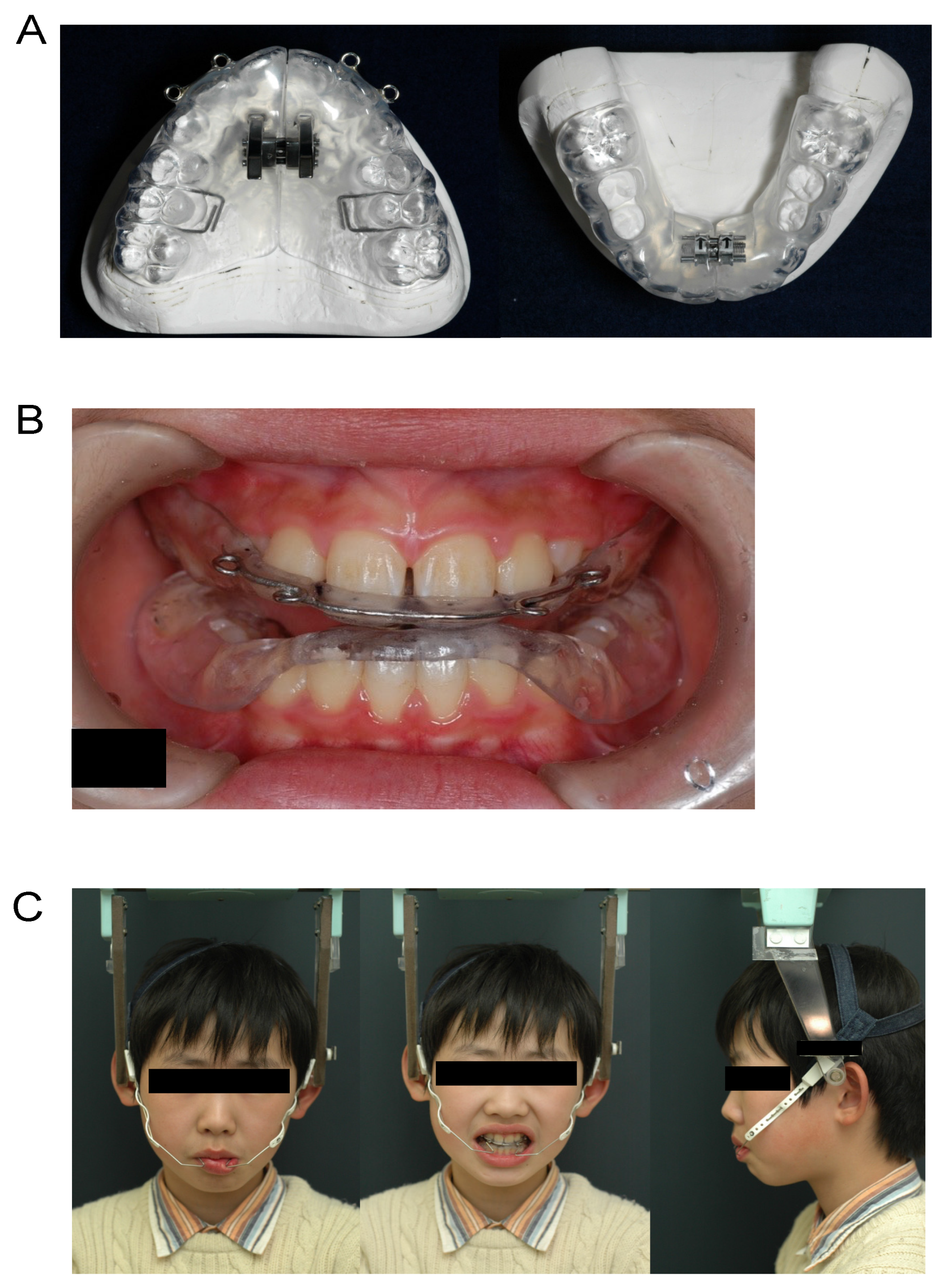
2.1. Differences between Simple Bite Jumping Appliance (SBJA) and BJA

Here, we used SBJA [15] rather than conventional BJA [16,17]. In SBJA, a resin plate covers the entire dentition and palate (Figure 1A). On the other hand, a conventional BJA resin plate covers only the palate and does not cover the entire dentition. Therefore, it was thought that the SBJA would be able to accept the force of the high-pull J-hook headgear with the entire upper jaw.

2.2. Attachment of Loop to SBJA

In order to improve gummy smiles caused by an overeruption of maxillary anterior teeth, we believe that it is necessary to apply upward orthodontic force to the maxillary anterior teeth, which is why we decided to use the high-pull J-hook headgear (TOMY INTERNATIONAL, Tokyo, Japan). A loop for combined use with the high-pull J-hook headgear was made using a 0.8 mm diameter cobalt-chromium alloy wire (JM Ortho, Tokyo), and attached using self-curing resin for orthodontic use (JM Ortho, Tokyo) onto the labial side of the maxillary anterior teeth of the SBJA (Figure 1A,B).

2.3. Combined Use of SBJA and High-Pull J-Hook Headgear

After mounting the SBJA in the oral cavity, the SBJA and the high-pull J-hook headgear were used together by attaching the high-pull J-hook to the loop attached to the SBJA, and patients were instructed to wear it for 12 hours a day (Figure 1C).
Figure 1. Combined use of SBJA and high-pull J-hook headgear. SBJA with loop attached to the labial side of maxillary anterior teeth (A); intra-oral photograph while wearing SBJA (B); facial photograph while wearing SBJA and high-pull J-hook headgear (C).
Applsci 12 12268 g001

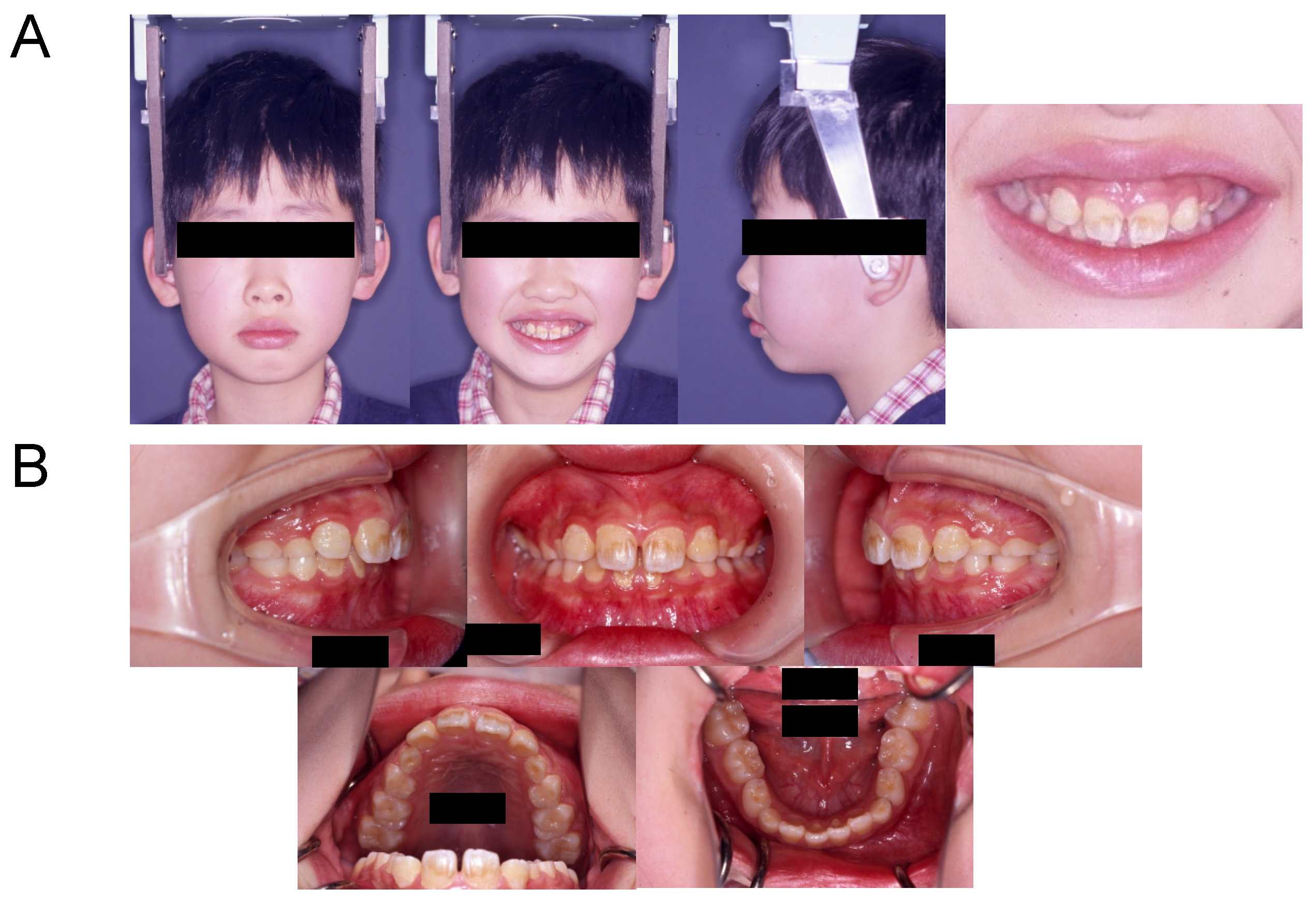
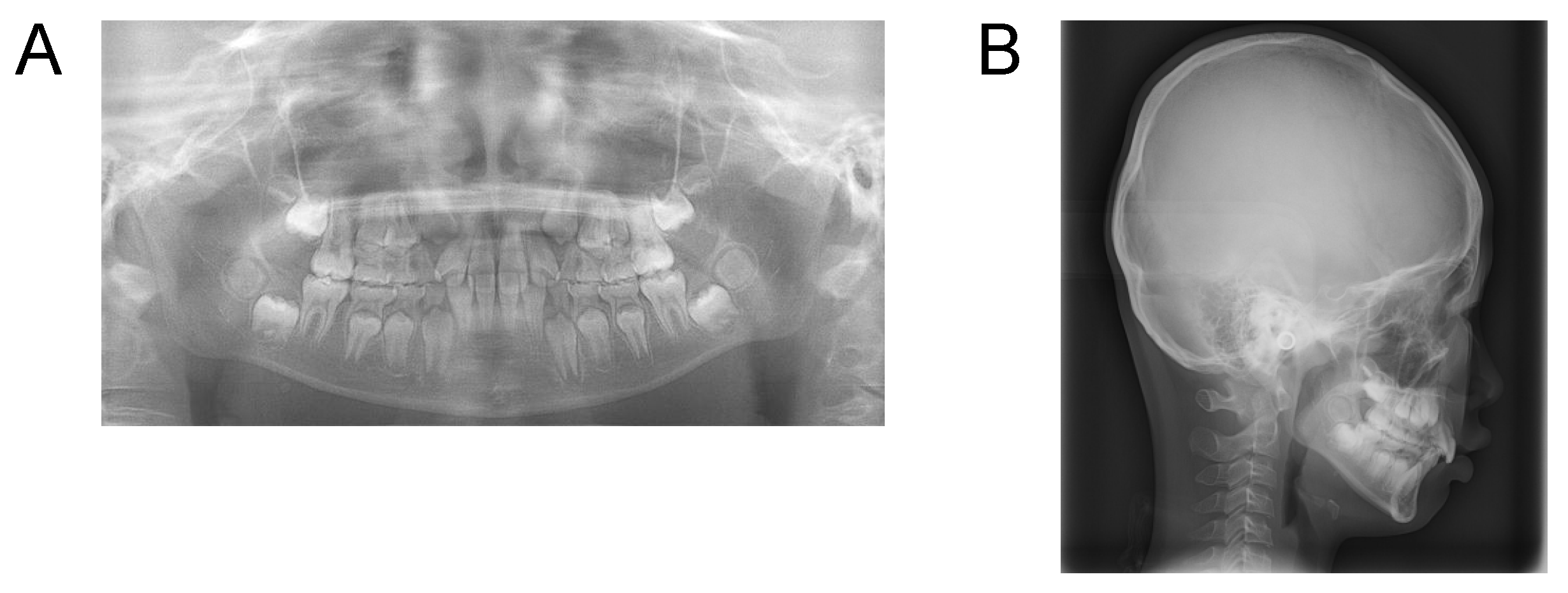
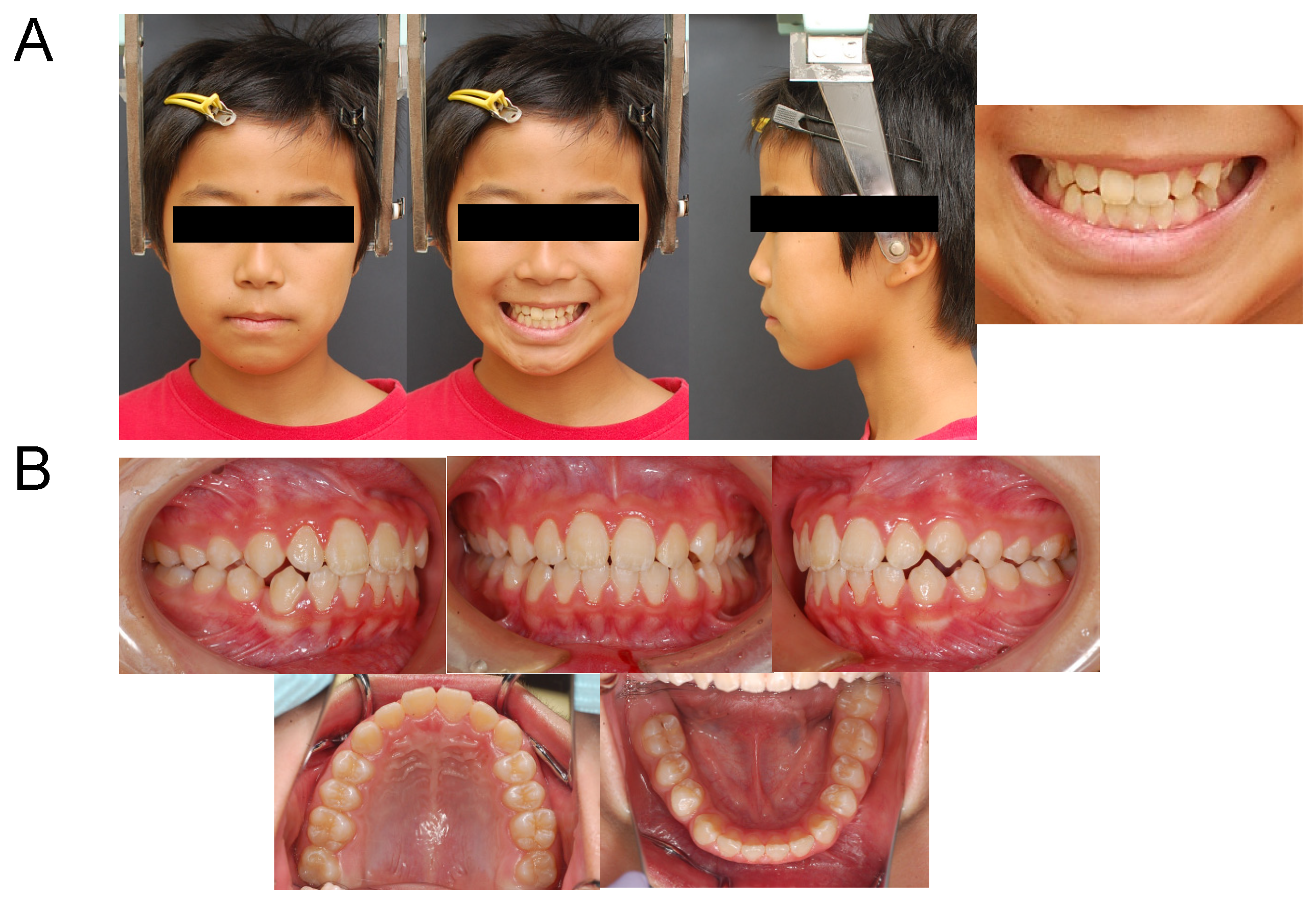
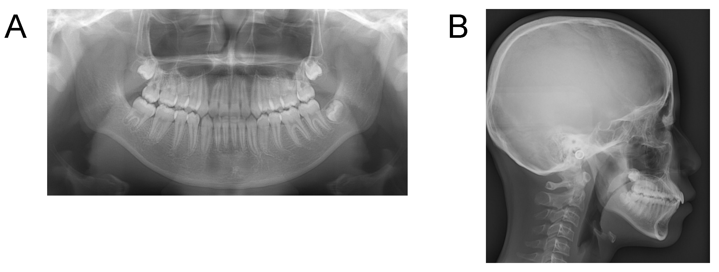
Case 1. Findings from initial examination

The patient was a Japanese boy of 10 years and 7 months at the time of his initial examination, with a chief complaint of overbite. In terms of family history, the patient’s father had a gap between his teeth. There were no items in the history of current illness or medical history that were particularly problematic. He had no notable habits.
In terms of facial findings (Figure 2A), the frontal view did not demonstrate any deviation of the mandible. When smiling, the distance between the upper lip to the maxillary left central incisor cervical line was 6.0 mm, and a gummy smile was notable. In the lateral view, a protrusion of the upper lip was observed.
In the intra-oral findings (Figure 2B), the occlusal relationship of the molars was Angle Class II on both the left and right sides, and the overjet was +6.5 mm. In addition, the deep overbite was +5.5mm. The maxillary and mandibular dentition midlines were coincident with the facial midline. The dental age was stage IIIA, and the width of the dental arches was small in both the upper and lower jaws. However, the length of the dental arch was large in both the upper and lower jaws. As for the dental arch morphology, the lower jaw exhibited a parabolic morphology. In addition, a midline diastema was noted in the maxilla.
In the panoramic radiograph (Figure 3), the maxillary bilateral central incisors had slightly short roots, and the tooth germs of the maxillary and mandibular bilateral third molars were observed. No other abnormalities in the number of teeth were observed.
In the cephalogram angle measurement (Table 1), the SNA angle was 85.0°, the SNB angle was 78.5°, and the ANB angle was +6.5° and large, thus exhibiting skeletal class II. The Frankfort-mandibular plane angle (FMA) was 30.5°. U1 to SN was 103.0° and the maxillary anterior axial inclination was normal. In addition, FMIA was 62.0°. In the cephalogram distance measurement (Table 2), the maxillary anterior tooth height (Is-Is’) was 31.5 mm. This indicates that the patient’s maxillary anterior teeth were higher than the average measurements obtained by Sakamoto et al. [18], who noted the average measurements of Japanese people. Wits appraisal was 0 mm.
Diagnosis
This case was diagnosed as a case of skeletal class II, accompanied by a gummy smile due to the vertical excessive growth of the upper jaw, and a deep overbite.
Treatment approach
We decided to use the SBJA for the purpose of improving the overbite, and skeletal maxillary prognathism for bite jumping, suppression of maxillary growth, and promotion of mandibular growth. Furthermore, we elected to suppress the vertical growth of the maxillary anterior teeth and to improve the gummy smile by using a combination of SBJA and high-pull J-hook headgear. To improve the narrowed maxillary arch, we planned to expand the maxillae laterally through enlargement of the SBJA expansion screw. The patient was instructed to use the appliance for more than 12 h per day.
Treatment course
The patient used SBJA (with expansion screw) and the high-pull J-hook headgear for 28 months, from 11 years and 5 months to 13 years and 9 months of age.
Treatment results
In this patient, as a result of treatment, SNA remained unchanged from 85.0°, while SNB increased from 78.5° to 80.5°. As a result, ANB changed from 6.5° to 4.5°, and the skeletal maxillary prognathism improved. The maxillary anterior teeth (Is-Is’), which had been high, returned to normal height. With growth, the maxillary molars protruded, but the protruding of the maxillary anterior teeth was suppressed, and the occlusal plane (occlusal plane to FH) changed from 19.0° to 11.5°, and altered to an anterior elevation (Figure 4, Table 1). Wits appraisal was −2.0 mm. The left central incisor cervical exposure while smiling decreased from 6.0 mm to 0.0 mm, thus improving the gummy smile (Figure 5A). In addition, the overbite improved, and the molar relationship changed from class II to class I (Figure 5B). No root resorption was observed in the maxillary anterior teeth (Figure 6).

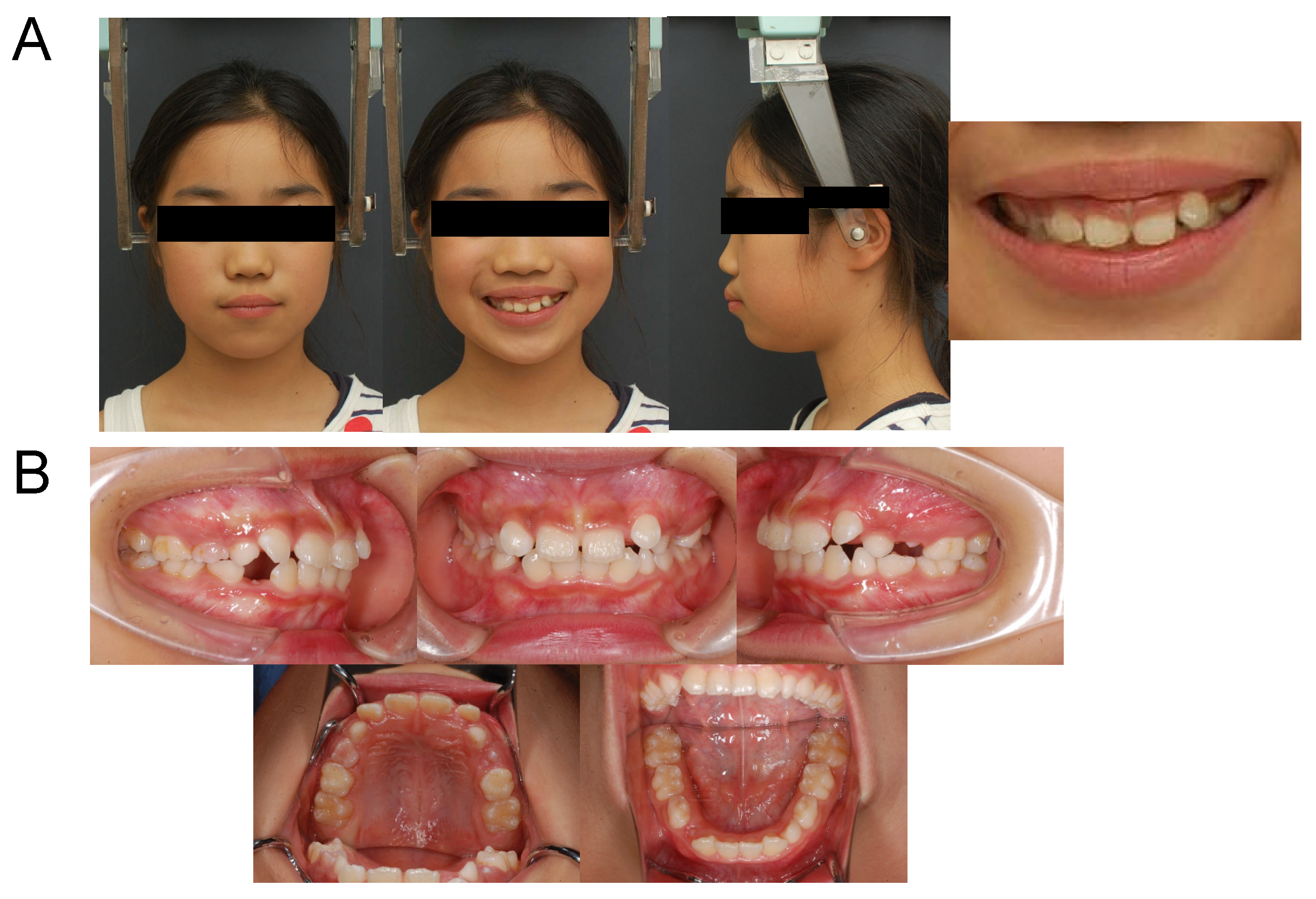
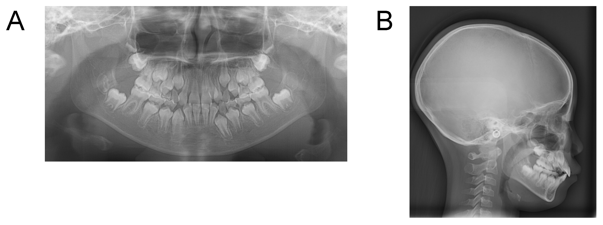
Case 2. Findings from initial examination

The patient was a Japanese boy of 10 years 0 months at the time of his initial examination, with a chief complaint of rattling anterior teeth. In terms of family history, the patient’s father had teeth crowding. There was no history of current illness or medical history that was particularly problematic. He had no notable habits.
In terms of facial findings (Figure 7A), the frontal view did not demonstrate any deviation of the mandible. When smiling, the distance between the upper lip to the maxillary left central incisor cervical line was 5.5 mm, and a gummy smile was notable. In the lateral view, a protrusion of the upper and lower lips was observed.
In the intra-oral findings (Figure 7B), the occlusal relationship of the molars was Angle Class II on both the left and right sides, and the overjet was +7.0 mm on the right side and +4.0 mm on the left side. In addition, the overbite was +2.0 mm on the right side and +5.5 mm on the left side, exhibiting a deep overbite. The maxillary anterior teeth were crowded, and the maxillary dentition midline was deviated 1.5 mm to the right from the facial midline. The dental age was stage IIIB, and the width of the dental arches was small in both the upper and lower jaws, exhibiting narrowing. The length of the dental arch was normal for both the upper and lower jaws. Furthermore, the dental arches exhibited a parabolic morphology in both the upper and lower jaws.
The panoramic radiographs (Figure 8) showed no abnormalities in the number of teeth.
In the cephalogram angle measurement (Table 3), the SNA angle was 85.0°, the SNB angle was 77.0°, and the ANB angle was +8.0° and large, thus exhibiting skeletal class II. The Frankfort-mandibular plane angle (FMA) was 33.5°. U1 to SN was 118.5° and the maxillary anterior teeth showed labial inclination. In addition, FMIA was 53.5°. In the cephalogram distance measurement (Table 4), the maxillary anterior tooth height (Is-Is’) was 31.0 mm indicating that, with reference to Sakamoto et al. [18], who recorded the measurements of Japanese people, the patient’s maxillary anterior teeth were higher than average. Wits appraisal was 2.0 mm.
Diagnosis
This patient was diagnosed as a case of skeletal class II, accompanied by a gummy smile due to the vertical excessive growth of the upper jaw, and a deep overbite and crowding.
Treatment approach
First, we decided to improve the crowding of the maxillary anterior teeth with a sectional arch. After that, we decided to use SBJA for the purpose of improving the overbite and skeletal maxillary prognathism with the aim of bite jumping, suppressing maxillary growth, and promoting mandibular growth. Furthermore, we decided to suppress the vertical growth of the maxillary anterior teeth and improve the gummy smile by using the SBJA and high-pull J-hook headgear together. To improve the narrowed maxillary and mandibular arch, we planned to expand the maxillae laterally through enlargement of the SBJA expansion screw. The patient was instructed to use the appliance for more than 12 h per day.
Treatment course
The patient used SBJA (with expansion screw) and high-pull J-hook headgear for 28 months from 10 years 5 months to 12 years 9 months of age.
Treatment results
In this patient, as a result of treatment, SNA remained unchanged from 85.0°, while SNB increased from 77.0° to 80.0°. As a result, ANB changed from 8.0° to 5.0°, and the skeletal maxillary prognathism improved. The maxillary anterior teeth height (Is-Is’), which had been high, returned to normal. With growth, the maxillary molars protruded, but the protruding of the maxillary anterior teeth was suppressed, and the occlusal plane (occlusal plane to FH) changed from 15.0° to 10.5°, and transformed into an anterior elevation (Figure 9, Table 3). Wits appraisal was −0.5 mm. The left central incisor cervical exposure while smiling decreased from 5.5 mm to 0.0 mm, thus improving the gummy smile (Figure 10A). In addition, the overbite improved, and the molar relationship changed from class II to class I (Figure 10B). No root resorption was observed in the maxillary anterior teeth (Figure 11).

Case 3. Findings from initial examination
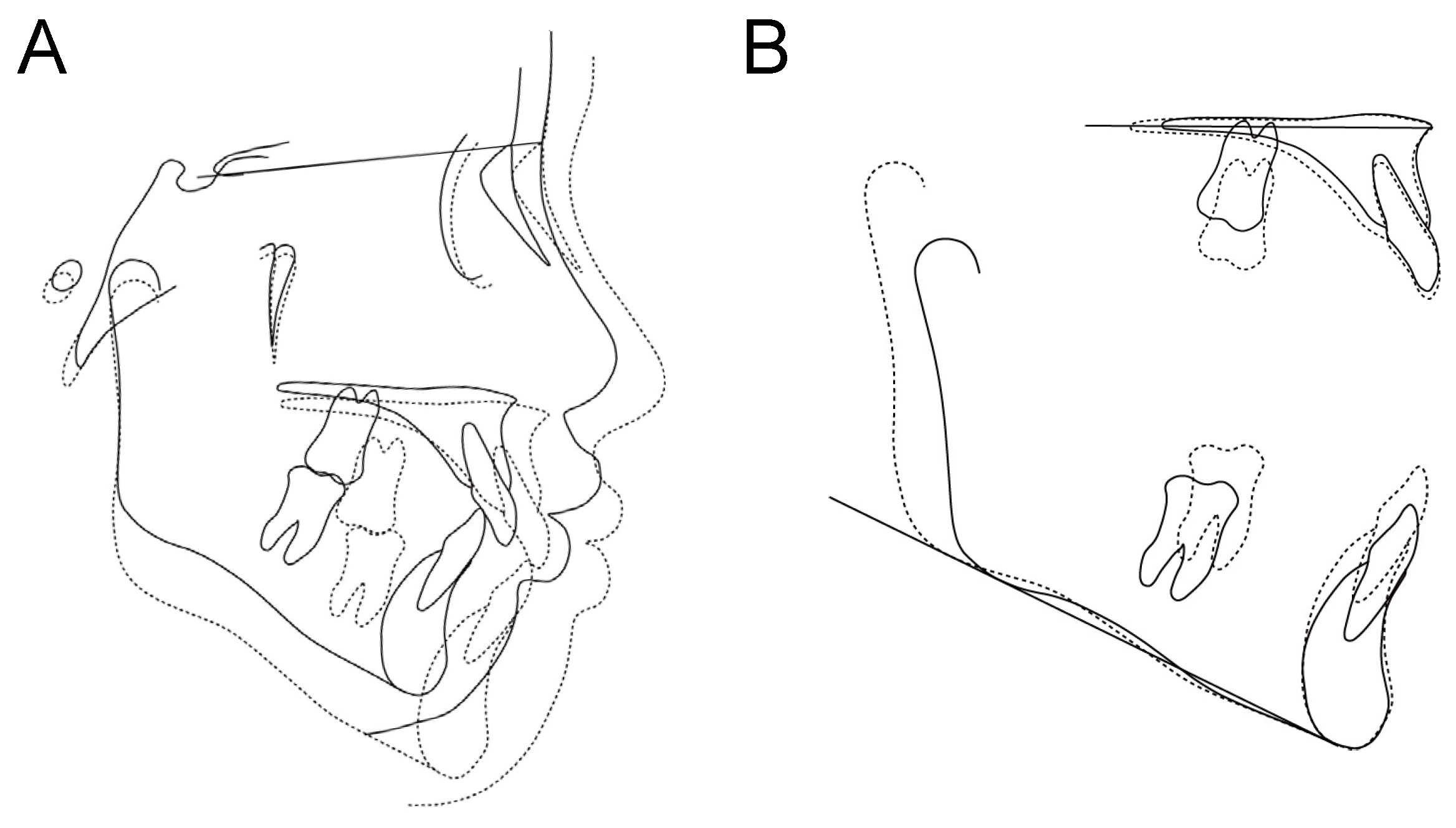
The patient was a Japanese girl of 8 years 10 months old at the time of the initial examination, with a chief complaint of rattling anterior teeth. In terms of family history, the patient’s mother had teeth crowding. There were no records in her history of current illness or medical history that were particularly problematic. She had no notable habits.
In terms of facial findings (Figure 12A), the frontal view did not demonstrate any deviation of the mandible. When smiling, however, the distance between the upper lip to the maxillary left central incisor cervical line was 4.0 mm, and a gummy smile was evident. In the lateral view, a protrusion of the upper lip was observed.
In the intra-oral findings (Figure 12B), the occlusal relationship of the molars was Angle Class II on both the left and right sides, and the overjet was +5.0 mm on both sides. In addition, the overbite was +3.0 mm. The maxillary anterior teeth were crowded, and the maxillary and mandibular dentition midlines were coincident with the facial midline. The dental age was stage IIIB, and narrowed dental arches were exhibited in both the upper and lower jaws. The length, however, of the dental arch was normal for both the upper and lower jaws, exhibiting a parabolic morphology.
The panoramic radiographs (Figure 13) showed no abnormalities in the number of teeth.
In the cephalogram angle measurement (Table 5), the SNA angle was 84.5°, the SNB angle was 78.5°, and the ANB angle was +6.0°, thus exhibiting skeletal class II. The Frankfort-mandibular plane angle (FMA) was 26.0°. U1 to SN was 109.0° and the maxillary anterior teeth showed labial inclination. In addition, FMIA was 52.0°. In the cephalogram distance measurement (Table 6), the maxillary anterior tooth height (Is-Is’) was 29.5 mm, so that with reference to Sakamoto et al. [18], the patient’s maxillary anterior teeth were high. Wits appraisal was −0.5 mm.
Diagnosis
This patient was diagnosed as a case of skeletal class II, accompanied by a gummy smile due to the vertical excessive growth of the upper jaw, and crowding.
Treatment approach
First, we decided to improve the crowding of the maxillary anterior teeth with a sectional arch. After that, we decided to use SBJA for the purpose of improving skeletal maxillary prognathism in order to suppress maxillary growth and promote mandibular growth. Furthermore, we decided to suppress the vertical growth of the maxillary anterior teeth and improve the gummy smile by using SBJA and a high-pull J-hook headgear together. To improve the narrowed maxillary and mandibular arch, we planned to expand the maxillae laterally through enlargement of the SBJA expansion screw. The patient was instructed to use the appliance for more than 12 h per day.
Treatment course
The patient used SBJA (with expansion screw) and high-pull J-hook headgear for 25 months from 9 years and 8 months of age to 11 years and 9 months of age.
Treatment results
In this patient, as a result of treatment, SNA remained unchanged from 84.5°, while SNB increased from 78.5° to 80.5°. As a result, ANB changed from 6.0° to 4.0°, and the skeletal maxillary prognathism improved. The maxillary anterior teeth height (Is-Is’) returned to normal height. With growth, the maxillary molars protruded, but the protruding of the maxillary anterior teeth was suppressed, and the occlusal plane (occlusal plane to FH) changed from 15.5° to 11.0°, and transformed to anterior elevation (Figure 14, Table 5). Wits appraisal was −1.5 mm. The left central incisor cervical exposure while smiling decreased from 4.0 mm to 0.0 mm, thus improving the gummy smile (Figure 15A). In addition, the molar relationship changed from class II to class I (Figure 15B), and no root resorption was observed in the maxillary anterior teeth (Figure 16).

3. Discussion

All three cases in this study were in their growth period, and gummy smiles caused by the overeruption of maxillary anterior teeth were observed. There have been various reports on the causes and ways of improving gummy smiles [2,4,5,6,9,10]. However, there have been no reports on the spontaneous disappearance of gummy smiles observed during growth. Rather, it has been reported that the amount of exposure of the maxillary anterior teeth does not change during the growth process [19]. We thought, therefore, that it was unlikely that overeruption of the anterior teeth would improve spontaneously during the growth process. In addition, even if orthodontic treatment was started in childhood, it was not desirable to postpone improvement of the gummy smile complaint until the use of multi-bracket appliances. Therefore, in order to improve gummy smiles, we decided to develop a treatment plan that would lead to improvement by means of the application of orthodontic force in the direction of suppressing the eruption of maxillary anterior teeth from the growth stage.
The high-pull J-hook headgear is a device that is connected to hooks attached to the anterior teeth of the arch wire of a multi-bracket device, pulling the maxillary anterior teeth upward. By pulling the maxillary anterior teeth upward, the maxillary anterior teeth are pushed down, and the overbite is reduced, thereby facilitating improvement of a deep overbite [20,21,22]. Furthermore, since it is often used in combination with a multi-bracket device, there have been no reports of the use of the high-pull J-hook headgear during the growth period.
Since the three cases examined here had skeletal maxillary prognathism in the growth period, we decided to improve their skeletal characteristics in the first stage of treatment. In addition, although we considered that pressing down the maxillary anterior teeth using a multi-bracket device after the end of growth would improve the gummy smiles, we thought that the combined use of high-pull J-hook headgear with a removable applianc, would render it possible to actively improve gummy smiles at an early stage by suppressing the eruption of maxillary anterior teeth before the end of the growth period.
As patients were in the mixed dentition stage, rather than using the headgear in combination with a multi-bracket device, we devised a plan to attach a loop to a part that is equivalent to the maxillary anterior teeth of a SBJA. This was then used to improve maxillary prognathism, and to use the high-pull J-hook headgear to draw the part corresponding to the maxillary anterior teeth upwards to suppress their eruption and improve the gummy smiles.
In general, the effects of BJA include a decrease in the ANB angle due to maxillary growth suppression and mandibular growth promotion, as well as a change in the anteversion of the occlusal plane, clockwise rotation of the mandible, and lingual inclination of the anterior maxillary teeth [23,24]. The common BJA effect seen in these three cases was the decrease in the ANB angle due to mandibular growth promotion.
It has been reported that normally, the SNA angle increases slightly with growth [25]. The general maxillary growth suppression effect of BJA is the reduction in the SNA angle by around 0.5°, but without a significant difference [23,24]. Here, the SNA angle remained almost unchanged in all three cases. This may be the result of the suppression of the SNA angle, which should increase with normal growth, due to the general effect of BJA, but this is difficult to ascertain.
In addition, the combined use of SBJA and high-pull J-hook headgear caused a change in the anterior elevation of the occlusal plane, which is generally not an observed effect of BJA. It is an opposite consequence of the general BJA effect, which is the anteversion of the occlusal plane. We found, however, that when an upward force was applied to the anterior part of the SBJA by the high-pull J-hook headgear, the entire maxillary dentition being covered with a plate, which is a feature of the SBJA, led to the application of an anti-clockwise force being applied to the entire maxillary dentition. This suppressed the eruption of maxillary anterior teeth and caused anterior elevation changes to the occlusal plane due to the normal growth of the maxillary molars. In addition, we conceived that the suppression of eruption of maxillary anterior teeth improved the gummy smile.
Furthermore, although BJA is generally associated with the clockwise rotation of the mandible [23], this was not observed in any of the three cases. Sakai et al. [24] reported that BJA generally involves a large amount of protrusion of the mandibular molars. In this study, the amount of protrusion of the mandibular molars was smaller than that of the maxillary molars in all the three cases observed, so it is possible that the clockwise rotation of the mandible was not noted. This mechanism will, however, need to be investigated in the future. Additionally, the treatment duration was long in all three cases. It took time for the effect of the appliance to appear. It is considered that long treatment duration is a demerit of the method of improving gummy smiles using SBJA and high-pull J-hook headgear.
In these cases, as a result of using SBJA in combination with high-pull J-hook headgear with the aim of improving the overeruption of maxillary anterior teeth, the eruption of maxillary anterior teeth was suppressed, and led to a result similar to those reported on treatments that improved gummy smiles using orthodontic anchor screws in patients in the growth stage [13,14]. It was thought that the method of improving gummy smiles using SBJA and high-pull J-hook headgear, as examined in this study, may be an option for school-age patients and their parents, who may be resistant to surgically invasive treatments. When an orthodontic anchor screw is used to improve a gummy smile during the growth period, the anchor screw may be lost, and it has been reported that the dropout rate from treatment after implanting orthodontic anchor screws is high in young patients [26]. We suspect that the improvement of gummy smiles using SBJA and high-pull J-hook headgears are an alternative treatment in such situations. However, the lengthening of the treatment period must be considered.
In conclusion, the combined use of SBJA with a loop attached to the labial side of maxillary anterior teeth and high-pull J-hook headgear may suppress the eruption of maxillary anterior teeth and improve gummy smiles in growth period patients with maxillary prognathism. However, this study was a case series, and therefore future studies are needed to assess the ideal treatment for specific subgroups of patients, the ideal timing and so on.

Author Contributions

Conceptualization, T.S. (Takuma Sato), M.T. (Masako Tabuchi), K.M. and S.G.; Methodology, T.S. (Takuma Sato) and K.M.; Validation, R.A.; Formal analysis, T.S. (Takuma Sato) and R.A.; Investigation, T.S. (Takuma Sato); Resources, S.G.; Data curation, R.A.; Writing—original draft, T.S. (Takuma Sato); Writing—review & editing, M.T. (Masako Tabuchi), M.K., T.S. (Takeo Sekiya) and M.T. (Mifumi Takahashi); Supervision, S.G.; Project administration, T.S. (Takuma Sato) and K.M. All authors have read and agreed to the published version of the manuscript.

Funding

This research received no external funding.

Institutional Review Board Statement

Not applicable.

Informed Consent Statement

Informed consent was obtained from all subjects involved in the study.

Conflicts of Interest

The authors declare no conflict of interest.

References

  1. Kokich, V.O.; Kokich, V.G.; Kiyak, H.A. Perceptions of dental professionals and laypersons to altered dental esthetics: Asymmetric and symmetric situations. Am. J. Orthod. Dentofacial. Orthop. 2006, 130, 141–151. [Google Scholar] [CrossRef] [PubMed]
  2. Proffit, W.R.; Fields, H.W.; Sarver, D.M. Contemporary Orthodontics; Mosby Year Book: St. Louis, MO, USA, 2007. [Google Scholar]
  3. Hunt, O.; Johnston, C.; Hepper, P.; Burden, D.; Stevenson, M. The influence of maxillary gingival exposure on dental attractiveness ratings. Eur. J. Orthod. 2002, 24, 199–204. [Google Scholar] [CrossRef] [PubMed] [Green Version]
  4. Robbins, J.W. Differential diagnosis and treatment of excess gingival display. Pract. Periodontontics Aesthet. Dent. 1999, 11, 265–272. [Google Scholar]
  5. Silberberg, N.; Goldstein, M.; Smidt, A. Excessive gingival display—Etiology, diagnosis, and treatment modalities. Quintessence Int. 2009, 40, 809–818. [Google Scholar] [PubMed]
  6. Monaco, A.; Streni, O.; Marci, M.C.; Marzo, G.; Gatto, R.; Giannoni, M. Gummy smile: Clinical parameters useful for diagnosis and therapeutical approach. J. Clin. Pediatr. Dent. 2005, 29, 19–25. [Google Scholar] [CrossRef] [PubMed]
  7. Evian, C.I.; Cutler, S.A.; Rosenberg, E.S.; Shah, R.K. Altered passive eruption: The undiagnosed entity. J. Am. Dent. Assoc. 1993, 124, 107–110. [Google Scholar] [CrossRef] [PubMed]
  8. Levine, R.A.; McGuire, M. The diagnosis and treatment of the gummy smile. Compend. Contin. Educ. Dent. 1997, 18, 757–762. [Google Scholar]
  9. Ohnishi, H.; Yagi, T.; Yasuda, Y.; Takata, K. A mini-implant for orthodontic anchorage in a deep overbite case. Angle Orthod. 2005, 75, 444–452. [Google Scholar] [CrossRef]
  10. Lin, J.C.Y.; Yeh, C.L.; Liou, E.J.W.; Bowman, S.J. Treatment of skeletal-origin gummy smiles with miniscrew anchorage. J. Clin. Orthod. 2008, 42, 285–296. [Google Scholar]
  11. Kokich, V. Esthetic and anterior tooth position: An orthodontic perspective. Part II: Vertical position. J. Esthet. Restor. Dent. 1993, 5, 200–207. [Google Scholar] [CrossRef]
  12. Peck, S.; Peck, L.; Kataja, M. The gingival smile line. Angle Orthod. 1992, 62, 92–100. [Google Scholar] [CrossRef]
  13. Kim, T.W.; Kim, H.; Lee, S.J. Correction of deep overbite and gummy smile by using a mini-implant with a segmented wire in a growing Class II Division 2 patient. Am. J. Orthod. Dentofacial Orthop. 2006, 130, 676–685. [Google Scholar] [CrossRef] [PubMed] [Green Version]
  14. Kim, T.W.; Freitas, B.V. Orthodontic treatment of gummy smile by using mini-implants (part I): Treatment of vertical growth of upper anterior dentoalveolar complex. Dental Press J. Orthod. 2010, 15, 42.e1–42.e9. [Google Scholar] [CrossRef]
  15. Miyazawa, K.; Sakai, N.; Tsutsui, T.; Tabuchi, M.; Goto, S. The simplified bite jumping appliance: An improved functional appliance. Orthod. Waves 2015, 74, 18–21. [Google Scholar] [CrossRef]
  16. Sander, F.G.; Lassak, C. The modification of growth with the Jumping-the-Bite plate compared to other functional orthodontic appliances. J. Orofac. Orthop. 1990, 51, 155–164. [Google Scholar] [CrossRef]
  17. Martina, R.; Cioffi, I.; Galeotti, A.; Tagliaferri, R.; Cimino, R.; Michelotti, A.; Valletta, R.; Farella, M.; Paduano, S. Efficacy of the Sander bite-jumping appliance in growing patients with mandibular retrusion: A randomized controlled trial. Orthod. Craniofacial Res. 2013, 16, 116–126. [Google Scholar] [CrossRef]
  18. Sakamoto, T.; Miura, F.; Iizuka, T. Linear Analyses on the Developmental Changes of Dentofacial Complex of Japanese by Means of Roentgenographic. Cephalometry. J. Stomatol. Soc. 1963, 30, 169–182. [Google Scholar] [CrossRef] [Green Version]
  19. Bergman, R.T.; Waschak, J.; Borzabadi-Farahani, A.; Murphy, N.C. Longitudinal study of cephalometric soft tissue profile traits between the ages of 6 and 18 years. Angle Orthod. 2014, 84, 48–55. [Google Scholar] [CrossRef] [Green Version]
  20. Burstone, C.J. Deep overbite correction by intrusion. Am. J. Orthod. 1977, 72, 1–22. [Google Scholar] [CrossRef]
  21. Janzen, E.K. A balanced smile—A most important treatment objective. Am. J. Orthod. 1977, 72, 359–372. [Google Scholar] [CrossRef]
  22. Deguchi, T.; Murakami, T.; Kuroda, S.; Yabuuchi, T.; Kamioka, H.; Takano-Yamamoto, T. Comparison of the intrusion effects on the maxillary incisors between implant anchorage and J-hook headgear. Am. J. Orthod. Dentofacial Orthop. 2008, 133, 654–660. [Google Scholar] [CrossRef] [PubMed]
  23. Burhan, A.S.; Nawaya, F.R. Dentoskeletal effects of the Bite-Jumping Appliance and the Twin-Block Appliance in the treatment of skeletal Class II malocclusion: A randomized controlled trial. Eur. J. Orthod. 2015, 37, 330–337. [Google Scholar] [CrossRef] [PubMed] [Green Version]
  24. Sakai, N.; Miyazawa, K.; Tsutsui, T.; Tabuchi, M.; Shibata, M.; Goto, S. Comparative study of the treatment effects off bionator and bite jumping appliances on Cllass II malocclusions. Orthod. Waves 2016, 75, 1–9. [Google Scholar] [CrossRef]
  25. Van Diepenbeek, A.F.; Buschang, P.H.; Prahl-Andersen, B. Age-dependant cephalometric standards as determined by multilevel modeling. Am. J. Orthod. Dentofacial Orthop. 2009, 135, 79–87. [Google Scholar] [CrossRef] [PubMed]
  26. Motoyoshi, M.; Matsuoka, M.; Shimizu, N. Application of orthodontic miniimplants in adolescents. Int. J. Oral Maxillofac. Surg. 2007, 36, 695–699. [Google Scholar] [CrossRef] [PubMed]
Figure 2. Facial photographs and intra-oral photographs at initial examination for Case 1; facial photograph and enlarged photograph of the mouth at initial examination (A); intra-oral photograph at initial examination (B).
Applsci 12 12268 g002
Figure 3. Panoramic radiograph (A) and lateral roentgenographic cephalogram (B) of Case 1 at initial examination.
Applsci 12 12268 g003
Figure 4. Superimposed images of Case 1; overall (SN plane, reference point S) (A); maxilla (palatal plane, reference point ANS) and mandible (mandibular marginal plane, reference point Me) (B).
Applsci 12 12268 g004
Figure 5. Facial photograph and intra-oral photograph of Case 1 at the end of the first stage of treatment; facial photograph and enlarged photograph of the mouth at the end of the first stage of treatment (A); intra-oral photograph at the end of the first stage of treatment (B).
Applsci 12 12268 g005
Figure 6. Panoramic radiograph (A) and lateral roentgenographic cephalogram (B) of Case 1 at the end of the first stage of treatment.
Applsci 12 12268 g006
Figure 7. Facial photographs and intra-oral photographs at initial examination for Case 2; facial photograph and enlarged photograph of the mouth at initial examination (A); intra-oral photograph at initial examination (B).
Applsci 12 12268 g007
Figure 8. Panoramic radiograph (A) and lateral roentgenographic cephalogram (B) of Case 2 at initial examination.
Applsci 12 12268 g008
Figure 9. Superimposed images of Case 2; overall (SN plane, reference point S) (A); maxilla (palatal plane, reference point ANS) and mandible (mandibular marginal plane, reference point Me) (B).
Applsci 12 12268 g009
Figure 10. Facial photograph and intra-oral photograph of Case 2 at the end of the first stage of treatment; facial photograph and enlarged photograph of the mouth at the end of the first stage of treatment (A); intra-oral photograph at the end of first stage of treatment (B).
Applsci 12 12268 g010
Figure 11. Panoramic radiograph (A) and lateral roentgenographic cephalogram (B) of Case 2 at the end of the first stage of treatment.
Applsci 12 12268 g011
Figure 12. Facial photographs and intra-oral photographs at initial examination for Case 3; facial photograph and enlarged photograph of the mouth at initial examination (A); intra-oral photograph at initial examination (B).
Applsci 12 12268 g012
Figure 13. Panoramic radiograph (A) and lateral roentgenographic cephalogram (B) of Case 3 at initial examination.
Applsci 12 12268 g013
Figure 14. Superimposed images of Case 3 Overall (SN plane, reference point S) (A) Maxilla (palatal plane, reference point ANS) and mandible (mandibular marginal plane, reference point Me) (B).
Applsci 12 12268 g014
Figure 15. Facial photograph and intra-oral photograph of Case 3 at the end of the first stage of treatment. Facial photograph and enlarged photograph of the mouth at the end of the first stage of treatment (A); intra-oral photograph at the end of the first stage of treatment (B).
Applsci 12 12268 g015
Figure 16. Panoramic radiograph (A) and lateral roentgenographic cephalogram (B) of Case 3 at the end of the first stage of treatment.
Applsci 12 12268 g016
Table 1. Cephalogram angle measurements of Case 1.
Pre-TreatmentPost SBJA + High-Pull Headgear
(10 y 7 m)(14 y 4 m)
Measurements (°)ValueValue
SNA85.085.0
SNB78.580.5
ANB6.54.5
FMA30.530.5
U-1 to SN103.0103.0
FMIA62.061.0
L-1 to mand.pl.87.588.5
Interincisal Angle126.0126.0
SN to Nasal floor9.07.5
Occ.pl.to FH19.011.5
Table 2. Cephalogram distance measurements of Case 1.
Pre-TreatmentPost SBJA + High-Pull Headgear
(10 y 7 m)(14 y 4 m)
Measurements (mm)ValueMeanDeviationValueMeanDeviation
N-ANS55.051.5 ± 3.1X58.057.0 ± 3.5N
ANS-Me68.566.2 ± 3.7N77.573.0 ± 4.6N
Is-Is’31.528.5 ± 2.1X33.030.7 ± 2.8N
Mo-Ms17.519.1 ± 1.7N25.523.7 ± 2.2N
Ii-Ii’42.041.4 ± 1.5N47.546.0 ± 1.9N
Mo-Mi29.029.9 ± 2.3N35.534.1 ± 2.9N
Wits appraisal0.0 −2.0
N: Within one standard deviation of the mean
X: Outside one standard deviation of the mean
Table 3. Cephalogram angle measurements of Case 2.
Pre-TreatmentPost SBJA + High-Pull Headgear
(10 y 0 m)(13 y 10 m)
Measurements (°)ValueValue
SNA85.085.0
SNB77.080.0
ANB8.05.0
FMA33.533.5
U-1 to SN118.5110.0
FMIA53.553.5
L-1 to mand.pl.92.593.0
Interincisal Angle117.0125.0
SN to Nasal floor7.04.0
Occ.pl.to FH15.010.5
Table 4. Cephalogram distance measurements of Case 2.
Pre-TreatmentPost SBJA + High-Pull Headgear
(10 y 0 m)(13 y 10 m)
Measurements (mm)ValueMeanDeviationValueMeanDeviation
N-ANS55.551.5 ± 3.1X58.557.0 ± 3.5N
ANS-Me66.566.2 ± 3.7N71.073.0 ± 4.6N
Is-Is’31.028.5 ± 2.1X32.530.7 ± 2.8N
Mo-Ms18.019.1 ± 1.7N25.023.7 ± 2.2N
Ii-Ii’40.041.4 ± 1.5N45.046.0 ± 1.9N
Mo-Mi28.029.9 ± 2.3N31.534.1 ± 2.9N
Wits appraisal2.0 −0.5
N: Within one standard deviation of the mean
X: Outside one standard deviation of the mean
Table 5. Cephalogram angle measurements of Case 3.
Pre-TreatmentPost SBJA + High-Pull Headgear
(8 y 10 m)(13 y 5 m)
Measurements (°)ValueValue
SNA84.584.5
SNB78.580.5
ANB6.04.0
FMA26.026.0
U-1 to SN109.0115.5
FMIA52.049.5
L-1 to mand.pl.102.0104.5
Interincisal Angle116.0107.0
SN to Nasal floor8.57.5
Occ.pl.to FH15.511.0
Table 6. Cephalogram distance measurements of Case 3.
Pre-TreatmentPost SBJA + High-Pull Headgear
(8 y 10 m)(13 y 5 m)
Measurements (mm)ValueMeanDeviationValueMeanDeviation
N-ANS51.047.0 ± 2.5X52.553.3 ± 2.8N
ANS-Me62.562.1 ± 2.9N69.070.4 ± 3.8N
Is-Is’29.526.3 ± 2.1X30.030.5 ± 2.1N
Mo-Ms18.517.3 ± 1.5N24.022.6 ± 1.7N
Ii-Ii’39.037.8 ± 1.6N44.043.2 ± 1.6N
Mo-Mi29.027.5 ± 1.7N33.031.9 ± 2.1N
Wits appraisal−0.5 −1.5
N: Within one standard deviation of the mean
X: Outside one standard deviation of the mean
Publisher’s Note: MDPI stays neutral with regard to jurisdictional claims in published maps and institutional affiliations.

