Article

Quercus robur Older Bark—A Source of Polyphenolic Extracts with Biological Activities

1
Department of Pharmacognosy and Phytotherapy, Faculty of Pharmacy, “George Emil Palade” University of Medicine, Pharmacy, Sciences and Technology of Târgu Mures, 38 Gheorghe Marinescu Street, 540139 Târgu Mures, Romania
2
Department of Microbiology, Faculty of Medicine, “George Emil Palade” University of Medicine, Pharmacy, Sciences and Technology of Târgu Mures, 38 Gheorghe Marinescu Street, 540139 Târgu Mures, Romania
3
Doctoral School of Medicine and Pharmacy, “George Emil Palade” University of Medicine, Pharmacy, Sciences and Technology of Târgu Mures, 38 Gheorghe Marinescu Street, 540139 Târgu Mures, Romania
4
Department of Pharmaceutical Botany, Faculty of Pharmacy, “Iuliu Hatieganu” University of Medicine and Pharmacy, 8 Victor Babes Street, 400012 Cluj-Napoca, Romania
5
Laboratory of Chromatography, Institute of Advanced Horticulture Research of Transylvania, University of Agricultural Sciences and Veterinary Medicine, 400372 Cluj-Napoca, Romania
6
Department of Pharmaceutical Botany, Faculty of Pharmacy, “George Emil Palade” University of Medicine, Pharmacy, Sciences and Technology of Târgu Mures, 38 Gheorghe Marinescu Street, 540139 Târgu Mures, Romania
7
Research Center of Medicinal and Aromatic Plants, “George Emil Palade” University of Medicine, Pharmacy, Sciences and Technology of Târgu Mures, 38 Gheorghe Marinescu Street, 540139 Târgu Mures, Romania
*
Author to whom correspondence should be addressed.
Appl. Sci. 2022, 12(22), 11738; https://doi.org/10.3390/app122211738
Submission received: 23 October 2022 / Revised: 7 November 2022 / Accepted: 16 November 2022 / Published: 18 November 2022

Abstract

:
Processing wood leads to the accumulation of considerable amounts of forest waste, such as branches, leaves and especially bark. These byproducts can be exploited for their high content of biomolecules. Our aim was to histo-anatomically and phytochemically characterize the older oak bark and evaluate the potential biological activities. After the histo-anatomical characterization and processing the plant material, the aqueous and hydroalcoholic extracts were obtained using microwave-assisted extraction (MAE) and ultrasound-assisted extraction (UAE). After the quantification of the total polyphenol content (TPC) and the total tannins content (TTC), the bark extracts were evaluated for antioxidant, antimicrobial and enzyme inhibitory activity. In the transverse section, the rhytidome (the succession of periderms) and the dead secondary phloem can be observed. The results of the polyphenol content of the oak bark extracts show that the hydroethanolic oak bark obtained using MAE has the highest absolute value of the TPC (347.74 mg/g). In comparison with the extracts obtained using UAE, the bark extracts obtained using MAE showed a significantly higher TPC. The greatest antioxidant and antibacterial activity were also recorded for the tested extracts. For gram-positive bacteria, the MBC values ranged from 0.3 (S. aureus) to 5 mg/mL and the oak bark extract obtained using MAE showed good activity against C. krusei (MIC—2.5 mg/mL). Among the assayed enzyme inhibitory activities, the extracts showed a significant antidiabetic effect in vitro with the highest activity corresponding to an IC50 of 3.88 µg/mL. The older oak bark represents a potential source of compounds with antibacterial, antioxidant or antidiabetic activity, which can be exploited through the biorefining processes.

1. Introduction

The bark is part of the vascular plant, consisting of all the external tissues (succession of periderms and the dead secondary phloem) of the vascular cambium [1]. The bark has an important role in the protection of woody plants. The diversity of the histo-anatomical structure and chemical composition of the barks makes them a particularly interesting potential source of biomolecules, which can be exploited in the context of biorefining [2]. To assess the potential of barks, it is important to know the specific information about their structure and anatomy.
Forest wastes are considered to be of low value due to the lack of efficient extraction techniques. Classical extraction methods may have certain limitations regarding energy, time and solvents. However, ultrasound-assisted extraction (UAE) and microwave-assisted extraction (MAE) can extract compounds in a short time with reduced energy and solvents, preserving the functionality of the biomolecules [3,4]. However, the associated variables (time, frequency, power, temperature, solvent ratio, etc.) must be optimized for each type of vegetal material. The goal of MAE for dried plant material is to heat the small amounts of moisture that exist in plant cells. When this moisture is heated using a microwave, it evaporates and puts considerable pressure on the cell wall. This pressure causes the cell wall to weaken and eventually break from within, causing the phytocompounds to exude from the cells and increasing the extraction yield [5]. In UAE, the ultrasound energy induces the formation of cavitation bubbles, which expand and further collapse, producing the extreme local conditions that cause rapid fragmentation in the cellular structure. This process further leads to an increased extraction yield [3]. Both the UAE and MAE methods are considered green extraction methods.
The Quercus genus (L.) belongs to the Fagaceae family and Fagales order and contains trees that are spread throughout the world with different morphology in terms of chemical compounds [6]. One of the most well-known species is Quercus robur L. (syn Quercus pedunculata Hoffm), known as common oak or pedunculate oak. It is found spontaneously in deciduous forests and is used in the wood industry and folk medicine [6,7] Q. robur bark also contains hydrolysable and condensed tannins. Elansary et al. [6] revealed several phenolic compounds in the composition of Q. robur bark, including catechin, ellagic acid, protocatechuic acid, gallic acid and vanillic acid. The bioactivity assay revealed antioxidant, antibacterial, antidiabetic and anticancer activity in the Q. robur bark extracts [6,8].
According to the European Pharmacopoeia [9], oak bark can be obtained from 10 to 15-year-old oak trees (Quercus robur, Q. petraea and Q. pubescens). However, these trees are not of interest to the wood processing industry, since their diameter is too small. Therefore, a question arises: what happens to the bark of the oak trees used in the forestry industry? According to the FAO (Food and Agriculture Organization), global wood production was about 5157 million m3 in 2020 (FAO, 2022), generating over 103 million m3 of wood residue [10]. In the wood industry, the bark is considered a waste and it is mainly used as fuel.
Thus, this study evaluated (1) the histo-anatomical structure of older oak bark; (2) the polyphenolic and tannins content of the aqueous and hydroalcoholic extracts that were obtained using MAE and UAE; and (3) the biological potential of older oak bark extracts (antioxidant capacity, antibacterial activity, enzyme inhibitory activity).
The novelty of the study consists of the valorization of old pedunculate oak bark (forest waste) by recommending it as a source for phenolic compounds with potential antidiabetic, antioxidant and antimicrobial biological actions.

2. Materials and Methods

2.1. Plant Sample

The pedunculate oak (Q. robur L.) bark was provided by the Botanical Garden of the George Emil Palade University of Medicine, Pharmacy, Science and Technology of Târgu Mureș (46°33′24″ N, 24°34′59″ E) in Mures County, Romania in April 2021. The tree’s age was approximately 90 years and a voucher specimen was deposited at the Herbarium with the reference number 116/8. The bark was dried at 50 °C for 24 h in a drying oven and milled with a cutting mill.

2.2. Histo-Anatomical Analysis

For the histo-anatomical observations, the bark was first fixed and preserved in 70% alcohol then prepared for further analysis by the classic procedure. The cross sections (ten sections) were made through the oak bark and were collected on a watch glass with water. Half of the obtained sections were stained green using iodine. The sections were mounted between slides in a few drops of water, analyzed using a microscope (Motic B series) and photographed using a digital camera (Nikon, Tōkyō, Japan).

2.3. Extraction

The two extraction methods used (UAE and MAE) were previously optimized on lignocellulosic matrices [11,12]. UAE was performed in an ultrasonic bath (Elma Schmidbauer GmbH, Singen, Germany) at 40 kHz ultrasonic power. 2.5 g of powdered bark material was weighed and placed into a volumetric flask with 100 mL of solvent (water or ethanol: water 50:50, v/v). The vessel was then covered to prevent evaporation. The extraction was performed at 70 °C for 15 min [11]. For the MAE, 10 g of older bark were placed into the microwave extractor vessel with 200 mL of solvent (water or ethanol: water 70:30, v/v). The extractions were performed in a microwave extractor (Ethos X, Milestone, Sorisole, Italy) for 18 min at 650 W for the hydroalcoholic extracts and 30 min at 850 W for the aqueous extracts [12]. After the extraction, the samples were centrifuged, concentrated and freeze-dried.

2.4. Total Phenolics and Tannins Content

The total polyphenolic content (TPC) was assessed using the Folin–Ciocâlteu method [13] and the quantification of tannins was performed using the classic method published in the 8th edition of the European Pharmacopoeia [9]. The final total tannin content (TTC) was expressed as a % of the pyrogallol/freeze-dried extract.

2.5. Antioxidant In Vitro Assays

For the in vitro assays, two complementary methods were used, based on the DPPH and ABTS reagents. For the free radical scavenging activity with DPPH, different concentrations of the tested oak extracts (50 µL of the freeze-dried bark extract in methanol 50%) and 200 µL DPPH (0.1 mM) methanolic solution were incubated in darkness for 30 min at room temperature. The absorbance was read at 517 nm with a microplate reader (Epoch, BioTek, Winooski, VT, USA). The formula for inhibition capacity was:
i n h i b i t i o n   % = A c A s A c × 100
where Ac is the absorbance of the control solution determined in the same conditions and As is the absorbance of the sample [14].
The IC50 was determined from the nonlinear regression plot of the inhibition percentage against the logarithms of concentrations.
For the free radical scavenging activity with ABTS (2,2′-azino-bis(3-ethylbenzothiazoline-6-sulfonic acid) 50 µL of the freeze-dried oak extract and 200 µL ABTS (methanolic solution, 10 mM) were incubated at room temperature. The absorbance was read at 734 nm. The formula for inhibition capacity was:
i n h i b i t i o n   % = A c A s A c × 100
where Ac is the absorbance of the control solution and As is the absorbance of the tested sample [15].

2.6. Assay of the Antimicrobial Activity

The antimicrobial activity was evaluated by determining the minimum inhibitory concentrations (MICs) [16]. For the bacterial strains, the freeze-dried oak bark extracts were dissolved in 5% DMSO and 200 µL was added to the wells in the first column of a 96-well plate. The samples were then binarily diluted. The bacterial suspensions were prepared using 10 µL of inoculum and 9990 µL of Muller–Hinton broth 2X. Then, 100 µL of the bacterial suspensions was added to each well. The oak bark extracts and bacterial strain suspension were added as controls (positive and negative). The plates were incubated for 24 h at 37 °C. For the fungal strains, the working protocol was followed with some differences. A solution of 100 μL of the inoculated fungal suspension was prepared by adding one milliliter of the fungal inoculum (0.5 McF) into 9 mL of the RPMI (Gibco Roswell Park Memorial Institute) medium. Subsequently, successive dilutions of the test substances were performed in the RPMI medium. The MIC readings were performed in each well with a 50% growth inhibition.

2.7. Alfa-Glucosidase Inhibitory Assay

The α-glucosidase inhibitory activity was measured according to a previously described method [17,18]. In brief, 50 μL of the resolubilized samples were diluted with 50 μL of the phosphate buffer (100 mM, pH 6.8) in a microplate, then mixed with 50 μL of yeast α-glucosidase and incubated for 10 min. After the incubation time, 50 μL of the substrate were added. The coloration was measured at 405 nm due to the release of p-nitrophenol. The acarbose was used as a standard. The results were expressed as IC50 using the equation:
i n h i b i t i o n   % = A c A s A c × 100
where Ac is the absorbance of the control solution and As is the absorbance of the tested sample.

2.8. Tyrosinase Inhibitory Activity

The tyrosinase inhibitory activity of each tested extract was determined by a previously described method [19]. In brief, 40 µL of the sample with different concentrations were mixed with 80 µL of the 5 mM phosphate buffer with a pH of 6.5, and then mixed with 40 µL of tyrosinase (125 U/mL in phosphate buffer). The reaction mix was incubated for 10 min at room temperature, and then 40 µL of L-DOPA (10 mM in phosphate buffer) was added. After another incubation of 20 min, the absorbance was measured at 475 nm. Kojic acid was used as positive control. The results were expressed as IC50 using the following equation:
i n h i b i t i o n   % = A c A s A c × 100
where Ac is the absorbance of the control solution and As is the absorbance of the tested sample.

2.9. Acetylcholinesterase Inhibitory Activity

The inhibitory activity was measured using the Ellman’s method, which was previously described [19,20]. A 25 μL sample was diluted with 50 μL of the Tris–HCl buffer (50 mM, with pH 8.0). It was then mixed with 125 μL of the DTNB solution in a Tris–HCl buffer (0.9 mM) and 25 μL of the enzyme solution (0.078 U/mL in Tris–HCl buffer). The reaction mix was incubated in a dark place for 15 min at room temperature. Then, 25 μL of the ATCI solution (4.5 mM in Tris–HCl buffer) was added and the samples were reincubated for 10 min. The absorbance of each sample was recorded at 405 nm and galantamine was used as positive control. The results were expressed as IC50 using the equation:
i n h i b i t i o n   % = A c A s A c × 100
where Ac is the absorbance of the control solution and As is the absorbance of the tested sample.
For all of the enzymatic inhibition assays, the results were expressed considering the dilution in the 96 wells for each sample.

2.10. Statistical Analysis

The determinations were performed in triplicate and the results were expressed as mean ± standard deviation. The statistical analysis was performed using GraphPad Prism 9.4.1. The data were subjected to a one-way analysis of variance (ANOVA) and the differences between the means were evaluated using the post-hoc Tukey’s test. The statistical significance was considered to be p < 0.05.

3. Results

3.1. Structure and Histo-Anatomy of Oak Bark

The aspects of the Q. robur outer bark are blackish-brown, rough and deeply furrowed. In the transverse section, the rhytidome (consisting of a succession of periderms) and the dead secondary phloem can be observed (Figure 1A–D).
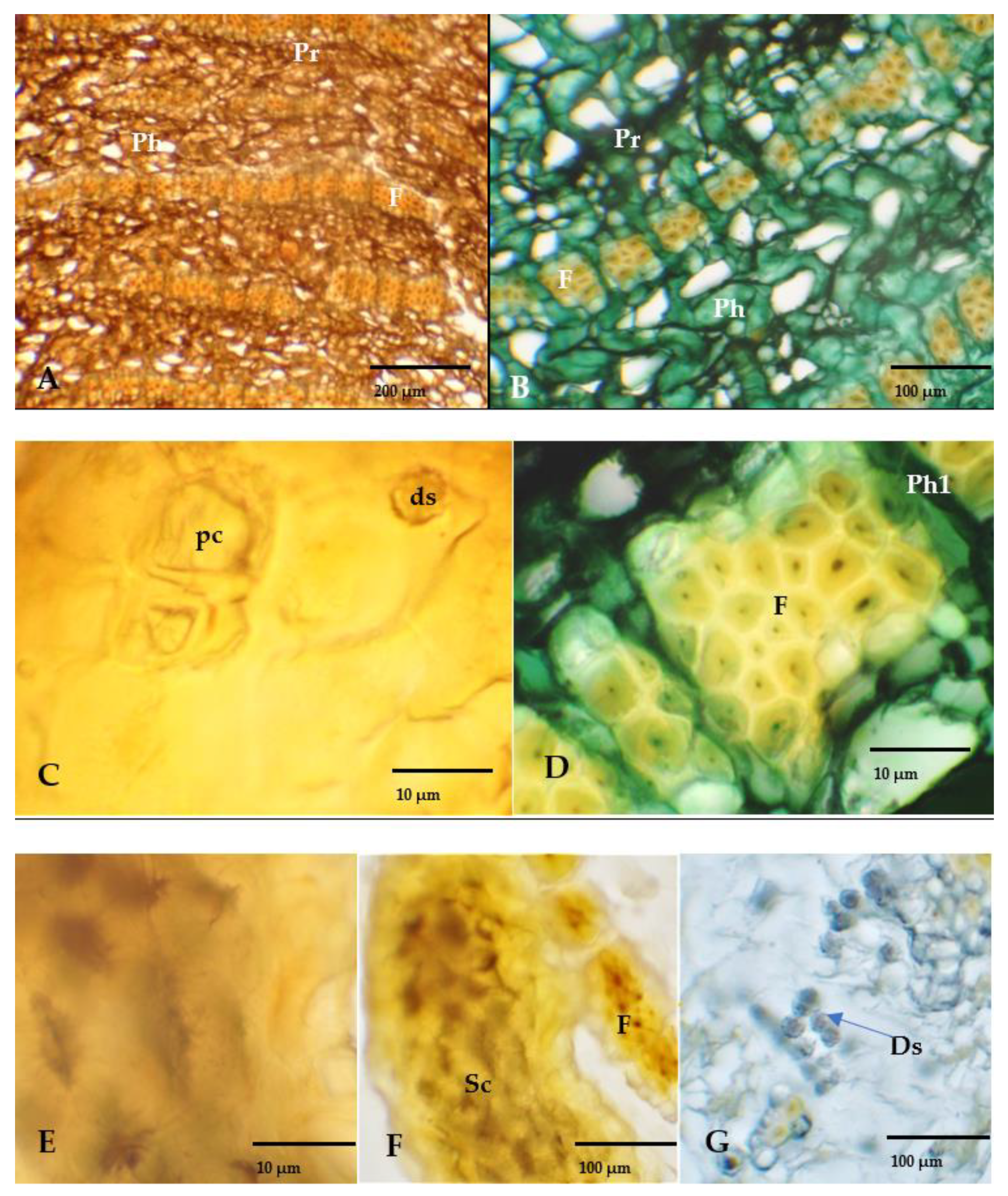
In the transverse section (Figure 2A–G), the tangential bands of the secondary phloem develop, accompanied by chambered crystalliferous cells with prismatic crystals or druses (Figure 2C,G). The rays of the uniseriate phloem cross the groups of fibers (Figure 2D). The sclereids develop solitarily or in groups (Figure 2E,F) with different forms and dimensions.

3.2. Oak Bark Extracts Characterization

The extraction yield (Table 1) of the hydroethanolic extracts is similar for both the MAE and UAE methods. On the other hand, the extraction yield of the aqueous extractions is higher for MAE compared to UAE.
The results from Table 1 show that the QREM variant (347.74 mg/g) has the highest value, followed by the QRAM (323.16 mg/g) and QRAUS (267.04 mg/g) variants. The bark extracts that were obtained using MAE, compared to bark extracts that were obtained using UAE, showed significantly higher TPC values. By comparing the aqueous extracts to the hydroalcoholic extracts that were obtained using MAE, it can be observed that the aqueous extract had a significantly lower TPC. The higher content of tannins was quantified in the aqueous extracts, but there were no significant differences between the results.

3.3. Antioxidant Activity of Oak Bark Extracts

The TPC results indicate the antioxidant effect of the oak bark extracts (Table 1). This result was also confirmed by the DPPH and ABTS methods, resulting in the antioxidant effect of the oak bark extracts (Table 2). In the case of the tested solutions, the DPPH reagent was reduced extremely quickly within the first 50 s. The antioxidant activity at the initial time were lower than the activity at the end of the reaction. The higher antioxidant activity using both radical scavenging assays was recorded for QREUS and QRAUS.

3.4. Antimicrobial Activity of Oak Bark Extracts

As can be observed in Table 3, the oak bark extracts exerted antibacterial activity against the tested bacteria strains. The MIC values for gram-positive bacteria ranged from 0.3 to 1.25 mg/mL. The MBC values ranged from 0.3 (S. aureus) to 5 mg/mL. The MBCs of QREM was significantly higher (0.3 mg/mL) compared to the other samples. Among the gram-negative bacteria, those most sensitive to the oak bark extracts were K. pneumoniae (MBC—0.6 mg/mL) and P. aeruginosa (MIC—0.6 mg/mL). The MICs for E. coli were not determined, since they were above the maximum tested concentrations.
The oak bark extract that was obtained using MAE showed good activity against Candida krusei (MIC—2.5 mg/mL) and weak activity against C. parapsilosis (MIC—5 mg/mL), (Table 4). The MICs were not recorded for C. albicans.

3.5. Enzyme Inhibitory Activity of Oak Bark Extracts

The oak bark extracts were evaluated for their alfa-glycosidase, tyrosinase and acetyl–cholinesterase inhibitory potential, and the results are presented in Table 5.
In all of the assayed activities, the anti-α-glucosidase potential was the strongest, which can be observed from the IC50 values and from the inhibition graph presented in Figure 3. In comparison to the positive control (acarbose), all the samples showed an inhibitory activity at least 10 times lower, and the best IC50 was identified in the ethanolic extract that was obtained using MAE (QREM). In this case, the IC50 values ranged from 3.88 to 5.60 µg/mL, significantly lower in comparison to the IC50 of acarbose (122.27 µg/mL). However, the activity against tyrosinase and acetyl–cholinesterase was modest. All the extracts showed a moderate activity against tyrosinase and the highest activity was observed for the ethanolic extract that was obtained using UAE (QREUS). Finally, the Q. robur extracts showed a very weak inhibition against acetyl–cholinesterase, exhibiting an insignificant activity in comparison to galantamine (used as positive control).
Regarding the correlation between the applied extraction method and the activity of the final extracts, MAE induced the highest inhibitory activity against α-glucosidase, while there was no clear correlation for the anti-tyrosinase activity since QREUS was the most active and QRAUS was the least active.

4. Discussion

In the European Pharmacopoeia, the oak bark is described as the bark of young branches and the lateral shoots of Q. robur, Q. petraea and/or Q. pubescens. On the other hand, older bark is considered to be forest waste [2]. Considering the enormous quantities of forest waste, particularly from older bark [7], it is essential to successfully recover the active biomolecules from these sources. The combination of environmental consciousness, financial motivations and international legislation provide incentives for the recovery of the organic waste phytocompounds and the subsequent secondary utilization of those compounds.
The present study describes the histo-anatomical structure of the older oak bark (Quercus robur bark) together with the phytochemical composition and biological activity of the extracts. Studies on the biological activity of some of the extracts obtained from oak bark do not present clear information about the histo-anatomical structure of the plant matrix that was studied. It is clear that the chemical composition of the extracts were influenced by the type of tissue correlated with the age of the tree and the other external factors acting on the plant [21]. The histo-anatomical structure was described in order to define the plant tissue that was subjected for extraction. Thus, the histo-anatomical structure of the oak bark consists of a sequence of periderm and secondary phloem in accordance with the descriptions previously observed in the specialized literature [22].
The polyphenols and tannins content were in line with the anatomical observations. The bark of Q. robur was rich in polyphenols that could be solubilized by different polarity solvents. In this study, a significantly higher TPC was observed for the hydroethanolic bark extracts that were obtained using MAE. On the other hand, the higher values of tannins were quantified in the aqueous extracts.
The TPC in the oak bark extracts indicates that the older oak bark is a valuable source of biomolecules. Similar reports were previously given for other oak bark extracts, even though the extraction procedures differed, or the plant matrix was not histo-anatomically characterized. For example, for ethanol–water extracts (50:50, v/v, bark from a 17-year-old Q. robur tree, UAE extraction) the TPC was 610.63 mg GAE/g for the dried bark extract [23], which was higher compared to the older oak bark extracts that were characterized in this work (50:50, v/v, bark from a 90 to100-year-old Q. robur tree, UAE/MAE extraction). Another study [24] reported a much lower TPC (54.3 mg GAE/g for the dried bark extract) compared to our results. In this case, the hydroethanolic extract was obtained from the bark of a 148-year-old tree using the Soxhlet extraction method. It can be considered that the extraction type and the age of the oak tree influenced the concentration of the biomolecules in the bark extracts., The differences between the TPC values could also be linked with a gradual decomposition of the phenolic compounds over time [24].
The presence of the phenolic compounds could be correlated with the biological potential (antioxidant, antimicrobial, antidiabetic etc.) of the bark extracts. Due to the differences in the methods and calculations, the comparison between the results and the literature data should be done with caution. However, our results are in agreement with those from the previously published literature [6,8]. As can be observed, the older oak bark extracts exerted a high antioxidant activity, an expected activity given the high polyphenolic content. According to research from the literature, ellagic acid—a key bioactive component—could be partially responsible for the antioxidant properties of the oak bark extracts [6].
With the exception of E. coli, the older oak bark extracts exhibited antibacterial activity against all of the tested species. A previous report documented the antimicrobial activity of the oak bark extract on S. aureus and C. albicans. Another recent study reported strong antibacterial activities against P. aeruginosa, Mariniluteicoccus flavus and E. coli. In the current study, antibacterial activities were found against S. aureus MRSA, K. pneumoniae and P. aeruginosa. In addition, the antifungal activity against C. krusei was highlighted. These antimicrobial activities might be attributed to phenolic compounds, such as ellagic acid, epicatechin and catechin, and compounds with strong antimicrobial activities [6,25].
Quercus species are known for their complex phytochemical composition, which allows for the possibility of using them as sources of bioactive compounds with pharmacological activity for chronic diseases, such as a metabolic syndrome [26,27]. Regarding the assayed enzyme inhibitory activities, the extracts showed a significant inhibitory activity on α-glucosidase. The different plant parts of Q. robur were previously shown to have a high α-glucosidase inhibitory activity, correlated in some cases to tannin content and tannin derivatives [28]. At the same time, the extracts that were obtained from different plant parts of Quercus species showed a strong α-glucosidase inhibitory activity. Specific examples of the compounds isolated from Quercus sp. with α-glucosidase inhibitory activity include hexagalloylglucose from the galls of Q. infectoria [29], tiliroside from the leaves of Q. gilva [30] and the acorn kernel-derived polyphenols from Q. variabilis [31]. Moreover, other studies showed a similar activity of the bark extracts for other species in the Quercus genus, such as Q. coccifera [32] and Q. rubra [33], which showed IC50 values similar to acarbose, an approved alpha-glucosidase inhibitor. These findings support for further preclinical and clinical studies where these effects must be validated [32,33]. In the current study, we have identified a correlation between the TPC values and the IC50 values for all the extracts, which highlights the idea that phenolic species contribute to bioactivity. The most active sample against α-glucosidase—the hydroethanolic extract that was obtained using MAE—also showed the highest TPC value. An optimization study regarding Q. robur bark has concluded that MAE acts as an efficient method for recovering higher TPC values, especially when using hydroalcoholic extraction, which is highlighted in the findings of the present study [34]. A similar trend has been observed for MAE and the hydroalcoholic extraction of Q. cerris bark, showing a higher yield of total polyphenols [12].
Phenolic derivatives, such as resveratrol, can act as tyrosinase inhibitors due to their chemical analogy with tyrosine. This class of phytochemical compounds has the potential to be used in diseases characterized by melanin hyperproduction [33,35]. Previously, certain research studies assessed the tyrosinase inhibitory activity of Quercus species extracts. Examples include the bark of Q. coccifera [32], the bark of Q. rubra [33] and the galls of Q. infectoria [36]. Hubert et al. showed that among 30 species of common temperate trees, the methanolic extract of Q. robur bark was the most active against tyrosinase [37]. Nonetheless, the older bark extracts showed modest activity against tyrosinase, much weaker in comparison to kojic acid as a positive control. This might indicate that the quantity of polyphenols in their composition are not enough to achieve sufficient inhibition. From all the types of extraction applied, hydroethanolic extraction mixed with UAE showed the highest anti-tyrosinase activity (with an IC50 of 79.8 µg/mL), suggesting that this combination would be suitable for obtaining the most active extract from older oak bark.

5. Conclusions

The general histo-anatomical structure of Q. robur (older tree) bark was described and was found to resemble most oak species. The main pattern showed a rhytidome with sequential periderms and dead phloem, including groups of sclereids and abundant druses in parenchyma cells. The concentration of total polyphenols indicate that the bark could be a good source of polyphenols, and it was concluded that the highest extraction yield was obtained with microwave-assisted extraction. The older oak bark extracts showed antioxidant, antimicrobial and α-glucosidase inhibitory activity, and also a modest activity against tyrosinase. The results of this study revealed the potential of Q. robur older bark as a source of pharmacologically active compounds that could be further tested in preclinical and clinical studies. Moreover, microwave treatment seemed to be a superior method for obtaining extracts with a higher inhibitory activity against α-glucosidase, in comparison to ultrasounds.
These results constitute the reasons for the valorization of older oak bark (forest waste), and integrating it into a biorefining scheme for the recovery of biomolecules. The potential efficiency of forest waste and the enhanced utilization of these byproducts can be integrated into a circular economic framework.

Author Contributions

Conceptualization, C.T.; methodology, C.T., A.N. (Adrian Nisca), A.N. (Alexandru Nicolescu), R.Ș., M.B., A.D.M., A.M. (Adrian Man), A.M. (Andrei Mocan) and C.N.C.; validation, C.T. and A.M. (Adrian Man); investigation, C.T., A.N. (Adrian Nisca), R.Ș., M.B., N.-A.C. and C.N.C.; resources, C.T.; data curation, C.T., A.N. (Alexandru Nicolescu), R.Ș., M.B., A.N. (Adrian Nisca), A.M. (Andrei Mocan), A.D.M. and C.N.C.; writing—original draft preparation, R.Ș., C.T. and A.N. (Alexandru Nicolescu); writing—review and editing A.M. (Adrian Man); supervision, C.T.; project administration, C.T.; funding acquisition, C.T. All authors have read and agreed to the published version of the manuscript.

Funding

This work was supported by a grant from the Romanian Ministry of Education and Research, CNS-UEFISCDI, project number PN-III-P1-1.1-TE-2019-1549, within PNCDI III.

Institutional Review Board Statement

Not applicable.

Informed Consent Statement

Not applicable.

Data Availability Statement

Not applicable.

Conflicts of Interest

The authors declare no conflict of interest.

References

  1. Niklas, K.J. The Mechanical Role of Bark. Am. J. Bot. 1999, 86, 465–469. [Google Scholar] [CrossRef] [PubMed]
  2. Leite, C.; Pereira, H. Cork-Containing Barks—A Review. Front. Mater. 2017, 3, 63. [Google Scholar] [CrossRef] [Green Version]
  3. Kumar, K.; Srivastav, S.; Sharanagat, V.S. Ultrasound Assisted Extraction (UAE) of Bioactive Compounds from Fruit and Vegetable Processing by-Products: A Review. Ultrason. Sonochem 2021, 70, 105325. [Google Scholar] [CrossRef] [PubMed]
  4. Llompart, M.; Garcia-Jares, C.; Celeiro, M.; Dagnac, T. Extraction|Microwave-Assisted Extraction☆. In Encyclopedia of Analytical Science, 3rd ed.; Worsfold, P., Poole, C., Townshend, A., Miró, M., Eds.; Academic Press: Oxford, UK, 2019; pp. 67–77. ISBN 978-0-08-101984-9. [Google Scholar]
  5. Bagade, S.B.; Patil, M. Recent Advances in Microwave Assisted Extraction of Bioactive Compounds from Complex Herbal Samples: A Review. Crit. Rev. Anal. Chem. 2021, 51, 138–149. [Google Scholar] [CrossRef] [PubMed]
  6. Elansary, H.O.; Szopa, A.; Kubica, P.; Ekiert, H.; Mattar, M.A.; Al-Yafrasi, M.A.; El-Ansary, D.O.; Zin El-Abedin, T.K.; Yessoufou, K. Polyphenol Profile and Pharmaceutical Potential of Quercus spp. Bark Extracts. Plants 2019, 8, 486. [Google Scholar] [CrossRef] [PubMed] [Green Version]
  7. Dróżdż, P.; Pyrzynska, K. Assessment of Polyphenol Content and Antioxidant Activity of Oak Bark Extracts. Eur. J. Wood Wood Prod. 2018, 76, 793–795. [Google Scholar] [CrossRef] [Green Version]
  8. Unuofin, J.O.; Lebelo, S.L. UHPLC-QToF-MS Characterization of Bioactive Metabolites from Quercus Robur L. Grown in South Africa for Antioxidant and Antidiabetic Properties. Arab. J. Chem. 2021, 14, 102970. [Google Scholar] [CrossRef]
  9. Council of Europe, E.D. for the Q. of M.& H. European Directorate for the Quality of Medicines & Health Care. In European Pharmacopoeia; Council of Europe: Strasbourg, France, 2014; ISBN 978-92-871-7525-0. [Google Scholar]
  10. FAO. FAOSTAT—Forestry Production and Trade; Food and Agriculture Organization of the United Nations: Rome, Italy, 2022. [Google Scholar]
  11. Tanase, C.; Domokos, E.; Coșarcă, S.; Miklos, A.; Imre, S.; Domokos, J.; Dehelean, C.A. Study of the Ultrasound-Assisted Extraction of Polyphenols from Beech (Fagus Sylvatica L.) Bark. BioResources 2018, 13, 2247–2267. [Google Scholar] [CrossRef]
  12. Nisca, A.; Ștefănescu, R.; Moldovan, C.; Mocan, A.; Mare, A.D.; Ciurea, C.N.; Man, A.; Muntean, D.-L.; Tanase, C. Optimization of Microwave Assisted Extraction Conditions to Improve Phenolic Content and In Vitro Antioxidant and Anti-Microbial Activity in Quercus Cerris Bark Extracts. Plants 2022, 11, 240. [Google Scholar] [CrossRef]
  13. Cicco, N.; Lanorte, M.T.; Paraggio, M.; Viggiano, M.; Lattanzio, V. A Reproducible, Rapid and Inexpensive Folin–Ciocalteu Micro-Method in Determining Phenolics of Plant Methanol Extracts. Microchem. J. 2009, 91, 107–110. [Google Scholar] [CrossRef]
  14. Skrypnik, L.; Grigorev, N.; Michailov, D.; Antipina, M.; Danilova, M.; Pungin, A. Comparative Study on Radical Scavenging Activity and Phenolic Compounds Content in Water Bark Extracts of Alder (Alnus glutinosa (L.) Gaertn.), Oak (Quercus Robur L.) and Pine (Pinus sylvestris L.). Eur. J. Wood Wood Prod. 2019, 77, 879–890. [Google Scholar] [CrossRef]
  15. Re, R.; Pellegrini, N.; Proteggente, A.; Pannala, A.; Yang, M.; Rice-Evans, C. Antioxidant Activity Applying an Improved ABTS Radical Cation Decolorization Assay. Free. Radic. Biol. Med. 1999, 26, 1231–1237. [Google Scholar] [CrossRef]
  16. Tănase, C.; Coşarcă, S.; Toma, F.; Mare, A.; Man, A.; Miklos, A.; Imre, S.; Boz, I. Antibacterial activities of Beech Bark (Fagus sylvatica L.) Polyphenolic Extract. Environ. Eng. Manag. J. (EEMJ) 2018, 17, 877–884. [Google Scholar] [CrossRef]
  17. Les, F.; Venditti, A.; Cásedas, G.; Frezza, C.; Guiso, M.; Sciubba, F.; Serafini, M.; Bianco, A.; Valero, M.S.; López, V. Everlasting Flower (Helichrysum Stoechas Moench) as a Potential Source of Bioactive Molecules with Antiproliferative, Antioxidant, Antidiabetic and Neuroprotective Properties. Ind. Crops Prod. 2017, 108, 295–302. [Google Scholar] [CrossRef]
  18. Spínola, V.; Castilho, P.C. Evaluation of Asteraceae Herbal Extracts in the Management of Diabetes and Obesity. Contribution of Caffeoylquinic Acids on the Inhibition of Digestive Enzymes Activity and Formation of Advanced Glycation End-Products (In Vitro). Phytochemistry 2017, 143, 29–35. [Google Scholar] [CrossRef] [PubMed]
  19. Nicolescu, A.; Babotă, M.; Zhang, L.; Bunea, C.I.; Gavrilaș, L.; Vodnar, D.C.; Mocan, A.; Crișan, G.; Rocchetti, G. Optimized Ultrasound-Assisted Enzymatic Extraction of Phenolic Compounds from Rosa Canina L. Pseudo-Fruits (Rosehip) and Their Biological Activity. Antioxidants 2022, 11, 1123. [Google Scholar] [CrossRef] [PubMed]
  20. Păltinean, R.; Ielciu, I.; Hanganu, D.; Niculae, M.; Pall, E.; Angenot, L.; Tits, M.; Mocan, A.; Babotă, M.; Frumuzachi, O.; et al. Biological Activities of Some Isoquinoline Alkaloids from Fumaria Schleicheri Soy. Will. Plants 2022, 11, 1202. [Google Scholar] [CrossRef]
  21. Sousa, V.; Ferreira, J.P.A.; Miranda, I.; Quilhó, T.; Pereira, H. Quercus Rotundifolia Bark as a Source of Polar Extracts: Structural and Chemical Characterization. Forests 2021, 12, 1160. [Google Scholar] [CrossRef]
  22. Trockenbrodt, M. Qualitative Structural Changes during Bark Development in Quercus Robur, Ulmus Glabra, Populus Tremula and Betula Pendula. IAWA J. 1991, 12, 5–22. [Google Scholar] [CrossRef]
  23. Sillero, L.; Prado, R.; Andrés, M.A.; Labidi, J. Characterisation of Bark of Six Species from Mixed Atlantic Forest. Ind. Crops Prod. 2019, 137, 276–284. [Google Scholar] [CrossRef]
  24. Dedrie, M.; Jacquet, N.; Bombeck, P.-L.; Hébert, J.; Richel, A. Oak Barks as Raw Materials for the Extraction of Polyphenols for the Chemical and Pharmaceutical Sectors: A Regional Case Study. Ind. Crops Prod. 2015, 70, 316–321. [Google Scholar] [CrossRef]
  25. De, R.; Sarkar, A.; Ghosh, P.; Ganguly, M.; Karmakar, B.C.; Saha, D.R.; Halder, A.; Chowdhury, A.; Mukhopadhyay, A.K. Antimicrobial Activity of Ellagic Acid against Helicobacter Pylori Isolates from India and during Infections in Mice. J. Antimicrob. Chemother. 2018, 73, 1595–1603. [Google Scholar] [CrossRef] [Green Version]
  26. Saini, R.; Patil, S.M. Anti-Diabetic Activity of Roots Of Quercus Infectoria Olivier in Alloxan Induced Diabetic Rats. Int. J. Pharm. Sci. Res. 2012, 3, 1318–1321. [Google Scholar] [CrossRef]
  27. Peesa, J. Herbal Medicine for Diabetes Mellitus: A Review. Int. J. Phytopharm. 2013, 3, 1–22. [Google Scholar] [CrossRef]
  28. Muccilli, V.; Cardullo, N.; Spatafora, C.; Cunsolo, V.; Tringali, C. α-Glucosidase Inhibition and Antioxidant Activity of an Oenological Commercial Tannin. Extraction, Fractionation and Analysis by HPLC/ESI-MS/MS and 1H NMR. Food Chem. 2017, 215, 50–60. [Google Scholar] [CrossRef] [PubMed]
  29. Hwang, J.-K.; Kong, T.-W.; Baek, N.-I.; Pyun, Y.-R. α-Glycosidase Inhibitory Activity of Hexagalloylglucose from the Galls of Quercus Infectoria. Planta Med. 2000, 66, 273–274. [Google Scholar] [CrossRef]
  30. Indrianingsih, A.W.; Tachibana, S.; Dewi, R.T.; Itoh, K. Antioxidant and α-Glucosidase Inhibitor Activities of Natural Compounds Isolated from Quercus Gilva Blume Leaves. Asian Pac. J. Trop. Biomed. 2015, 5, 748–755. [Google Scholar] [CrossRef] [Green Version]
  31. Wu, M.; Yang, Q.; Wu, Y.; Ouyang, J. Inhibitory Effects of Acorn (Quercus Variabilis Blume) Kernel-Derived Polyphenols on the Activities of α-Amylase, α-Glucosidase, and Dipeptidyl Peptidase IV. Food Biosci. 2021, 43, 101224. [Google Scholar] [CrossRef]
  32. Sari, S.; Barut, B.; Özel, A.; Kuruüzüm-Uz, A.; Şöhretoğlu, D. Tyrosinase and α-Glucosidase Inhibitory Potential of Compounds Isolated from Quercus Coccifera Bark: In Vitro and in Silico Perspectives. Bioorganic Chem. 2019, 86, 296–304. [Google Scholar] [CrossRef]
  33. Tanase, C.; Nicolescu, A.; Nisca, A.; Ștefănescu, R.; Babotă, M.; Mare, A.D.; Ciurea, C.N.; Man, A. Biological Activity of Bark Extracts from Northern Red Oak (Quercus Rubra L.): An Antioxidant, Antimicrobial and Enzymatic Inhibitory Evaluation. Plants 2022, 11, 2357. [Google Scholar] [CrossRef]
  34. Bouras, M.; Chadni, M.; Barba, F.J.; Grimi, N.; Bals, O.; Vorobiev, E. Optimization of Microwave-Assisted Extraction of Polyphenols from Quercus Bark. Ind. Crops Prod. 2015, 77, 590–601. [Google Scholar] [CrossRef]
  35. Działo, M.; Mierziak, J.; Korzun, U.; Preisner, M.; Szopa, J.; Kulma, A. The Potential of Plant Phenolics in Prevention and Therapy of Skin Disorders. Int. J. Mol. Sci. 2016, 17, 160. [Google Scholar] [CrossRef] [PubMed] [Green Version]
  36. Sharififar, F.; Dehghan-Nudeh, G.; Raeiat, Z.; Amirheidari, B.; Moshrefi, M.; Purhemati, A. Tyrosinase Inhibitory Activity of Major Fractions of Quercus Infectoria Galls. Pharmacogn. Commun. 2013, 3, 21–26. [Google Scholar] [CrossRef] [Green Version]
  37. Hubert, J.; Angelis, A.; Aligiannis, N.; Rosalia, M.; Abedini, A.; Bakiri, A.; Reynaud, R.; Nuzillard, J.-M.; Gangloff, S.C.; Skaltsounis, A.-L.; et al. In Vitro Dermo-Cosmetic Evaluation of Bark Extracts from Common Temperate Trees. Planta Med. 2016, 82, 1351–1358. [Google Scholar] [CrossRef] [PubMed]
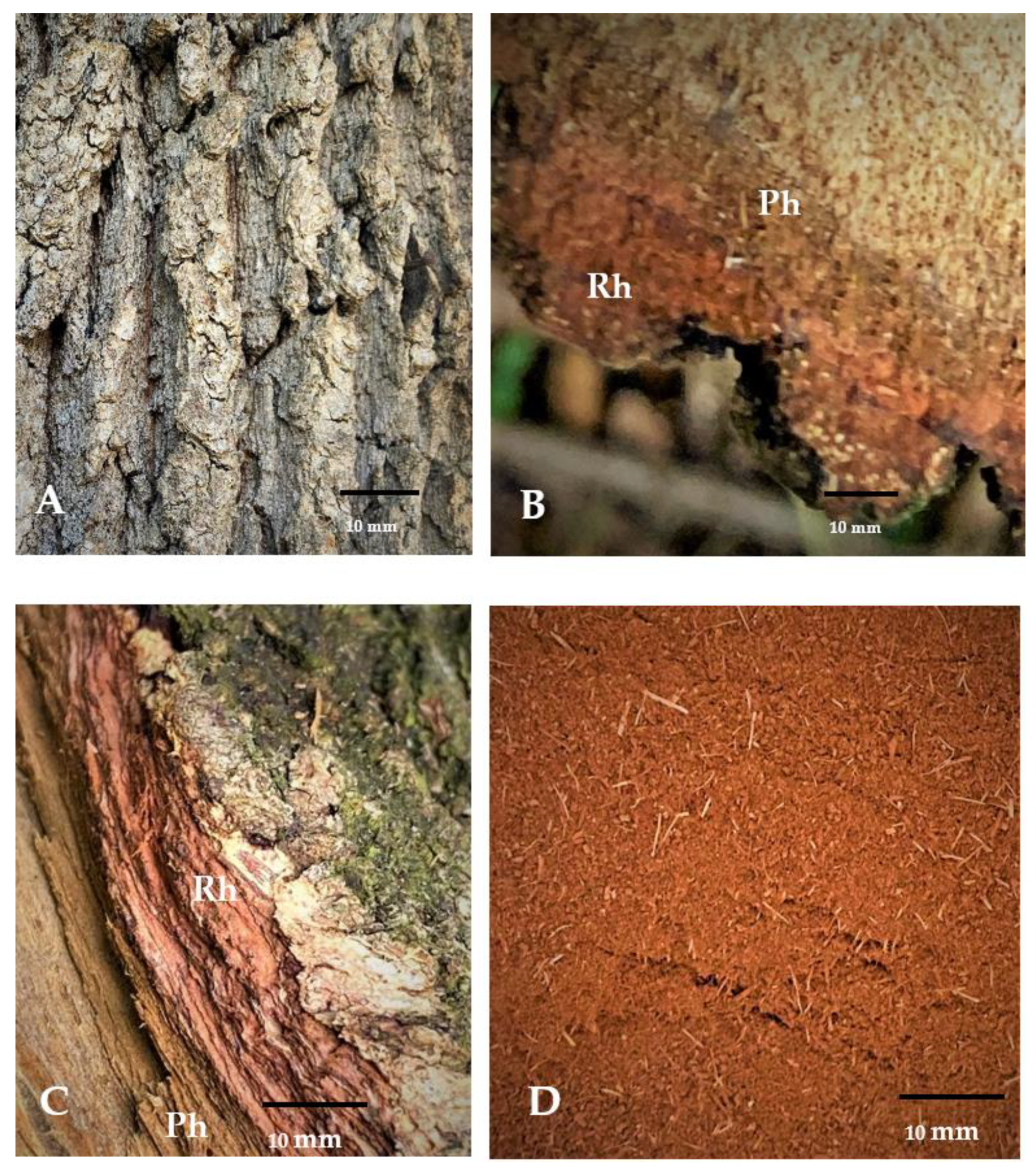
Figure 1. The bark structure of Quercus robur (A); General aspects of the transverse section, rhytidome (Rh) and phloem (Ph), (B); General aspects of the rhytidome in the fracture (C); General aspect of the powder after milling (D).
Applsci 12 11738 g001
Figure 2. The histo-anatomical aspects of the older bark of Quercus robur in the transverse section. (A) Rhytidome (old bark) includes various periderms (Pr), dead secondary phloem (Ph) between them and sclerenchimatic fibers and sclereids (FS); (B) portions of rhytidome—periderm stained green with iodine; (C) chambered crystalliferous cells with prismatic crystals (pc) and druses (ds); (D) detail with sclerenchimatic fibers (F) and uniseriate phloem rays (Ph1); (E) sclereids (Sc); (F) sclereids group and fibers (F); (G) druses (ds).
Applsci 12 11738 g002
Figure 3. Graphical representation of the dependence between the logC (of the concentration expressed as μg/mL) and the inhibition percentage against the α-glucosidase of the Q. robur older bark extracts, using the activity of acarbose as the positive control.
Applsci 12 11738 g003
Table 1. The extraction yield, total content of phenolic compounds and tannins in oak bark extracts.
Code SampleExtraction Yield (%)TPC (mg GAE/g DW) ± SD *TTC (% m/m) *
QREM5.66347.74 ± 8.66 a26.02 ± 8.88
QRAM4.57323.16 ± 3.17 b37.16 ± 4.48
QREUS5.32240.99 ± 1.49 c33.88 ± 5.53
QRAUS2.72267.04 ± 4.21 d33.85 ± 4.77
* TPC—total polyphenol content; DW—dry weight; TTC—total tannins content; GAE—gallic acid equivalent; SD—standard deviation (n = 3); QREM—hydroalcoholic extract obtained using MAE; QRAM—aqueous extract obtained using MAE; QREUS—hydroalcoholic extract obtained using UAE; QRAUS—aqueous extract obtained using UAE. The different letters in the same column note significant statisticaldifferences at p < 0.05.
Table 2. The values of IC50 for antioxidant activity of the oak bark extracts.
Code SampleIC50 DPPH (µg/mL) *IC50 ABTS (µg/mL) *
QREM11.99 ± 0.348.68 ± 0.31
QRAM1.94 ± 0.176.06 ± 0.00
QREUS2.43 ± 0.132.76 ± 0.12
QRAUS2.89 ± 0.832.77 ± 0.42
* ABTS—2,2′-azino-bis(3-ethylbenzothiazoline-6-sulfonic acid; DPPH—2,2-diphenyl-1-picrylhydrazyl; SD—standard deviation (n = 3); QREM—hydroalcoholic extract obtained using MAE; QRAM—aqueous extract obtained using MAE; QREUS—hydroalcoholic extract obtained using UAE; QRAUS—aqueous extract obtained using UAE.
Table 3. The antimicrobial activity (mg/mL) of the Q. robur bark extracts.
QREMQRAMQREUSQRAUS
MICMBCMICMBCMICMBCMICMBC
S. aureusGram-positive
bacteria
0.30.30.350.30.60.60.6
MRSA1.251.250.61.251.251.250.60.6
E. coliGram-negative
bacteria
>5>5>5>5>5>5>5>5
K. pneumoniae1.2550.60.61.252.50.60.6
P. aeruginosa2.52.5550.62.51.25>5
MIC: minimum inhibitory concentration; MBC: minimum bactericidal concentration; MRSA: methicillin-resistant Staphylococcus aureus; QREM—hydroalcoholic extract obtained using MAE; QRAM—aqueous extract obtained using MAE; QREUS—hydroalcoholic extract obtained using UAE; QRAUS—aqueous extract obtained using UAE.
Table 4. Antifungal activity (MIC in mg/mL) of the oak bark extracts.
QREMQRAMQREUSQRAUS
C. albicans>5>5>5>5
C. parapsilosis5>5>5>5
C. krusei2.52.555
MIC: minimum inhibitory concentration; QREM—hydroalcoholic extract obtained using MAE; QRAM—aqueous extract obtained using MAE; QREUS—hydroalcoholic extract obtained using UAE; QRAUS—aqueous extract obtained using UAE.
Table 5. The in vitro enzyme inhibitory activity of the oak bark extracts.
EnzymeControlTested SolutionsValues
Acetyl–cholinesterase
(IC50, µg/mL)
QREM143.4
QRAM152.8
QREUS148.4
QRAUS159.2
Galantamine 0.0002
Alfa-glucosidase (IC50, µg/mL) QREM3.88
QRAM4.07
QREUS5.60
QRAUS4.945
Acarbose 122.27
Tyrosinase
(IC50, µg/mL)
QREM145.74
QRAM172.22
QREUS79.8
QRAUS234
Kojic acid 4.44
QREM—hydroalcoholic extract obtained using MAE; QRAM—aqueous extract obtained using MAE; QREUS—hydroalcoholic extract obtained using UAE; QRAUS—aqueous extract obtained using UAE.
Publisher’s Note: MDPI stays neutral with regard to jurisdictional claims in published maps and institutional affiliations.