Article

Preliminary Study on the Larval Development of Calliphora vicina (Diptera: Calliphoridae) on Different Types of Substrates Used as Reference in Forensic Entomology

by
Cristiana Manea (Amariei)
1,
Ion Sandu
2,3,4,5,*,
Diana Bulgaru Iliescu
6,*,
Norina Consuela Forna
7,
Viorica Vasilache
4,5,* and
Vasile Sîrbu
8
1
Faculty of Geography and Geology, Alexandru Ioan Cuza University of Iași, Carol I Blvd., 20A, 700505 Iași, Romania
2
Academy of Romanian Scientists (AORS), 54 Splaiul Independenței St., Sector 5, 050094 Bucharest, Romania
3
National Institute for Research and Development in Environmental Protection, 294 Splaiul Independenței, 6th District, 060031 Bucharest, Romania
4
Science Department, Interdisciplinary Research Institute, Alexandru Ioan Cuza University of Iași, 11 Carol I Bulevard, 700506 Iași, Romania
5
Romanian Inventors Forum, 3 Sf. Petru Movilă St., L11, III/3, 700089 Iași, Romania
6
Institute of Legal Medicine, Medicine Faculty, Grigore T. Popa Medicine and Farmacy University of Iași, 4 Buna Vestire Street, 700450 Iași, Romania
7
Dental Medicine Faculty, Grigore T. Popa Medicine and Farmacy University of Iași, 16 Universitatii Street, 700115 Iași, Romania
8
Faculty of Biology, Alexandru Ioan Cuza University of Iași, Carol I Blvd., 20A, 700505 Iași, Romania
*
Authors to whom correspondence should be addressed.
Appl. Sci. 2022, 12(21), 10907; https://doi.org/10.3390/app122110907
Submission received: 8 October 2022 / Revised: 21 October 2022 / Accepted: 25 October 2022 / Published: 27 October 2022
(This article belongs to the Section Biomedical Engineering)

Abstract

:
Necrophagous insects are indicators of corpse decay. Analyses of their development stages give us indications on the degree of decay of corpses and contribute to the determination of the post-mortem interval. The differences in structure, biochemical composition and nutritive capacity of the cadaveric substratum can impair the duration of the developmental stages and dimensions of the organisms that metamorphose. This work tackles the study of the development, in laboratory conditions, of Calliphora vicina in correlation with different nutritive substrates (from areas of the corpse strongly affected by the incipient attack of Diptera) in order to obtain preliminary data, aiming to determine the post-mortem interval.

1. Introduction

Forensic entomology is involved in several segments of forensic investigations: the determination of the probable time of death through the analysis of entomological fauna succession on the corpse (exogenous factors); its infestation with parasites, virus, bacteria, yeasts, etc.; and the presence of pesticides and other substances in the insects coming from different biotopes, etc. [1,2,3].
The colonization and developmental rates of blowfly species are used to estimate the minimum post-mortem interval (mPMI) [4]. The fauna of interest is represented by the Diptera, Calliphoridae, Sarcopagidae and Muscidae families, specific to the areas where the investigation takes place and according to the respective biotopes.
The order Diptera predominates, in most cases, on corpses undergoing decay under the influence of biotic exogenous factors, combined with microclimatic factors (temperature, humidity, and illumination). We must mention that they can only infest the corpse in favorable climatic conditions. Excluding cold periods, the activity period of Calliphora vicina starts with temperatures of between 13 and 16 °C (which places the activity threshold of this species at a lower temperature than other species of flies) [5]. Their activity can be accelerated by high temperatures and levels of humidity in the environment, as well as the presence of other endogenous factors of cadaveric system decay (infestation prior to the time of death). The presence or absence of bacteria on a corpse may affect the development and survival of maggots that infest it. Understanding the microbial interactions with the maggot’s system is important for the improvement in the interpretation of PMI [4,5]. For example, a series of authors have studied the effect of two bacteria (Escherichia coli and Staphylococcus aureus) [6] on the rate of development and survival of three species of Diptera of forensic importance (Lucilia sericata, Calliphora vicina, and Calliphora vomitoria). It is well known that different endogenous contaminations of a corpse indicate the potential of bacteria on maggots during their development by the alteration of maggot development and weight. This inter regnum interaction is of great importance for forensic entomology, where the development data of certain fly species are used to estimate the minimum post-mortem interval (mPMI) [7,8].
Flies are usually the primary colonizers of a human corpse [9,10]. The colonization interval (the time between death and colonization) of an exposed corpse may vary from minutes to days. If a corpse has been hidden, it may vary even more [11]. As we have shown before, environmental factors, such as temperature [12,13,14], humidity [13,15], and solar light [13,16], directly influence the rate of colonization and development. Bacteria that populate the corpse may also be extremely influential in terms of the regulation of the attraction and selection of the sites for oviposition [4,17]. A recent study mentioned that Calliphora vicina maggots develop significantly slower on decayed than on fresh liver and suggested that temporal changes in the bacteria colonizing the liver are responsible for that [18]. Other studies indicate that microbial infections modify significantly during the post mortem interval based on the rate of decay [19]. The initial bacteria community varies based on the age [20] and health of the victim [21], the colonization area [22], environmental temperature and humidity [23], as well as depending on the season [24,25].
Taking these into consideration, this study has been conducted on the Calliphoridae family and its development on two organs, different in terms of biochemical content, but frequently exposed in cases of suspect deaths, humid tissues (orifices of the nose, mouth, eyes, ears, genitals, etc.) being the first ones targeted in the succession of necrophagous insects. The tongue and eye of pigs were chosen because the sampling of specimens undergoing the entomological study does not involve the movement, transportation, or any other action that could lead to the alteration or denaturation of a forensic investigation [26,27,28]. The eye of the pig is similar to the human eye. The presence of external mucus, conjunctiva, and eyelids makes it a wet environment, favorable for the development of necrophagous insect larvae. The thin wall makes it easy to penetrate, and it allows the elimination of the glassy humor inside the eye, which contributes to the maintenance of humidity and nutrients favorable for the development of necrophagous insects [29,30]. In terms of calories, for each 100 g of substance, the two organs contain the following calories: tongue, 271 calories, and eye, 60 calories. From a biochemical point of view, the pig tongue contains 64 g proteins, 18.6 g lipids, and 0 carbohydrates; the pig eye contains 27 g proteins, 2 g lipids, and the rest is blood plasma [31]. To this end, the aim of this work consists of obtaining preliminary data regarding the development, in laboratory conditions, of Calliphora vicina on two nutritional substrates (pig’s tongue and eye), different in terms of anatomical structure and biochemical composition/nutrients, so as to suggest potential directions of data use in forensic expertise in the calculation of the post-mortem interval. Analyzing the literature, we noticed that several studies carried out experiments on the species Calliphora vicina using a varied range of nutritional substrates (for example, different organs of an animal species [32], the same organ from different animals [33,34,35], different treatments for a single organ type, e.g., frozen/thawed vs. fresh [34]). All these studies have made significant contributions to the understanding of developmental mechanisms and offer a significant perspective on the effects of nutrition on the development of dipteran larvae, but the element of novelty that this study presents is actually constituted by the fact that pig’s tongue and eye constitute two nutritional substrates that have not been studied until this moment.

2. Materials and Methods

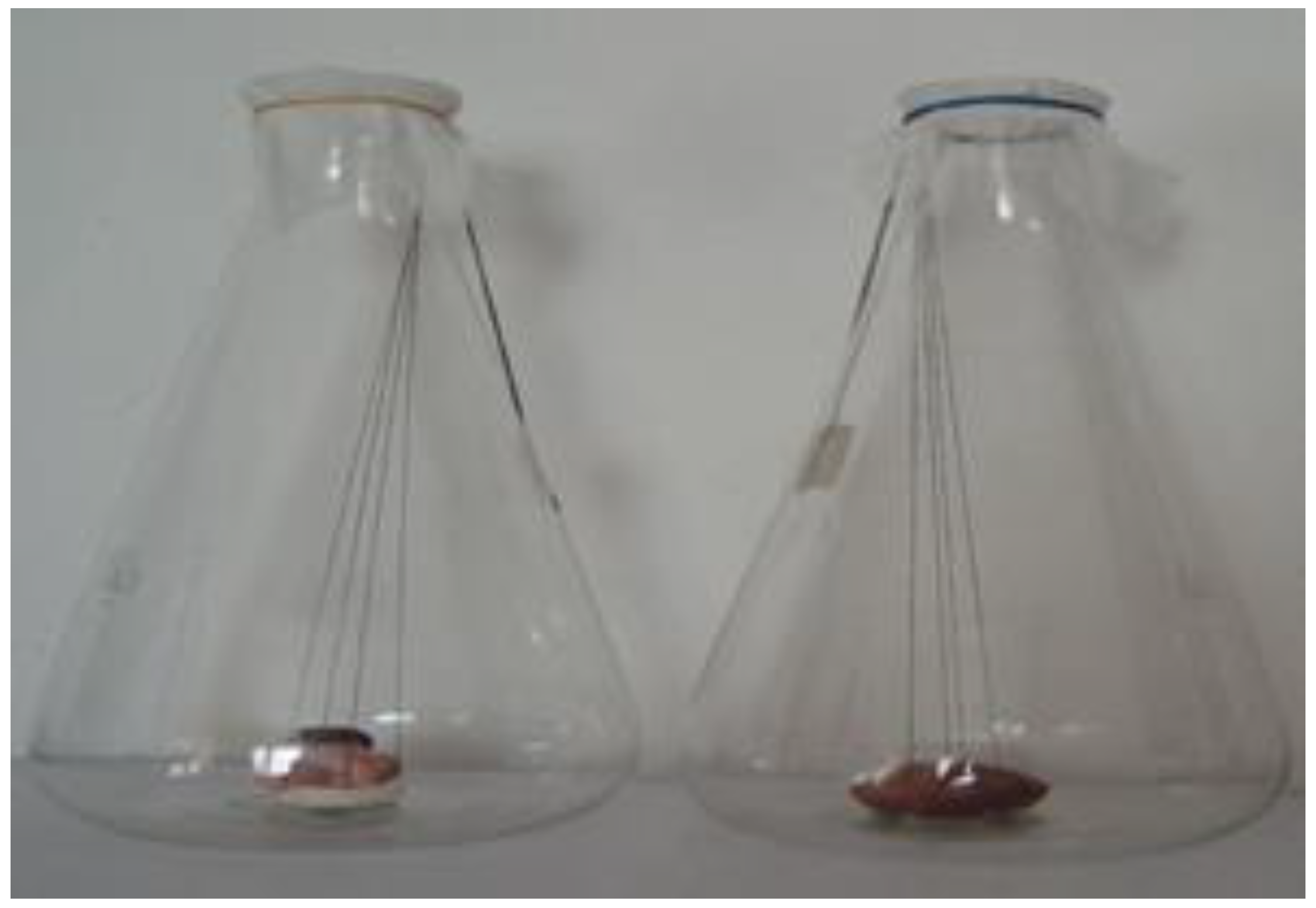
The study was conducted over 11 days. The development of necrophagous Diptera is influenced by the atmospheric conditions; thus, temperatures above 35 °C and beneath 12 °C are considered factors that limit the activity of insects [36,37,38,39]. In our case, temperature was constantly maintained at 25 °C. Females were captured using a previously prepared experimental trap, consisting of a chicken meat support. Females were identified/determined and placed in the Erlenmeyer glasses used in the experiment. They were allowed to lay the eggs, later being released. Species and stages of insect development were determined based on Romanian fauna [26,27]. After this, the monitoring of the evolution of larval development was started. An adult female was placed in each glass. The evolution of larval development was monitored closely, hour by hour, until the onset of the first larval stage and then at an interval of 5 h to follow the onset of the other stages, establishing the time for each subsequent stage. For each development stage, ten individuals were analyzed gradually, according to the study of maggots. The experimental protocol consisted of capturing adult Calliphora vicina females and placing them in the experimental containers for spawning. Eggs were obtained by introducing adult insects in two Erlenmeyer glasses with a narrow entry measuring 165 × 285 mm, where substratum samples (pig tongue and eye) had been introduced previously. Because the Erlenmeyer glasses were tall, the organs were tied with strings in such a way so that they could be easily lifted at any time in order to be studied. The glasses were placed in an oven with a constant temperature of 25 °C, where the growth of the maggots was studied and periods for their development were established (Figure 1).
The fresh tongue and eye of a healthy adult pig were used as substrata. Before placing the larvae in the preservation solution, they were killed by immersion in boiling water. In this study, only one developmental characteristic was measured, namely, larval length. The samples collected during each developmental stage of the studied Diptera (from the first larval stage to the emergence of adult flies) were kept in containers with Kahle solution for the measurements. Survival rates were also determined stepwise in pupae and adults by reference exclusively to specimens remaining on the substrate and live adults resulting from each pupa, respectively.
In order to conduct measurements of the eggs, larvae, pupa, and flies, we used a special ribbon millimetric scale used in forensic investigations.
We took photographs with a Panasonic camera. By means of a Zeiss Imager a.1M microscope, with an AXIOCAM camera attached to it, we took photographs of the developmental stages, against clear and dark backgrounds and at different magnifications. The microscope works with a specialized Axion Vision Release 4.7.1. software.
The statistical processing of the data was conducted using the SPSS program. The average length, standard deviation, and confidence interval were expressed.

3. Results and Discussions

In this section, characteristics of the larvae in different developmental stages are presented.

3.1. Characteristics of the Development of Caliphora vicina on Different Substrates

The Calliphora vicina species studied prefer protein substrates for development because they cannot develop on every type of substrate. As could be noted during the study, the development in the species Calliphora vicina occurs through complete metamorphosis, with stages clearly determined by morphological and structural characteristics.
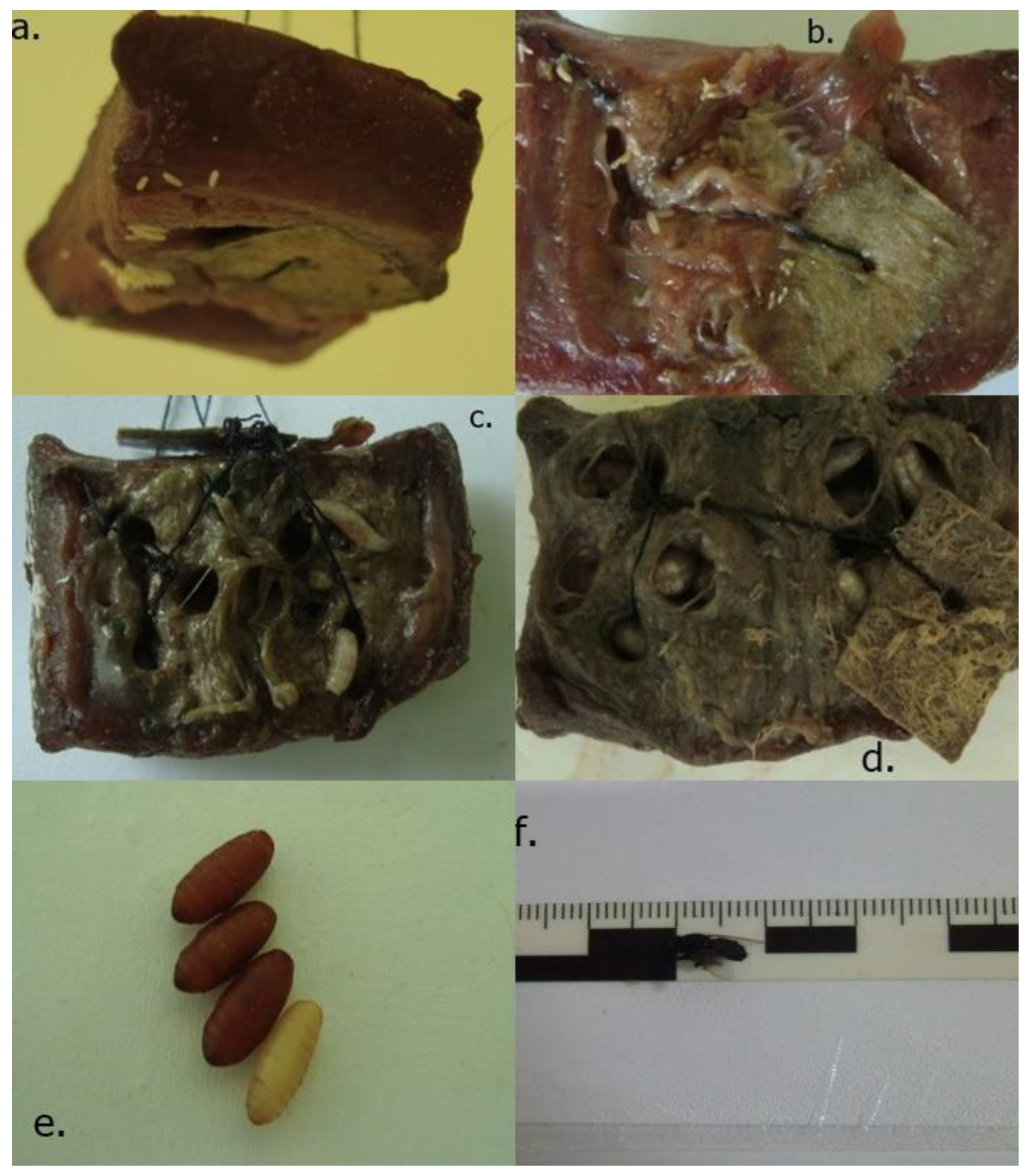
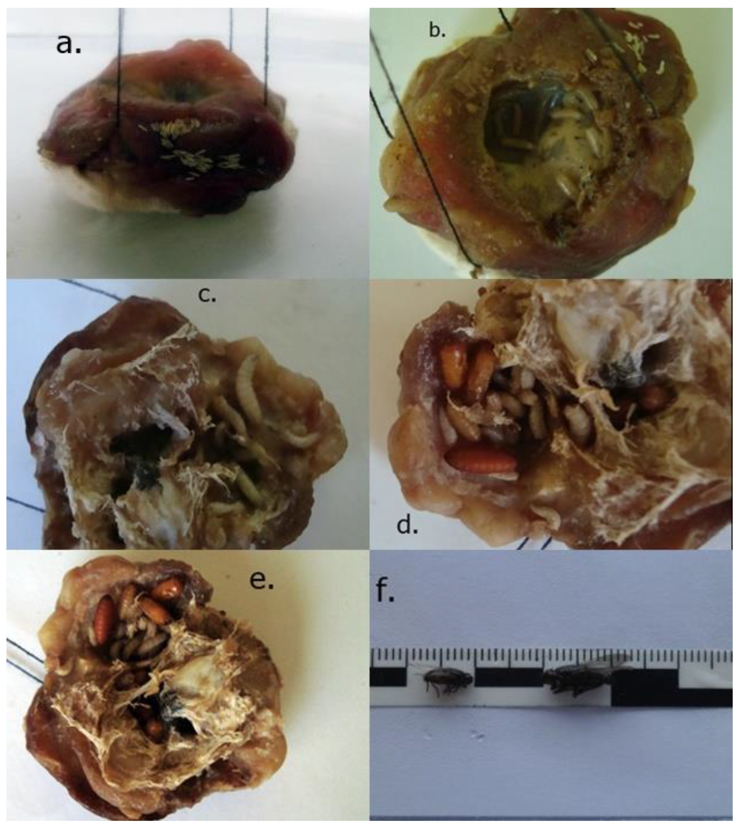
The analysis of the material observed has shown that the insect used for colonization preferred laying eggs on the softer and more humid surfaces of the organs [40,41,42,43]. The larvae developed by executing perforations in substratum while feeding. In the case of samples from the tongue substratum, development in early stages took place in the middle part of the sample in the cross-section, after which the larvae migrated towards the surface of the sample. In the case of samples from the eye, larvae in all developmental stages used the interior of the eye to develop, not its exterior (Figure 2 and Figure 3).
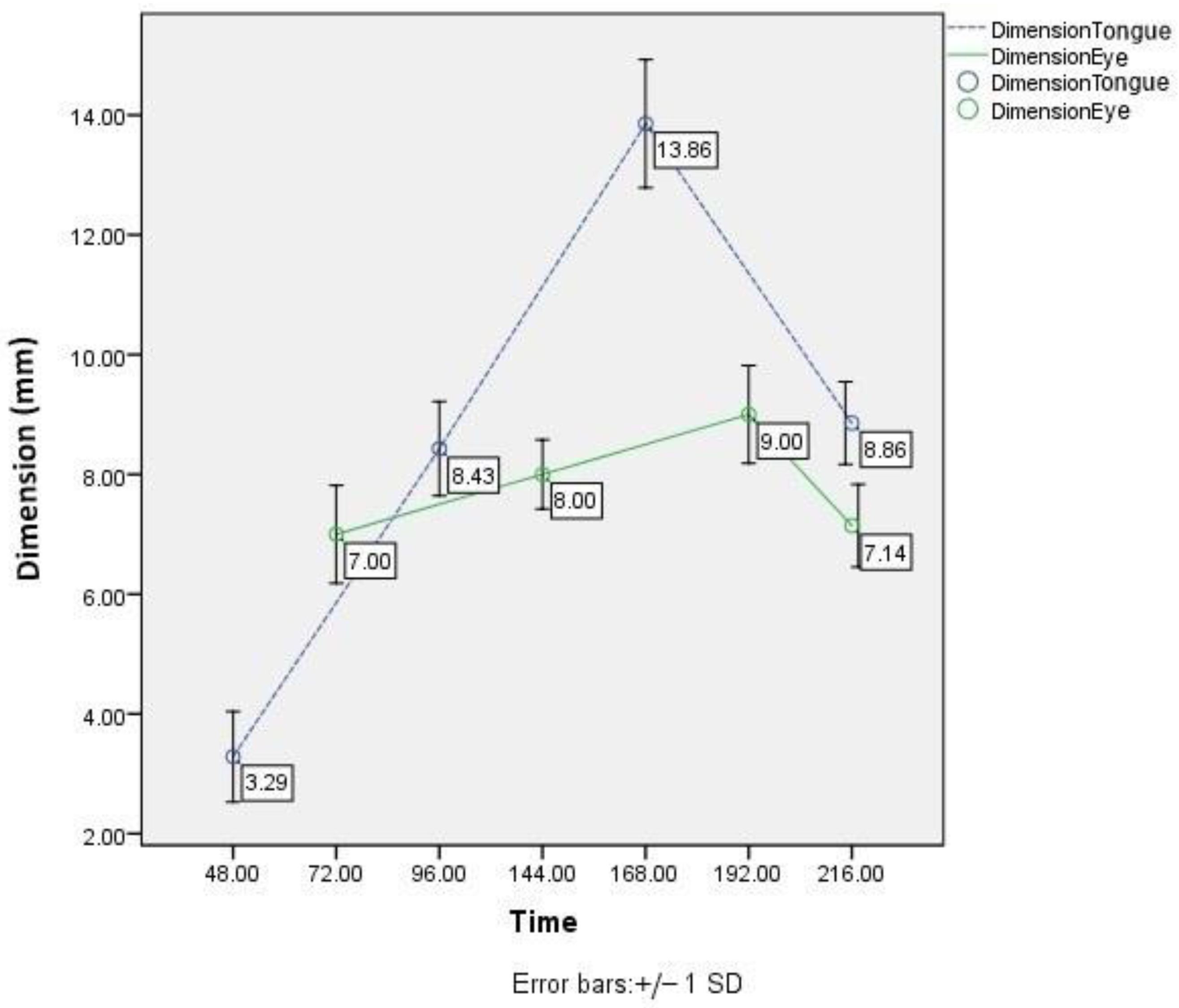
The acceleration of development in larvae stages depending on substratum is characterized by differences in terms of time, in favor of the tongue substratum during the first three stages, except the pupa stage, the difference being: stage I, 24 h; stage II, 48 h; and stage III, 24 h (Table 1 and Table 2). Using these data and comparing them to the literature, we can conclude that the pig tongue substratum is more favorable for the development of Calliphora vicina because it ensures a more nutritious content. Statistical differences of the average sizes on each substrate in each stage of the development cycle in Calliphora vicina are shown in Figure 4.
The first larval stage in the nutritionally richer substrate, i.e., on the pig’s tongue, begins after approximately 48 h, while in the case of the pig’s eyes, it starts slower, after 72 h. It should be noted that the actual development time compared to the time when the larval measurements were made varied by +/− 2–3 h.
From the analysis of the variability in the body length of Calliphora vicina in metamorphoses, where the variables are the development duration and the substratum type, we can notice that the larvae stage II and III on pig tongue have much greater dimensions compared to the same stages of development on pig eye. The variability in the body length of the ten individuals measured for each stage is given by the different structural components of the studied organs and the different approaches used by the larvae in the feeding process.

3.2. Pig Tongue and Eye

Although the Erlenmeyer glasses were covered with gauze, the pupae formed on the tongue were parasitized by a hornet species. Hornets are part of the Pteromalidae family, specifically the species Nasonia vitripennis [44]. Along with other species from the same family, Nasonia vitripennis Walker (Hymenoptera: Pteromalidae) is used for the biological control of synanthrope flies. However, reporting this specificity within the forensic expertise is harmful because it can compromise the correct estimation of the post-mortem interval [45].
They lay eggs on fly pupa areas, and their larvae feed on them [46,47,48]. It is known from the literature [49,50] that parasitoid species have a series of strategies in finding their host and laying eggs. The selection process takes place in several stages and ensures that the parasitoid offspring can reach maturity. Among the selection criteria, we can mention: response to olfactory cues, aspects of locomotor activity, and behavioral response to the host. The number of infected pupae was small (2 pupae out of 16); in the pupae developed on pig tongue, we identified two Nasonia vitripennis individuals (Figure 5).

4. Conclusions

By referring to the previous results found in the specialized literature which indicated that no difference is noted in terms of the duration of the specific stages of the development cycle on all the substrates studied (respectively, camel meat, goat meat, and fish), in the present study, we can conclude by pointing out some differences, namely, that the onset from stage I, in similar environmental conditions, was different. At the same time, from the data obtained, we can presume that pig’s tongue is more favorable for the development of the species Calliphora vicina, presumably due to its structure, which provides the different types of tissues necessary for all stages of larval development. In the field of forensic entomology, necrophagous Diptera such as Calliphora vicina are important in determining the postmortem interval. The duration of the development cycle in the species Calliphora vicina under laboratory-controlled conditions at a constant temperature of 25 degrees Celsius on the substrates used for experimental purposes was a minimum of 10 days (on pig’s tongue) and a maximum of 11 days (on pig’s eyes). In the study, we wanted to use pig liver as a nutrient substrate, too, but the experiment failed multiple times, with either the adult fly not laying eggs, or, after the first-stage, all larvae dying and thus the development process being interrupted. Regarding the two substrates used, it can be said that the initial development on the pig eye substrate occurs at a longer time interval (after 72 h) compared to the larval stage I on the pig tongue substrate, which starts after 48 h. However, if we analyze their development from the perspective of the larval size in the case of stage I development, a larger average size is noted in the case of the pig eye developmental substrate (7.00 mm) compared to the pig tongue substrate, where the average size reaches 3.29 mm.
The analysis of the larvae on another substratum is suggested based on the data obtained because larval stage I is characterized by smaller dimensions, nearly half the size, on the substratum with higher levels of nutrients. We consider the abovementioned comparison necessary because if the estimation of the PMI is determined according to the stages and dimensions of the larvae, taking into consideration the larvae only from one type of substratum, its accuracy will be altered by significant errors.
We can conclude from the study that the substrate and its biochemical composition directly influence the larval development and the size of the larvae at each stage of the development cycle, so this aspect is important to take into account as it may compromise the calculation of the post-mortem interval in forensic cases. The obtained values indicate that a high-calorie diet (and an increased protein content) has an effect on the larval size, causing reduced sizes in the first two stages of development (Stage I—3.29 mm; Stage II—8.43 mm), while in the third stage, the size on the highly nutritious substrate (pig’s tongue) is much larger (13.85 mm) than on the substrate with a low nutrient supply (pig’s eye), where the sizes in the early stages of development were larger (Stage I—7.00 mm; Stage II—8.00 mm), and in stage III of development, the size was, on average, only 9.00 mm.

Author Contributions

Conceptualization, C.M., D.B.I., V.S., and I.S.; methodology, I.S. and V.S.; formal analysis, C.M., D.B.I., I.S., V.V., and V.S.; data curation, D.B.I. and N.C.F.; writing—original draft preparation, C.M., V.S., V.V., and N.C.F.; writing—review and editing, C.M., D.B.I., V.V., and I.S.; data acquisition, C.M., D.B.I., V.V., and I.S.; C.M., I.S., D.B.I., N.C.F., V.V., and V.S. are equally the main authors of this paper. All authors have read and agreed to the published version of the manuscript.

Funding

Authors are thankful to Romanian Ministry of Research, Innovation and Digitization, within Program 1—Development of the national RD system, Subprogram 1.2—Institutional Performance—RDI excellence funding projects, Contract no.11PFE/30.12.2021, for financial support.

Institutional Review Board Statement

Not applicable.

Informed Consent Statement

Not applicable.

Data Availability Statement

All required data are provided in the manuscript.

Conflicts of Interest

The authors declare no conflict of interest.

References

  1. Matei, G. Importanța entomologiei în investigația criminalistică. Rev. Română Crim. 2005, 6, 14–16. [Google Scholar]
  2. Gennard, E.D. Forensic Entomology: An Introduction; Wiley and Sons: West Sussex, UK, 2007. [Google Scholar]
  3. Gunn, A. Essential Forensic Biology, 2nd ed.; Wiley and Sons: Liverpool, UK, 2009. [Google Scholar]
  4. Thompson, C.R.; Brogan, R.S.; Scheifele, L.Z.; Rivers, D.B. Bacterial interactions with necrophagous flies. Ann. Entomol. Soc. Am. 2013, 106, 799–808. [Google Scholar] [CrossRef]
  5. Leccese, A.M. Insects as forensic indicators: Methodological aspects. Anil Aggarwal’s Internet J. Forensic Med. Toxicol. 2004, 5, 26–32. [Google Scholar]
  6. Crooks, E.R.; Bulling, M.T.; Barnes, K.M. Microbial effects on the development of forensically important blow fly species. Forensic Sci. Int. 2016, 266, 185–190. [Google Scholar] [CrossRef] [Green Version]
  7. Amendt, J.; Richards, C.S.; Campobasso, C.P.; Zehner, R.; Hall, M.J.R. Forensic entomology: Applications and limitations. Forensic Sci. Med. Pathol. 2011, 7, 379–392. [Google Scholar] [CrossRef] [PubMed]
  8. Hall, M.J.R. The Relationship between Research and Casework in Forensic Entomology. Insects 2021, 12, 174. [Google Scholar] [CrossRef]
  9. Rodriguez, W.C.; Bass, W.M. Insect Activity and its Relationship to Decay Rates of Human Cadavers in East Tennessee. J. Forensic Sci. 1983, 28, 423–432. [Google Scholar] [CrossRef] [Green Version]
  10. Greenberg, B. Flies as forensic indicators. J. Med. Entomol. 1991, 28, 565–577. [Google Scholar] [CrossRef]
  11. Vanin, S.; Gherardi, M.; Bugelli, V.; Di Paolo, M. Insects found on a human cadaver in central Italy including the blowfly Calliphora loewi (Diptera, Calliphoridae), a new species of forensic interest. Forensic Sci. Int. 2011, 207, e30–e33. [Google Scholar] [CrossRef]
  12. George, K.A.; Archer, M.S.; Toop, T. Abiotic environmental factors influencing blowfly colonization patterns in the field. Forensic Sci. Int. 2013, 229, 100–107. [Google Scholar] [CrossRef]
  13. Tomberlin, J.K.; Benbow, M.E. Forensic Entomology; CRC Press: Boca Raton, FL, USA; Taylor & Francis Group: Milton Park, UK, 2015. [Google Scholar]
  14. Barnes, K.M.; Grace, K.A.; Bulling, M. Nocturnal Oviposition Behavior of Forensically Important Diptera in Central England. J. Forensic Sci. 2015, 60, 1601–1604. [Google Scholar] [CrossRef] [PubMed] [Green Version]
  15. Verma, K.; Reject, P. Assessment of Post Mortem Interval, (PMI) from Forensic Entomotoxicological Studies of Larvae and Flies. Entomol. Ornithol. Herpetol. 2013, 2, 104–108. [Google Scholar]
  16. Kirkpatrick, R.S.; Olson, J.K. Nocturnal light and temperature influences on necrophagous, carrion-associating blowfly species (Diptera: Calliphoridae) of forensic importance in Central Texas. Southwest. Entomol. 2007, 32, 1601–1604. [Google Scholar] [CrossRef]
  17. Tomberlin, J.K.; Crippen, T.L.; Tarone, A.M.; Singh, B.; Adams, K.; Rezenom, Y.H.; Benbow, M.E.; Flores, M.; Longnecker, M.; Pechal, J.L.; et al. Interkingdom responses of flies to bacteria mediated by fly physiology and bacterial quorum sensing. Anim. Behav. 2012, 84, 1449–1456. [Google Scholar] [CrossRef]
  18. Richards, C.S.; Rowlinson, C.C.; Cuttiford, L.; Grimsley, R.; Hall, M.J.R. Decomposed Liver has a significantly adverse effect on the development rate of the blowfly Calliphora vicina. Int. J. Leg. Med. 2013, 127, 259–262. [Google Scholar] [CrossRef] [PubMed] [Green Version]
  19. Metcalf, J.L.; Xu, Z.Z.; Weiss, S.; Lax, S.; Van Treuren, W.; Hyde, E.R.; Song, S.J.; Amir, A.; Larsen, P.; Sangwan, N.; et al. Microbial community assembly and metabolic function during mammalian corpse decomposition. Science 2016, 351, 158–162. [Google Scholar] [CrossRef] [PubMed] [Green Version]
  20. Somerville, D.A. The Normal Flora of the Skin in Different Age Groups. Br. J. Dermatol. 1969, 81, 248–258. [Google Scholar] [CrossRef]
  21. Hau, T.C.; Hamzah, N.H.; Lian, H.H. Decomposition Process and Post Mortem Changes: Review. Sains Malays. 2014, 43, 1873–1882. [Google Scholar]
  22. Metcalf, J.L.; Parfrey, L.W.; Gonzalez, A.; Lauber, C.L.; Knights, D.; Ackermann, G.; Humphrey, G.C.; Gebert, M.J.; Van Treuren, W.; Berg-Lyons, D.; et al. A microbial clock provides an accurate estimate of the postmortem interval in a mouse model system. eLife 2013, 2, e01104. [Google Scholar] [CrossRef]
  23. Carter, D.O.; Yellowlees, D.; Tibbett, M. Temperature affects microbial decomposition of cadavers (Rattus rattus) in contrasting soils. Appl. Soil Ecol. 2008, 40, 129–137. [Google Scholar] [CrossRef] [Green Version]
  24. Janaway, R.C.; Percival, S.L.; Wilson, A.S. Decomposition of Human Remains. In Microbiology and Aging; Percival, S.L., Ed.; Springer: New York, NY, USA, 2009; pp. 313–334. [Google Scholar]
  25. Carter, D.O.; Metcalf, J.A.; Bibat, A.; Knight, R. Seasonal variation of post-mortem microbial communities. Forensic Sci. Med. Pathol. 2015, 11, 202–207. [Google Scholar] [CrossRef] [PubMed]
  26. Crisan, A.; Muresan, D. Clasa Insecte. In Manual de Entomologie Generală; Presa Universitara Clujană: Cluj-Napoca, Romania, 1999. [Google Scholar]
  27. Lehrer, Z.A. Fauna Republicii Socialiste Romania-Insecta: Diptera, Familia; Academiei Republicii Socialiste România: Bucuresti, Romania, 1972; Volume XI, p. 12. [Google Scholar]
  28. Iwasaki, S. Evolution of the structure and function of the vertebrate tongue. J. Anat. 2002, 201, 1–13. [Google Scholar] [CrossRef] [PubMed]
  29. Coțofan, V.; Hrițcu, V. Anatomia Animalelor Domestice. In Organologie; Orizonturi Universitare: Timișoara, Romania, 2007; Volume II, pp. 77–82. [Google Scholar]
  30. Pastea, E.; Cotofan, V.; Chitescu, S.; Miclea, M.; Cornelia, N.; Niculescu, V.; Radu, C.; Popovici, I.; Palicica, R. Anatomia Comparata o Animalelor Domestice; Didactica si Pedagocica: Bucuresti, Romania, 1985; Volume I, pp. 251–253. [Google Scholar]
  31. Donovan, S.E.; Hall, M.J.R.; Turner, B.D.; Moncrieff, C.B. Larval growth rates of the blowfly, Calliphora vicina, over a range of temperatures. Med. Vet. Entomol. 2006, 20, 106–114. [Google Scholar] [CrossRef] [PubMed]
  32. Hadjer-Kounouz, S.; Kamel, L. Development of Calliphora vicina (Robineau-Desvoid)(Diptera: Calliphoridae) under dif-ferent biotic and abiotic conditions. J. Entomol. Zool. Stud. 2017, 5, 683–691. [Google Scholar]
  33. Kaneshrajah, G.; Turner, B. Calliphora vicina larvae grow at different rates on different body tissues. Int. J. Leg. Med. 2004, 118, 242–244. [Google Scholar] [CrossRef]
  34. Bernhardt, V.; Schomerus, C.; Verhoff, M.A.; Amendt, J. Of pigs and men—Comparing the development of Calliphora vicina (Diptera: Calliphoridae) on human and porcine tissue. Int. J. Leg. Med. 2017, 131, 847–853. [Google Scholar] [CrossRef]
  35. Day, D.M.; Wallman, J.F. Influence of Substrate Tissue Type on Larval Growth in Calliphora augur and Lucilia cuprina (Diptera: Calliphoridae). J. Forensic Sci. 2006, 51, 657–663. [Google Scholar] [CrossRef]
  36. Niederegger, S.; Pastuszek, J.G. Preliminary studies of the influence of fluctuating temperatures on the development of various forensically relevant flies. Forensic Sci. Int. 2010, 199, 72–78. [Google Scholar] [CrossRef]
  37. Matuszewski, S.; Szafałowicz, M.; Grzywacz, A. Temperature-dependent appearance of forensically useful flies on carcasses. Int. J. Leg. Med. 2013, 128, 1013–1020. [Google Scholar] [CrossRef] [Green Version]
  38. Defilippo, F.; Bonilauri, M.S.P.; Dottori, M. Effect of Temperature on Six Different Developmental Landmarks within the Pupal Stage of the Forensically Important Blowfly Calliphora vicina (Robineau-Devoidy) (Diptera: Calliphoridae). Forensic Sci. Int. 2013, 58, 1554–1557. [Google Scholar]
  39. Priscilla, H.S. The spectral sensitivity of Calliphora maggots. J. Exp. Biol. 1961, 38, 237–248. [Google Scholar]
  40. Singh, D.; Bala, M. Studies on larval dispersal in two species of blowflies (Diptera: Calliphoridae). J. Forensic Res. 2010, 102, 2. [Google Scholar]
  41. Davies, L. Lifetime reproductive output of Calliphora vicina and Lucilia sericata in outdoor caged and field populations; flight vs. egg production? Med. Vet. Entomol. 2006, 20, 453–458. [Google Scholar] [CrossRef] [PubMed]
  42. Salanitro, L.B.; Massaccesi, A.C.; Urbisaglia, S.; Peria, M.E.; Centeno, N.D.; Chirino, M.G. Calliphora vicina (Diptera: Calliphoridae): Growth rates, body length differences, and implications for the minimum post-mortem interval estimation. Rev. Soc. Entomol. Argent. 2022, 81, 39–48. [Google Scholar] [CrossRef]
  43. Clark, K.; Evans, L.; Wall, R. Growth rates of the blowfly, Lucilia sericata, on different body tissues. Forensic Sci. Int. 2006, 156, 145–149. [Google Scholar] [CrossRef] [PubMed]
  44. Day, D.M.; Wallman, J.F. A comparison of frozen/thawed and fresh food substrates in development of Calliphora augur (Diptera: Calliphoridae) larvae. Int. J. Leg. Med. 2006, 120, 391–394. [Google Scholar] [CrossRef]
  45. Jones, T.H.; Turner, B.D. The effect of host spatial distribution on patterns of parasitism by Nasonia vitripennis. Èntomol. Exp. Appl. 1987, 44, 169–175. [Google Scholar] [CrossRef]
  46. King, P.E.; Askew, R.R.; Sanger, C. The detection of parasitized host by male of Nasonia vitripennis (Walker) (Hymenoptera: Pteromalidae) and some possible implications. Physiol. Entomol. 1969, 44, 85–90. [Google Scholar]
  47. Vinogradova, E.B.; Zinovjeva, K.B. Maternal induction on larval diapauses in the blowefly, Calliphora vicina. J. Insect Physiol. 1972, 18, 2401–2409. [Google Scholar] [CrossRef]
  48. Kalyanaraman, D.; Gadau, J.; Lammers, M. The generalist parasitoid Nasonia vitripennis shows more behavioural plasticity in host preference than its three specialist sister species. Ethology 2021, 127, 964–978. [Google Scholar] [CrossRef]
  49. Bates, L.N.; Wescott, D.J. Comparison of decomposition rates between autopsied and non-autopsied human remains. Forensic Sci. Int. 2016, 261, 93–100. [Google Scholar] [CrossRef] [PubMed]
  50. Frederickx, C.; Dekeirsschieter, J.; Verheggen, F.J.; Haubruge, E. Depth and type of substrate influence the ability of Nasonia vitripennis to locate a host. J. Insect Sci. 2014, 14, 58. [Google Scholar] [CrossRef] [PubMed]
Figure 1. Erlenmeyer glasses with biological samples.
Applsci 12 10907 g001
Figure 2. Developmental stages, Calliphora vicina, pig tongue: (a)—eggs; (b)—larva stage I; (c)—larva stage II; (d)—larva stage III; (e)—pupa; (f)—juvenile.
Applsci 12 10907 g002
Figure 3. Developmental stages, Calliphora vicina, pig eye: (a)—eggs; (b)—larva stage I; (c)—larva stage II; (d)—larva stage III; (e)—pupa; (f)—juvenile.
Applsci 12 10907 g003
Figure 4. Developmental stages, Calliphora vicina, variation in the body length.
Applsci 12 10907 g004
Figure 5. Nasonia vitripennis: (a)—adult (OM 100×); (b)—adult and millimetric scale.
Applsci 12 10907 g005
Table 1. Developmental stages, Calliphora vicina (temperature, time, and body length), pig tongue, p ≤ 0.05, survival rates 87.5 %.
Developmental StageTemperature (°C)Time (Hours)Dimensions
Average (mm)
Standard Deviation95% Confidence Interval between Limits
Inferior Limit (mm)Superior Limit (mm)
Eggs25---
Larva stage I25483.290.752.593.98
Larva stage II25968.430.787.709.16
Larva stage III2516813.851.0612.8614.84
Pupa252168.850.698.219.49
Fly25240Different-
Table 2. Developmental stages, Calliphora vicina (temperature, time, and body length), pig eye, p ≤ 0.05, survival rates 100%.
Developmental StageTemperature (°C)Time (Hours)Dimensions
Average (mm)
Standard Deviation95% Confidence Interval between Limits
Inferior Limit (mm)Superior Limit (mm)
Eggs25---
Larva stage I25727.000.816.247.76
Larva stage II251448.000.577.478.53
Larva stage III251929.000.818.249.76
Pupa 252167.140.696.507.78
Fly25264Different-
Publisher’s Note: MDPI stays neutral with regard to jurisdictional claims in published maps and institutional affiliations.