Article

Celector®: An Innovative Technology for Quality Control of Living Cells

1
Stem Sel Srl, 40127 Bologna, Italy
2
Department of Chemistry “G. Ciamician”, University of Bologna, 40126 Bologna, Italy
3
Department of Experimental, Diagnostic and Specialty Medicine, University of Bologna, 40126 Bologna, Italy
*
Authors to whom correspondence should be addressed.
Appl. Sci. 2022, 12(19), 9967; https://doi.org/10.3390/app12199967
Submission received: 10 August 2022 / Revised: 23 September 2022 / Accepted: 28 September 2022 / Published: 4 October 2022

Abstract

:
Among the in vitro and ex vivo models used to study human cancer biology, cancer cell lines are widely utilized. The standardization of a correct tumor model including the stage of in vitro testing would allow for the development of new high-efficiency drug systems. The poor correlation between preclinical in vitro and in vivo data and clinical trials is still an open issue, hence the need for new systems for the quality control (QC) of these cell products. In this work, we present a new technology, Celector®, capable of the label-free analysis and separation of cells based on their physical characteristics with full preservation of their native properties. Two types of cancer cell lines were used: HL60 as cells growing in suspension and SW620 as adherent cells. Cell lines in general show a growth variability depending on the passage and method of culture. Celector® highlights physical differences that can be correlated to cell viability. This work demonstrates the use of Celector® as an analytical platform for the QC of cells used for drug screening, with fundamental improvement of preclinical tests. Cells with a stable doubling time under analysis can be collected and used as standardized systems for high-quality drug monitoring.

1. Introduction

Cell therapy is one of the innovative medical approaches used to treat disorders from osteo-articular reconstructive surgery and tissue engineering to cancer. Advanced therapy medicinal products (ATMPs) are cells and tissue products that are considered to be a new type of drug. In particular, stem cells can be differentiated into specialized cell types to substitute damaged tissue, to damp immunoreactions, or to release factors to stimulate the regenerative process as a cell-free approach. All of these applications require good standard conditions for cell isolation and expansion [1,2,3]. In the advent of personalized cell therapies, manufacturers need to isolate, manipulate, and expand patient-derived cells in vitro, thus requiring stringent and especially independent quality control measures. The most important requirements for a good manufacture practice (GMP) standard for ATMP production are sterility, identity, purity, viability, potency, and reproducibility (Agency, European Medicine (2007), Committee for Human Medicinal Products (CHMP): Guideline on Human Cell-Based Medicinal Products). Stem cells are a heterogeneous population that can differ depending on the origin and procurement of the starting material; thus, it is critical to determine the yield and identity/purity of the final cell population. The use of appropriate markers and measures of cell population heterogeneity are nowadays considered fundamental to standardize the isolation and quality control (QC) of cell products. Moreover, once cells are expanded to an adequate number, vials are cryopreserved and stored until their use for cell-based applications [4]. Cell products need to be validated before cryopreservation and their quality must be checked after thawing to prove they have maintained viability, purity, and homogeneity [5]. The purity of cell products should minimize the presence of undesirable contaminants such as differentiated or senescent cells, non-cellular impurities, and cell debris that are not required and could impair the function of the cell product. The viability of the cell product is an important aspect to take into consideration for the efficacy and integrity of cell-based products. Viable cells are directly correlated with their biological activity, and the percentage of living cells should be around 70%. Regardless, dead cells should be cleared from the solution to improve the quality of the final cell product.
A standard operation procedure (SOP) supports an increase in the manufacture process robustness to assure the reproducibility of the cell product, including between different donors. Automation is a strategy to maximize reproducibility, reduce costs, and avoid labor-intensive procedures, which can generate errors [6]. Automated cell culture systems are available and QC platforms, especially miniaturized QCs, are used in cell factories [7]. These technologies are very interesting because they simultaneously reduce the manual steps, and thus also the costs, generated by the use of media and reagents and specific requirements. However, these technologies mostly involve single-cell analysis, and they lack the ability to control the homo/heterogeneity of biological samples.
In addition to stem cells and other primary cells for ATMP, cancer cells are widely used to study human cancer biology. Patient tumor-derived cells are an interesting cell source; however, they represent a fraction of the tumor itself at a specific stage, usually an advanced one. To investigate the initiation and progression of tumors, cancer cell lines have been produced. Cancer cell lines are tumor-derived cells which are used worldwide as in vitro platforms to study cancer mechanisms, enable drug discovery, and test new innovative therapies. These cells have the capacity for exponential growth and can maintain their morphology and characteristics. However, during duplication, mutations can occur, as well as a senescence phenomenon. QC systems to report the health state of cells are time-consuming and dependent on the ability of operators. Differences in the culture medium, percentage of serum, growing cell density, type of plastic, training of operators, and much more can affect the quality and vitality of cells, in addition to the risk of microbial contamination. Proliferative capacity is one of the characteristics measured to assess the cells’ quality and vitality. However, new quality systems need to be implemented to fulfil the lack of QC for these types of cells.
Analytical methods applied for QC aspects, such as chromatography-like technology for small molecules, could be a powerful tool for QC in ATMPs. Microfluidic systems have been used lately for bio-analytes and single-cell analysis, and among them, field-flow fractionation (FFF) has proven its capacity to analyze, discriminate, and separate a wide size range of biological samples based on their native physical characteristics with high resolution and throughput, and to correlate physical properties to biological cell activity [8,9,10,11,12,13,14,15,16]. Among FFF techniques, the gravitational field-flow fractionation (GrFFF) variant is the most employed for whole-cell analysis with advantages related to the simple instrumental set-up of the separative device which allows its easy decontamination and guarantees sterile and clean conditions. In GrFFF, the separation occurs in a capillary channel due to the combined action of the flow of the mobile phase and Earth’s gravity field [17,18]. Despite the interesting potentialities of GrFFF, the technique shows some limitations, particularly related to the low throughput for adherent cells. We developed a novel method, non-equilibrium Earth-gravity-assisted dynamic fractionation (NEEGA-DF) to work with adherent cells to avoid their attachment to capillary walls [19]. Cells with different physical characteristics acquire different velocities inside the capillary channel and elute at different times. An automated instrument was developed to implement the NEEGA-DF method (Celector®, Stem Sel srl, Bologna, Italy). We proved that different subpopulations could be defined in amniotic fluid stem cells, and we could select the most potent cells and identify the mesenchymal stem cell component in fresh bone marrow samples [20,21]. In this work, we demonstrated for the first time the use of Celector® as a quality control system for cell culture procedure standardization in order to monitor changes in cell behavior during in vitro culturing. The instrument is equipped with a micro-camera for cell detection, and specifically designed software for image acquisition, post-processing, and data analysis was employed. The output of the instrument is a multiparametric fractogram representing number, size, and shape of the eluted cells as a function of fractionation time, and it is the fingerprint of the cell sample. This instrument possesses two functions, as a QC device and as a sorter to enrich the homogenous target cell population.
We analyzed two types of cell lines, the promyeloblasts HL60 and the colorectal cancer cells SW620, as models of in vitro expansion. Both cell lines were monitored for their doubling time, and each passage was analyzed by the instrument to obtain the cell viability and dimensions, the presence of dead cells, and the evolution of possible heterogeneity of the cell population.

2. Materials and Methods

2.1. In Vitro Cell Culture

As a quality control system, we focused the analysis on two tumor cell lines, one growing in suspension and the second adherent to a plastic surface.
HL60 are promyeloblasts, and they can be expanded in vitro with a cell density of 100,000 cells/mL in growth medium consisted of RPMI, 10% FBS, and 1% penicillin and streptomycin. The SW620 cell line comprises colorectal cancer cells which proliferate on plastic surfaces. They grow in Iscove MEM, 10% FBS, and 1% PS at a cell density of 10,000 cells/cm2(all reagents were from Gibco). Both cell lines were kept in a humidified incubator at 37 °C and 5% CO2 and expanded to obtain the correct number of cells to be analyzed.
In parallel, both cell lines were cryopreserved using a freezing solution constituted of 10% DMSO and 90% of FBS at a concentration of 700,000 cells/100 µL (all reagents were from Gibco).

2.2. Instrumentation

2.2.1. Fractionation Principle and Procedure

Cell samples were fractionated through Celector®, an instrument consisting of a fluidic system, a biocompatible capillary separation device, a micro-camera detector placed at the exit of the separation channel (MER-U3 camera, DAHENG IMAGING, Beijing, China) to monitor the elution process, and a fraction collector connected to the separation device. The instrument was placed inside a laminar flow cabinet to provide sterile working conditions. The system generates a recorded plot of the eluted cell number as a function of time (fractogram) and for frame acquisition.
The separation is obtained in the capillary device (channel) with a rectangular cross-section made of inert and biocompatible plastic material of 40 cm length, 4 cm width, and 300 µm thickness. A physiological buffer is used as the mobile phase. The cell suspension is injected at a flow rate of 1 mL/min, and during the elution, cells reach a specific position across the channel thickness due to the combined action of Earth’s gravity, acting perpendicular to the flow, and opposing lift forces related to the morphological features of the sample. Due to the laminar flow of the mobile phase, cells at a specific position in the channel acquire well-defined velocities and are therefore eluted at specific times. The fractionation mechanism allows the maintenance of native properties and a high sample recovery. The implementation of an in-flow injection with the absence of stop-flow cell sedimentation, and the use of the elution flow rate values generates hydrodynamic forces that are intense enough to lift and keep cells away from the channel wall, reducing cell adhesion.
The system was decontaminated at first by flushing with a cleaning solution at a 1 mL/min flow rate. Next, the system was washed copiously with sterile, demineralized water at the same flow rate, and then the fractionation system was flushed at 0.5 mL/min with a sterile coating solution to block non-specific interaction sites on the plastic walls of the fluidic system. Finally, a sterile mobile phase was used to fill the system. All solutions were provided by Stem Sel srl.

2.2.2. Optical Analysis

Eluted cells were monitored using a micro-camera detector (MER-U3 camera, DAHENG IMAGING, Beijing, China) that was placed at the outlet of the fractionation channel. The imaging software (Celector Optics, Stem Sel srl, Bologna, Italy) generates a real-time fractogram representing the number of cells versus recorded time. The software imaging data are then post-processed to obtain the geometrical features of the eluted cells as a function of time. In this work, we focused on cell diameter to obtain information on population heterogeneity during cell expansion. These geometrical features were then visualized as curves using dedicated data processing (Stem Sel Analyzer, Stem Sel srl, Bologna, Italy) to obtain the average of all parameters in a selected time interval (cell fraction).

2.3. Cell Analysis and Collection

For every sample, cells were firstly analyzed to obtain a specific fractogram and identify the fraction to collect. Consecutive analyses were run to increase the number of collected cells per fraction. The fractionated cells were centrifuged, counted by erythrosine dye (Sigma, St. Louis, MO, USA), and then plated in an appropriate culture medium for cell expansion. Cells were then monitored for several passages, and the doubling time was calculated using the following formula: DT = hours of culture × LOG(2)/(LOG(final n. cells)-LOG(initial n. cells plated)).
Frozen cells were thawed at room temperature and divided into two equal parts: one part for analysis using Celector, the other washed in PBS before cells were centrifuged and resuspended in PBS and analyzed using Celector at a concentration of 300,000 cells/100 µL. For both conditions, cells were divided and collected into two fractions, the initial minutes of the analysis (void) and the main population. Both fractions were centrifuged, then cells were counted and plated to check metabolic activity using the Alamar blue assay (Invitrogen, Carlsbad, CA, USA).

Immunofluorescence Staining and Flow Cytometry Analysis

Pre- and post- selection cells from HL60 and SW620 were counted and plated onto glass slides for immunofluorescence staining for the proliferative marker Ki67 (Anti-KI-67-Vio515, Miltenyi) at a concentration of 1:50, and F-actin at a concentration of 1:100 in SW620 cells. Briefly, cells were fixed in 4% PFA for 10 min after 5 days of culture post selection, permeabilized with 0.1% triton and 0.1% BSA in PBS for 10 min, blocked for 1 h at RT in 1% BSA in PBS, and then incubated with antibodies which were already labeled. Images were then taken using a fluorescent microscope (Leica), and for Ki67 quantification, staining of the samples was measured using Image J software and then compared to the area of nuclei stained with DAPI to obtain the estimation of its expression.
Part of the collected cells and the control cells were collected and stained for the apoptotic marker Annexin V (BD Pharmingen) and immediately read using a flow cytometer (S3e Cell sorter, Bio-Rad Laboratories, Hercules, CA, USA). Data were analyzed using Flow Jo.

2.4. Celector® Test Procedure

Analyses in Celector® are carried out according to the following procedure.
Cells in adhesion are enzymatically detached, centrifuged at 1200 rpm, resuspended in 1 mL of PBS or culture medium, and counted by erythrosine staining to exclude dead cells; cells in suspension are collected and immediately centrifuged at 1200 rpm, resuspended in 1 mL of PBS, and counted by erythrosine staining to exclude dead cells. The cell suspensions are prepared to obtain a final concentration of 300,000 cells/100 µL, the volume to be injected into the system. In the meantime, the Celector® instrument is set for the experimental procedure. A sterile solution of mobile phase, coating, distilled water, and cleaning solution, distributed by Stem Sel, is placed on the instrument and connected to the fluidics and fractionator. Using the software connected to the instrument (Celector® Mover), sterile conditioning is launched to prepare the system for cell analysis and once the conditioning is completed and on idle mode, the system is ready for cell analysis. Before the analysis starts, Celector® Optics, a software designed by Stem Sel, is switched on and connected, in auto mode setting, to the Celector® Mover. An experiment folder is created with the reference of name of the biological sample to be analyzed and the background image is acquired. Using Celector® Mover, the flow rate, time of analysis, time of collection, and injection volume are set. When the run starts, acquisition of images begins, and the live fractogram appears on the screen. Once the time of analysis ends, the acquisition will automatically end. Consecutive analysis can be performed to collect a higher number of cells per fraction.
The acquired data are post-processed by the Celector® Optics software, and the physical parameters visualized using the Stem Sel analyzer software.

2.5. Statistical Analysis

Statistical analysis was performed using Graph Pad Prism, running the 2way ANOVA test and mean and standard deviation were graphed (*, p < 0.5; **, p < 0.01, ***; p < 0.001; **** p < 0.0001).

3. Results and Discussion

3.1. Characterization of Expanded Cell Line

The cancer cell lines HL60 and SW620 were analyzed using Celector® to monitor their physical composition and the possible modification of their homogeneity. These two cell lines were chosen to observe possible differences in culture expansion because of their adhesion properties: SW620 are adherent cells, small in dimension with a star shape, and they grow in very tight connection with each other and with a slower proliferative rate; HL60 cells grow in suspension with a higher proliferative rate compared to SW620, and differences in morphology and dimension can be observed during passages in culture.
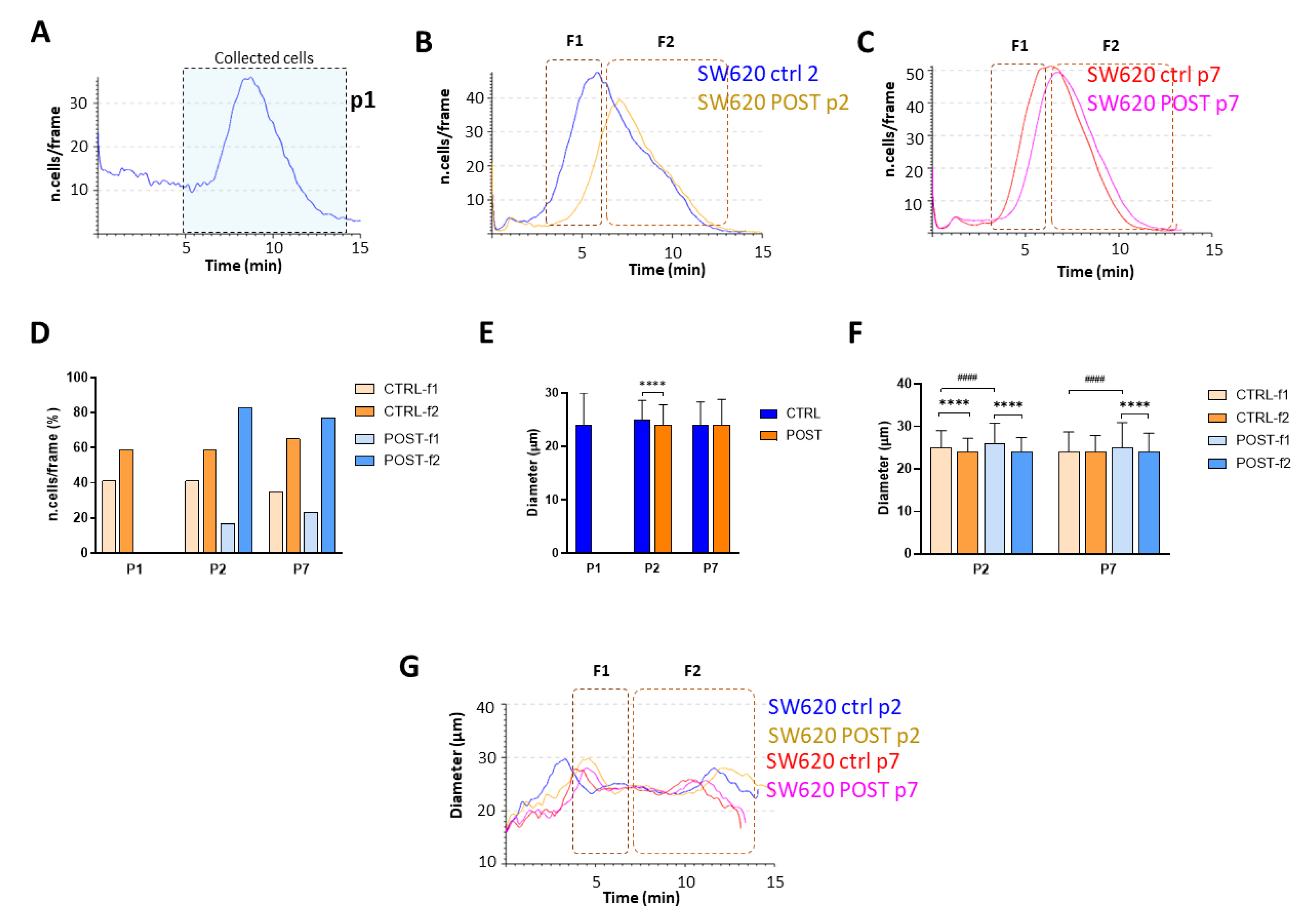
The first run served to obtain the sample fractogram, the fingerprint of the sample, and to decide the time interval of sorting. Successively, further runs were performed to obtain results in triplicate, and one fraction was consecutively collected to obtain the right number of cells. The time interval for HL60 was set between the 4th and 10th minutes of analysis, depleting the living cells from the unwanted material such as cell debris and dead cells (Figure 1A). The curve representing the number of cells as a function of time showed a similar-normal distribution, with the peak in the middle of the curve: 60% of cells eluting in the ascending portion and 40% in the descending portion of the curve. The collected cells (POST) were centrifuged and expanded in vitro following the same culture condition of the control (CTRL), the entire cell population. At every passage in culture, after enzymatic detachment, CTRL- and POST-derived cells were counted and analyzed at the instrument to monitor the fractogram, and successively, their physical characteristics. One passage after the sorting, CTRL and POST cells did not show differences in their fractograms (Figure 1B), but the ratio between the initial eluate compared to the final (F1 vs. F2) (Figure 1D) was reversed: 40% to F1, and 60% of cells were now in the second half, namely F2. This trend increased further after a couple of passages. At p5, 20% of cells were in F1 for the CTRL and 15% for the POST cells (Figure 1D), and the majority in F2. The total time of elution between p2 and p5 persisted. However, at p5, there was a clear shift in the cells’ presence towards the fraction F2 (Figure 1C). This shift denoted a change in the process of in vitro cell expansion. Due to the fact that FFF is based on physical characteristics, we investigated the dimensions of cells that can be obtained by post-processing image analysis. The average of all eluted cells showed a diameter of 25 µm at p1, 26 µm and 27 µm at p2 for CTRL- and POST-cells, respectively, and an even higher enlargement at p5, with the CTRL reaching 30 µm and POST 28 µm (Figure 1E). Splitting the population into the two hypotheticals fractions, cells were slightly larger in F2 at p2, and this difference was intensified at p5 (Figure 1F). The curve representing the average of the diameter, in Figure 1G, represents the cell culture behavior observed in the cell fractogram, showing a similar-normal distribution for cells at p1 and a p2, while the majority of cells in F2 exhibited a higher diameter at p5. Generally, based on FFF principles, larger cells exit at the beginning of the analysis, followed by smaller cells. A HL60 dimensional analysis showed a higher dimension of F2 cells compared to F1. Therefore, the density of the cells could be the parameter to distinguish F1 cells from F2 cells. A deeper investigation of the intracellular composition should be performed to understand the state of the cells, such as cell-cycle phase, mitochondrial activity, and protein content [22,23].
A different behavior was observed when adherent SW620 cells were analyzed during their in vitro expansion. SW620 cells showed a similar-normal distribution as a rather homogenous population (Figure 2A) with an opposite trend compared to HL60 cells: 40% of cells eluted in the first part of the curve, F1, and 60% in the descendant slope, F2 (Figure 2D). Dimensionally, SW620 cells were smaller than HL60, with an average diameter of 24 µm (Figure 2E). The main population was collected in the time interval of 6–14 min of analysis, centrifuged, and then expanded. One passage later, at p2, POST-derived cells showed a higher retention time, with a delay of the fractogram (Figure 2B). This difference was lost at passage 7 in culture (Figure 2C), retrieving their homogeneity. To analyze cell composition at p2, when the population was divided into two equal fractions based on the CTRL fractogram, the CTRL cells maintained the same division: 40% in the F1 and 60% in the F2, whereas the POST cells reversed with 80% of cells in the second fraction (Figure 2D). Dimensionally, POST-derived cells were generally slightly smaller compared to CTRL at p2, but identical at the later passage. For both passages, F1 cells were always bigger compared to F2 cells, proving the general principles of the fractionation method.

3.2. Cell Analysis Post-Selection

Viable cells were counted pre- and post-analysis using the exclusion dye erythrosine, and afterwards through flow cytometry analysis to detect expression of Annexin V, the apoptotic marker.
Before Celector® analysis, HL60 cells showed a viability of 80%, whereas SW620 had a viability of 92% (Figure 3A). After the analysis and sorting procedure, collected cells, excluding the void, were counted and showed a higher viability—with most of the dead cells eliminated in the void, the first two minutes of the run. The viability of HL60 rose to 92%, and of SW620 from 92% to 99% (Figure 3A).
In addition, to prove Celector®’s ability to maintain cell viability, and to enrich the most vital cell population without cell damage, staining for the apoptotic marker Annexin V was performed and confirmed the exclusion dye counting data: the HL60 cell control population possesses 22% apoptotic cells, and the sorted cells only 5% (Figure 3B); SW620, as shown in the fractogram (Figure 3C), did not show an elevated void and the population was mostly alive with a viability of 92%. Annexin V staining showed that the percentage of early apoptotic cells was stable, around 2%, in both conditions.
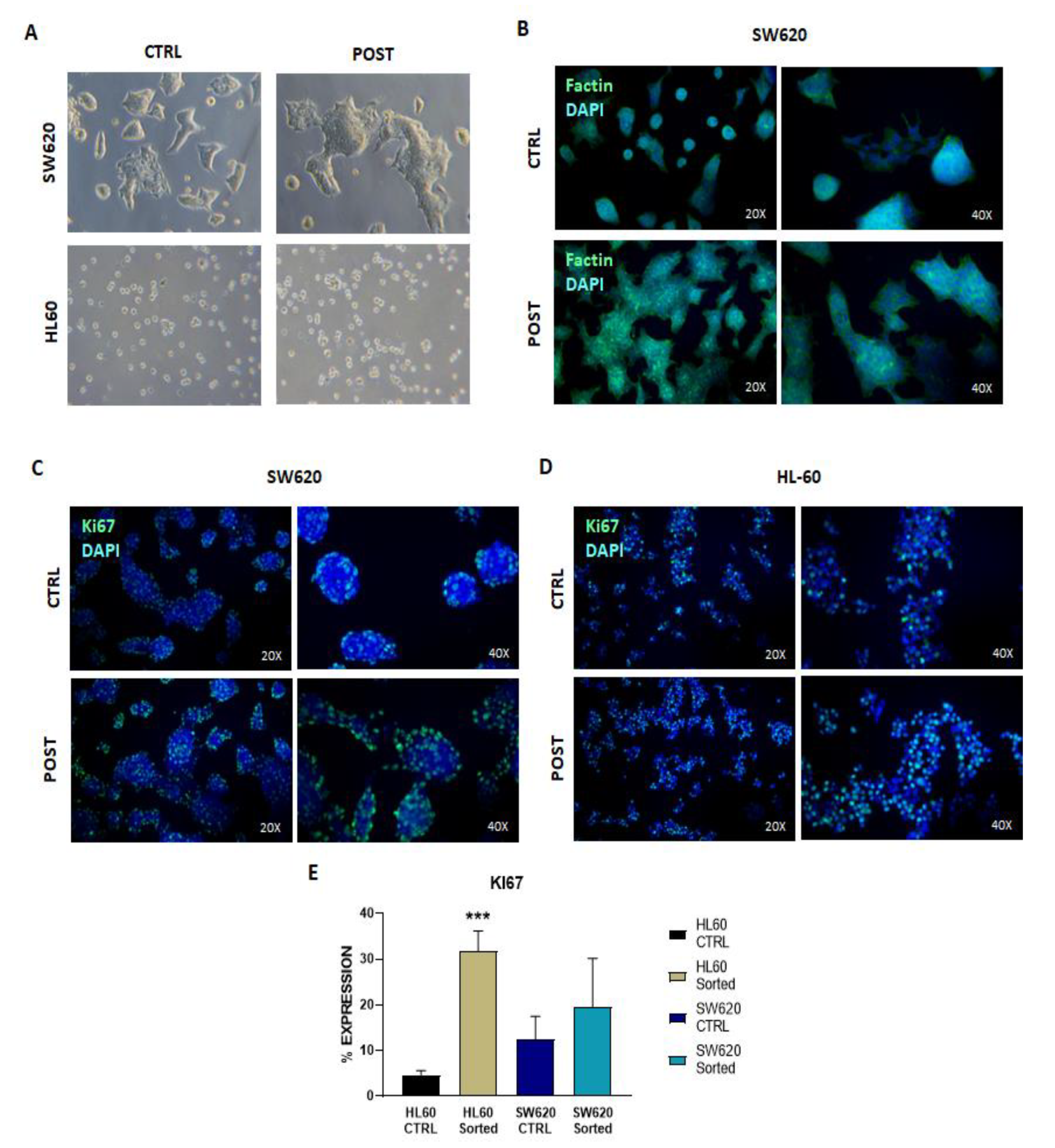
Moreover, to prove the maintenance of morphology and proliferative potential, images were captured via light microscope and immunofluorescence staining for specific markers were performed. SW620 cells showed a similar morphology using the bright-field microscope (Figure 4A) and by the expression of F-actin filaments to the stress cytoskeleton (Figure 4B). We could observe bigger colonies of cells, probably caused by the presence of more viable cells in the selected population compared to the control population due to the removal of the dead cells in the void. HL60 showed the same morphology, having the same ability to form small cell aggregates during culture conditions (Figure 4A).
When the proliferation marker Ki67 was investigated, the staining showed a statistically higher presence of positive nuclei in the HL60 selected cells compared to controls (Figure 4C,D), while for the SW620, even though there is a trend towards a higher percentage of positive nuclei, no difference was seen between the control population and the selected cells (Figure 4B,C). We can define the NEEGA-DF method as a gentle technique that does not alter the morphology or state of the selected cells.

3.3. In Vitro Expansion of Tumor Cell Line

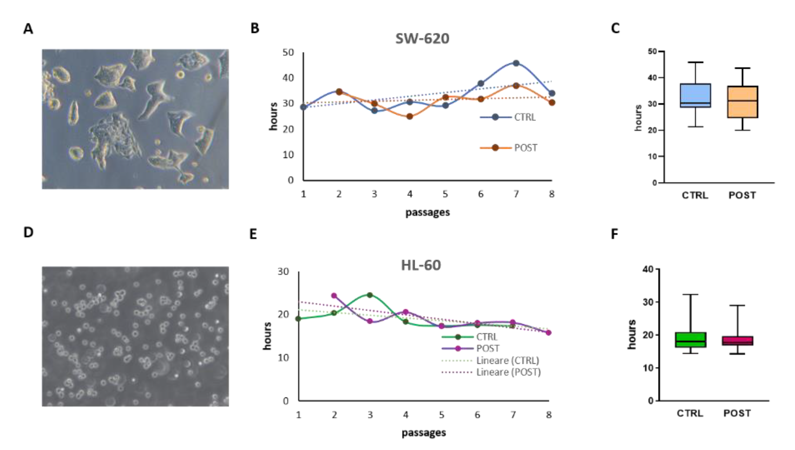
Tumor cell lines are expanded for many passages in culture, and their duplication time can vary depending on the accuracy of the procedure, type of media, and reagents. The in vitro expansion is monitored using the duplication time. SW620 cells are adherent cells which grow in colonies (Figure 5A). The adherent SW620 cell lines were more unstable compared to the HL60 cells (Figure 3B). Even though there is no statistical difference between the control and the collected cells (POST), POST-SW620 cells showed a more stable trend in their duplication time, as shown by the linear tendency. To expand in cell culture, SW620 cells need to be enzymatically detached from plastic surfaces and from each other. This procedure could affect vitality and adhesion characteristics much more than cells growing in suspension such as the HL60 (Figure 5D), which did not show this difference in the duplication time, or rather, showed a stabilization of DT during culture (Figure 3E).

3.4. Analysis of Cell Cryo-Preservation

In laboratories and cell banks, cells are routinely cryo-preserved in several batches as a stock for later purposes. Quality control on thawed cells is fundamental for cell production. We tested the feature of Celector® on frozen SW620 cells. Cells were analyzed using Celector® immediately after thawing, and after the washing step in PBS and centrifugation to remove frozen solution. We monitored the morphological changes of the cells in the freezing solution of 10% DMSO in FBS compared to the cells that were moved to a physiological solution. Thawed cells exit earlier, at the same time as the void (Figure 6A). A fractogram overlay of the two conditions showed the presence of two peaks in the thawed cells, with the first peak between 2 and 6 min of analysis (F1) that was missing in the washed cells, and the second from 6 to 15 min of analysis (F2) (Figure 6B). Even in the washed sample, a high void composed of dead cells and debris is present and can be removed using this separation technique. Since the diameter was very similar along the analysis (Figure 6C), other physical characteristics, such as density and intracellular composition, are involved.
We tested cell viability using an Alamar blue assay on the total population (Ftot), on the collected main peak excluding the void which was separately collected. Ftot and the control population behaved similarly. The void derived from the thawed cells showed a faster growth, whereas very few cells were present in the void after centrifugation and these showed only some reactivity after 3 days (Figure 6D). The presence of live and proliferative cells collected in the void was predictable by the fractogram, which clearly showed the presence of half of the injected cells eluting during the void and in the hypothetical fraction F1. The analysis of thawed cells is very useful when different cryopreservation reagents, methods of freezing, and thawing are tested on cells to define the best protocol to avoid loss and damage to the cells.

4. Conclusions

This work showed the ability of the new instrument Celector®, which exploits FFF principles to analyze cell composition during culture expansion and cryopreservation methods. This protocol could be adopted in lab and cell banks to monitor their cell production processes as a quality control platform to standardize the cell-culture process.

5. Patents

Celector® is based on technology patented in Italy (no. IT1371772, “Method and Device to Separate Totipotent Stem Cells”), in the USA, and in Canada (no. 8263359 US en. CA2649234, “Method and Device to Separate Stem Cells”). Stem Sel® also has an Italian patent (IT1426514, “Device for the Fractionation of Objects and Fractionation Method”, allowed in 2016).

Author Contributions

Conceptualization, S.Z. and B.R.; methodology, B.R., A.Z. and P.R.; investigation, S.Z. and A.M.; data curation, S.Z.; writing—original draft preparation, S.Z.; writing—review and editing, S.Z., B.R., V.M., P.M., V.P., L.B. and F.A.; visualization, S.Z.; supervision, B.R. All authors have read and agreed to the published version of the manuscript.

Funding

This research received no external funding.

Institutional Review Board Statement

Not applicable.

Informed Consent Statement

Not applicable.

Data Availability Statement

Not applicable.

Conflicts of Interest

Andrea Zattoni, Barbara Roda, and Pierluigi Reschiglian are associates of the academic spinoff company Stem Sel Srl (Bologna, Italy). The company mission includes the development and production of novel technologies and methodologies for the separation and characterization of cells and biosamples. All the other authors report no conflict of interest, since nobody has commercial associations that might create a conflict of interest in connection with the submitted manuscript.

References

  1. Cimino, M.; Gonçalves, R.M.; Barrias, C.C.; Martins, M.C.L. Xeno-Free Strategies for Safe Human Mesenchymal Stem/Stromal Cell Expansion: Supplements and Coatings. Stem Cells Int. 2017, 2017, 6597815. [Google Scholar] [CrossRef] [PubMed] [Green Version]
  2. de Peppo, G.M. GMP-Compatible, Xeno-Free Culture of Human Induced Mesenchymal Stem Cells. Methods Mol. Biol. 2021, 2286, 121–129. [Google Scholar] [CrossRef] [PubMed]
  3. Aghayan, H.R.; Payab, M.; Mohamadi-Jahani, F.; Aghayan, S.S.; Larijani, B.; Arjmand, B. GMP-Compliant Production of Human Placenta-Derived Mesenchymal Stem Cells. Methods Mol. Biol. 2021, 2286, 213–225. [Google Scholar] [CrossRef] [PubMed]
  4. Diekmann, U.; Spindler, R.; Wolkers, W.F.; Glasmacher, B.; Müller, T. Cryopreservation and quality control of mouse embryonic feeder cells. Cryobiology 2011, 63, 104–110. [Google Scholar] [CrossRef]
  5. Yuan, Z.; Lourenco Sda, S.; Sage, E.K.; Kolluri, K.K.; Lowdell, M.W.; Janes, S.M. Cryopreservation of human mesenchymal stromal cells expressing TRAIL for human anti-cancer therapy. Cytotherapy 2016, 18, 860–869. [Google Scholar] [CrossRef] [Green Version]
  6. Ornelas-González, A.; González-González, M.; Rito-Palomares, M. Microcarrier-based stem cell bioprocessing: GMP-grade culture challenges and future trends for regenerative medicine. Crit. Rev. Biotechnol. 2021, 41, 1081–1095. [Google Scholar] [CrossRef]
  7. Wartmann, D.; Rothbauer, M.; Kuten, O.; Barresi, C.; Visus, C.; Felzmann, T.; Ertl, P. Automated, Miniaturized, and Integrated Quality Control-on-Chip (QC-on-a-Chip) for Cell-Based Cancer Therapy Applications. Front. Mater. 2015, 2, 60. [Google Scholar] [CrossRef] [Green Version]
  8. Reschiglian, P.; Zattoni, A.; Roda, B.; Casolari, S.; Moon, M.H.; Lee, J.; Jung, J.; Rodmalm, K.; Cenacchi, G. Bacteria sorting by field-flow fractionation. Application to whole-cell Escherichia coil vaccine strains. Anal. Chem. 2002, 74, 4895–4904. [Google Scholar] [CrossRef]
  9. Marassi, V.; Maggio, S.; Battistelli, M.; Stocchi, V.; Zattoni, A.; Reschiglian, P.; Guescini, M.; Roda, B. An ultracentrifugation—hollow-fiber flow field-flow fractionation orthogonal approach for the purification and mapping of extracellular vesicle subtypes. J. Chromatogr. A 2021, 1638, 461861. [Google Scholar] [CrossRef]
  10. Marrazzo, P.; Pizzuti, V.; Zia, S.; Sargenti, A.; Gazzola, D.; Roda, B.; Bonsi, L.; Alviano, F. Microfluidic Tools for Enhanced Characterization of Therapeutic Stem Cells and Prediction of Their Potential Antimicrobial Secretome. Antibiotics 2021, 10, 750. [Google Scholar] [CrossRef]
  11. Marassi, V.; Roda, B.; Casolari, S.; Ortelli, S.; Blosi, M.; Zattoni, A.; Costa, A.L.; Reschiglian, P. Hollow-fiber flow field-flow fractionation and multi-angle light scattering as a new analytical solution for quality control in pharmaceutical nanotechnology. Microchem. J. 2018, 136, 149–156. [Google Scholar] [CrossRef]
  12. Roda, B.; Marassi, V.; Zattoni, A.; Borghi, F.; Anand, R.; Agostoni, V.; Gref, R.; Reschiglian, P.; Monti, S. Flow field-flow fractionation and multi-angle light scattering as a powerful tool for the characterization and stability evaluation of drug-loaded metal-organic framework nanoparticles. Anal. Bioanal. Chem. 2018, 410, 5245–5253. [Google Scholar] [CrossRef] [PubMed]
  13. Wang, X.-B.; Yang, J.; Huang, Y.; Vykoukal, J.; Becker, F.F.; Gascoyne, P.R.C. Cell Separation by Dielectrophoretic Field-flow-fractionation. Anal. Chem. 2000, 72, 832–839. [Google Scholar] [CrossRef] [PubMed] [Green Version]
  14. Hervieu, C.; Verdier, M.; Barthout, E.; Begaud, G.; Christou, N.; Sage, M.; Pannequin, J.; Battu, S.; Mathonnet, M. A Label-Free Cell Sorting Approach to Highlight the Impact of Intratumoral Cellular Heterogeneity and Cancer Stem Cells on Response to Therapies. Cells 2022, 11, 2264. [Google Scholar] [CrossRef] [PubMed]
  15. Faye, P.A.; Vedrenne, N.; De la Cruz-Morcillo, M.A.; Barrot, C.C.; Richard, L.; Bourthoumieu, S.; Sturtz, F.; Funalot, B.; Lia, A.S.; Battu, S. New Method for Sorting Endothelial and Neural Progenitors from Human Induced Pluripotent Stem Cells by Sedimentation Field Flow Fractionation. Anal. Chem. 2016, 88, 6696–6702. [Google Scholar] [CrossRef]
  16. Cailleteau, C.; Micallef, L.; Lepage, C.; Cardot, P.J.; Beneytout, J.L.; Liagre, B.; Battu, S. Investigating the relationship between cell cycle stage and diosgenin-induced megakaryocytic differentiation of HEL cells using sedimentation field-flow fractionation. Anal. Bioanal. Chem. 2010, 398, 1273–1283. [Google Scholar] [CrossRef]
  17. Melucci, D.; Roda, B.; Zattoni, A.; Casolari, S.; Reschiglian, P.; Roda, A. Field-flow fractionation of cells with chemiluminescence detection. J. Chromatogr. A 2004, 1056, 229–236. [Google Scholar] [CrossRef]
  18. Lattuada, D.; Roda, B.; Pignatari, C.; Magni, R.; Colombo, F.; Cattaneo, A.; Zattoni, A.; Cetin, I.; Reschiglian, P.; Bolis, G. A tag-less method for direct isolation of human umbilical vein endothelial cells by gravitational field-flow fractionation. Anal. Bioanal. Chem. 2013, 405, 977–984. [Google Scholar] [CrossRef]
  19. Roda, B.; Lanzoni, G.; Alviano, F.; Zattoni, A.; Costa, R.; Di Carlo, A.; Marchionni, C.; Franchina, M.; Ricci, F.; Tazzari, P.L.; et al. A novel stem cell tag-less sorting method. Stem Cell Rev. Rep. 2009, 5, 420–427. [Google Scholar] [CrossRef]
  20. Casciaro, F.; Zia, S.; Forcato, M.; Zavatti, M.; Beretti, F.; Bertucci, E.; Zattoni, A.; Reschiglian, P.; Alviano, F.; Bonsi, L.; et al. Unravelling Heterogeneity of Amplified Human Amniotic Fluid Stem Cells Sub-Populations. Cells 2021, 10, 158. [Google Scholar] [CrossRef]
  21. Zia, S.; Cavallo, C.; Vigliotta, I.; Parisi, V.; Grigolo, B.; Buda, R.; Marrazzo, P.; Alviano, F.; Bonsi, L.; Zattoni, A.; et al. Effective Label-Free Sorting of Multipotent Mesenchymal Stem Cells from Clinical Bone Marrow Samples. Bioengineering 2022, 9, 49. [Google Scholar] [CrossRef] [PubMed]
  22. Matthews, H.K.; Bertoli, C.; de Bruin, R.A.M. Cell cycle control in cancer. Nat. Rev. Mol. Cell Biol. 2022, 23, 74–88. [Google Scholar] [CrossRef] [PubMed]
  23. Diab, K.A.; Fahmy, M.A.; Hassan, E.M.; El-Toumy, S.A. Evaluation of the cytotoxic, anticancer, and genotoxic activities of Acacia nilotica flowers and their effects on N-methyl-N-nitrosourea-induced genotoxicity in mice. Mol. Biol. Rep. 2022, 49, 8439–8448. [Google Scholar] [CrossRef] [PubMed]
Figure 1. Celector analysis for HL60 cells. Fractogram of HL60 cells at passage 1 in culture and the collection time interval to exclude cell debris and dead cells, namely the void, including all elements that cannot reach the equilibrium and are immediately flushed out of the channel (A). The cell profile was monitored after in vitro culture for the CTRL cell population (ctrl) and for the collected cells (POST). After every enzymatic detachment, cells were analyzed for both conditions, and at p2 any differences were noticed (B), while at passage 5, the peak of the fractogram was shifted towards a higher elution time (C). The distribution of the percentage of cells during the analysis showed a difference in behavior during culture conditions, from a mostly homogenous situation to a higher presence of cells in the last eluate (D). The cell diameter was measured by post-processing image analysis, and CTRL cells showed an increase in dimensions during culture, while POST cells showed a slower increase in dimension with a statistically significant difference between the CTRL and POST cells (E). When the population was decomposed into 2 fractions, F1 and F2, CTRL cells showed a minor difference in dimension at p5 with F2 cells bigger than F1 cells, whereas POST cells showed the same trend but with a wider difference between the two fractions (F). One of the Celector® outputs, as a post-process image, is the curve representing the average of dimensions as a function of time (G). Statistical analysis was performed using the 2way ANOVA test and mean and standard deviation were graphed (**** and #### p < 0.0001).
Applsci 12 09967 g001
Figure 2. Celector analysis for SW620 cells. Representative fractogram of SW620 cells at passage 1 in culture and collection time of the main population (A). The cell profile was monitored after in vitro culture for the CTRL cell population and for the collected cells, post-culture. After every enzymatic detachment, cells were analyzed for both conditions, and at p2, CTRL cells exit the channel earlier than POST cells (B), while at passage 7, both conditions showed similar elution times and fractogram profiles (C). The distribution of the percentage of cells during the analysis showed a difference in behavior during culture conditions, from a mostly homogenous situation to a higher presence of cells in F2 for the POST-derived cells (D). The cell diameter was measured, and CTRL cells showed a higher dimension at p2, and POST cells a stable dimension over the passages (E). When the population was decomposed into 2 fractions, F1 and F2, the resulting F1 cells are bigger than F2 cells at p2 and p7 (F). The representation of the average diameter as a function of time showed similar dimensions between the two conditions (G). Statistical analysis was performed using the 2way ANOVA test and mean and standard deviation were graphed (**** and #### p < 0.0001).
Applsci 12 09967 g002
Figure 3. Viability count (A), apoptotic cells positive for Annexin V staining (B,C) in pre- and post-processed cells for both cell line HL60 and SW620, and a representative fractogram of the two (D,E).
Applsci 12 09967 g003
Figure 4. Representative images of HL60 and SW620 cells by light microscope (A) and expression of F-actin (B) in SW620 cells by immunofluorescence did not show any alteration in the cytoskeleton of selected cells. The proliferative characteristics of cells were analyzed by immunofluorescence staining for Ki67 (C,D) and quantified by Image J post-processing analysis by measuring the percentage of the stained area (E). Statistical analysis was performed using the 2way ANOVA test and mean and standard deviation were graphed (*** p < 0.001).
Applsci 12 09967 g004
Figure 5. Representative image of SW620 cells in culture growing in colonies (A), and HL60 cells growing in suspension (D). The doubling time (DT) for both cell lines was monitored during passages in culture, and the line of tendency represents the average (B,E). The mean DTs for both cell lines in all passages analyzed are represented by scatter plot graphs for the CTRL- and POST-derived cells (C,F).
Applsci 12 09967 g005
Figure 6. Representative figure of the fractogram of SW620 thawed cells in freezing medium (thaw in the black curve) and after the washing step (washed in the red curve) (A), showing a different retention time between the conditions. The profile was analyzed on three fractions: void (F1), F2, and F3. Thawed cells suspended in the freezing medium showed a two-peak distribution, splitting the main population into the two hypothetical fractions F2 and F3, while 80% of washed cells exit in F3 (B). Dimensional analysis showed smaller cells in F3 compared to F2 and no difference between the two conditions (C). When proliferation was monitored using Alamar blue assay, absorbance was measured very 24 hours and plotted for each fraction (D).
Applsci 12 09967 g006
Publisher’s Note: MDPI stays neutral with regard to jurisdictional claims in published maps and institutional affiliations.