Article

Protection of Phytoextracts against Rotenone-Induced Organismal Toxicities in Drosophila melanogaster via the Attenuation of ROS Generation

by
Mahendra P. Singh
1,*,†,
Ranjana Himalian
1,†,
Shabnam Shabir
1,†,
Ahmad A. Obaid
2,
Abdulhakeem S. Alamri
3,4,
Charis M. Galanakis
5,6,7,
Sandeep Kumar Singh
8,* and
Emanuel Vamanu
9,*
1
School of Bioengineering and Biosciences, Lovely Professional University, Delhi-Jalandhar GT Road, Phagwara 144411, India
2
Laboratory Medicine Department, Faculty of Applied Medical Sciences, Umm Al-Qura University, Makkah 24382, Saudi Arabia
3
Department of Clinical Laboratory Sciences, the Faculty of Applied Medical Sciences, Taif University, Taif 21944, Saudi Arabia
4
Centre of Biomedical Sciences Research (CBSR), Deanship of Scientific Research, Taif University, Taif 21944, Saudi Arabia
5
Research & Innovation Department, Galanakis Laboratories, 73131 Chania, Greece
6
Food Waste Recovery Group, ISEKI Food Association, 1190 Vienna, Austria
7
Department of Biology, College of Science, Taif University, Taif 26571, Saudi Arabia
8
Indian Scientific Education and Technology Foundation, Lucknow 226002, India
9
Faculty of Biotechnology, University of Agricultural Sciences and Veterinary Medicine, 011464 Bucharest, Romania
*
Authors to whom correspondence should be addressed.
These authors contributed equally to this work.
Appl. Sci. 2022, 12(19), 9822; https://doi.org/10.3390/app12199822
Submission received: 21 July 2022 / Revised: 26 September 2022 / Accepted: 27 September 2022 / Published: 29 September 2022
(This article belongs to the Special Issue Bioactive Compounds from Natural Products - Volume II)

Abstract

:
Nutraceuticals play an essential role in the reduction in free radical generation in cells. A similar idea was used in the present study to determine the effects of aqueous extracts on the organismal toxicities in a nontarget organism, Drosophila melanogaster, known as the fruit fly. Punica granatum (peel and pulp), Carica papaya (peel), Foeniculum vulgare (seeds), Trigonella foenum-graecum (seeds), and Urtica dioica (leaves) extracts were employed in this study. The organismal or behavioral effects in rotenone-, and rotenone- and phytoextract-treated flies were evaluated using wild-type Drosophila melanogaster (Oregon R+). Reactive oxygen species (ROS) and behavioral parameters (climbing ability, memory power, emergence, and reproductive potential) were investigated. Urtica dioica leaves, Punica granatum peel, and pulp elicited maximal amelioration in Drosophila, although not at the same intensity, and all exhibited a varied degree of improvement in different assays. Most extracts with their potent active components (phenols, tannins, flavonoids, and amino acids) revealed a protective action against rotenone-induced toxicities at the organismal level in the stated parameters above. Interestingly, different strains and parameters had varied improvement tendencies. Thus, Drosophila may be used as a suitable in vivo animal model for such investigations, and the usage of phytoextracts may prevent a variety of disorders, including neurodegeneration. The results of this study may help in the use of specific herbs as reliable sources of phytoingredients that may be useful in developing nutraceuticals and in other clinical uses.

1. Introduction

Free radical damage to lipids, polypeptides, and DNA, and its impact on apoptosis, cell proliferation, and ion transport contribute to diseases including neurodegenerative disorders. The loss of dopaminergic neurons in the substantia nigra is a hallmark of Parkinson’s disease (PD), one of the most prevalent slow-progressing, age-related neurodegenerative disorders [1]. Oxidative stress is a key factor in the beginning and progression of dopaminergic degeneration, even though the exact pathogenic pathways underlying PD are still unknown. Oxidative stress is greatly elevated in the PD brain because of its high lipid content [2]. Reactive oxygen species (ROS), a result of oxidative metabolism, can damage lipids, proteins, and DNA. Additionally, the overproduction of ROS causes cellular damage through protein oxidation, lipid peroxidation, and mitochondrial dysfunction after glial cells are activated [3]. The outcomes of all these biochemical occurrences are dopaminergic neurodegeneration and the onset of PD motor symptoms. To treat PD, the pharmacological manipulation of oxidative stress, inflammation, and apoptosis is an important therapeutic target [4]. Many serious diseases are currently resistant to conventional treatments, including antibiotics. There is no known cure for PD [5]. Therefore, it is pertinent to develop an effective medicine-based solution from natural compounds such as herbal extracts that have no or fewer side effects and can further stop the progression of the disease [6].
In the Ayurvedic medicinal system, Punica granatum (PG), Carica papaya (CP), Trigonella foenum-graecum (TFG), Foeniculum vulgare (FV), and Urtica dioica (UD) are used as well-known nerve relaxants and cognition enhancers [7]. Epidemiological studies showed a correlation between the increased consumption of antioxidant-rich foods such as fruits, vegetables, and herbs, and a reduced chance of developing chronic illnesses [8,9]. The bioactive components of these herbs, such as alkaloids, saponins, carotenoids, quercetin, and polyphenols, are well-known for their protective effects [10,11,12]. These phytochemicals have a wide variety of bioactivities, such as altering the metabolism and release of dopamine (DA), inducing reduced inflammation, controlling mitochondrial homeostasis and growth factors, and restoring proteostasis by controlling the activity of heat-shock proteins (HSPs), and cell-clearing enzymes autophagy and proteasome [13]. Heat-shock proteins evolved from prokaryotic bacteria into mammals and are classified by their molecular weight [14,15,16], and help in the transmembrane transport of proteins and protect cells from thermal or oxidative stress [17]. In the case of many diseases, the apoptosis brought on by stress or any other environmental element is connected to the decreased activity of heat-shock proteins such as HSP-70 and HSP-90 [18]. Secondary metabolites from plants are molecules with antioxidant characteristics that shield cells from oxidative stress, reducing the risk of oxidative-stress-related diseases developing. Additionally, these characteristics lower the risk of some diseases, including those that increase with age, including cancer, obesity, and cardiovascular and neurological diseases [19].
Rotenone, a widely used ketonic insecticide, was extensively examined in Drosophila to imitate Parkinson’s motor impairments, and several previous studies reported the same [20,21,22,23,24]. By disrupting the oxidative phosphorylation pathway inside the organism, rotenone causes endogenous oxidative damage and, in extreme cases, cell death [25]. Dopaminergic neurons are more prone to damage due to rotenone-induced toxicities in motor neurons through chemically mediated effects [20,25]. We can better understand the disease in this model by artificially inducing symptomatic Parkinson’s disease in flies with the use of drugs [24,26,27,28,29], which could further help in the management of symptoms. The Drosophila model has the potential to aid in studying several human diseases, including cancer and neurodegenerative disorders. This animal model is useful in studying the phenotype of neurodegenerative illnesses because it is genetically tractable, simple to maintain, and has a brief life cycle. Rotenone was utilized in the study as a test chemical because it impairs Drosophila’s short-term frighten locomotion and lessens the negative geotaxis response [21,23,24]. Pioneering studies demonstrated that chronic rotenone exposure causes the selective loss of dopaminergic neurons and severe locomotor dysfunctions in fly models [20,30,31]. We attempted to investigate the ethnomedicinal effects of phytoextracts on rotenone-treated fruit flies (Oregon R+) through organismal assays (developmental and behavioral assays).

2. Materials and Methods

2.1. Drosophila Strains

In this study, wild-type Drosophila melanogaster (Oregon R+) was used; the flies and larvae were fed a standard D. melanogaster diet [14] containing propyl-p-hydroxybenzoate, propionic acid, yeast, agar-agar, maize powder, sulfur-free sugar, and propionic acid, grown and maintained at 25 ± 1 °C under biochemical oxygen demand (BOD) incubator conditions.

2.2. Plant Materials

Origin and identification of plant materials: In this study, we used different plant materials procured from different regions of India (Figure 1). Among them, fenugreek and fennel seeds are used in Indian cuisine, and pomegranate and papaya are also globally consumed fruits. Nettle leaves are used as saag (dry curry) in the Himalayan region. Fennel and fenugreek seeds were procured from Haridwar, Uttarakhand, India, pomegranate and papaya were procured from Palampur, Himachal Pradesh, India, and nettle leaves were procured from Dharamshala, Himachal Pradesh, India. All plant materials were identified by the Indian Central Institute (CSIR-IHBT, Palampur, India), and the following voucher numbers are provided for them.
Nettle leaves (Urtica dioica): voucher no. PLP-16697.
Papaya (Carica papaya): voucher no. PLP-PLP-16696.
Pomegranate (Punica granatum): voucher no. PLP-16695.
Fennel seeds (Foeniculum vulgare): voucher no. PLP-16700.
Fenugreek seeds (Trigonella foenum-graecum): voucher no. PLP-16699.

2.3. Phytoextract Preparation

A 10% aqueous extract was prepared for each selected plant extract (phytoextract). The extracts were prepared by using the following plants: Punica granatum (PG) peel and juice, Carica papaya (CP) peel, whole Trigonella foenum-graecum (TFG) seeds, crushed Foeniculum vulgare (FV) seeds, and Urtica dioica (UD) leaves. The selected plant material was washed under tap water and allowed to air dry for a week. All dried plant parts were manually ground into a fine powder after drying, and the resulting fine particles were sieved through a sieve.
We made a 10% aqueous extract by combining 5 grams of the powdered material with 50 ml distilled water, steeping the result at 95–100 °C for 10–15 min.
The final concentration of the plant extract was 0.001%, and the same concentration was used for all the groups along with rotenone at 0.05 ppm (final conc.) as a test chemical for comparison in the study.

2.4. Treatment Schedule

For the generation of neurodegeneration behavioral models or phenotypes, a control diet (untreated or not chemically combined), rotenone (ROT) treatment alone (0.05 ppm), and ROT together with coexposure to several phytoconstituents, including PG peel, PG pulp, CP peel, UD leaves, crushed FV seeds, and entire TFG seeds were administered. For the purpose of testing the flies’ climbing and memory abilities, they were exposed for 120 h [17]. For the emergence assay, the first instar larvae were allowed to develop on the control ROT, ROT alone, and ROT mixed with various phytoextracts in the Drosophila food media. PG-P represents Punica granatum peel, PG-p is Punica granatum juice, CP is Carica papaya peel, TFG is whole Trigonella foenum-graecum seeds, FV is crushed Foeniculum vulgare seeds, UD is Urtica dioica leaves, and ROT represents rotenone. The organisms were also evaluated within the context of this reproductive capacity according to Gayathri and Krishnamurthy’s descriptions (1981) [32]. In order to determine the exposure for the main experiments, Drosophila were divided into eight groups: Group 1 represented the control (untreated), Group 2 was rotenone (ROT; 0.05 ppm), Group 3 comprised ROT (0.05 ppm) + PG-P (0.001%), Group 4 was ROT (0.05 ppm) + PG-p (0.001%), Group 5 was ROT (0.05 ppm) + CP-P (0.001%), Group 6 represented ROT (0.05 ppm) + UD (0.001%), Group 7 comprised ROT (0.05 ppm) + FV (0.001%), and Group 8 consisted of ROT (0.05 ppm) + TFG (0.001%).

2.4.1. Preparation of Tissue Homogenate and Measurement of Intracellular ROS Production

The third instar larvae were given 24 h of treatment with various dietary media: untreated (control), ROT, and ROT + phytoextracts. After 24 h, larvae were rinsed, and a 10% homogenate was obtained by homogenizing the larvae in cold 0.1 M phosphate buffer with 0.15 M KCl. At 4 °C for 20 min, the homogenate was centrifuged at 12,000 g. To evaluate ROS production, the supernatant was obtained and used [14].
The amount of ROS produced in the control, and rotenone- and ROT + phytoextracts-treated larvae was measured using fluorescent dye 2′,7′-dichlorodihydrofluorescein diacetate (DCFHD) (Sigma, St. Louis, USA). In a quantitative examination, 10% of the tissue from the third instar larvae from the control, and rotenone- and ROT + phytoextract-exposed groups was taken and incubated for 45 min with 10 µm DCFHD at ambient temperature in the dark. The absorbance was measured with a spectrofluorometer (Jasco, Japan, FP-8300) at an emission wavelength of 519 nm, and standardized with the amount of total protein present [17].

2.4.2. Climbing Assay

The climbing ability was evaluated in freshly eclosed virgin Drosophila melanogaster flies. Five pairs of virgin flies were transferred to untreated food (normal), rotenone (ROT)-mixed food, and ROT + phytoextract-mixed food (3 vials for each group, each with 10 flies). The flies in each group were given treatment for 120 h. After the completion of the 120 h treatments, the flies were acclimatized in the cylinder for 10 min and then evaluated at random for a total of 10 trials each [33]. The flies were gently banged to the base of the cylinder. The Drosophila were scored for escaping the 10 cm mark in 10 s in 10 trials each. Group means and standard error means were statistically compared for each group with the control and rotenone. The percentage of flies crossing the 10 cm mark in 10 s was calculated [30,34,35].

2.4.3. Memory Assay

The initial steps of the memory assay were identical to those of the 120 h climbing assay (5 days). The adult flies of Oregon R+ were alternatively conditioned in the light without food, and dark with food in 30 min cycles for 8 h in total. After the conditioning (light + no food, and dark + food) treatment, the flies were again kept in their respective vials with food-under-dark conditions until the assessment of memory through the T-maze assay. On the following morning, flies of each group were assayed to judge their memory (10 flies per vial, 3 vials per group). Before the experiment, flies were starved for 30 min, and after light and dark conditioning, the flies were not assayed immediately, as we had to check their memory. The percentage of flies moving toward the light and dark was calculated [34].

2.4.4. Emergence

The synchronous eggs laid by female flies for 1 h were collected on Petri plates containing normal food. After 24 (±2) h of egg laying, newly eclosed first instar larvae of Oregon R+ were transferred to the different groups (controls, ROT, and ROT along with different phytoextracts), with 5 vials per group and 50 larvae in each vial. The total number of flies emerging from different groups was noted from the day in which first fly emerged until all the flies were eclosed. The development of the flies was assessed in the different groups as explained previously [32].

2.4.5. Reproductive Capacity

For this assay, the protocol established by Gayathri and Krishnamurthy (1981), followed by Singh et al. (2009) [14] was used with slight modifications. A pair of newly emerging flies from each normal and treatment meal were chosen. Freshly eclosed virgin males and females of Oregon R+ emerging from control, rotenone alone, and ROT + phytoextracts were used in this assay. Five pairs were used for each group in five different vials, with one pair (1 male + 1 female) per vial. They were allowed to feed on food, lay eggs, and were shifted to fresh vials of normal food daily (after 24 h), and this process was repeated for the next 9 days (a total of 10 days).
Each vial was considered for the number of eggs laid by females, the total number of eggs laid in a 10-day period, total fecundity, and the total number of the eggs that females laid daily. Fertility was calculated using the formula below:
Number of flies emerged in 10 days × 100
Number of eggs laid in 10 days
The total number of flies that had emerged from all of the eggs laid throughout the course of these 10 days was counted. The average number of flies that had matured per pair during a 10-day period was used to calculate reproductive performance.

2.5. Statistical Analysis

Using one-way analysis of variance followed by Tukey’s test, the means ± SEM were compared for any significant differences. Significance was ascribed at a p-value < 0.05 or less.

3. Results

3.1. ROS Measurement in Control, Rotenone, and Rotenone along with Plant-Extract-Fed Groups in Wild-Type Oregon R+

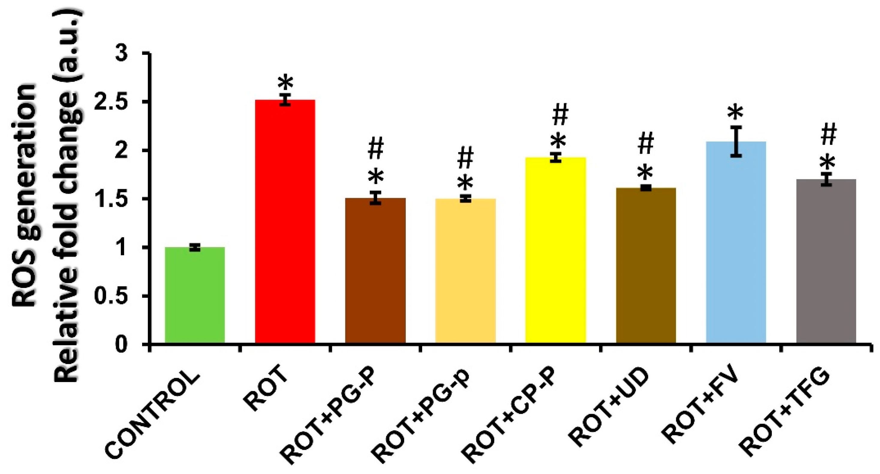
ROS generation in rotenone-exposed and rotenone cotreated with phytoconstituent groups: Figure 2 depicts the comparative fold changes in ROS generation in Drosophila melanogaster larvae fed on the test chemical rotenone (ROT) and the control (no treatment) diet. When rotenone, a test chemical, was compared to the control, the largest fold change in ROS formation was seen (2.52-fold). In comparison to the control larvae, the third instar larvae’s ROS generation increased after 24 h of exposure. The significance was ascribed at p-value ≤ 0.05.

3.2. Climbing Assay of Control, Rotenone, and Rotenone along with Plant-Extract-Fed Groups in Wild-Type Oregon R+

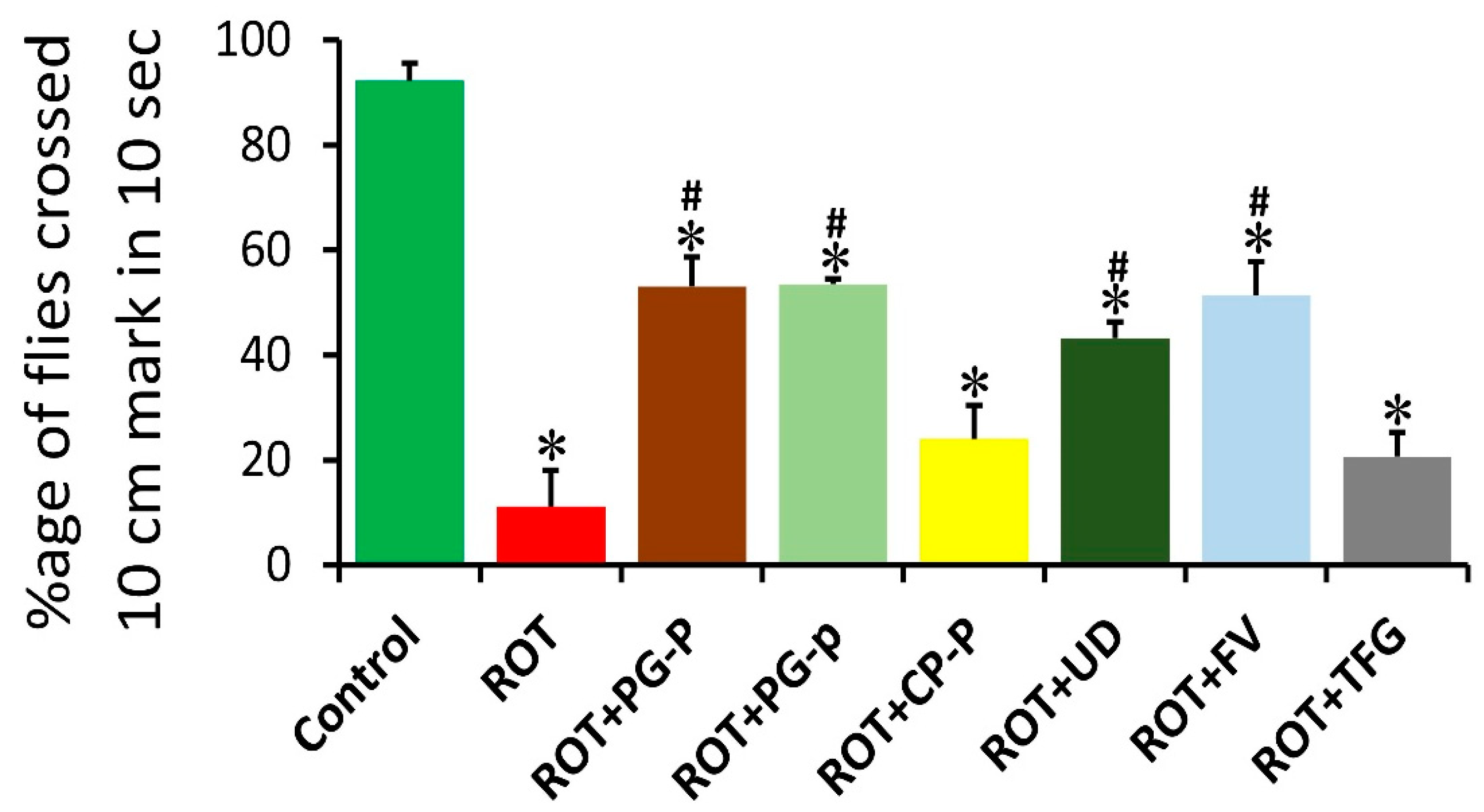
The control flies (untreated) showed a maximal escape ability after 10 s compared to that of 0.05 ppm ROT and ROT with different plant extracts. ROT-fed flies (exposed for 5 days; 120 h) exhibited significant difficulty in the climbing assay, and only 11.5% of the flies were successful in crossing the mark of the apparatus compared to the control (92.3%). Drosophila fed on test chemicals along with the plant extract showed a variable pattern in the climbing abilities of adult flies when compared to that of the control or rotenone-treated groups. Statistically significant improvement was evident in ROT + PG-p (4.80-fold increase), ROT + PG-P (4.75-fold increase), ROT + UD (3.88-fold increase), and ROT + FV (4.61-fold increase) compared to that of ROT-exposed flies. Interestingly, we did not observe statistically significant amelioration in the climbing activity of ROT + CP-P (2.15-fold increase)- and ROT + TFG (1.85-fold increase)-treated flies compared to the ROT-treated flies, although they showed some improvement in climbing ability (p < 0.05). Altogether, different groups showed different percentages of improvement in climbing ability (Figure 3).

3.3. Memory Assay of Drosophila Exposed to ROT and ROT Cotreated with Plant Extract on Oregon R+

Control adult flies (untreated) showed 96% memory retention. The differences in the control flies in achieving 100% memory retention could be attributed to their inherent ability to move, climb, and escape toward the light. Only 30% of the adult ROT-fed flies could memorize the offered dark condition, covering very little distance because they were moving too slowly to reach the target. Adult Drosophila fed on test chemicals along with the plant extract showed variable memory improvement (food + dark) when compared to the control or rotenone-treated groups. Amazingly, we observed statistically significant (p < 0.05) improvement in the memory retention of adult flies, with 100% memory retention in ROT + FV and ROT + TFG when compared to ROT alone. A statistically significant (p < 0.05) improvement (food + dark) was evident in ROT + UD (96.67%), ROT + CP-P (96.66%), ROT + PG-p (93.33%) and ROT + PG-P (93.33%) compared to ROT-fed flies. All the groups showed a statistically significant (p < 0.05) difference in the movement of the flies toward the light with no food compared to the untreated and ROT-treated flies. The flies were starved for 30 min before being placed in the T-shaped setup to test their memory capacity (Figure 4).

3.4. The Emergence of Drosophila Melanogaster in ROT, and ROT and Phytoextract-Fed Groups on the Wild-Type Strain (Oregon R+)

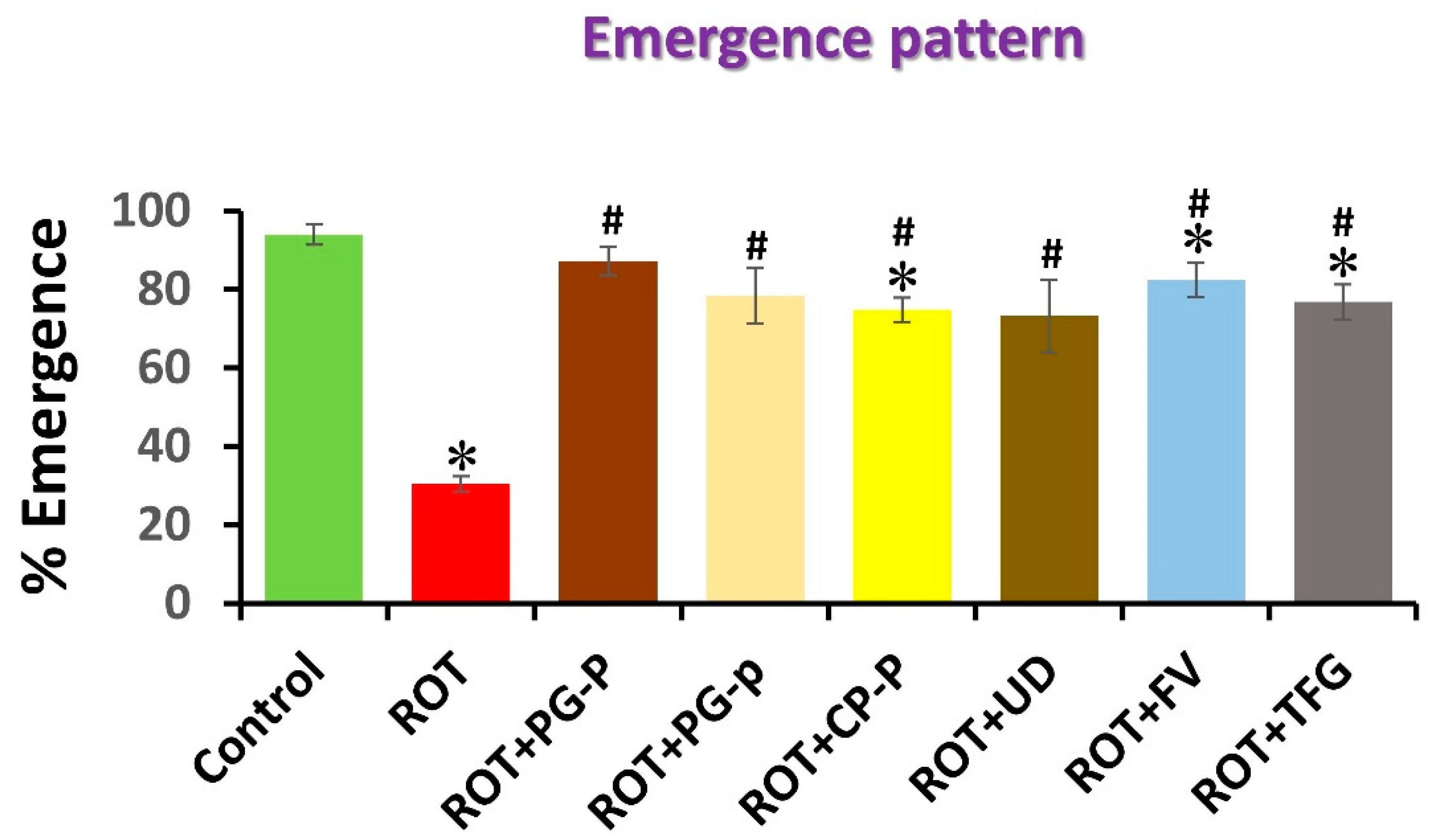
The number of flies that emerged increased (by 94.8%) in the wild-type control flies. Only 30.4% of the total number of rotenone-fed flies emerged. There was a statistically significant increase in the overall number of flies that emerged in all of the groups of flies fed on plant extracts together with ROT compared to the rotenone treatment, although none of the groups reached the threshold of the control (untreated flies). In comparison to the control group, the ROT + PG-P (87.2%), ROT + PG-p (78.4%), ROT + CP-P (74.8%), and ROT + FV (82.4%) groups all demonstrated full emergence; however, fewer flies emerged than in those left untreated: ROT + TFG (77.2%) and RO + UD (75.6%). All the groups having plant extracts along with the ROT had the same day of emergence. The number of flies that emerged each day was significantly higher in the flies that had been fed the plant extracts (PG-P, PG-p, CP-P, FV, TFG, and UD) and ROT than that in the flies fed ROT alone (Figure 5).

3.5. Reproductive Capacity of Control, ROT, and ROT Coexposed to Phytoextracts in Oregon R+

In wild-type Drosophila melanogaster, fecundity was decreased in positive control and rotenone-treated flies compared to that in control untreated flies. All the groups having a combination of plant extracts with ROT showed a reduction in total fecundity compared to that of the untreated flies, and more than that of the ROT-fed flies. The mean daily egg laying/female/10 days in all the groups of flies that had been fed the ROT and plant extract combination had statistically significant results concerning both control and ROT-fed flies. The fertility percentage in ROT + PG-p, ROT + CP-P, and ROT + UD showed statistically significant improvement compared to ROT, and was significant with control-fed flies, but did not attain the fertility of control flies. The fertility percentage of ROT + PG-P flies was significant compared to that of the control flies only, and ROT + FV and ROT + TFG were significant compared to ROT-fed flies. Reproductive performance in all ROT + phytoextract groups showed statistically significant values compared to that of the control and ROT-fed flies (Table 1).

4. Discussion

Oxidative stress is a well-known element contributing to the death of neuronal cells. Free radical species significantly influence the deterioration of the CNS because they lack the ability to regenerate, and brain cells are particularly susceptible to such damage [36]. Even though the body’s natural defenses can handle free radicals, they are sometimes ineffective, and cellular damage occurs. Considering the above, strengthening the defense system using herbal remedies would be a useful strategy to resolve this crisis [37].
PG, CP, TFG, FV, and UD are well-known for their abilities to improve cognition, memory, and learning in the Ayurvedic medical system [8,9,10,11,12]. Recent studies primarily used animal models to examine the protective potential of selected herbs against many neurodegenerative diseases. The various phytochemicals of the selected herbs that are responsible for their antioxidant and protective potential were well-identified [38]. This comprehension is effective in regulating the risk factors that lead to the development of diseases. The current study was conducted to determine the neuroprotective potential of PG, CP, TFG, FV, and UD extracts.
The adverse consequences of ROT and its improvement were tested on wild-type Drosophila melanogaster (Oregon R+) by administering aqueous extracts of phytoextracts PG peel, PG pulp (juice), CP peel, FV seeds, TFG seeds, and UD leaves along with ROT, mixed with the diet. All the selected phytoextracts reversed the effect of ROT in the organismal investigation with various potential levels after treating the flies concurrently.
The Drosophila PD phenotype was generated through chemical induction by exposing them to a rotenone mixed diet. After exposure to rotenone, PD-like pathology, as reported in a previous study [30], was analyzed in the wild type (Oregon R+). The characterized pathologies in PD fly models render them useful in identifying therapeutic targets and revealing pathological mechanisms [29,39,40,41]. The age-dependent loss in motor performance that defines the Drosophila PD model is typically measured with negative geotaxis climbing, which is more relevant to Parkinson’s disease (PD) and assesses motor capacity differently than just a measure of walking ability. In order to examine for locomotor impairment, we evaluated the geotaxis climbing of rotenone-treated flies in this study. Following confirmation that the PD model had been generated, additional studies were conducted to determine how different treatments affected memory, emergence, reproductive factors, and the measurement of ROS.
In this study, the comparative fold changes in ROS production in larvae exposed to rotenone (ROT), the control (no treatment), and the plant extract and test chemical (ROT) groups were all measured. Rotenone showed the highest fold change in ROS formation (2.52-fold) when compared to the control. In comparison to ROT alone, the combination of ROT and plant extracts reduced the production of ROS. Rotenone showed that, with the exception of the ROT + FV group, there was a substantial difference between the ROT + PG Pl, ROT + PG J, ROT + CP Pl, ROT + UD, and ROT + TFG groups when compared to the rotenone group in terms of ROS generation in third instar larvae. Increased ROS production was linked to DNA oxidative damage, including strand breakage, and base and nucleotide alterations [18]. A strong positive connection between ROS and DNA damage in the exposed organism supports the idea that the pesticide-induced ROS formation may be a significant factor in the organism’s DNA damage. Natural antioxidants have been investigated for several years in relation to diseases caused by oxidative stress. On H2O2-treated MCF-7 cells (breast cancer), a study of five antioxidant fractions from the Fucus spiralis seaweed revealed a decrease in ROS generation, the induction of apoptosis by caspase 9 activation, and the depolarization of the mitochondrial membrane. Plant metabolites with cytoprotective activities against hydrogen peroxide toxicity, and an intriguing prospective utility as an oxidative stress modulator were found in the five antioxidant fractions that were examined [42].
Our research demonstrates that Drosophila melanogaster’s climbing ability and memory were improved by all nutraceutical treatments in combination with rotenone. Compared to the ROT-treated group, the nutritional supplement treatment in all strains significantly improved climbing ability. The presence of specific substances in plants, such as flavonoids and terpenoids, may shield cells from DNA damage and is likely what causes this phytotherapeutic agent’s modulatory function [43]. Recent investigations discovered an improvement in locomotor dysfunction with the administration of different nutraceuticals than those in the current study [44].
In a previous study, B. monnieri greatly improved climbing ability, reduced dopamine depletion, decreased rotenone-induced mortality, and protected against oxidative stress in D. melanogaster treated with rotenone [45]. Antioxidant-rich foods and supplements should be consumed regularly to extend an organism’s life [46,47,48]. Accordingly, the early emergence and improvement in reproductive parameters observed in the present study in flies fed with phytoextracts may be attributable to the antioxidant-boosting property linked to them. Major classes of secondary metabolites such as flavonoids, glycosides, tannins, alkaloids, proteins, steroids, amino acids, and sugars function as effective antioxidant compounds that can counteract the harmful effects of rotenone. Due to the wide variety of phytoconstituents they contain, these nutraceuticals have rich pharmacological utility in conventional treatments. Moreover, the abnormal accumulation of Fe, Mn, and Cu metals in a fly’s head was linked to a reduction in life expectancy and mobility [49]. The fact that D. melanogaster has impaired metal homeostasis in PD and Parkinsonism suggests that it is unable to effectively control cognitive impairment [50,51,52].
The wild-type flies (control group) in this study showed the highest overall emergence. When compared to flies exposed to ROT, ROT and phytoextracts triggered early emergence, and the number of flies that emerged on the first days increased [53,54,55]. The total number of flies that emerged was lower overall in the ROT and phytoextract-treated groups, but the number of flies that emerged early was higher than that in the control flies, and higher than that in the ROT-treated flies [56]. Additionally, due to the effective antioxidant capacity, secondary metabolites may have therapeutic effects. Therefore, in the present work, the protective effects of nutraceutical (PG, CP, TFG, FV, and UD) aqueous extracts against functional damages generated by ROT on Drosophila total emergence were studied. The results support the preventive role of nutraceutical extracts by restoring the fly emergence rate.
Although statistically significant results for the above parameters demonstrate the significance of these nutraceuticals (PG, CP, TFG, FV, and UD) as potent antitoxic agents that could reverse the toxic effects imposed by ROT, this illustrates the significance of drug repurposing as a technique and calls for careful investigation. The study also emphasizes the value of the PD Drosophila model as a crucial instrument for creating in vivo drug discovery models. Combining in vivo drug discovery with phenotypic screening is a promising approach for overcoming the limitations of in vitro discovery methods [57,58]. It is possible to successfully scale up Drosophila models to provide more throughput than that in other in vivo PD models.

5. Conclusions

The phytoextracts used in this study showed better climbing ability using phytoconstituents than that of those with rotenone alone, which showed a substantial reduction in climbing ability. Once the visible symptoms of climbing disability had been confirmed, we further evaluated memory power, emergence, reproductive capacity, and the measurement of ROS in the flies treated with ROT alone and ROT coexposed to different phytoextracts. This demonstrates that the ability of nutraceuticals (bioactive substances of phytoextracts) to control antioxidant defense mechanisms and have antioxidative characteristics has the potential to alleviate oxidative stress caused by ROT. The phytoconstituents, which showed good climbing and memory-enhancing power, improved the eclosion and overall reproductive capacity of the flies. Phytoconstituents could significantly attenuate rotenone-induced organismal toxicities in the Drosophila model. The results of this research help in the quality control of raw herbaceous material to confirm its potential for phytopharmaceutical applications and health-promoting qualities that might be utilized in medicine discovery.

Author Contributions

Conceptualization, M.P.S., S.K.S. and R.H.; methodology, R.H.; software, R.H. and M.P.S.; validation, M.P.S. and R.H.; formal analysis, R.H. and M.P.S.; resources, M.P.S.; writing—original draft preparation, M.P.S., R.H., E.V., A.A.O., A.S.A., C.M.G. and S.S.; supervision, M.P.S. All authors have read and agreed to the published version of the manuscript.

Funding

This research received no external funding.

Institutional Review Board Statement

Not applicable.

Informed Consent Statement

Not applicable.

Data Availability Statement

Data are available in the manuscript.

Acknowledgments

We acknowledge the Lovely Professional University for the infrastructure and the chemicals required for the research. We are also grateful to Anurag Sharma, NITTE University, Mangalore, India for providing the Drosophila stock and for the assistance in ROS estimation. ASA and CMG highly acknowledge the Taif University Researchers Supporting Project number TURSP-2020/288, Taif University, Taif, Saudi Arabia.

Conflicts of Interest

The authors declare that they have no conflicts of interest in the publication.

References

  1. Javed, H.; Meeran, M.F.N.; Azimullah, S.; Eddin, L.B.; Dwivedi, V.D.; Jha, N.K.; Ojha, S. α-Bisabolol, a Dietary Bioactive Phytochemical Attenuates Dopaminergic Neurodegeneration through Modulation of Oxidative Stress, Neuroinflammation and Apoptosis in Rotenone-Induced Rat Model of Parkinson’s disease. Biomolecules 2020, 10, 1421. [Google Scholar] [CrossRef] [PubMed]
  2. Ong, W.-Y.; Leow, D.M.-K.; Herr, D.R.; Yeo, C.J.-J. What Do Randomized Controlled Trials Inform Us about Potential Disease-Modifying Strategies for Parkinson’s Disease? NeuroMolecular Med. 2022, 1–13. [Google Scholar] [CrossRef] [PubMed]
  3. Chakrabarty, R.; Yousuf, S.; Singh, M.P. Contributive Role of Hyperglycemia and Hypoglycemia Towards the Development of Alzheimer’s Disease. Mol. Neurobiol. 2022, 59, 4274–4291. [Google Scholar] [CrossRef] [PubMed]
  4. Booth, H.D.; Hirst, W.D.; Wade-Martins, R. The Role of Astrocyte Dysfunction in Parkinson’s Disease Pathogenesis. Trends Neurosci. 2017, 40, 358–370. [Google Scholar] [CrossRef] [PubMed]
  5. Beckers, M.; Bloem, B.R.; Verbeek, M.M. Mechanisms of peripheral levodopa resistance in Parkinson’s disease. NPJ Park. Dis. 2022, 8, 56. [Google Scholar] [CrossRef]
  6. Thirumalaisamy, R.; Bhuvaneswari, M.; Haritha, S.; Jeevarathna, S.; Janani, K.; Suresh, K. Curcumin, Naringenin and Resveratrol from Natural Plant Products Hold Promising Solution for Modern World Diseases–A Recent Review. S. Afr. J. Bot. 2022. [Google Scholar] [CrossRef]
  7. Himalian, R.; Singh, S.K.; Singh, M.P. Ameliorative Role of Nutraceuticals on Neurodegenerative Diseases Using the Drosophila melanogaster as a Discovery Model to Define Bioefficacy. J. Am. Nutr. Assoc. 2021, 41, 511–539. [Google Scholar] [CrossRef]
  8. Moradi, A.; Nezamoleslami, S.; Nezamoleslami, S.; Clark, C.C.; Sohouli, M.H.; Ghiasvand, R. The association between Dietary total antioxidant capacity with risk of Rheumatoid Arthritis in adults: A case-control study. Clin. Nutr. ESPEN 2022, 2405–4577. [Google Scholar] [CrossRef]
  9. Dhalaria, R.; Verma, R.; Kumar, D.; Puri, S.; Tapwal, A.; Kumar, V.; Nepovimova, E.; Kuca, K. Bioactive Compounds of Edible Fruits with Their Anti-Aging Properties: A Comprehensive Review to Prolong Human Life. Antioxidants 2020, 9, 1123. [Google Scholar] [CrossRef]
  10. Kaveh, S.; Mahoonak, A.S.; Ghorbani, M.; Jafari, S.M. Fenugreek seed (Trigonella foenum graecum) protein hydrolysate loaded in nanosized liposomes: Characteristic, storage stability, controlled release, and retention of antioxidant activity. Ind. Crops Prod. 2022, 182, 114908. [Google Scholar] [CrossRef]
  11. Khammassi, M.; Mighri, H.; Ben Mansour, M.; Amri, I.; Jamoussi, B.; Khaldi, A. Metabolite profiling and potential antioxidant activity of sixteen fennel (Foeniculum vulgare Mill.) populations wild-growing in Tunisia. S. Afr. J. Bot. 2022, 148, 407–414. [Google Scholar] [CrossRef]
  12. Chandrasekara, A.; Shahidi, F. Herbal beverages: Bioactive compounds and their role in disease risk reduction—A review. J. Traditional Complement. Med. 2018, 8, 451–458. [Google Scholar] [CrossRef] [PubMed]
  13. Balakrishnan, R.; Azam, S.; Cho, D.-Y.; Su-Kim, I.; Choi, D.-K. Natural Phytochemicals as Novel Therapeutic Strategies to Prevent and Treat Parkinson’s Disease: Current Knowledge and Future Perspectives. Oxid. Med. Cell. Longev. 2021, 2021, 6680935. [Google Scholar] [CrossRef]
  14. Singh, M.P.; Reddy, M.K.; Mathur, N.; Saxena, D.; Chowdhuri, D.K. Induction of hsp70, hsp60, hsp83 and hsp26 and oxidative stress markers in benzene, toluene and xylene exposed Drosophila melanogaster: Role of ROS generation. Toxicol. Appl. Pharmacol. 2009, 235, 226–243. [Google Scholar] [CrossRef]
  15. Singh, M.P.; Mishra, M.; Sharma, A.; Shukla, A.; Mudiam, M.; Patel, D.; Ram, K.R.; Chowdhuri, D.K. Genotoxicity and apoptosis in Drosophila melanogaster exposed to benzene, toluene and xylene: Attenuation by quercetin and curcumin. Toxicol. Appl. Pharmacol. 2011, 253, 14–30. [Google Scholar] [CrossRef] [PubMed]
  16. Singh, M.P.; Ram, K.R.; Mishra, M.; Shrivastava, M.; Saxena, D.K.; Chowdhuri, D.K. Effects of co-exposure of benzene, toluene and xylene to Drosophila melanogaster: Alteration in hsp70, hsp60, hsp83, hsp26, ROS generation and oxidative stress markers. Chemosphere 2010, 79, 577–587. [Google Scholar] [CrossRef]
  17. Sharma, A.; Mishra, M.; Shukla, A.; Kumar, R.; Abdin, M.; Chowdhuri, D.K. Organochlorine pesticide, endosulfan induced cellular and organismal response in Drosophila melanogaster. J. Hazard. Mater. 2012, 221–222, 275–287. [Google Scholar] [CrossRef]
  18. Hu, C.; Yang, J.; Qi, Z.; Wu, H.; Wang, B.; Zou, F.; Mei, H.; Liu, J.; Wang, W.; Liu, Q. Heat shock proteins: Biological functions, pathological roles, and therapeutic opportunities. MedComm 2022, 3, e161. [Google Scholar] [CrossRef]
  19. Deepika; Maurya, P.K. Health Benefits of Quercetin in Age-Related Diseases. Molecules 2022, 27, 2498. [Google Scholar] [CrossRef]
  20. Coulom, H.; Birman, S. Chronic exposure to rotenone models sporadic Parkinson’s disease in Drosophila melanogaster. J. Neurosci. 2004, 24, 10993–10998. [Google Scholar] [CrossRef] [Green Version]
  21. Hosamani, R.; Ramesh, S.R.; Muralidhara. Attenuation of Rotenone-Induced Mitochondrial Oxidative Damage and Neurotoxicty in Drosophila melanogaster Supplemented with Creatine. Neurochem. Res. 2010, 35, 1402–1412. [Google Scholar] [CrossRef] [PubMed]
  22. Islam, R.; Yang, L.; Sah, M.; Kannan, K.; Anamani, D.; Vijayan, C.; Kwok, J.; Cantino, M.E.; Beal, M.F.; Fridell, Y.-W.C. A neuroprotective role of the human uncoupling protein 2 (hUCP2) in a Drosophila Parkinson’s Disease model. Neurobiol. Dis. 2012, 46, 137–146. [Google Scholar] [CrossRef] [PubMed]
  23. Lawal, H.O.; Chang, H.-Y.; Terrell, A.N.; Brooks, E.S.; Pulido, D.; Simon, A.F.; Krantz, D.E. The Drosophila vesicular monoamine transporter reduces pesticide-induced loss of dopaminergic neurons. Neurobiol. Dis. 2010, 40, 102–112. [Google Scholar] [CrossRef]
  24. Shabir, S.; Yousuf, S.; Singh, S.K.; Vamanu, E.; Singh, M.P. Ethnopharmacological Effects of Urtica dioica, Matricaria chamomilla, and Murraya koenigii on Rotenone-Exposed D. melanogaster: An Attenuation of Cellular, Biochemical, and Organismal Markers. Antioxidants 2022, 11, 1623. [Google Scholar] [CrossRef] [PubMed]
  25. Sherer, T.; Betarbet, R.; Testa, C.M.; Seo, B.B.; Richardson, J.R.; Kim, J.H.; Miller, G.W.; Yagi, T.; Matsuno-Yagi, A.; Greenamyre, J.T. Mechanism of Toxicity in Rotenone Models of Parkinson’s Disease. J. Neurosci. 2003, 23, 10756–10764. [Google Scholar] [CrossRef]
  26. Muñoz-Soriano, V.; Paricio, N. Drosophila Models of Parkinson’s Disease: Discovering Relevant Pathways and Novel Therapeutic Strategies. Park. Dis. 2011, 2011, 520640. [Google Scholar] [CrossRef]
  27. Steffan, J.S.; Bodai, L.; Pallos, J.; Poelman, M.; McCampbell, A.; Apostol, B.L.; Kazantsev, A.; Schmidt, E.; Zhu, Y.-Z.; Greenwald, M.; et al. Histone deacetylase inhibitors arrest polyglutamine-dependent neurodegeneration in Drosophila. Nature 2001, 413, 739–743. [Google Scholar] [CrossRef]
  28. Auluck, P.K.; Chan, H.Y.E.; Trojanowski, J.Q.; Lee, V.M.-Y.; Bonini, N.M. Chaperone Suppression of alpha -Synuclein Toxicity in a Drosophila Model for Parkinson’s Disease. Science 2002, 295, 865–868. [Google Scholar] [CrossRef]
  29. Whitworth, A.J.; Theodore, D.A.; Greene, J.C.; Beneš, H.; Wes, P.D.; Pallanck, L.J. Increased glutathione S -transferase activity rescues dopaminergic neuron loss in a Drosophila model of Parkinson’s disease. Proc. Natl. Acad. Sci. USA 2005, 102, 8024–8029. [Google Scholar] [CrossRef]
  30. Feany, M.B.; Bender, W.W. A Drosophila model of Parkinson’s disease. Nature 2000, 404, 394–398. [Google Scholar] [CrossRef]
  31. Hirth, F. Drosophila melanogaster in the study of human neurodegeneration. CNS Neurol. Disord. Drug Targets 2010, 9, 504–523. [Google Scholar] [CrossRef] [PubMed]
  32. Gayathri, M.; Krishnamurthy, N. Studies on the toxicity of the mercurial fungicide Agallol 3 in Drosophila melanogaster. Environ. Res. 1981, 24, 89–95. [Google Scholar] [CrossRef]
  33. Pendleton, R.G.; Parvez, F.; Sayed, M.; Hillman, R. Effects of Pharmacological Agents upon a Transgenic Model of Parkinson’s Disease in Drosophila melanogaster. J. Pharmacol. Exp. Ther. 2002, 300, 91–96. [Google Scholar] [CrossRef]
  34. Ali, Y.O.; Escala, W.; Ruan, K.; Zhai, R.G. Assaying Locomotor, Learning, and Memory Deficits in Drosophila Models of Neurodegeneration. J. Vis. Exp. 2011, 49, 2504. [Google Scholar] [CrossRef]
  35. Madabattula, S.T.; Strautman, J.C.; Bysice, A.M.; O’Sullivan, J.A.; Androschuk, A.; Rosenfelt, C.; Doucet, K.; Rouleau, G.; Bolduc, F. Quantitative Analysis of Climbing Defects in a Drosophila Model of Neurodegenerative Disorders. J. Vis. Exp. JoVE 2015, 100, e52741. [Google Scholar] [CrossRef] [PubMed]
  36. Gandhi, S.; Abramov, A.Y. Mechanism of oxidative stress in neurodegeneration. Oxid. Med. Cell. Longev. 2012, 2012, 428010. [Google Scholar] [CrossRef] [PubMed]
  37. Yousuf, S.; Shabir, S.; Singh, M.P. Protection Against Drug-Induced Liver Injuries Through Nutraceuticals via Amelioration of Nrf-2 Signaling. J. Am. Nutr. Assoc. 2022, 1–21. [Google Scholar] [CrossRef]
  38. Himalian, R.; Singh, M.P. A Comparative account on Antioxidant activities, Total phenolic and Flavonoid contents of Punica granatum, Carica papaya, Foeniculum vulgare, Trigonella foenum-graecum, and Urtica dioica: An in vitro Evaluation. Res. J. Pharm. Technol. 2022, 15, 1175–1183. [Google Scholar] [CrossRef]
  39. Auluck, P.K.; Bonini, N.M. Pharmacological prevention of Parkinson disease in Drosophila. Nat. Med. 2002, 8, 1185–1186. [Google Scholar] [CrossRef]
  40. Bilen, J.; Bonini, N.M. Drosophila as a Model for Human Neurodegenerative Disease. Annu. Rev. Genet. 2005, 39, 153–171. [Google Scholar] [CrossRef] [Green Version]
  41. Whitworth, A.J.; Wes, P.D.; Pallanck, L.J. Drosophila models pioneer a new approach to drug discovery for Parkinson’s disease. Drug Discov. Today 2006, 11, 119–126. [Google Scholar] [CrossRef]
  42. Vallejo, M.J.; Salazar, L.; Grijalva, M. Oxidative Stress Modulation and ROS-Mediated Toxicity in Cancer: A Review on In Vitro Models for Plant-Derived Compounds. Oxid. Med. Cell. Longev. 2017, 2017, 4586068. [Google Scholar] [CrossRef]
  43. Golla, U.; Bhimathati, S.S.R. Evaluation of Antioxidant and DNA Damage Protection Activity of the Hydroalcoholic Extract of Desmostachya bipinnata L. Stapf. Sci. World J. 2014, 2014, 215084. [Google Scholar] [CrossRef] [PubMed]
  44. Kumar, A.; Christian, P.K.; Panchal, K.; Guruprasad, B.R.; Tiwari, A.K. Supplementation of Spirulina (Arthrospira platensis) Improves Lifespan and Locomotor Activity in Paraquat-Sensitive DJ-1βΔ93 Flies, a Parkinson’s Disease Model in Drosophila melanogaster. J. Diet. Suppl. 2017, 14, 573–588. [Google Scholar] [CrossRef] [PubMed]
  45. Hosamani, R.; Muralidhara. Neuroprotective efficacy of Bacopa monnieri against rotenone induced oxidative stress and neurotoxicity in Drosophila melanogaster. Neurotoxicology 2009, 30, 977–985. [Google Scholar] [CrossRef]
  46. Carey, J.R.; Harshman, L.G.; Liedo, P.; Müller, H.G.; Wang, J.L.; Zhang, Z. Longevity–fertility trade-offs in the tephritid fruit fly, Anastrepha ludens, across dietary-restriction gradients. Aging Cell 2008, 7, 470–477. [Google Scholar] [CrossRef]
  47. Lee, K.P.; Simpson, S.J.; Clissold, F.J.; Brooks, R.; Ballard, J.W.O.; Taylor, P.W.; Soran, N.; Raubenheimer, D. Lifespan and reproduction in Drosophila: New insights from nutritional geometry. Proc. Natl. Acad. Sci. USA 2008, 105, 2498–2503. [Google Scholar] [CrossRef]
  48. Skorupa, D.A.; Dervisefendic, A.; Zwiener, J.; Pletcher, S.D. Dietary composition specifies consumption, obesity, and lifespan in Drosophila melanogaster. Aging Cell 2008, 7, 478–490. [Google Scholar] [CrossRef]
  49. Bonilla-Ramirez, L.; Jimenez-Del-Rio, M.; Velez-Pardo, C. Acute and chronic metal exposure impairs locomotion activity in Drosophila melanogaster: A model to study Parkinsonism. BioMetals 2011, 24, 1045–1057. [Google Scholar] [CrossRef]
  50. Lee, D.W.; Andersen, J.K. Iron elevations in the aging Parkinsonian brain: A consequence of impaired iron homeostasis? J. Neurochem. 2010, 112, 332–339. [Google Scholar] [CrossRef]
  51. Guilarte, T.R. Manganese and Parkinson’s Disease: A Critical Review and New Findings. Environ. Health Perspect. 2010, 118, 1071–1080. [Google Scholar] [CrossRef] [PubMed]
  52. Huster, D. Wilson disease. Best Pract. Res. Clin. Gastroenterol. 2010, 24, 531–539. [Google Scholar] [CrossRef] [PubMed]
  53. Margulies, C.; Tully, T.; Dubnau, J. Deconstructing Memory in Drosophila. Curr. Biol. 2005, 15, R700–R713. [Google Scholar] [CrossRef]
  54. Batool, Z.; Sadir, S.; Liaquat, L.; Tabassum, S.; Madiha, S.; Rafiq, S.; Tariq, S.; Batool, T.S.; Saleem, S.; Naqvi, F.; et al. Repeated administration of almonds increases brain acetylcholine levels and enhances memory function in healthy rats while attenuates memory deficits in animal model of amnesia. Brain Res. Bull. 2016, 120, 63–74. [Google Scholar] [CrossRef]
  55. Zhao, Y.; Sun, H.; Lu, J.; Li, X.; Chen, X.; Tao, D.; Huang, W.; Huang, B. Lifespan extension and elevated hsp gene expression in Drosophila caused by histone deacetylase inhibitors. J. Exp. Biol. 2005, 208, 697–705. [Google Scholar] [CrossRef] [PubMed]
  56. Sharma, D.; Singh, M.P.; Vimal, D.; Kumar, S.; Jha, R.R.; Chowdhuri, D.K. Benzene induced resistance in exposed Drosophila melanogaster: Outcome of improved detoxification and gene modulation. Chemosphere 2018, 201, 144–158. [Google Scholar] [CrossRef] [PubMed]
  57. Finkbeiner, S.; Skibinski, G. Drug discovery in Parkinson’s disease: Update and developments in the use of cellular models. Int. J. High Throughput Screen. 2011, 2, 15–25. [Google Scholar] [CrossRef]
  58. Adnew, W.; Asmare, B.; Mekuriaw, Y. Review on knowledge gap in Brachiaria grass research and utilization: Ethiopian perspective. AgroLife Sci. J. 2021, 10, 9–26. Available online: https://agrolifejournal.usamv.ro/index.php/scientific-papers/567-review-on-knowledge-gap-in-brachiaria-grass-research-and-utilization-ethiopian-perspective-567#spucontentCitation1 (accessed on 20 September 2022). [CrossRef]
Figure 1. Different nutraceuticals used in this study. (A) Punica granatum, (B) Carica papaya, (C) Urtica dioica, (D) Trigonella foenum-graecum, and (E) Foeniculum vulgare.
Applsci 12 09822 g001
Figure 2. Drosophila third instar larvae exposed to ROT and ROT cotreated with plant extract for only 24 h showed a relative fold shift in ROS production. ROS generation was examined in control, ROT, and ROT along with phytoextracts groups (pomegranate peel (PG-P), pomegranate pulp (PG-p), papaya peel (CP-P), nettle leaves (UD), fennel (FV), and fenugreek (TFG)). All observations are expressed as the mean ± SEM, n = 3, with each group having 50 larvae per vial, and were performed 3 times. Significance was ascribed to * p < 0.05 compared to the control; # p < 0.05 compared to rotenone.
Applsci 12 09822 g002
Figure 3. Wild-type D. melanogaster’s capability for climbing after 120 h. Rotenone, ROT; pomegranate peel, PG-P; pomegranate pulp, PG-p; papaya peel, CP-P; nettle leaves, UD; fennel, FV; fenugreek, TFG. All observations are expressed as the mean ± SEM, n = 10, and were performed 3 times. Results were significant at p < 0.05. Significance was ascribed to * p <0.05 compared to the control; # p < 0.05 compared to rotenone.
Applsci 12 09822 g003
Figure 4. Drosophila melanogaster (Oregon R+) memory after a 16-h interval in the presence of the conditions. Fenugreek, FV; nettle leaves, UD; fennel, FV; pomegranate peel, PG-P; papaya peel, CP-P; rotenone, ROT; TFG, Trigonella foenum-graecum. Each observation was performed three times and is expressed as the mean ± SEM. Results were significant at p < 0.05. Significance was * p < 0.05 compared to control; # p < 0.05 compared to rotenone (light vs. light and dark vs. dark).
Applsci 12 09822 g004
Figure 5. Emergence of flies of Drosophila melanogaster in Oregon R+. All observations are expressed as the mean of 5 vials per group with 50 larvae per vial. Rotenone, ROT; pomegranate peel, PG-P; pomegranate pulp, PG-p; papaya peel, CP-P; nettle leaves, UD; fennel, FV; and fenugreek, TFG. Each observation was performed three times and is shown as the mean ± SEM. Results were significant at p < 0.05. Significance was * p < 0.05 compared to control; # p < 0.05 compared to rotenone.
Applsci 12 09822 g005
Table 1. Reproductive capacity of Oregon flies in control, ROT, and combinations of phytoextracts.
GroupsTotal FecundityMean Daily Egg Laying/Female/10 DaysFertility PercentageReproductive Performance
CONTROL1186237.2 ± 2.8780.99 ± 1.1596.1 ± 0.44
ROT23947.8 ± 1.74 *35.96 ± 3.07 *8.5 ± 0.08 *
ROT + PG-P948189.6 ± 2.80 *,#40.21 ± 0.70 *38.1 ± 0.10 *,#
ROT + PG-p709141.8 ± 3.63 *,#54.22 ± 1.35 *,#38.4 ± 0.19 *,#
ROT + CP-P855171 ± 4.90 *,#44.01 ± 1.36 *,#37.5 ± 0.03 *,#
ROT + ND733146.6 ± 2.73 *,#49.79 ± 0.08 *,#36.5 ± 0.13 *,#
ROT + FV751150.2 ± 2.28 *,#78.61 ± 1.40 #59 ± 0.17 *,#
ROT + TFG588117.6 ± 1.96 *,#77.85 ± 1.43 #45.8 ± 0.26 *,#
Note: All experiments were conducted three times, and all observations are expressed as the mean ± SEM, n = 3. The results were significant at p ≤ 0.05. Significance: * with control, and # with rotenone of a particular group within the same strains of flies. Rotenone, ROT; pomegranate peel, PG-P; pomegranate pulp, PG-p; papaya peel, CP-P; nettle leaves, UD; fennel, FV; and fenugreek, TFG.
Publisher’s Note: MDPI stays neutral with regard to jurisdictional claims in published maps and institutional affiliations.