
Analysis of Coating Loss from Coated Stainless Steel Orthodontic Wire


Abstract
1. Introduction
2. Materials and Methods
2.1. Specimens
2.2. Wire Bending
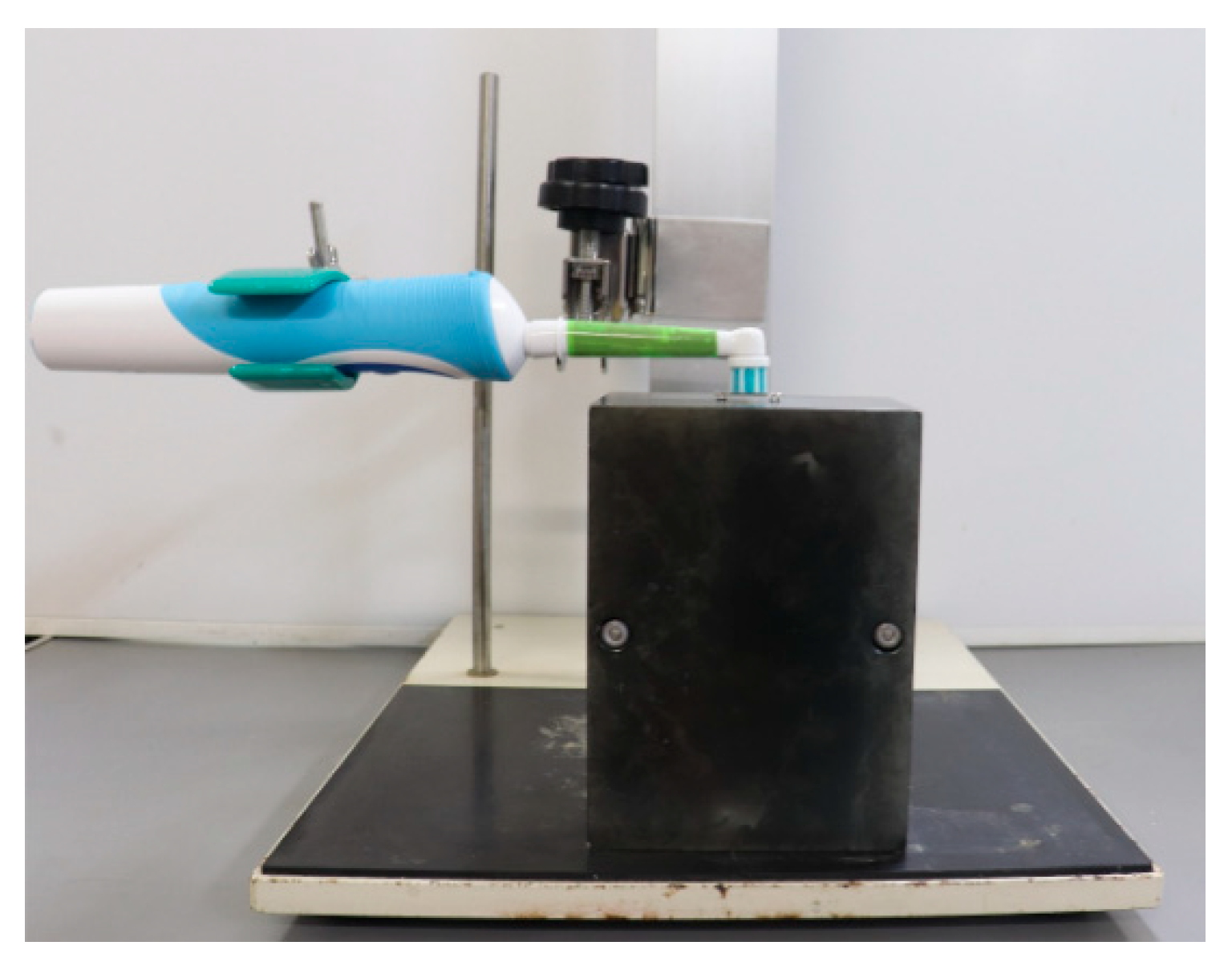
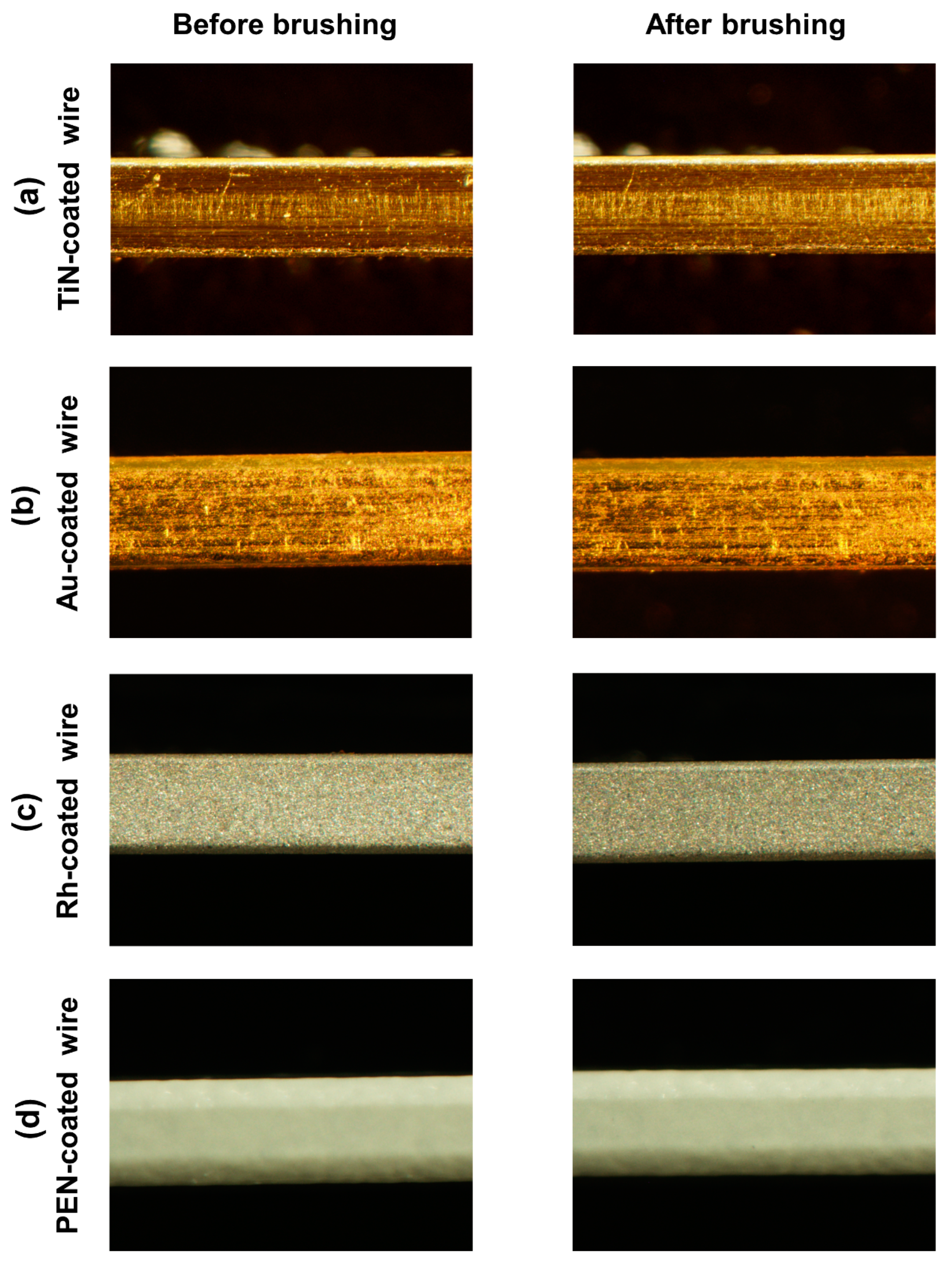
2.3. Brushing Tests
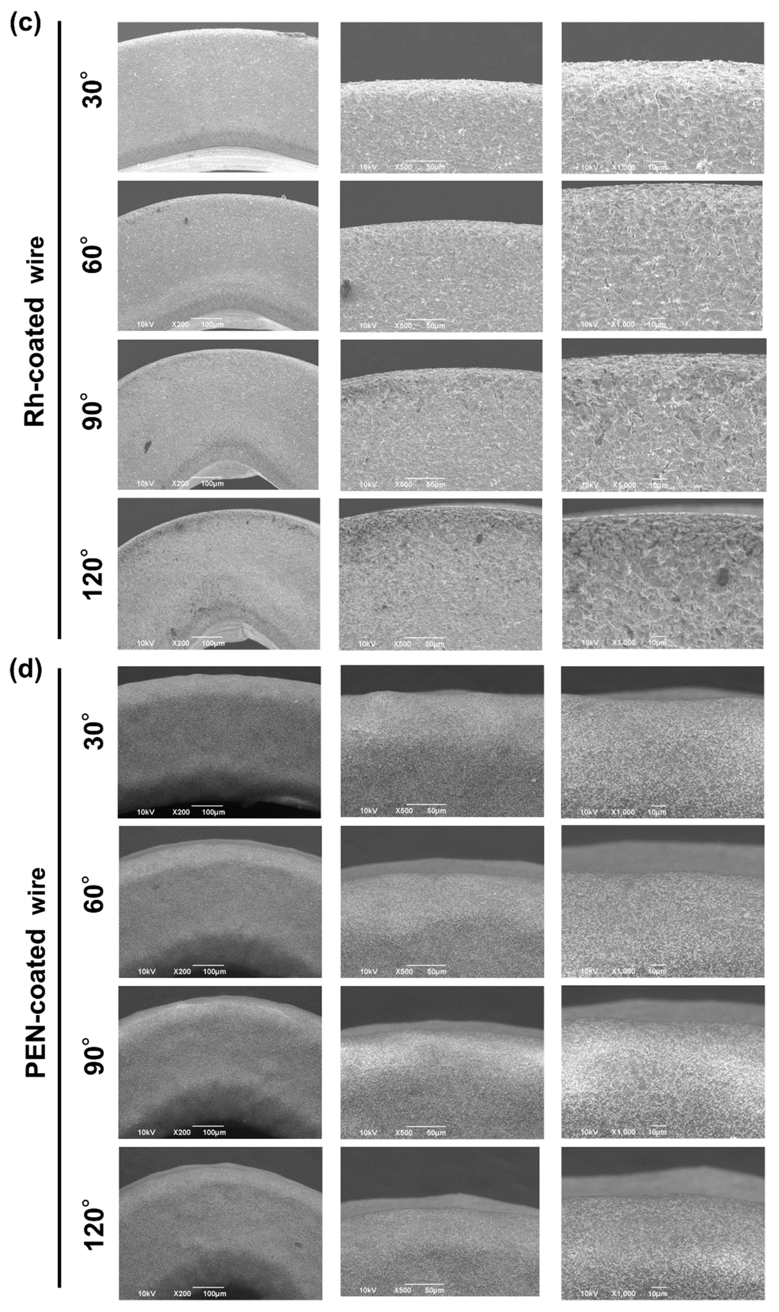
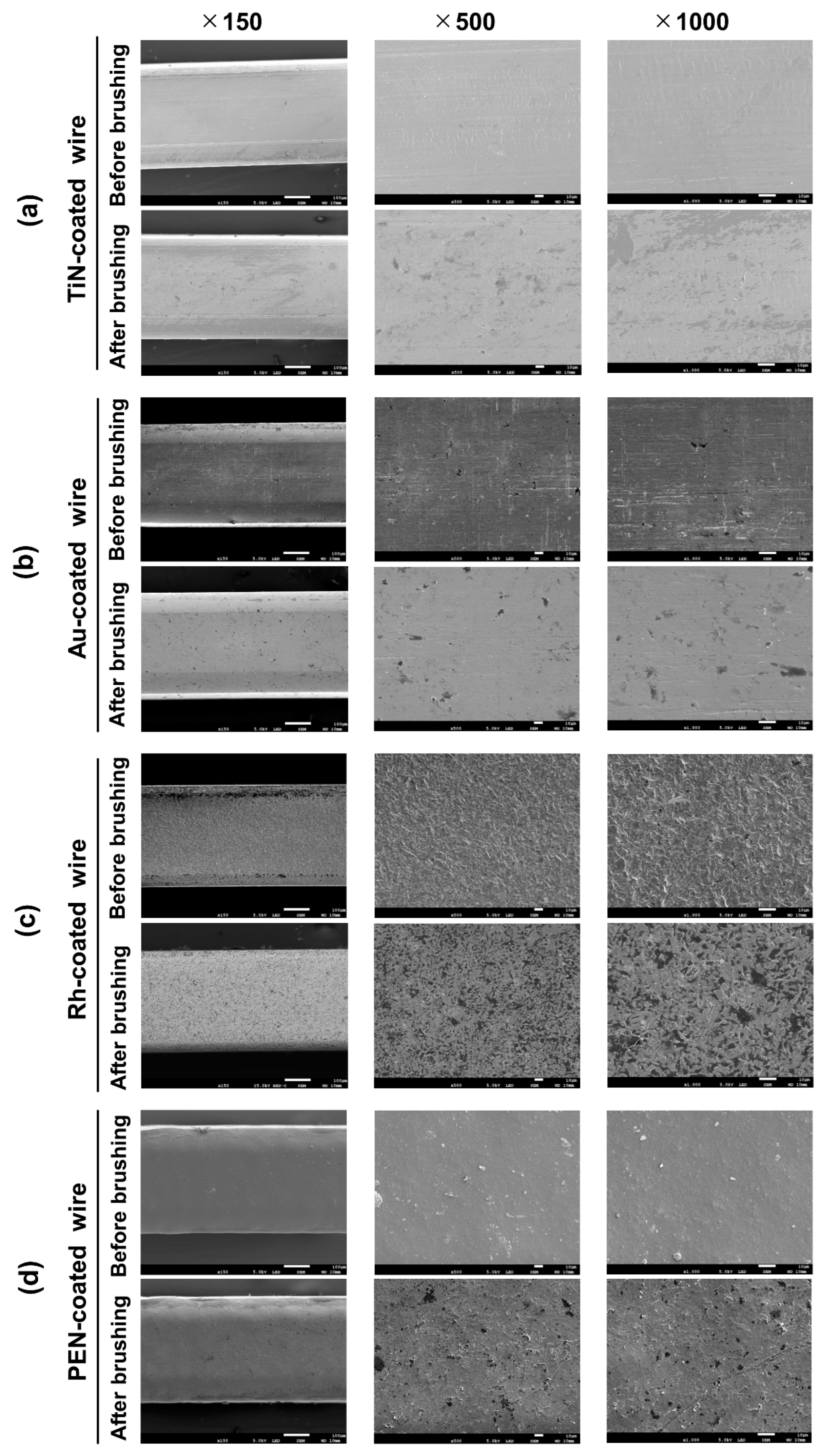
2.4. Observation of the Wire Surface
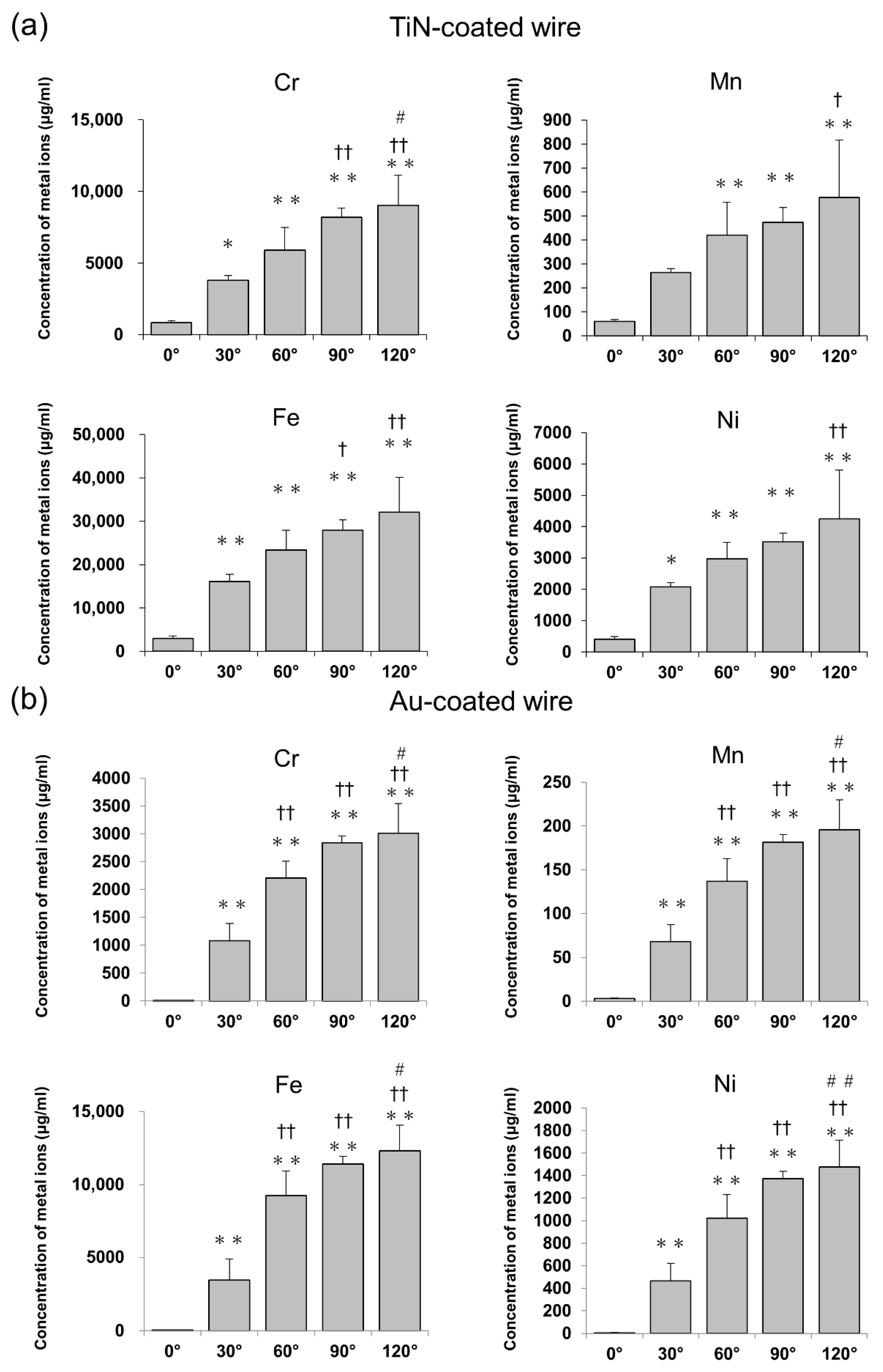
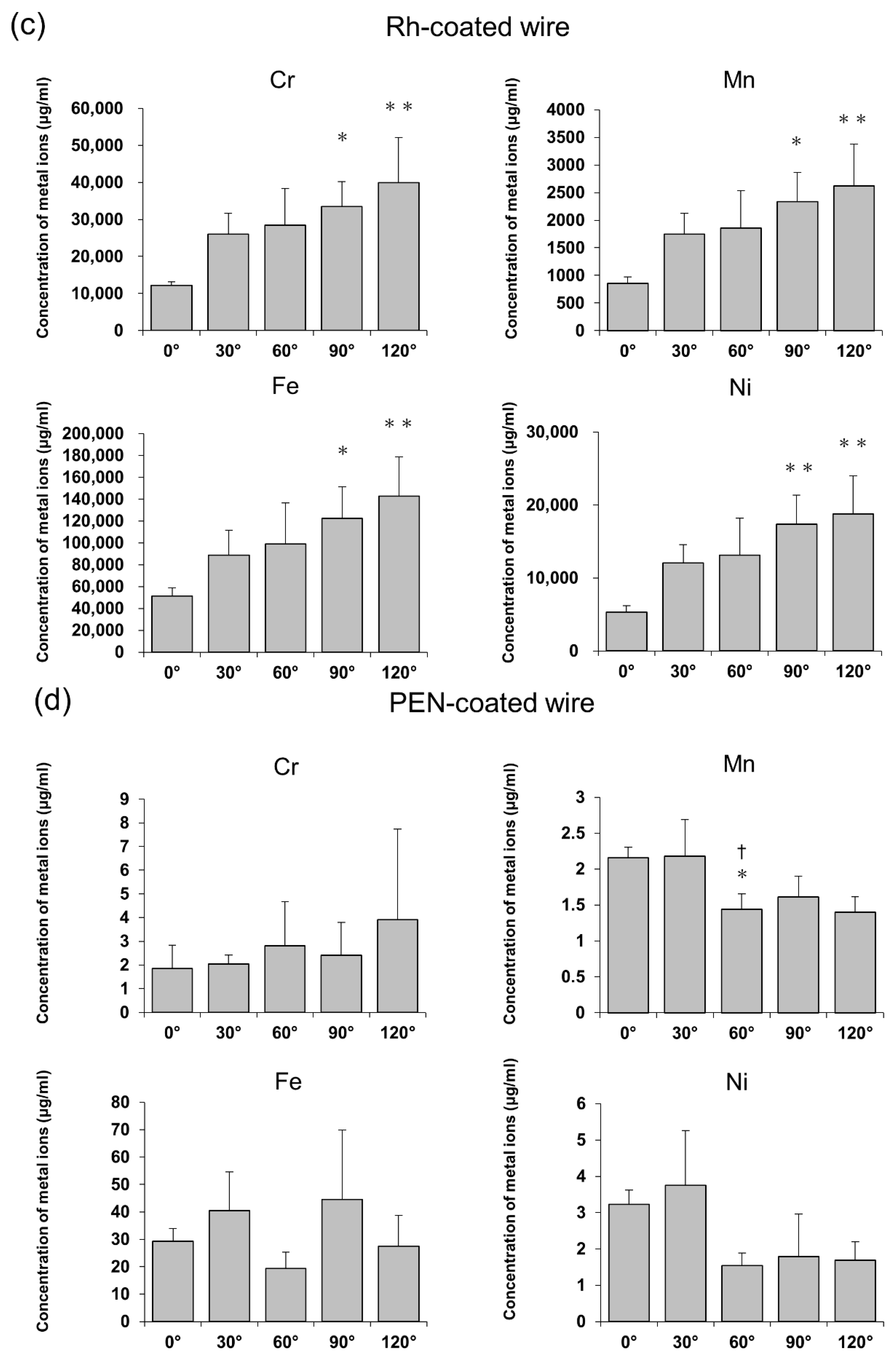
2.5. Metal Ion Release Tests
2.6. EDS Analysis
2.7. Measurement of Surface Roughness
2.8. Statistics
3. Results
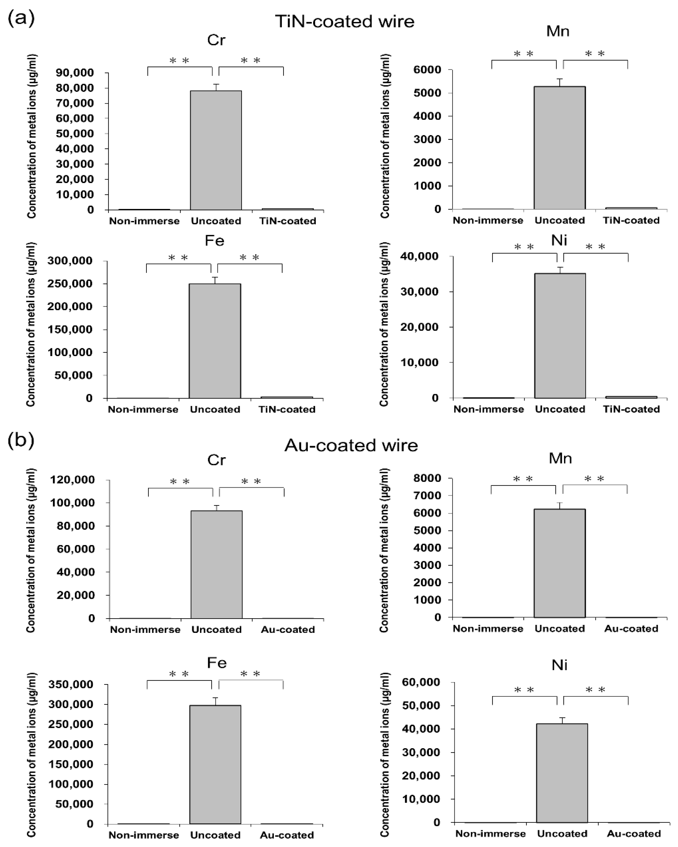
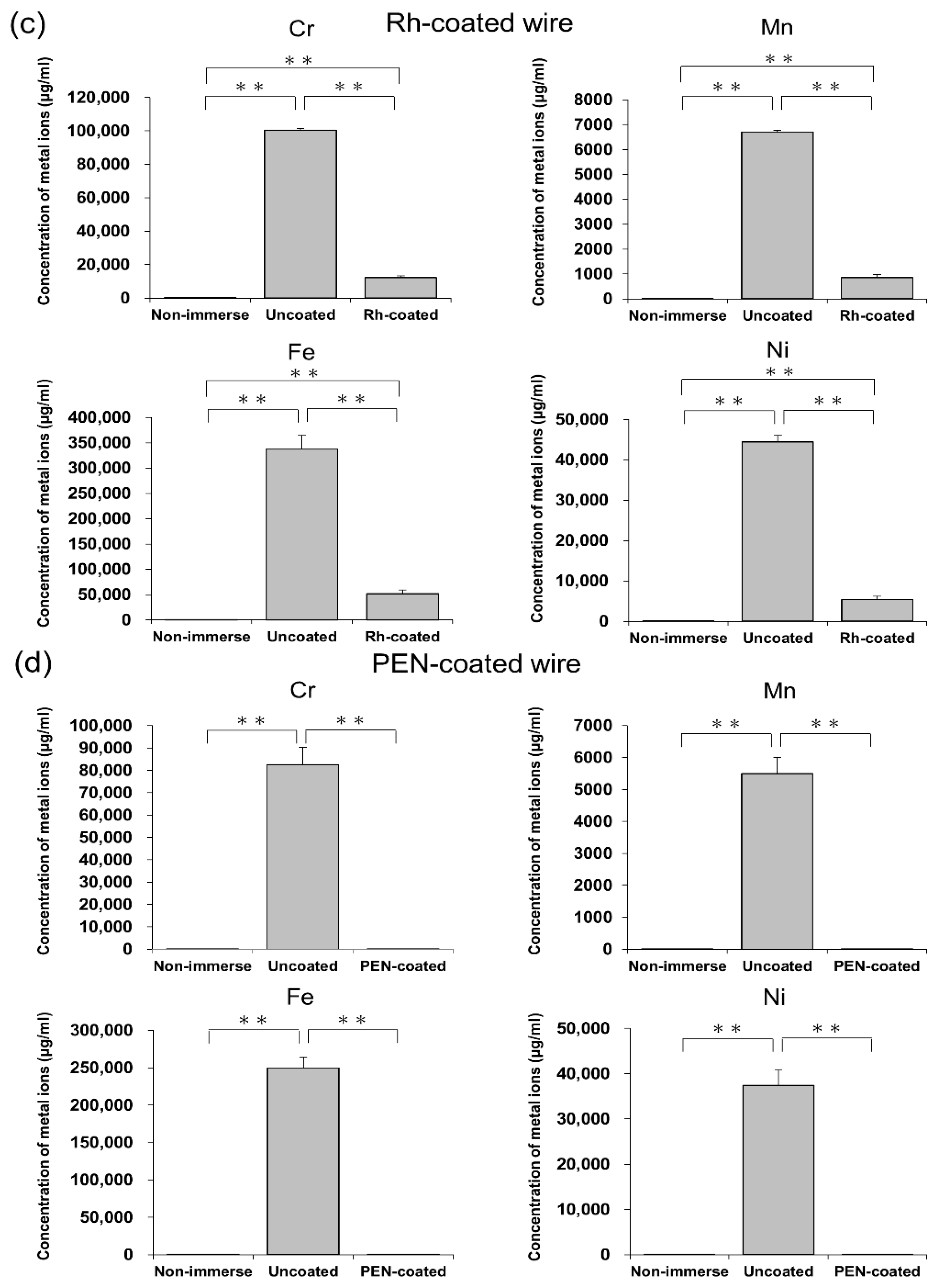
3.1. Analysis of Acid Corrosion
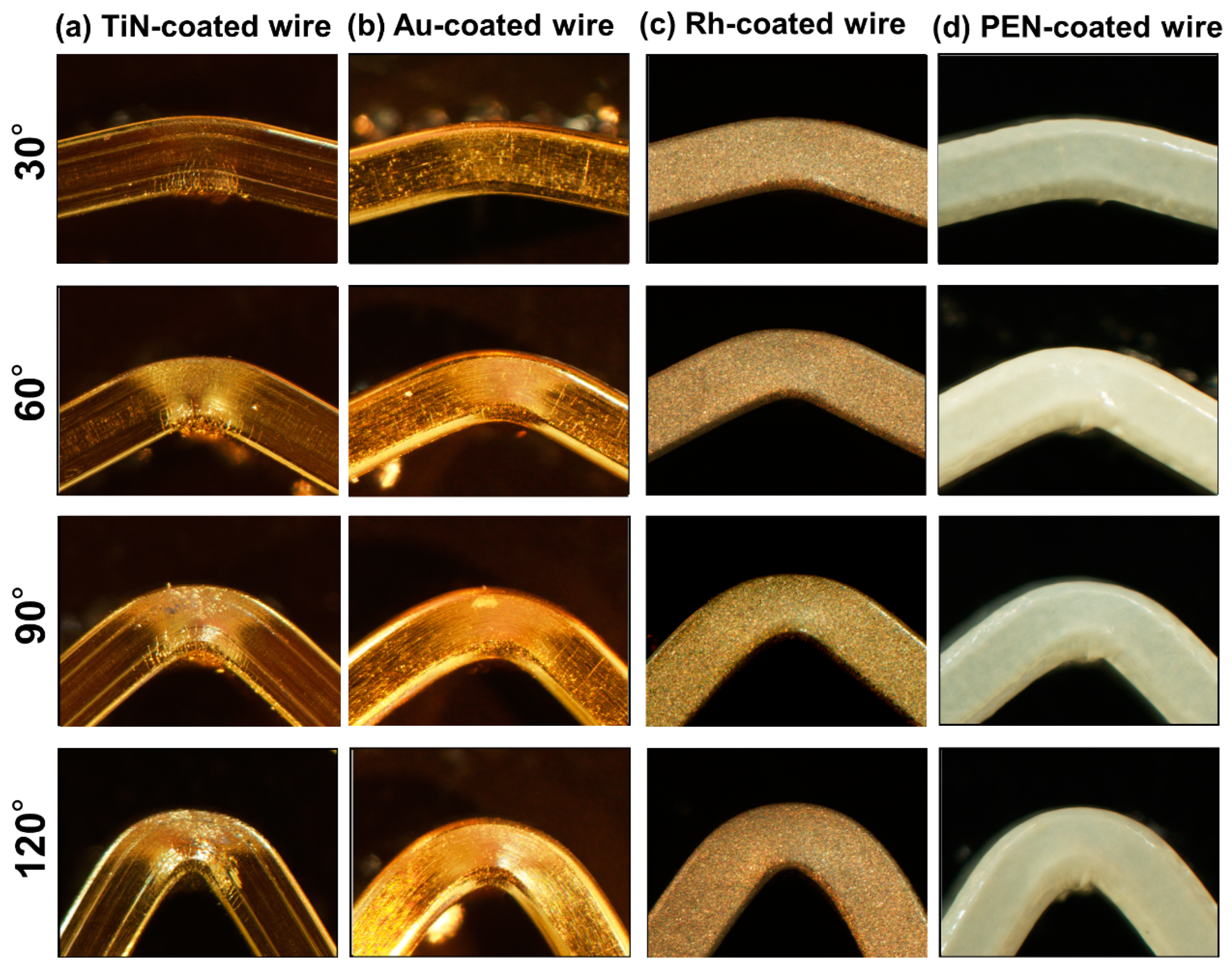
3.2. Analysis of Coating Loss by Wire Bending
3.3. Analysis of Coating Loss by Brushing
3.4. Analysis of Surface Roughness
4. Discussion
5. Conclusions
Author Contributions
Funding
Institutional Review Board Statement
Informed Consent Statement
Data Availability Statement
Conflicts of Interest
Abbreviations
| Au | gold |
| Co | cobalt |
| Co-Cr | cobalt-chromium |
| Cr | chromium |
| CrN | chromium nitride |
| EDS | energy dispersive spectrometry |
| Fe | iron |
| ICP–MS | inductively coupled plasma mass spectrometry |
| Mn | manganese |
| Ni | nickel |
| Ni-Ti | nickel titanium |
| PEN | polyethylene naphthalate |
| Rh | rhodium |
| SEM | scanning electron microscopy |
| SS | stainless steel |
| TiN | titanium nitride |
| β-Ti | β-titanium |
References
- Mortz, C.; Bindslev-Jensen, C.; Andersen, K. Prevalence, incidence rates and persistence of contact allergy and allergic contact dermatitis in The Odense Adolescence Cohort Study: A 15-year follow-up. Br. J. Dermatol. 2012, 168, 318–325. [Google Scholar] [CrossRef]
- Kusy, R.P. A review of contemporary archwires: Their properties and characteristics. Angle Orthod. 1997, 67, 197–207. [Google Scholar] [CrossRef] [PubMed]
- Hida, M.; Miyazawa, K.; Tsuruta, S.; Kurosawa, M.; Hata, Y.; Kawai, T.; Goto, S. Effect of heat treatment conditions on the mechanical properties of Ti-6Mo-4Sn alloy for orthodontic wires. Dent. Mater. J. 2013, 32, 462–467. [Google Scholar] [CrossRef]
- Hwang, C.-J.; Shin, J.-S.; Cha, J.Y. Metal release from simulated fixed orthodontic appliances. Am. J. Orthod. Dentofac. Orthop. 2001, 120, 383–391. [Google Scholar] [CrossRef]
- Fors, R.; Persson, M. Nickel in dental plaque and saliva in patients with and without orthodontic appliances. Eur. J. Orthod. 2006, 28, 292–297. [Google Scholar] [CrossRef]
- Arango, S.; Peláez-Vargas, A.; García, C. Coating and Surface Treatments on Orthodontic Metallic Materials. Coatings 2012, 3, 1–15. [Google Scholar] [CrossRef]
- Kishore, S.; Felicita, A.S.; Siva, S. Evaluation of Nickel Release in Blood and Periodontal Tissue with the Use of NiTi Wires, Bands and Brackets in Orthodontics—A Systematic Review. J. Evol. Med. Dent. Sci. 2021, 10, 1539–1546. [Google Scholar] [CrossRef]
- Al Jabbari, Y.S.; Fehrman, J.; Barnes, A.C.; Zapf, A.M.; Zinelis, S.; Berzins, D.W. Titanium Nitride and Nitrogen Ion Implanted Coated Dental Materials. Coatings 2012, 2, 160–178. [Google Scholar] [CrossRef]
- Ito, A.; Kitaura, H.; Sugisawa, H.; Noguchi, T.; Ohori, F.; Mizoguchi, I. Titanium Nitride Plating Reduces Nickel Ion Release from Orthodontic Wire. Appl. Sci. 2021, 11, 9745. [Google Scholar] [CrossRef]
- Sugisawa, H.; Kitaura, H.; Ueda, K.; Kimura, K.; Ishida, M.; Ochi, Y.; Kishikawa, A.; Ogawa, S.; Takano-Yamamoto, T. Corrosion resistance and mechanical properties of titanium nitride plating on orthodontic wires. Dent. Mater. J. 2018, 37, 286–292. [Google Scholar] [CrossRef]
- Katic, V.; Curkovic, L.; Bosnjak, M.U.; Peros, K.; Mandic, D.; Spalj, S. Effect of pH, fluoride and hydrofluoric acid concentration on ion release from NiTi wires with various coatings. Dent. Mater. J. 2017, 36, 149–156. [Google Scholar] [CrossRef][Green Version]
- de Amorim, M.C.; Gomes, S.d.R.; da Silva, B.P.; Aoki, I.V.; Basting, R.T. Surface Micromorphology, Ion Release and Resistance to Corrosion of Orthodontic Wires Aesthetic Coating Subject to Degradation. J. Bio-Tribo-Corrosion 2021, 8, 22. [Google Scholar] [CrossRef]
- Da Silva, D.L.; Mattos, C.T.; Simão, R.A.; Ruellas, A.C.D.O. Coating stability and surface characteristics of esthetic orthodontic coated archwires. Angle Orthod. 2013, 83, 994–1001. [Google Scholar] [CrossRef]
- Wu, H.; Yang, J.; Yan, Y.; Zheng, B.; Algahefi, A.L.; Ma, S.; Liu, Y. Study of Al–SiO2 aesthetic composite coating on orthodontic metal archwire. Coatings 2022, 12, 746. [Google Scholar] [CrossRef]
- Abdulkader, Y.C.; Kamaruddin, A.F.; Mydin, R.B.S. Effects of salivary pH on coating durability of two different aesthetic archwire coatings under a simulated intraoral environment. Saudi Dent. J. 2019, 32, 306–313. [Google Scholar] [CrossRef]
- Osmani, Z.J.; Poljšak, B.; Zelenika, S.; Kamenar, E.; Marković, K.; Perčić, M.; Katić, V. Ion release and surface changes of nickel–titanium archwires induced by changes in the pH value of the saliva—Significance for human health risk assessment. Materials 2022, 15, 1994. [Google Scholar] [CrossRef]
- Muguruma, T.; Iijima, M.; Kawaguchi, M.; Mizoguchi, I. Effects of Sp2/Sp3 ratio and hydrogen content on in vitro bending and frictional performance of DLC-coated orthodontic stainless steels. Coatings 2018, 8, 199. [Google Scholar] [CrossRef]
- Shirakawa, N.; Iwata, T.; Miyake, S.; Otuka, T.; Koizumi, S.; Kawata, T. Mechanical properties of orthodontic wires covered with a polyether ether ketone tube. Angle Orthod. 2018, 88, 442–449. [Google Scholar] [CrossRef]
- Krishnan, V.; Krishnan, A.; Remya, R.; Ravikumar, K.; Nair, S.A.; Shibli, S.; Varma, H.; Sukumaran, K.; Kumar, K.J. Development and evaluation of two PVD-coated β-titanium orthodontic archwires for fluoride-induced corrosion protection. Acta Biomater. 2011, 7, 1913–1927. [Google Scholar] [CrossRef]
- Arici, N.; Akdeniz, B.S.; Oz, A.A.; Gencer, Y.; Tarakci, M.; Arici, S. Effectiveness of medical coating materials in decreasing friction between orthodontic brackets and archwires. Korean J. Orthod. 2021, 51, 270–281. [Google Scholar] [CrossRef]
- Bard, A.J.; Parsons, R.; Jordan, J. Standard Potentials in Aqueous Solution, 1st ed.; Routledge: London, UK, 2017; ISBN 978-0-203-73876-4. [Google Scholar]
- Leonard, L. Advances in engineering plastics. Adv. Mater. Process 1997, 152, 29–32. [Google Scholar]
- Lillwitz, L. Production of dimethyl-2,6-naphthalenedicarboxylate: Precursor to polyethylene naphthalate. Appl. Catal. A Gen. 2001, 221, 337–358. [Google Scholar] [CrossRef]
- Hilbert, L.R.; Bagge-Ravn, D.; Kold, J.; Gram, L. Influence of surface roughness of stainless steel on microbial adhesion and corrosion resistance. Int. Biodeterior. Biodegrad. 2003, 52, 175–185. [Google Scholar] [CrossRef]
- Gornostyrev, Y.N.; Katsnelson, M.I.; Medvedeva, N.I.; Mryasov, O.N.; Freeman, A.J.; Trefilov, A.V. Peculiarities of defect structure and mechanical properties of iridium: Results of ab initio electronic structure calculations. Phys. Rev. B 2000, 62, 7802–7808. [Google Scholar] [CrossRef]
- Latella, B.; Gan, B.; Davies, K.; McKenzie, D.; McCulloch, D. Titanium nitride/vanadium nitride alloy coatings: Mechanical properties and adhesion characteristics. Surf. Coat. Technol. 2006, 200, 3605–3611. [Google Scholar] [CrossRef]
- Morita, T.; Takahashi, H.; Shimizu, M.; Kawasaki, K. Factors controlling the fatigue strength of nitrided titanium. Fatigue Fract. Eng. Mater. Struct. 1997, 20, 85–92. [Google Scholar] [CrossRef]
- Karayannidis, G.P.; Papachristos, N.; Bikiaris, D.N.; Papageorgiou, G.Z. Synthesis, crystallization and tensile properties of poly(ethylene terephthalate-co-2,6-naphthalate)s with low naphthalate units content. Polymer 2003, 44, 7801–7808. [Google Scholar] [CrossRef]
- Collier, S.; Pandis, N.; Johal, A.; Qureshi, U.; Sharma, P.K.; Fleming, P.S. A prospective cohort study assessing the appearance of retrieved aesthetic orthodontic archwires. Orthod. Craniofacial Res. 2017, 21, 27–32. [Google Scholar] [CrossRef] [PubMed]
- Ozkomur, A.; Erbil, M.; Akova, T. Diamondlike Carbon Coating as a Galvanic Corrosion Barrier Between Dental Implant Abutments and Nickel-Chromium Superstructures. Int. J. Oral Maxillofac. Implant. 2013, 28, 1037–1047. [Google Scholar] [CrossRef] [PubMed][Green Version]
- Li, M.; Luo, S.; Zeng, C.; Shen, J.; Lin, H.; Cao, C. Corrosion behavior of TiN coated type 316 stainless steel in simulated PEMFC environments. Corros. Sci. 2004, 46, 1369–1380. [Google Scholar] [CrossRef]


| TiN-Coated Wire | Au-Coated Wire | Rh-Coated Wire | PEN-Coated Wire | |
|---|---|---|---|---|
| Ra (μm) | 0.061 | 0.028 | 0.074 | 0.532 |
| SD | 0.007 | 0.004 | 0.002 | 0.014 |
Publisher’s Note: MDPI stays neutral with regard to jurisdictional claims in published maps and institutional affiliations. |
© 2022 by the authors. Licensee MDPI, Basel, Switzerland. This article is an open access article distributed under the terms and conditions of the Creative Commons Attribution (CC BY) license (https://creativecommons.org/licenses/by/4.0/).
Share and Cite
Ito, A.; Kitaura, H.; Noguchi, T.; Ohori, F.; Mizoguchi, I. Analysis of Coating Loss from Coated Stainless Steel Orthodontic Wire. Appl. Sci. 2022, 12, 9497. https://doi.org/10.3390/app12199497
Ito A, Kitaura H, Noguchi T, Ohori F, Mizoguchi I. Analysis of Coating Loss from Coated Stainless Steel Orthodontic Wire. Applied Sciences. 2022; 12(19):9497. https://doi.org/10.3390/app12199497
Chicago/Turabian StyleIto, Arata, Hideki Kitaura, Takahiro Noguchi, Fumitoshi Ohori, and Itaru Mizoguchi. 2022. "Analysis of Coating Loss from Coated Stainless Steel Orthodontic Wire" Applied Sciences 12, no. 19: 9497. https://doi.org/10.3390/app12199497
APA StyleIto, A., Kitaura, H., Noguchi, T., Ohori, F., & Mizoguchi, I. (2022). Analysis of Coating Loss from Coated Stainless Steel Orthodontic Wire. Applied Sciences, 12(19), 9497. https://doi.org/10.3390/app12199497