Article

Protective Effect of Ectoin on UVA/H2O2-Induced Oxidative Damage in Human Skin Fibroblast Cells

1
Beijing Key Laboratory of Plant Resource Research and Development, College of Chemistry and Materials Engineering, Beijing Technology and Business University, Beijing 100048, China
2
Institute of Cosmetic Regulatory Science, Beijing Technology and Business University, Beijing 100048, China
3
Yunnan Baiyao Group Co., Ltd., Kunming 650000, China
*
Authors to whom correspondence should be addressed.
These authors contributed equally to this work.
Appl. Sci. 2022, 12(17), 8531; https://doi.org/10.3390/app12178531
Submission received: 3 August 2022 / Revised: 18 August 2022 / Accepted: 24 August 2022 / Published: 26 August 2022

Abstract

:
Ectoin is an amino acid derivative that can create a balance between the osmotic pressure of cells and can protect enzymes, DNA proteins, and nucleic acids under extreme conditions. Ectoin has also been reported to slow skin aging. However, there are few reports on the protective effect of Ectoin on oxidative damage, especially on the regulation of PI3K/AKT-pathway-related genes at the mRNA level. UVA-induced oxidative stress injury and H2O2-induced oxidative stress injury are two common oxidative stress injury models. Skin fibroblasts produce a large number of ROS following excessive UV radiation or oxidative stimulation by H2O2, which further inhibits cell proliferation and causes cell apoptosis. In this study, UVA- and H2O2-induced oxidation models of human skin fibroblasts were established separately to investigate the protective effect of Ectoin. Further studies on the mechanisms involved, for example, the expression levels of genes associated with the PI3K/AKT signaling pathway and levels of antioxidant enzymes in cells, were determined. We found that Ectoin upregulated genes associated with the PI3K/AKT signaling pathway, including COL1A1, COL1A2, FN1, IGF2, NR4A1, and PIK3R1, but decreased intracellular ROS levels and malondialdehyde (MDA), while increasing the activities of superoxide dismutase (SOD) and glutathione peroxidase (GSH-Px). In conclusion, our results indicate that Ectoin exerts protective properties by the upregulated genes COL1A1, COL1A2, FN1, IGF2, NR4A1, and PIK3R1 and upregulating antioxidative enzyme levels.

1. Introduction

Fibroblasts play a major role in supporting many tissue structures and are important cells in the dermis that can secrete cytokines and thus participate in multiple healing stages [1,2,3]. Human skin fibroblasts (HSFs) are the main cell component in the dermis of human skin and form the main body of the dermis together with its own secreted elastic fibers, collagen fibers, and matrix components [4]. Numerous studies have demonstrated that HSF cells are important for the skin aging process [5,6,7]. UVA-induced oxidative stress damage and H2O2-induced oxidative stress damage are two common models of oxidative stress damage. When UVA/H2O2 stimulates HSF cells, the cells undergo oxidative injury, and the composition of antioxidant-related enzymes in the cells changes. Therefore, scientific research often establishes oxidative damage models to evaluate the efficacy of active substances. Fibroblasts produce a large amount of active oxygen following excessive ultraviolet radiation based on the dose of ultraviolet light, while H2O2 is the main component of active oxygen [8,9]. At very high concentrations, reactive oxygen species inhibit cell proliferation and lead to cell apoptosis [10]. UVA and H2O2 eventually cause oxidative stress damage to the skin [11,12,13,14].
Protecting the tissues from unwanted side effects is one strategy to lessen the unfavorable consequences of UVA and H2O2 in medical practice. As a result, the therapeutic benefits of natural compounds with antioxidant capabilities that may lessen the severity of UVA and H2O2-induced toxicities could be advantageous. Antioxidants are widely utilized as nutrients, and their effectiveness in reducing tissue and organ toxicity from various medicines was studied. Numerous studies have looked at the effectiveness of antioxidants as nutrients in reducing tissue and organ toxicity caused by a variety of conditions. Reactive oxygen species damage can be avoided and repaired by endogenous and exogenous antioxidants (ROS). They are referred to as “free radical scavengers” because they can strengthen the immune system and reduce the danger of sickness and toxins. Superoxide and other peroxides are chelated by enzyme-based antioxidants, such as catalase (CAT), glutathione peroxidase (GPx), and superoxide dismutase (SOD). They serve as defense mechanisms against endogenous antioxidants, clearing ROS activity and buildup in cells and preserving redox balance [15,16,17,18].
The PI3K/AKT signaling pathway is implicated in many key cell functions, such as protein synthesis, cell cycle, apoptosis, drug-resistant growth factors (EGF, NGF, and VEGF), hormones (prostaglandins and PGE2), and cytokine stimulation [19,20,21]. Collagen type I α 1 chain (COL1A1) and Collagen type I α 2 chain (COL1A2) encode the front α1 and α2 chains of type I collagen [22]. Collagen type IV α 5 chain (COL4A5) encodes one of the six subunits of type IV collagen, which is the main structural component of the basement membrane. Fibronectin 1 (FN1) is a kind of high-molecular-weight glycoprotein involved in a variety of biological functions, and its overexpression inhibits cell apoptosis by activating the PI3K/AKT signaling pathway [23,24].
Ectoin is an amino acid derivative that can balance the osmotic pressure of cells and can also protect enzymes, DNA proteins, and nucleic acids under extreme conditions, such as high levels of radiation [25]. Ectoin can reduce the effects of UVA-induced damage in human keratinocytes and fibroblasts by preventing the release of secondary messengers, the activation of AP-2, the expression of intercellular adhesion molecule-1, and the mutation of mitochondrial DNA [26]. Researchers suggest that Ectoin may also play an important role in increasing the activity of antioxidant enzymes and the level of nonenzymatic antioxidants. Research also shows that Ectoin plays an important role in stabilizing the cell membrane, reducing inflammation, and mitigating DNA damage induced by ultraviolet, infrared, and visible radiations. However, the detailed mechanisms involved in these processes have still not been fully understood [27,28].
Topically applied to the skin, Ectoin not only exerts a cellular protective effect but also exerts a skin-protective barrier function. Heinrich et al. assessed an Ectoin formulation with regard to its antiaging properties and found that 2% Ectoin was more effective in terms of skin hydration, skin elasticity, and skin surface structure than the vehicle treatment [29]. Furthermore, Ectoin can be used for the treatment of mild to moderate atopic dermatitis by improving skin moisture and strengthening skin barrier function [30,31].
However, the skin-protective effects of Ectoin are well documented, and the underlying mechanisms have still not been fully understood. There is a lack of reports that discuss insight into the mechanisms of Ectoin, especially changes in the expressions of genes involved in the PI3K/AKT pathway.
In this study, we established UVA-induced and H2O2-induced oxidative models of human skin fibroblast cells separately, discuss the protective effects exerted by Ectoin in the two models, and further studied the molecular mechanisms. The cellular levels of the antioxidant enzymes and oxidative products were also measured. Overall, this study provides a theoretical basis for the application of Ectoin for the treatment of antiskin oxidative damage.

2. Materials and Methods

2.1. Materials

HSFs were bought from the Cell Resource Center, Institute of Basic Medicine, Chinese Academy of Medical Sciences. Ectoin (purity ≥ 98%; Bloomage Biotechnology Co., Ltd., Beijing, China), Dulbecco’s Modified Eagle Medium (DMEM), fibroblast medium, fetal bovine serum (FBS), phosphate buffer saline (PBS), penicillin, streptomycin, and 0.25% (with EDTA) trypsin were all bought from Gibco Life Technologies (Carlsbad, CA, USA). RIPA buffer total protein extract (Sinogene, Beijing, China), 100× phosphatase inhibitors (Sinogene, Beijing, China), Bradford protein assay reagent (Sinogene, Beijing, China), and a TransStart® Top Green qPCR SuperMix kit were obtained from Beijing TransGen Biotech Co., Ltd. ROS, MDA, SOD, and GSH-Px kits were bought from Beyotime Biotechnology Co., Ltd.
All other chemicals were of analytical grade or complied with the standards required for cell culture experiments.

2.2. Cell Culture

Cells were cultured in DMEM, supplemented with 10% FBS, 1% fibroblast growth additives, and 1% penicillin (1 × 10⁵ U/L)–streptomycin (100 mg/L). The cells were incubated at 37 °C in a humidified atmosphere with 5% CO2. The culture medium was renewed when cell confluence reached 80%, and the cells were digested using 0.25% (with EDTA) trypsin.

2.3. Cell Viability

Cell viability was determined using the colorimetric CCK-8 method [32,33,34], following the manufacturer’s instructions. HSF cells were seeded into a 96-well plate at a concentration of 1 × 10⁵ cells per well at 37 °C and were incubated in a cell incubator (Shanghai Shengke Instrument Equipment Co., Ltd., Shanghai, China) with 5% CO2 for 12 h. The cells were treated with Ectoin at different concentrations ranging from 8 to 500 μg/mL for 24 h.

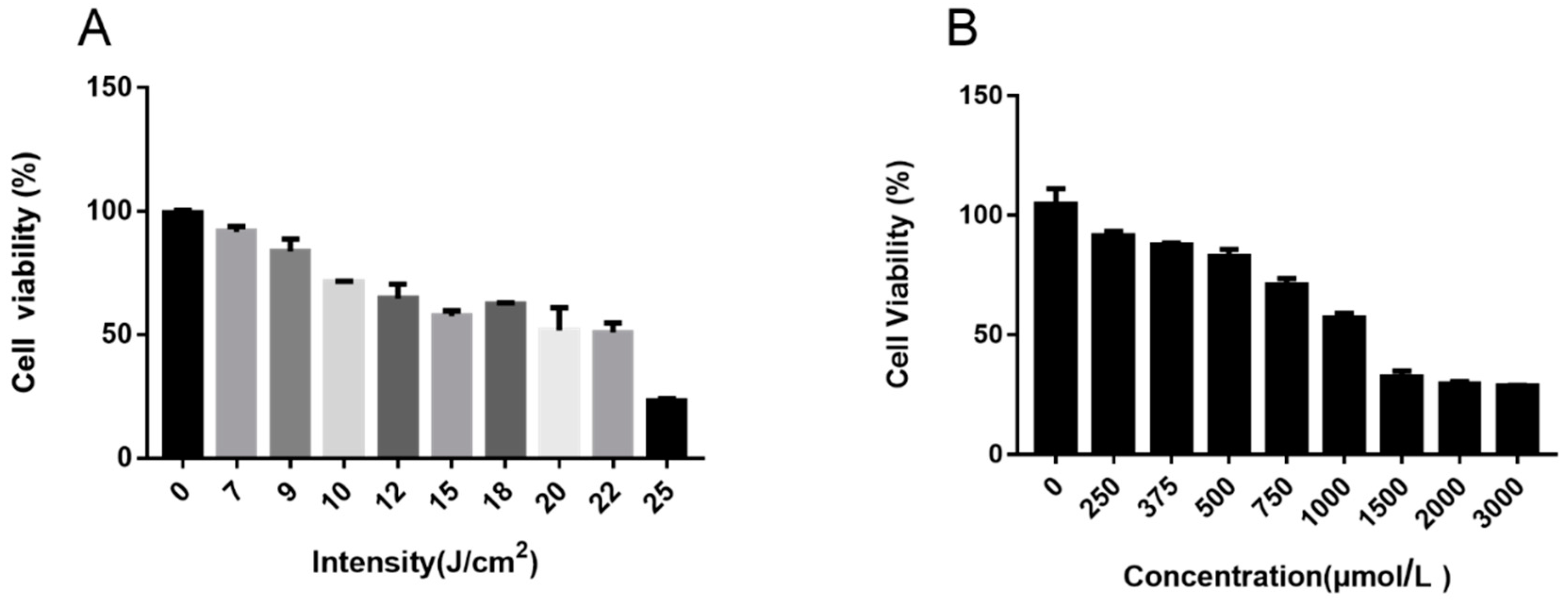
2.4. UVA and H2O2-Induced Model Establishment

UVA-induced model establishment: HSF cells were seeded into each well of a 96-well plate at a density of 1 × 10⁴ cells and cultured in a cell incubator for 12 h. The medium was replaced with 100 μL of PBS (pH 7.4, 0.01 M), and the cells were stimulated under different doses of UVA ranging from 7 to 25 J/cm2. UVA (365 nm) radiation was created using an UVA lamp (T8 40W, Royal Dutch Philips Electronics Ltd., Eindhoven, The Netherlands), and intensity was measured using an UV power meter (LS125, Shenzhen Shanglin Technology Co., Ltd., Shenzhen, China). IC50 parameters were chosen to establish the UVA-induced model.
H2O2-induced model establishment: The H2O2-induced model was established using the same method described above using different concentrations of H2O2 (250–3000 μmol·L−1) instead of PBS for different periods ranging up to 0.5 h. IC50 parameters were chosen to establish the H2O2-induced model.
The HSF cell viability was determined using the CCK-8 (Biorigin (Beijing) Inc., Beijing, China) method. Images of the cells were captured under a microscope (Shanghai Tucsen Vision Technology Co., Shanghai, China) to observe morphological changes.

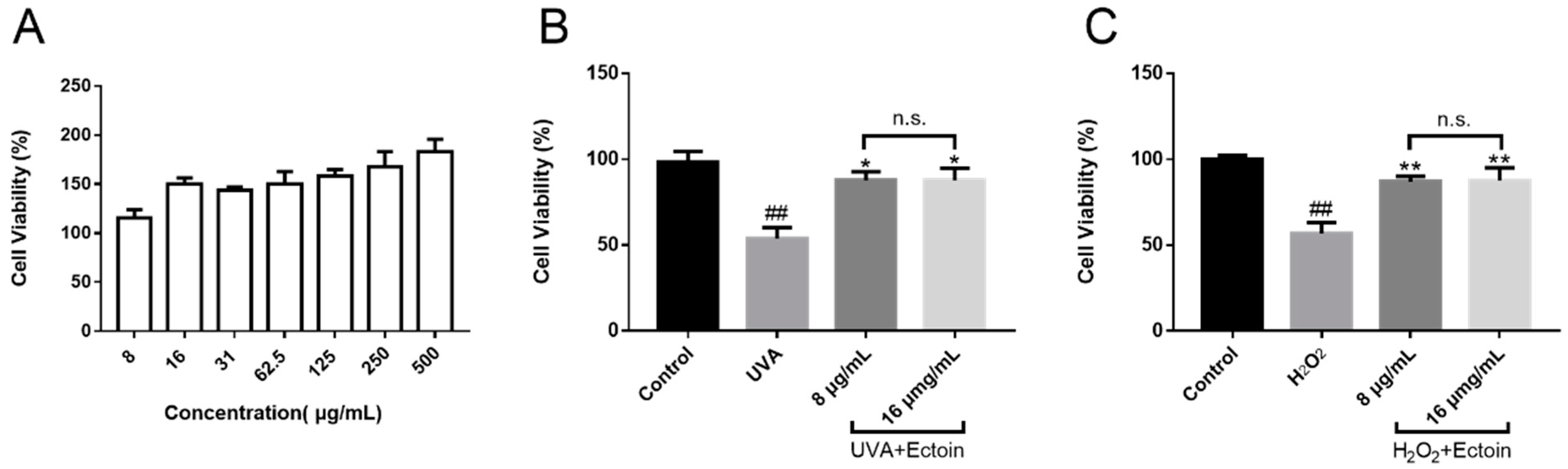
2.5. Protective Effects of Ectoin on UVA and H2O2-Induced Model

The cells were inoculated into 96-well plates at a density of 1 × 10⁴ and kept for 12 h to establish the UVA-induced or H2O2-induced oxidative models.
Then, different concentrations of Ectoin ranging from 8 to 500 μg/mL were added and incubated for different durations (24 h). Cell viability was calculated using the CCK-8 method.

2.6. RT-qPCR

The cells were collected after being subjected to the different treatments. Total RNA was extracted using the TriQuick Reagent (Beijing Solarbio Science and Technology Co., Ltd., Beijing, China), and the cDNA was synthesized following the manufacturer’s instructions. Reverse transcription-qPCR (RT-qPCR) was performed according to the instructions given in the TransStart® Top Green qPCR SuperMix kit. The reaction system was 20 μL in total. The specific reagents and dosages used are presented in Table S1, while the primers used are presented in Table 1. GAPDH was used as a normalization control.
The cyclic parameters used were predenaturation at 94 °C for 30 s and PCR reaction (45 cycles of 94 °C for 15 s, 60 °C for 15 s, and 72 °C for 10 s), and fluorescence data were collected at 72 °C. Analyses of the relative gene expression levels were performed using the 2−ΔΔCT method [35].

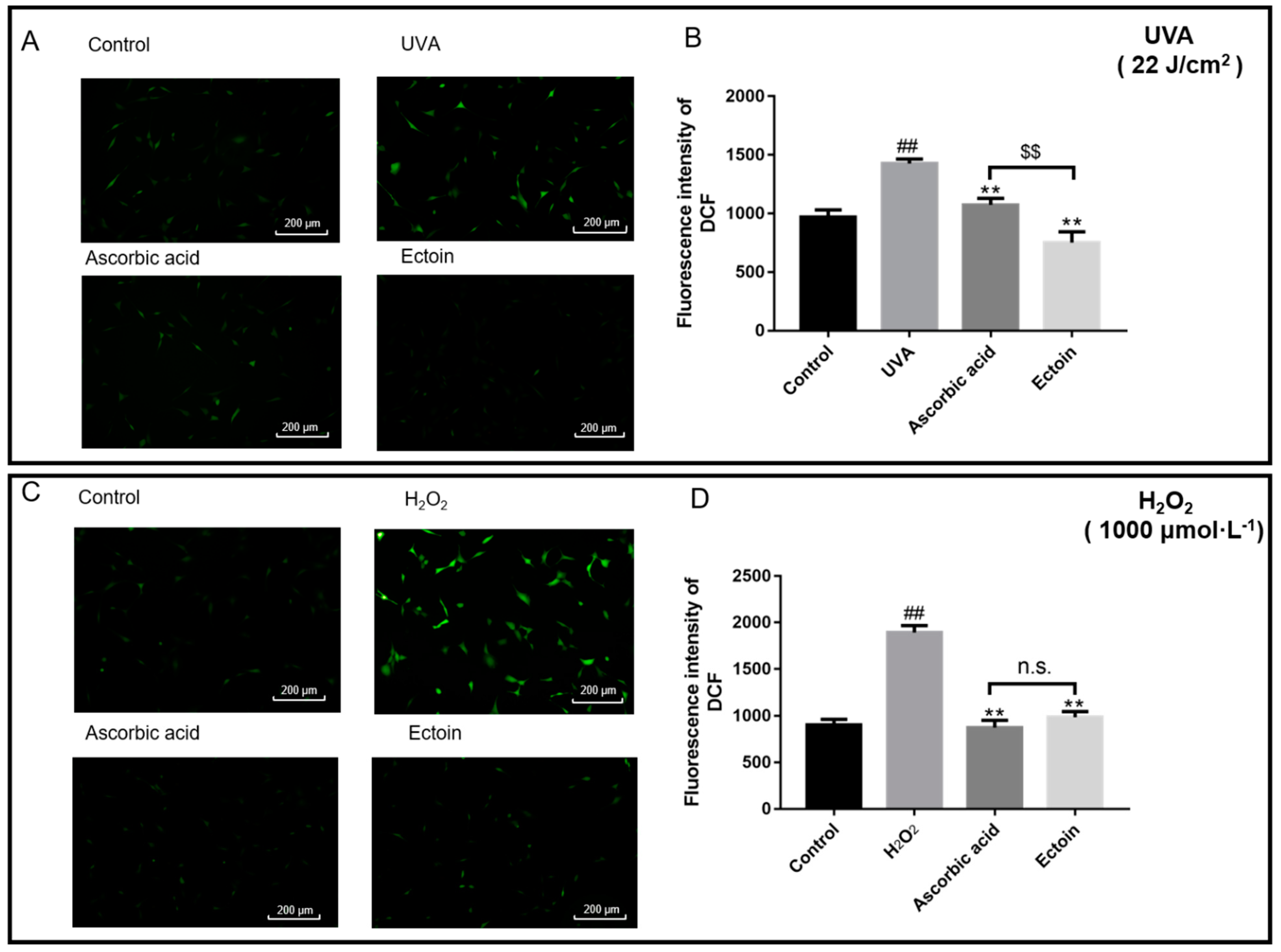
2.7. Determination of Intracellular Reactive Oxygen Species (ROS)

The production of intracellular ROS was determined using a fluorescence microscopy kit (Shanghai Tulsen Vision Technology Co., Ltd., Shanghai, China), following the manufacturer’s instructions. Then, 2′,7′-dichlorofluorescein diacetate (DCFH2-DA), which is readily taken up by cells and can be de-esterified to 2′,7′-dichlorodihydrofluorescein (DCFH2), was subsequently used. Thereafter, DCFH2 can be oxidized by intracellular ROS to form fluorescent 2′,7′-dichlorofluorescein (DCF). The cells were treated with different concentrations of Ectoin, which affected ROS levels and finally induced fluorescent changes.

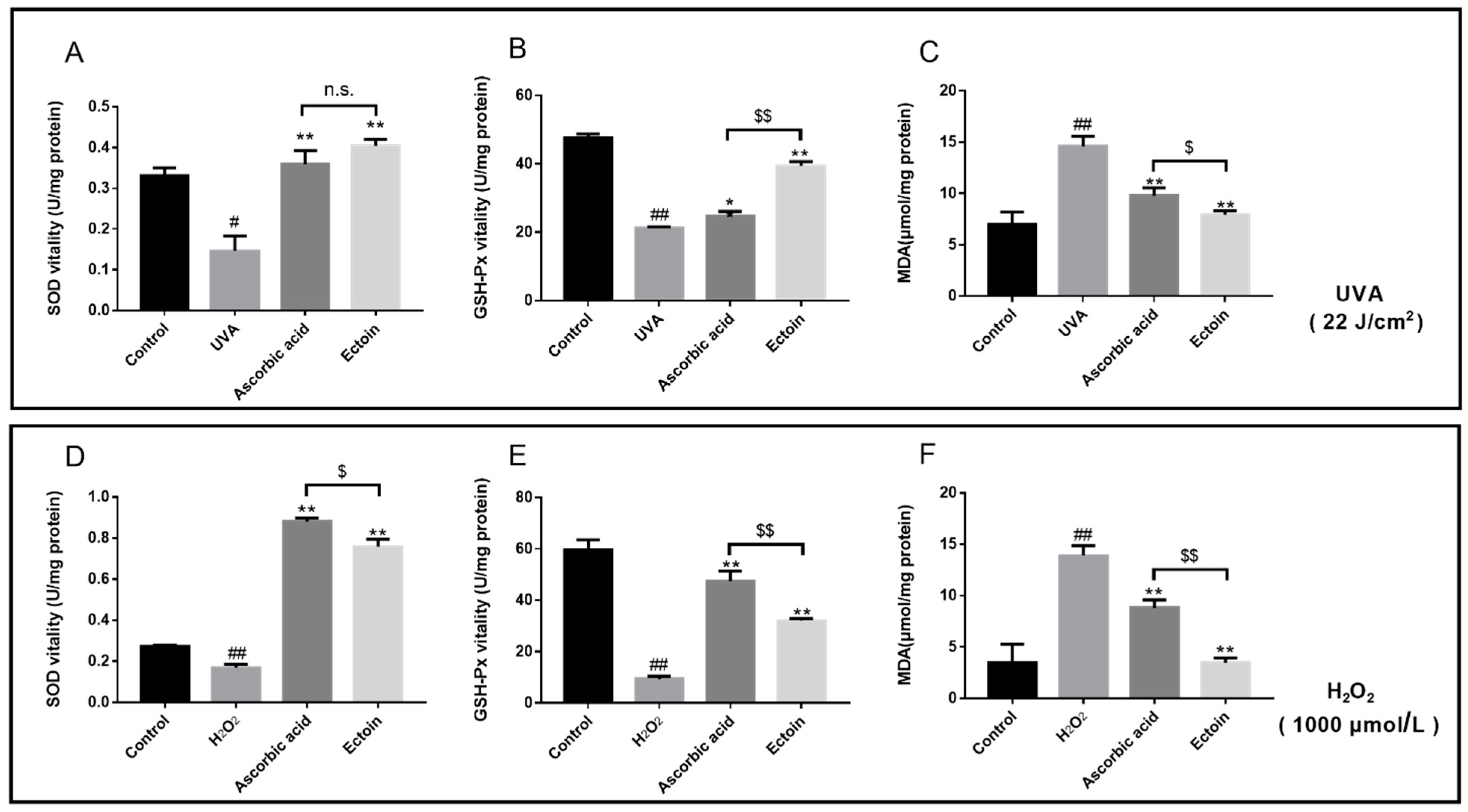
2.8. Determination of Intracellular GSH-Px, MDA, and SOD

The cells subjected to the different treatments were collected. GSH-Px, MDA, and SOD levels were detected using colorimetric reagent kits (Nanjing Jiancheng, Nanjing, China) following the manufacturer’s instructions. Ascorbic acid (60 μg/mL; Supplementary Figures S1 and S2) was chosen as a positive control. The cell viability after ascorbic acid treatment is given in the supplementary materials.

2.9. Statistics

All experiments were performed in triplicate at least, and data were expressed as mean ± standard deviation (SD). The data were analyzed using SPSS 17.0 (SPSS, Armonk, NY, USA) and GraphPad Prism 9.0 (GraphPad Software, San Diego, CA, USA) software. One-way analysis of variance was used to determine the significance of differences between the groups (#, * p < 0.05; ##, ** p < 0.01).

3. Results

3.1. Cell Viability of Ectoin and Oxidative Stress Damaged Cells

UVA/H2O2 can decrease cell viability and cause the oxidative damage of cells. As the dose of UVA or H2O2 was increased, the survival rate of the HSF cells gradually decreased (Figure 1).
Figure 1A shows the survival rates of the HSF cells after treatment with different doses of UVA. When the UVA radiation dose was 22 J/cm2, the cell survival rate was (50.89 ± 3.84)%. Figure 1B shows the survival rate of the HSF cells treated with 250–3000 μmol·L−1 H2O2 for 0.5 h. After treatment with 1000 μmol·L−1 H2O2 for 0.5 h, the cell survival rate decreased to (57 ± 2.03)%. Therefore, we chose to use IC50 parameters to establish the damage models. UVA at a dose of 20 J/cm2 was used to establish the UVA-induced oxidative stress damage model. The modeling condition of the H2O2-induced oxidative stress damage model was 1000 μmol·L−1 H2O2 for 0.5 h.
Ectoin protects cells from damage by UVA/H2O2 and improves cell viability. Ectoin at a concentration ranging from 8 to 500 μg/mL is nontoxic to HSF cells (Figure 2A) and is able to exert a good promotion effect on cell proliferation. Ectoin at a concentration ranging from 8 to 125 μg/mL for 24 h was able to significantly promote the proliferation of the HSF cells in a time-dependent manner. Two different concentrations (8 and 16 μg/mL for 24 h, separately) were used in subsequent experiments. Both 8 and 16 μg/mL of Ectoin enhanced cell viability under UVA or H2O2 stimulation, and there was no significant difference in cell viability between either of the two concentrations of Ectoin (Figure 2B,C).
Therefore, to facilitate the comparison of the protective effect of the same concentration of Ectoin exerted during UVA/H2O2 oxidative damage, 8 μg/mL of Ectoin was selected as the treatment concentration to be used on subsequent UVA and H2O2 damage models.

3.2. Antioxidant Enzymes and ROS Levels of Oxidative Stress Damaged Cells

As shown in Figure 3A,B,D,E, the SOD and GSH-Px vitalities reduced significantly following UVA/H2O2 damage (both p < 0.05), while MDA levels increased significantly, compared with the UVA-induced or H2O2-induced models (both p < 0.05, Figure 3C,F). Both Ectoin and ascorbic acid could significantly increase the SOD and GSH-Px activities, which had decreased due to UVA/H2O2 damage (p < 0.05, Figure 3A,B,D,E). In addition, there was no significance difference between the abilities of Ectoin and ascorbic acid to increase SOD activity, compared with the UVA-induced model. Moreover, Ectoin exerted a stronger increasing effect (p < 0.01, Figure 3B) on the UVA-induced decrease in GSH-Px levels than ascorbic acid.
In addition, Ectoin and ascorbic acid caused a significant change in reducing MDA levels, compared with the UVA-induced model. A more significant decrease in Ectoin treatment was observed on the UVA/H2O2-induced accumulation of MDA, compared with the positive control (Figure 3F).
UVA/H2O2 treatment could induce the production of cellular ROS. The fluorescent changes represented showed the changes in ROS levels. Fluorescence intensity was observed to have increased along with the formation of ROS. After UVA/H2O2 stimulation, the release of ROS in the cells of the model groups increased (Figure 4A,C). DCF fluorescence intensity is shown in Figure 4B,D. Then, a decrease in cell fluorescence intensity was observed after the damaged cells were treated with Ectoin or ascorbic acid, which indicates that Ectoin could reduce ROS levels.

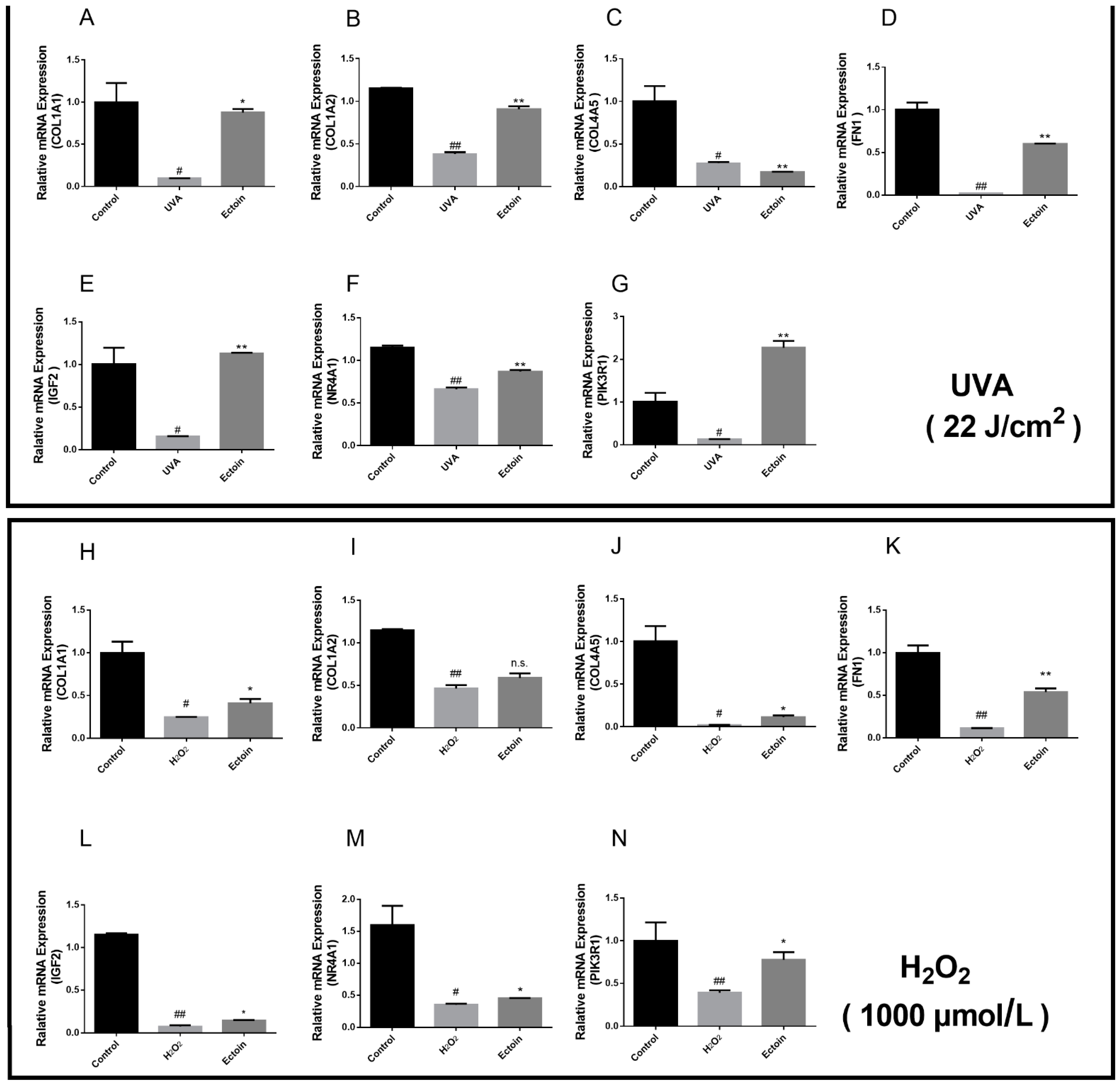
3.3. The Effects of Ectoin on the Expression of Related Genes in the PI3K/AKT Signaling Pathway

The UVA/H2O2-injured cells were treated with Ectoin for 24 h, and then the expression levels of COL1A1, COL1A2, COL4A5, FN1, IGF2, NR4A1, and PIK3R1 were detected. The results are shown in Figure 5. It can be observed that all seven genes were significantly downregulated after UVA injury or H2O2 injury (Figure 4).
Ectoin increased the expression levels of COL1A1 (Figure 5A), COL1A2 (Figure 5B), FN1 (Figure 5D), IGF2 (Figure 5E), NR4A1 (Figure 5F), and PIK3R1 (Figure 5G) in the model induced by UVA irradiation. However, the expression of COL4A5 (Figure 5C) decreased after Ectoin treatment.
In the model induced by H2O2, Ectoin increased the expression levels of COL1A1 (Figure 5H), COL4A5 (Figure 5J), FN1 (Figure 5K), IGF2 (Figure 5L), NR4A1 (Figure 5M), and PIK3R1 (Figure 5N). There was no significance of the time when Ectoin was added to the injured cells induced by H2O2. The PI3K/AKT pathway was accelerated and caused upregulation of the downstream genes, which played a role in the response to oxidative damage [36,37].

4. Discussion

One of the main causes of skin aging is oxidative damage to the skin [38]. ROS are oxygen-containing small species, such as 1O2, O3, OH•, H2O2, and O2•− [39]. Previous studies have shown that a large amount of ROS production can lead to oxidative stress, and oxidative damage can be caused when excessive ROS cannot be properly removed [39]. H2O2 is the main ROS, and it can directly or indirectly cause cell injury and induce cell death [40,41]. In addition, ultraviolet radiation is another important source of ROS production. Long-term exposure to ultraviolet radiation will damage the structure and function of DNA and proteins and cause other macromolecular damages. Although UVB energy is greater, UVA light can penetrate deeper into the skin to cause DNA damage. Much effort has been made to fight against skin aging as focus on cosmetic beauty has increased [42]. Therefore, H2O2 and UVA can cause oxidative damage by stimulating cells to produce excessive ROS.
Ectoin is a natural vital substance that was developed for cosmetic applications [29]. A study conducted using an UVA stress model showed that Ectoin protects the skin from the harmful effects of UVA-induced cell damage in a number of different ways. The study showed that UVA-induced secondary messenger release, transcription factor AP-2 activation, intercellular adhesion molecule-1 expression, and mitochondrial DNA mutation could be prevented by Ectoin [26]. We hypothesized that Ectoin could reduce the ROS produced by H2O2 stimulation, thus achieving the protective effect of cells. Furthermore, we evaluated the protective effects of Ectoin using both UVA-induced and H2O2-induced oxidative HSF models. Ectoin significantly increased the cell viability of the damaged cells, indicating the effect of Ectoin in promoting HSF cell proliferation. The PI3K/AKT pathway plays an important protective role against oxidative stress and resulting apoptosis [43]. PI3Ks are members of the lipid kinase family [36] and are activated by growth factor receptors, cell adhesion molecules, and G-protein-coupled receptors [44]. The PI3K/AKT signaling pathway is involved in many biological processes, including cell proliferation, apoptosis, angiogenesis, and glucose metabolism [45,46,47]. In our study, the expression levels of COL1A1, COL1A2, COL4A5, FN1, IGF2, NR4A1, and PIK3R1 were detected using RT-qPCR. Genes, such as IGF2, NR4A1, and PIK3R1, were upregulated, accelerating the PI3K/AKT pathway and downstream genes, such as COL1A1, COL1A2, COL4A5 (not in the UVA model), and FN1. The results showed that Ectoin could regulate the PI3K/AKT pathway at the molecular level. Resveratrol, an antioxidant additive, can partially inhibit oxidative stress and induce apoptosis by activating the PI3K/AKT pathway [48]. Zhang et al. found that Angelica polysaccharide can promote the activation of the PI3K/AKT signaling pathway, improve cell viability, reduce cell apoptosis and ROS production, and thus reduce the level of cell oxidative damage caused by H2O2 at a concentration of 300 μM [49]. Furthermore, the antioxidant enzymes play an important role in the defense against oxidative damage. Apart from the enzymes (such as SOD, CAT, and GSH-Px), ROS and MDA level are usually discussed in related studies. In this study, ROS, MDA, SOD, and GSH-Px were selected to represent levels of oxidative stress in cells. The MDA content of the experimental group treated with Ectoin decreased significantly, while levels of SOD and GSH-Px showed a significant increase, proving that Ectoin protects cells from UVA and H2O2 oxidative damage. Many potential functional materials play a role in accelerating the activities of antioxidant enzymes and decreasing levels of peroxide products. Skin-derived precursors (SKPs) significantly reduced the levels of the UV-induced apoptosis of cutaneous cells in the 3D skin model, which decreased the ROS and MDA levels and increased the GPX, SOD, and CAT levels [37]. In addition, the curcumin-treated groups showed a decrease in ROS and MDA content [50].
In our research study, Ectoin activated the genes in the PI3K/AKT pathway and enhanced the levels of antioxidant enzymes to protect HSF cells from oxidative stress, the damage it mediates, and even apoptosis. Our research provides a direction for the development of antioxidants and other active substances that protect against oxidative damage.

5. Conclusions

The results indicate that Ectoin can protect cells from oxidative damage caused by UVA and H2O2 by regulating antioxidant-related enzymes and upregulating genes involved in the PI3K/AKT signaling pathway and promoting antioxidant stress resistance of skin fibroblasts. Overall, this study provides a theoretical basis for the application of Ectoin as an antioxidant used in cosmetics. In general, the results of this study can be used as a basis to reduce damage caused by oxidative stress in the skin. However, more research is needed to identify other potential mechanisms of Ectoin action.

Supplementary Materials

The following supporting information can be downloaded at: https://www.mdpi.com/article/10.3390/app12178531/s1, Figure S1: Effects of different concentrations of Ascorbic acid on cell viability (n = 6).; Figure S2: The protective effects of Ascorbic acid on UVA-induced (A) and H2O2-induced (B) oxidative stress damage in HSF cells. Table S1: Reagents and dosage.

Author Contributions

Conceptualization, W.C. and Q.A.; methodology, M.L.; software, X.S.; validation, W.C., Q.A. and D.Z.; formal analysis, C.W.; investigation, M.L.; resources, Q.A.; data curation and visualization, W.C. and Q.A.; writing of the manuscript, W.C.; writing—review and editing, W.C. and Q.A.; supervision, J.Z.; project administration, J.Z. and C.W. All authors have read and agreed to the published version of the manuscript.

Funding

Zhejiang Province Leading Innovation and Entrepreneurship Team Project (2020R01018) Zhejiang Provincial Science and Technology Department, Shaoxing City “Top Ranking” System Science and Technology Project (2021B42001) Zhuji Science and Technology Bureau, and Zhejiang Province Science and Technology Plan Project (2022C02037) Science and Technology Department of Zhejiang Province.

Institutional Review Board Statement

Not applicable.

Informed Consent Statement

Not applicable.

Data Availability Statement

Such information is available from the corresponding author upon reasonable request.

Acknowledgments

We would like to thank the members of the panel for their guidance and contribution to this study, and various funds for their support.

Conflicts of Interest

The authors have no conflict of interest.

References

  1. Vistejnova, L.; Safrankova, B.; Nesporova, K.; Slavkovsky, R.; Hermannova, M.; Hosek, P.; Velebny, V.; Kubala, L. Low Molecular Weight Hyaluronan Mediated CD44 Dependent Induction of IL-6 and Chemokines in Human Dermal Fibroblasts Potentiates Innate Immune Response. Cytokine 2014, 70, 97–103. [Google Scholar] [CrossRef] [PubMed]
  2. Lynch, M.D.; Watt, F.M. Fibroblast heterogeneity: Implications for human disease. J. Clin. Investig. 2018, 128, 26–35. [Google Scholar] [CrossRef]
  3. Driskell, R.R.; Watt, F.M. Understanding fibroblast heterogeneity in the skin. Trends Cell Biol. 2015, 25, 92–99. [Google Scholar] [CrossRef] [PubMed]
  4. Wang, X.; Li, L. Fibroblasts and skin aging. Chin. J. Aesthetic Med. 2005, 14, 243–245. [Google Scholar]
  5. Yim, J.H.; Jang, M.S.; Mi, Y.M.; Lee, H.; Kim, S.; Lee, N. Constituents from the branches of Sambucus sieboldiana var. pendula with the properties of collagen synthesis activation. J. Appl. Pharm. Sci. 2015, 5, 119–122. [Google Scholar] [CrossRef]
  6. Gao, W.; Wang, Y.S.; Hwang, E.; Lin, P.; Bae, J.; Seo, S.A.; Yan, Z.; Yi, T.-H. Rubus idaeus. L. (red raspberry) blocks UVB-induced MMP production and promotes type I procollagen synthesis via inhibition of MAPK/AP-1, NF-κβ and stimulation of TGF-β/Smad, Nrf2 in normal human dermal fibroblasts. J. Photochem. Photobiol. B Biol. 2018, 185, 241–253. [Google Scholar] [CrossRef] [PubMed]
  7. Huang, C.-Y.; Lin, Y.-T.; Kuo, H.-C.; Chiou, W.-F.; Lee, M.-H. Compounds isolated from Eriobotrya deflexa leaves protect against ultraviolet radiation B-induced photoaging in human fibroblasts. J. Photochem. Photobiol. B Biol. 2017, 175, 244–253. [Google Scholar] [CrossRef] [PubMed]
  8. Marionnet, C.; Pierrard, C.; Golebiewski, C.; Bernerd, F. Diversity of biological effects induced by longwave UVA rays (UVA1) in reconstructed skin. PLoS ONE 2014, 9, e105263. [Google Scholar] [CrossRef]
  9. Zhong, J.; Li, L. Skin-derived precursors against UVB-induced apoptosis via Bcl-2 and Nrf2 up-regulation. BioMed Res. Int. 2016, 2016, 6894743. [Google Scholar] [CrossRef]
  10. Fu, H.; You, S.; Zhao, D.; An, Q.; Zhang, J.; Wang, C.; Wang, D.; Li, M. Tremella fuciformis polysaccharides inhibit UVA-induced photodamage of human dermal fibroblast cells by activating up-regulating Nrf2/Keap1 pathways. J. Cosmet. Dermatol. 2021, 20, 4052–4059. [Google Scholar] [CrossRef]
  11. Terra, V.A.; Souza-Neto, F.P.; Pereira, R.C.; Silva, T.N.X.; Costa, A.C.C.; Luiz, R.C.; Cecchini, R.; Cecchini, A.L. Time-dependent reactive species formation and oxidative stress damage in the skin after UVB irradiation. J. Photochem. Photobiol. B Biol. 2012, 109, 34–41. [Google Scholar] [CrossRef]
  12. Alafiatayo, A.A.; Lai, K.-S.; Ahmad, S.; Mahmood, M.; Shaharuddin, N.A. RNA-Seq analysis revealed genes associated with UV-induced cell necrosis through MAPK/TNF-α pathways in human dermal fibroblast cells as an inducer of premature photoaging. Genomics 2020, 112, 484–493. [Google Scholar] [CrossRef] [PubMed]
  13. Berneburg, M.; Gattermann, N.; Stege, H.; Grewe, M.; Vogelsang, K.; Ruzicka, T.; Krutmann, J. Chronically ultraviolet-exposed human skin shows a higher mutation frequency of mitochondrial DNA as compared to unexposed skin and the hematopoietic system. Photochem. Photobiol. 1997, 66, 271–275. [Google Scholar] [CrossRef] [PubMed]
  14. Premi, S.; Brash, D.E. Chemical excitation of electrons: A dark path to melanoma. DNA Repair 2016, 44, 169–177. [Google Scholar] [CrossRef] [PubMed]
  15. Zengin, G.; Mahomoodally, M.F.; Aktumsek, A.; Jeko, J.; Cziaky, Z.; Rodrigues, M.J.; Custodio, L.; Polat, R.; Cakilcioglu, U.; Ayna, A.; et al. Chemical Profiling and Biological Evaluation of Nepeta Baytopii Extracts and Essential Oil: An Endemic Plant from Turkey. Plant 2021, 10, 1176. [Google Scholar] [CrossRef] [PubMed]
  16. Su, Y.; Zhang, Y.; Fu, H.; Yao, F.; Liu, P.; Mo, Q.; Wang, D.; Zhao, D.; Wang, C.; Li, M. Physicochemical and Anti-UVB-Induced Skin Inflammatory Properties of Lacticaseibacillus Paracasei Subsp. Paracasei SS-01 Strain Exopolysaccharide. Fermentation 2022, 8, 198. [Google Scholar] [CrossRef]
  17. Fu, H.; Zhang, Y.; An, Q.; Wang, D.; You, S.; Zhao, D.; Zhang, J.; Wang, C.; Li, M. Anti-Photoaging Effect of Rhodiola Rosea Fermented by Lactobacillus Plantarum on UVA-Damaged Fibroblasts. Nutrients 2022, 14, 2324. [Google Scholar] [CrossRef] [PubMed]
  18. Huang, P.; Luo, F.-J.; Ma, Y.-C.; Wang, S.-X.; Huang, J.; Qin, D.-D.; Xue, F.-F.; Liu, B.-Y.; Wu, Q.; Wang, X.-L.; et al. Dual Antioxidant Activity and the Related Mechanisms of a Novel Pentapeptide GLP4 from the Fermented Mycelia of Ganoderma Lingzhi. Food Funct. 2022, 1–17. [Google Scholar] [CrossRef]
  19. Qiu, X.; Cheng, J.-C.; Chang, H.-M.; Leung, P.C.K. COX2 and PGE2 mediate EGF-induced E-cadherin-independent human ovarian cancer cell invasion. Endocr. Relat. Cancer 2014, 21, 533–543. [Google Scholar] [CrossRef]
  20. Ahn, I.E.; Ju, J.H.; Lee, S.Y.; Park, J.S.; Oh, H.J.; Kim, H.R.; Lee, S.H.; Park, S.H.; Kim, H.Y.; Cho, M.L. Upregulation of stromal cell-derived factor by IL-17 and IL-18 via a phosphatidylinositol 3-kinase-dependent pathway. Scand. J. Immunol. 2012, 76, 433–439. [Google Scholar] [CrossRef]
  21. Domin, J. Phosphoinositide 3-kinase signalling pathways. J. Cell Sci. 2001, 114, 1439–1445. [Google Scholar]
  22. Franck, V.; Wagner, E.F.; Alain, M. Distinct involvement of the Jun-N-terminal kinase and NF-κB pathways in the repression of the human COL1A2 gene by TNF-alpha. Embo Rep. 2002, 3, 1069–1074. [Google Scholar]
  23. Xu, T.; Huang, M.; Xia, R.; Liu, X.; Sun, M.; Yin, L.; Chen, W.; Han, L.; Zhang, E.; Kong, R.; et al. Decreased expression of the long non-coding RNA FENDRR is associated with poor prognosis in gastric cancer and FENdRR regulates gastric cancer cell metastasis by affecting fibronectin1 expression. J. Hematol. Oncol. 2014, 7, 63–77. [Google Scholar] [CrossRef] [PubMed]
  24. Ji, J.; Chen, L.; Zhuang, Y.; Han, Y.; Tang, W.; Xia, F. Fibronectin 1 inhibits the apoptosis of human trophoblasts by activating the PI3K/Akt signaling pathway. Int. J. Mol. Med. 2020, 46, 1908–1922. [Google Scholar] [CrossRef]
  25. Wang, F.X. Ecdoin, a natural skin care factor from the Egyptian desert salt lake. China Cosmet. 2020, 5, 88–93. [Google Scholar]
  26. Buenger, J.; Dsiller, H. Ectoin: An effective natural substance to prevent UVA-induced premature photoaging. Ski. Pharmacol. Physiol. 2004, 17, 232–237. [Google Scholar] [CrossRef]
  27. Schroeter, M.-A.; Meyer, S.; Hahn, M.B.; Solomun, T.; Sturm, H.; Kunte, H.J. Ectoine protects DNA from damage by ionizing radiation. Sci. Rep. 2017, 7, 15272. [Google Scholar] [CrossRef]
  28. Botta, C.; Di Giorgio, C.; Sabatier, A.-S.; De Méo, M. Genotoxicity of visible light (400–800 nm) and photoprotEctoin assessment of ectoin, L-ergo-thioneine and mannitol and four sunscreens. J. Photochem. Photobiol. B Biol. 2008, 91, 24–34. [Google Scholar] [CrossRef]
  29. Heinrich, U.; Garbe, B.; Tronnier, H. In vivo Assessment of Ectoin:A Randomized, Vehicle-Controlled Clinical Trial. Ski. Pharmacol. Physiol. 2007, 20, 211–218. [Google Scholar] [CrossRef]
  30. Marini, A.; Reinelt, K.; Krutmann, J.; Bilstein, A. Ectoine-containing cream in the treatment of mild to moderate atopic dermatitis: A randomised, comparator-controlled, intra-individual double-blind, multi-center trial. Ski. Pharmacol. Physiol. 2014, 27, 57–65. [Google Scholar] [CrossRef]
  31. Addor, F.A.S.A. Topical effects of sCa® (Cryptomphalus aspersasecretion) associated with regenerative and antioxidant ingredients on aged skin: Evaluation by confocal and clinical microscopy. Clin. Cosmet. Investig. Dermatol. 2019, 12, 133–140. [Google Scholar] [CrossRef] [PubMed]
  32. Song, Y.-R.; Han, A.-R.; Park, S.-G.; Cho, C.-W.; Rhee, Y.-K.; Hong, H.-D. Effect of enzyme-assisted extraction on the physicochemical properties and bioactive potential of lotus leaf polysaccharides. Int. J. Biol. Macromol. 2020, 153, 169–179. [Google Scholar] [CrossRef] [PubMed]
  33. Hseu, Y.-C.; Lo, H.-W.; Korivi, M.; Tsai, Y.-C.; Tang, M.-J.; Yang, H.-L. Dermato-protective properties of ergothioneine through induction of Nrf2/ARE-mediated antioxidant genes in UVA-irradiated Human keratinocytes. Free Radic. Biol. Med. 2015, 86, 102–117. [Google Scholar] [CrossRef]
  34. Li, Q.; Bai, D.; Qin, L.; Shao, M.; Zhang, S.; Yan, C.; Yu, G.; Hao, J. Protective effect of D-tetramannuronic acid tetrasodium salt on UVA-induced photo-aging in HaCaT cells. Biomed. Pharmacother. 2020, 126, 110094. [Google Scholar] [CrossRef] [PubMed]
  35. Livak, K.J.; Schmittgen, T.D. Analysis of relative gene expression data using real-time quantitative PCR and the 2(-Delta Delta C(T)) Method. Methods 2001, 25, 402–408. [Google Scholar] [CrossRef] [PubMed]
  36. Liao, Y.-X.; Zhang, Z.-P.; Zhao, J.; Liu, J.-P. Effects of Fibronectin 1 on Cell Proliferation, Senescence and Apoptosis of Human Glioma Cells Through the PI3K/AKT Signaling Pathway. Cell. Physiol. Biochem. 2018, 48, 1382–1396. [Google Scholar] [CrossRef] [PubMed]
  37. Xian, D.; Xiong, X.; Xu, J.; Xian, L.; Lei, Q.; Song, J.; Zhong, J. Nrf2 Overexpression for the Protective Effect of Skin-Derived Precursors against UV-Induced Damage: Evidence from a Three-Dimensional Skin Model. Oxidative Med. Cell. Longev. 2019, 2019, 7021428. [Google Scholar] [CrossRef]
  38. Sosa, V.; Moline, T.; Somoza, R.; Paciucci, R.; Kondoh, H.; LLeonart, M.E. Oxidative stress and cancer: An overview. Ageing Res. Rev. 2013, 12, 376–390. [Google Scholar] [CrossRef]
  39. Sohal, R.S.; Allen, R.G. Oxidative stress as a causal factor in differentiation and aging: A unifying hypothesis. Exp. Gerontol. 1990, 25, 499–522. [Google Scholar] [CrossRef]
  40. Bedard, K.; Krause, K.H. The NOX family of ROS-generating NADPH oxidases: Physiology and pathophysiology. Physiol. Rev. 2007, 87, 245–313. [Google Scholar] [CrossRef]
  41. Morry, J.; Ngamcherdtrakul, W.; Yantasee, W. Oxidative stress in cancer and fibrosis: Opportunity for therapeutic intervention with antioxidant compounds, enzymes, and nanoparticles. Redox Biol. 2017, 11, 240–253. [Google Scholar] [CrossRef] [PubMed]
  42. Fisher, G.; Kang, S.; Varani, J.; Bata-Csorgo, Z.; Wan, Y.; Datta, S.; Voorhees, J. Mechanisms of photoaging and chronological skin aging. Arch. Dermatol. 2002, 138, 1462–1470. [Google Scholar] [CrossRef] [PubMed]
  43. Kang, K.A.; Wang, Z.H.; Zhang, R.; Piao, M.J.; Kim, K.C.; Kang, S.S.; Kim, Y.W.; Lee, J.; Park, D.; Hyun, J.W. Myricetin Protects Cells against Oxidative Stress-Induced Apoptosisvia Regulation of PI3K/AKT and MAPK Signaling Pathway. Int. J. Mol. Sci. 2010, 11, 4348–4360. [Google Scholar] [CrossRef] [PubMed]
  44. Wong, P.S.; Ko, Y.; Sklar, G.E. Identification and evaluation of pharmacists’ commonly used drug information sources. Ann. Pharmacother. 2009, 43, 347–352. [Google Scholar] [CrossRef]
  45. Han, L.; Yang, Y.; Yue, X.; Huang, K.; Liu, X.; Pu, P.; Jiang, H.; Yan, W.; Jiang, T.; Kang, C. Inactivation of PI3K/AKT signaling inhibits glioma cell growth through modulation of β-catenin-mediated transcription. Brain Res. 2010, 1366, 9–17. [Google Scholar] [CrossRef]
  46. Datta, S.R.; Brunet, A.; Greenberg, M.E. Cellular survival: A play in three AKTs. Genes Dev. 1999, 13, 2905–2927. [Google Scholar] [CrossRef]
  47. Xie, Y.; Shi, X.; Sheng, K.; Han, G.; Li, W.; Zhao, Q.; Jiang, B.; Feng, J.; Li, J.; Gu, Y. PI3K/AKT signaling transduction pathway, erythropoiesis and glycolysis in hypoxia (Review). Mol. Med. Rep. 2019, 19, 783–791. [Google Scholar] [CrossRef]
  48. Yang, S.S.; Zhou, L.; Nephrology, D.O. Resveratrol Inhibits Oxidative Stress-mediated Apoptosis in Renal Tubular Epithelial Cells by Activating PI3K/AKT Pathway. Mod. Chin. Med. 2019, 21, 913–919. [Google Scholar]
  49. Zhang, X.; Xue, H.; Zhou, P.; Liu, L.; Yu, J.; Dai, P.; Qu, M. Angelica polysaccharide alleviates oxidative response damage in HaCaT cells through up-regulation of miR-126. Exp. Mol. Pathol. 2019, 110, 104281. [Google Scholar] [CrossRef]
  50. Wu, J.; Ibtisham, F.; Niu, Y.F.; Wang, Z.; Li, G.H.; Zhao, Y.; Nawab, A.; Xiao, M.; An, L. Curcumin inhibits heat-induced oxidative stress by activating the MAPK-Nrf2/ARE signaling pathway in chicken fibroblasts cells. J. Therm. Biol. 2019, 79, 112–119. [Google Scholar] [CrossRef]
Figure 1. The toxicity of UVA (A) and H2O2 (B) to HSF cells. The results are expressed as mean ± SD (n = 3). Different superscripts mean significant difference (uppercase for 24 h). There are six replicates in each group.
Applsci 12 08531 g001
Figure 2. The toxicity of Ectoin (A) to HSF cells and the repair effects of Ectoin on UVA-induced (B) and H2O2-induced (C) oxidative stress damage in HSF cells. The results are expressed as mean ± SD (n = 3). The model in (B) was established by UVA (22 J/cm2), and the model in (C) was established by H2O2 (1000 μmol·L−1). The discussed concentrations of Ectoin are 8 and 16 μg/mL. Statistical significance was determined by ANOVA test. (## p < 0.01, compared with the DMEM-treated control. * p < 0.05, and ** p < 0.01, compared with the Model. n.s., p > 0.05).
Applsci 12 08531 g002
Figure 3. The antioxidant activities of Ectoin on the total SOD activities (A,D), GSH-Px activities (B,E), MDA contents (C,F). The uppercase (AC) represents the UVA-induced damaged study, while the lowercase (DF) represents the H2O2-induced damaged study. Ascorbic acid (60 μg/mL, 24 h) was chosen as the positive control. The model in (AC) was established by UVA (22 J/cm2), and the model in (DF) was established by H2O2 (1000 μmol·L−1). Statistical significance was determined by ANOVA test. (## p < 0.01, and # p < 0.05, compared with the Control. * p < 0.05, and ** p < 0.01, compared with the Model. $ p < 0.05, and $$ p < 0.01, compared with the Ascorbic acid).
Applsci 12 08531 g003
Figure 4. (A,B) Effects of Ectoin and ascorbic acid on ROS production induced by UVA/H2O2 stimulation; (C,D) effects of Ectoin and ascorbic acid on ROS content in HSF cells stimulated by UVA/H2O2. Statistical significance was determined by ANOVA test. (## p < 0.01, compared with the Control. ** p < 0.01, compared with the Model. n.s., p > 0.05).
Applsci 12 08531 g004
Figure 5. The relative gene expression changes after the repair of Ectoin on UVA and H2O2 injury. Genes, COL1A1 (A,H), COL1A2 (B,I), COL4A5 (C,J), FN1 (D,K), IGF2 (E,L), NR4A1 (F,M), and PIK3R1 (G,N) were measured. The model in (AG) was established by UVA (22 J/cm2), and the model in (HN) was established by H2O2 (1000 μmol·L−1). Statistical significance was determined by ANOVA test. (## p < 0.01, and # p < 0.05, compared with the Control. * p < 0.05, and ** p < 0.01, compared with the Model. n.s., p > 0.05).
Applsci 12 08531 g005
Table 1. Primer sequences for real-time PCR.
Primer NameDirectionPrimer Sequences (5′-3′)
GAPDHFTCAGACACCATGGGGAAGGT
RTCCCGTTCTCAGCCATGTAG
COL4A5FCAAGGTCTACCAGGTCCAGAA
RTCATTCCATTGAGACCCGGC
FN1FCCCAATTGAGTGCTTCATGCC
RCCTCCAGAGCAAAGGGCTTA
IGF2FTCCTGTGAAAGAGACTTCCAG
RGTCTCACTGGGGCGGTAAG
COL1A1FGAGGGCCAAGACGAAGACATC
RCAGATCACGTCATCGCACAAC
COL1A2FGTTGCTGCTTGCAGTAACCTT
RAGGGCCAAGTCCAACTCCTT
PIK3R1FACCACTACCGGAATGAATCTCT
RGGGATGTGCGGGTATATTCTTC
NR4A5FATGCCCTGTATCCAAGCCC
RGTGTAGCCGTCCATGAAGGT
Publisher’s Note: MDPI stays neutral with regard to jurisdictional claims in published maps and institutional affiliations.

