Article

Assessing the Phytochemical Profile and Potential of Traditional Herbal Infusions against Aldose Reductase through In Silico Studies and LC-MS/MS Analysis

by
Thalia Tsiaka
1,2,*,
Eftichia Kritsi
1,2,
Dimitra Z. Lantzouraki
1,2,
Paris Christodoulou
2,
Diamantina Tsigrimani
2,
Irini F. Strati
1,
Vassilia J. Sinanoglou
1 and
Panagiotis Zoumpoulakis
1,*
1
Department of Food Science and Technology, University of West Attica, Ag. Spyridonos, 12243 Egaleo, Greece
2
Institute of Chemical Biology, National Hellenic Research Foundation, 48, Vas. Constantinou Ave., 11635 Athens, Greece
*
Authors to whom correspondence should be addressed.
Appl. Sci. 2022, 12(16), 8361; https://doi.org/10.3390/app12168361
Submission received: 22 July 2022 / Revised: 17 August 2022 / Accepted: 18 August 2022 / Published: 21 August 2022
(This article belongs to the Special Issue Antioxidants in Natural Products II)

Abstract

:
In the current market, there is a growing interest in traditional herbal nutraceuticals. Therefore, herbal formulations have re-emerged as products with sought-after nutraceutical and disease-preventing properties. The health-promoting effects of herbal bioactives are attributed to the active phytoconstituents of these plants. Thus, the aim of the present study was to evaluate the putative nutraceutical effectiveness of the preparations of ten herbs (chamomile, purple coneflower, lemon verbena, pennyroyal, spearmint, oregano, marjoram, headed savory, sea buckthorn, and St. John’s wort) by combining in silico techniques and LC-MS/MS analysis. The binding potential of the selected phenolic compounds, according to literature and web databases, was investigated by using molecular target prediction tools. Aldose reductase (AR), an enzyme of polyol pathway which is related to hyperglycemic-induced pathologies, emerged as the most promising molecular target. The molecular docking results showed that rosmarinic acid, caftaric acid, naringenin, and quercetin presented the highest binding affinity. In a further step, the phytochemical profile of the examined infusions, obtained by LC-MS/MS analysis, revealed that the abovementioned compounds were present, mainly in the herbs of the Lamiaceae family, designating headed savory as the herbal infusion with possible significant inhibitory activity against AR.

1. Introduction

In the last few years, especially after the outbreak of SARS-CoV-2, traditional herbal infusions [1,2] entered the global market by gaining consumers’ acceptance and by re-shaping the sales patterns due to their ascribed health-promoting properties; their sensory profile; and their simple, fast, and low-price preparation [3,4,5]. Current trends were confirmed by the outcomes of a recent observational study, which showed that over 70% of consumers drink at least one herbal infusion per week, while the majority of them relate the uptake of herbal products to mental and physical wellbeing [6].
The beneficial effects of plant preparations against several pathological conditions (oxidative stress, cancer types, diabetes, osteoarthritis, inflammation, etc.) and against food spoilage and deterioration [7] are mainly attributed to their bioactive constituents. Flavonoids, phenolic acids, anthocyanins, terpenoids, tocopherols, and carotenoids are the major groups of bioactive herb phytochemicals [8].
Because herbal products are key components of ethnopharmacology and folklore medicine, the consumption of herbal infusion depends on the cultural habits of each country and varies among different continents (i.e., Europe vs. Asia) [6]. Emphasizing the plant infusions that hold the biggest share in the European market, herbs such as headed savory (Thymus capitatus (L.) Hoffmanns. & Link), spearmint (Mentha spicata L.), oregano (Origanum vulgare ssp. hirtum), lemon verbena (Lippia citriodora or Aloysia citrodora Paláu), chamomile (Matricaria chamomilla L.), purple coneflower (Echinacea purpurea (L.) Moench), etc., are the most commonly used for such preparations [9] with designated nutraceutical attributes [10].
Specifically, the most characteristic aromatic plants of the Mediterranean region, such as headed savory (Thymus capitatus (L.) Hoffmanns. & Link), marjoram (Origanum majorana L.), oregano (Origanum vulgare ssp. hirtum), pennyroyal (Mentha pulegium L.), and spearmint (Mentha spicata L), belong to the Lamiaceae family and are widely used in the cosmetic, food, and health industries due to their antimicrobial, antitumor, and anti-inflammatory properties [11,12]. The species of the Asteraceae family, for instance, purple coneflower (Echinacea purpurea (L.) Moench) and wild chamomile (Matricaria chamomilla L.), also present significant biological activities, which are principally assigned to their phenolic content [12,13]. The extracts of lemon verbena (Lippia citriodora or Aloysia citrodora Paláu), a member of the Verbenaceae family, have been employed as antioxidant agents in edible coatings and nanoformulations [14], modulators of the gut microbiome [15], components for food enrichment [16], and antioxidant constituents [17]. Sea-buckthorn (Hippophae rhamnoides L.) of the Elaeagnaceae family has similar uses to lemon verbena. Particularly, sea-buckthorn shows therapeutic effects against respiratory and skin diseases, antibacterial and antifungal activity, and it is incorporated as a bioactive component in fortified foods [18,19]. Moreover, St. John’s wort (Hypericum perforatum L.), a species of the Hypericaceae family, is known for various clinical properties, such as neuroprotective, hypoglycemic, antioxidant, antimicrobial, and antidepressant activities [20,21,22].
Currently, the phytochemical fingerprint of herbal preparations is assessed by implementing and combining several analytical techniques, such as infrared spectroscopy (IR), liquid chromatography mass spectrometry (LC-MS), or gas chromatography (GC) with or without MS [23,24]. Furthermore, the hyphenation of analytical methodologies with in silico tools (i.e., molecular docking) and open web servers (i.e., TargetNet, SwissTarget Prediction, MolTarPred, etc.) [1,25] is extensively employed in order to predict the inhibition activity of several phytochemicals against disease-related enzymes and target proteins (i.e., carbonic anhydrase family, enzymes of arachidonic pathway, spike glycoprotein-ACE2 complex, RNA-dependent RNA-polymerase (RdR), kinase family, etc.). These enzymes and protein complexes are involved in the onset of various pathological conditions (i.e., inflammation, neurogenerative disease, SARS-CoV-2, diabetes, etc.) [26,27,28,29].
Thus, the aim of the present study was to scrutinize the potential of reintroducing herbal infusions as vital players in the field of nutraceuticals and health-promoting plant products by integrating in silico and LC-MS/MS analyses. Towards this aim, first, the phenolic compounds’ profile of the ten examined herbs were recorded after thoroughly reviewing the bibliographic data and screening natural products’ databases. The selected compounds were examined against various targets using molecular target prediction tools in order to identify the most promising ones. Second, molecular docking was implemented to explore the binding affinity of these phytoconstituents against the most potent targets. Finally, LC-MS/MS analysis was performed to elucidate the phenolic fingerprint of the studied infusions based on a developed in-house library and literature data in order to confirm the presence of the compounds, which exhibited the most significant binding activity in the herbal preparations, underlining their future use as putative nutraceutical agents.

2. Materials and Methods

2.1. Reagents and Standards

All infusions were prepared in deionized water. The LC-MS-grade water, methanol, acetonitrile and acetic acid were acquired by Thermo Fischer Scientific (Waltham, MA, USA), Merck KGaA (Darmstadt, Germany), Chem-Lab (Zedelgem, Belgium), and LGC Promochem (Teddington, UK), respectively. The phenolic standards (+)-catechin hydrate, quercetin, kaempferol, luteolin, eriodictyol, cinnamic acid, gallic acid, ellagic acid, taxifolin, rosmarinic acid, o-coumaric acid, and caffeic acid were obtained from Extrasythese (Genay, France). Rutin, hydroxytyrosol, and acetosyringone were purchased from ACROS Organics (Geel, Belgium), while hesperidin, 2-4-dihydroxybenzoic acid, syringic acid, sinapic acid, 4-hydroxybenzaldehyde, salicylic acid, naringenin, resveratrol, and naringin were obtained from Alfa Aesar (Ward Hill, MA, USA). Reference standards of p-coumaric acid, (−)-catechin, vanillic acid, vanillin, protocatehuic acid, gentisic acid, 4-hydroxybenzoic acid, syringaldehyde, ferulic acid, trans-3-hydroxycinnamic acid, benzoic acid, oleuropein, lariciresinol, isorhamnetin, and chlorogenic acid were acquired from Sigma-Aldrich (St. Louis, MO, USA).

2.2. Plant Material and Sample Preparation

Two hundred grams of ten dried herbs, belonging to the Lamiaceae, Asteraceae, Verbenaceae, Elaeagnaceae and Hypericaceae families, were provided by the biofarm Bioagroktima-Menekos in Chalkidiki, Greece. Particularly, the herbs under study were Origanum majorana L. (marjoram), Origanum vulgare ssp. hirtum (oregano), Thymus capitatus (L.) Hoffmanns. & Link (headed savory), Mentha pulegium L. (pennyroyal), Mentha spicata L. (spearmint), Echinacea purpurea (L.) Moench (purple coneflower), Matricaria chamomilla L. (chamomile), Lippia citriodora or Aloysia citrodora Paláu (lemon verbena), Hippophae rhamnoides L. (sea buckthorn), and Hypericum perforatum L. (St. John’s wort). The certification of the aromatic plants was conducted by the Inspection Institute for Organic Products BIOHELLAS, (103 Tatoiou Avenue & Souri, 144 51, Metamorphosis, Greece certification code B-168572), which is approved by the Greek Ministry of Rural Development and Food as Inspection and Certification body for organic products (European Union code GR-BIO-03).
For the herbal infusion preparation, 2 g of each sample was steeped in 60 mL of boiling distilled water in a stainless-steel pot and left at 25 °C for 15 min, then filtered under reduced pressure. When the infusions reached room temperature, the final volume was recorded in order to estimate any water losses caused during boiling. Then, the extracts were evaporated to dryness and the dry residues were kept at −20 °C for further analysis.

2.3. In Silico Studies

2.3.1. Phenolic Compounds Collection and Protein Target Prediction

Our extensive literature review and usage of the freely available resource FooDB (https://foodb.ca/ (accessed on 1 July 2022) produced a set of 86 phenolic compounds [30,31,32,33,34,35,36,37,38,39,40] contained by the ten examined herbs. Particularly, the phenolic chemical compositions of Origanum majorana L., Origanum vulgare ssp. hirtum, Thymus capitatus (L.) Hoffmanns. & Link, Mentha pulegium L., Mentha spicata L., Echinacea purpurea (L.) Moench, Matricaria chamomilla L., Lippia citriodora or Aloysia citrodora Paláu, Hippophae rhamnoides L., and Hypericum perforatum L. are presented in Table S1. Subsequently, the obtained compounds were sketched in 2D format (SMILES) and were subjected to potential target prediction by applying the freely accessible web-based prediction server TargetNet (http://targetnet.scbdd.com (accessed on 1 July 2022). Specifically, ensemble target net calculations were simultaneously performed to all collected compounds across 623 human proteins, including different combinations of a series of molecular fingerprint types (FP2, FP4, Daylight, MACCS, ECFP2, ECFP4, and ECFP6 fingerprints) [41]. The results evaluation was based on the area under the receiver operating characteristic curve (AUC) value, which provides an indication of the model to prioritize active over inactive compounds (values greater than 0.5). Therefore, the relevance of the targets was ranked according to the AUC values and indicated aldose reductase (AR) enzyme as the most promising target (Table S2).

2.3.2. Molecular Docking Studies

The crystal structures of human AR (PDB ID: 4LAU) enzyme were retrieved from the Protein Data Bank (PDB) and subjected to Protein Preparation Wizard [42]. All water molecules were removed, missing residues and hydrogen atoms were added, and restrained energy minimization was followed using an OPLS2005 force field to produce a geometrically stable structure. A grid box with dimensions 10 × 10 × 10 Å was also generated.
The RMSD value, expressing the similarity between the overlapping of cocrystallized ligand and docking poses, was utilized for the validation process. In Figure S1, the superimposition of the crystallographic and predicted pose of {2-[(4-bromobenzyl) carbamoyl]-5-chlorophenoxy} acetic acid ligand is presented for the case of AR enzyme. All phenolic compounds were prepared at the optimum pH = 7.0 ± 0.5, using LigPrep [43] of MAESTRO [44]. Μaestro software uses the following criteria to define hydrogen bonds (HBs): 1) the maximum distance between the H atom of the HB donor to the acceptor atom must be less than 2.8 Å; (2) the angle between the HB donor hydrogen and the acceptor atom must be greater than 120° (minimum value); and (3) the angle between the HB donor hydrogen, the acceptor atom, and another neighbor atom bonded to acceptor must be greater than 90° (minimum value). Maestro software also uses an automated approach to measure pi–pi interactions. The minimum distance between pi–pi planes is <3.5 Å.
Finally, molecular docking simulations were carried out on all phenolic compounds by applying the Glide Standard Precision (SP) mode [45] to identify their favorable binding poses. The maximum number of docking poses was set to 10, each of which was visually inspected and analyzed.

2.4. LC-ESI(−)-MS/MS Analysis

The identification of phenolic compounds was performed with a Thermo Finnigan Surveyor HPLC System (Thermo Fisher Scientific, Waltham, MA, USA) coupled with an LCQ Fleet Ion Trap Mass Spectrometer (Thermo Fisher Scientific, Waltham, MA, USA). Separation of the standard compounds was carried out at a column and autosampler temperature of 25 °C with a Kromasil (Nouryon AB, Inorganic specialties, 402 58 Göteborg, Sweden) C18 Hypersil Gold C18 column (length: 100.0 mm, internal diameter: 2.1 mm, particle size: 3.5 μm). The mobile phase consisted of acetonitrile and 0.1% acetic acid (Solvent A) and water and 0.2% acetic acid (Solvent B).
Phenolic standards (concentration = 10 μg/mL) were eluted and separated according to the following gradient elution program: 0–1 min at flow rate 0.300 mL/min with 10% A, 1.1–8 min at flow rate 0.300 mL/min with 20% to 30% A, 8.1–9 min at flow rate 0.350 mL/min with 50% A, 9.1–10.2 min at flow rate 0.350 mL/min with 50% to 65% A, 10.3–14 min at flow rate 0.350 mL/min with 65% to 100% A, 14.1–15 min at flow rate 0.350 mL/min with 100% A and, finally, recondition of the column from 15.1–18 min at flow rate 0.300 mL/min with 10% A. The injection volume was set at 10 μL. Four milligrams of the dried extracts was diluted at 1000 μL of methanol:water 70:30 v/v (injection solvent). Samples were filtered through Mini-UniPrep 0.45 μm, Nylon Filter Media with Polypropylene (GE Healthcare Companies, Chicago, IL, USA).
The electrospray ionization (ESI) MS/MS analysis was conducted in negative ion mode after the optimization of the following conditions: spray voltage = 3.94 kV, capillary voltage = −48.49 V, capillary temperature = 300 °C, sheath gas flow rate = 4 a.u., and sweep gas flow rate = 20 a.u. Full-scan MS was performed from m/z 80 to 700. Multiple reaction monitoring (MRM) mode was applied for the MS/MS transitions. The processing of the data was performed by using Xcalibur software (version 2.1, Thermo Scientific, Waltham, MA, USA).
The annotation of the phenolic compounds in herbal infusions was based on the LC-MS/MS in-house library of the 40 phenolic standards and on published data. The latter was performed by recording and comparing the retention times and MS/MS data of the peaks at mass tolerance of 5 ppm.

3. Results and Discussion

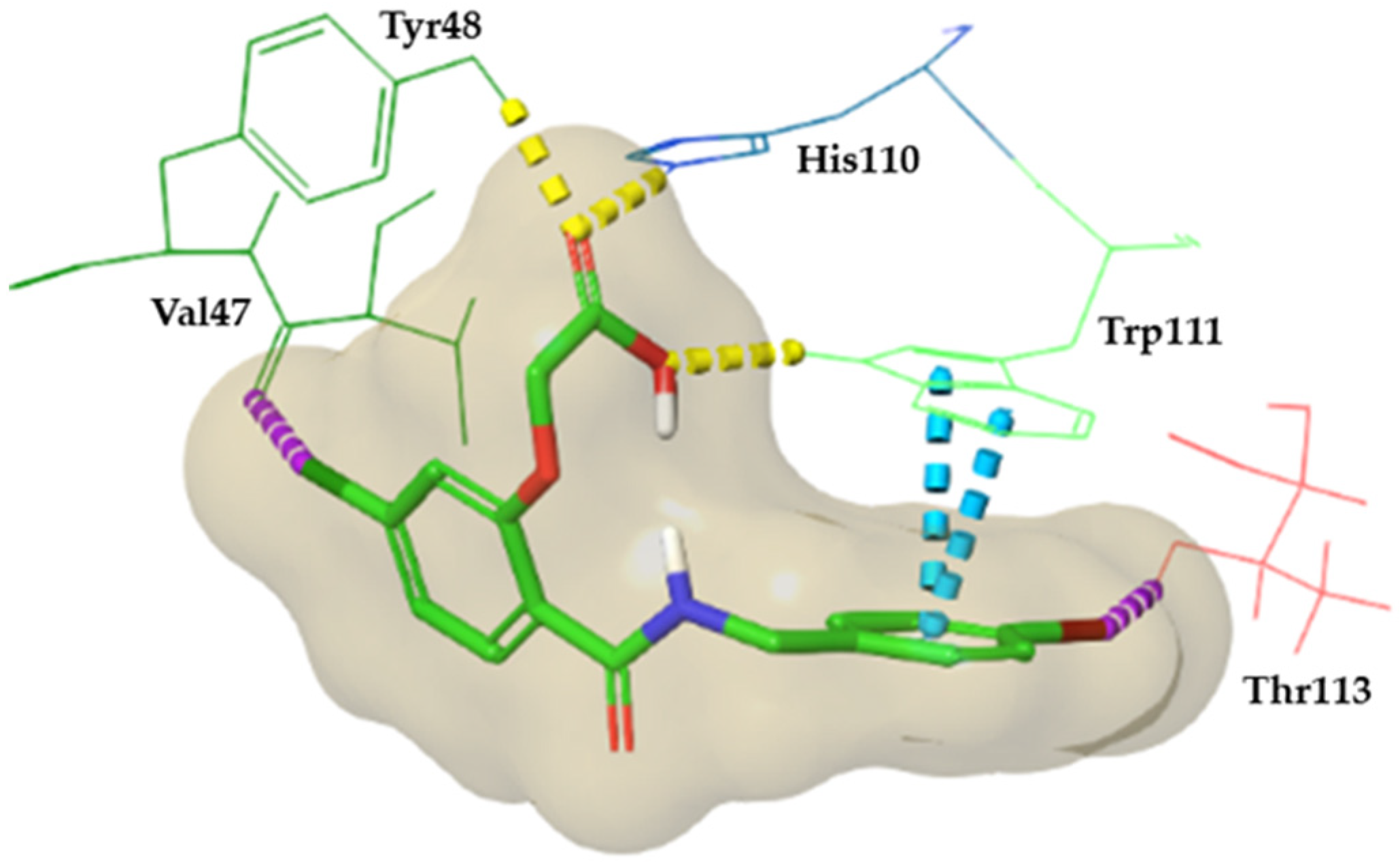
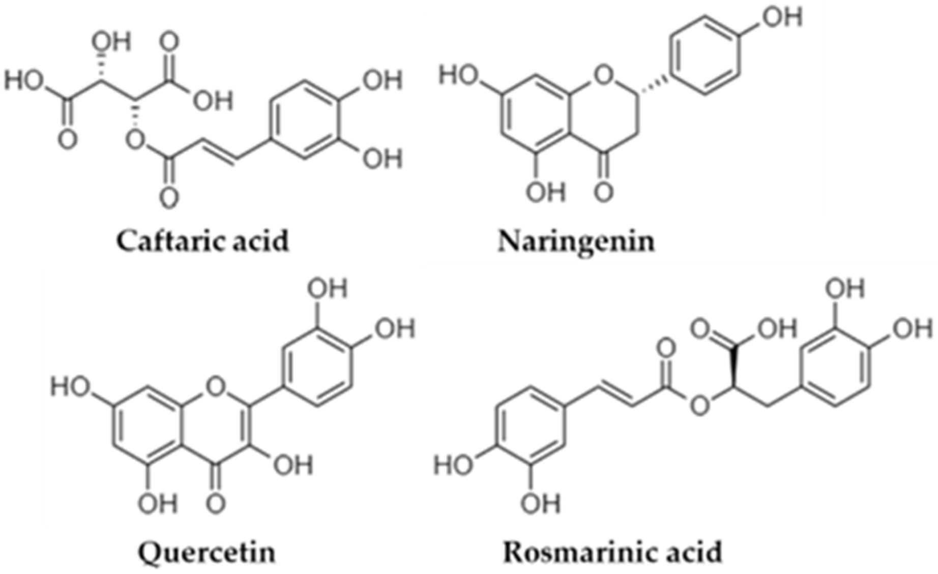
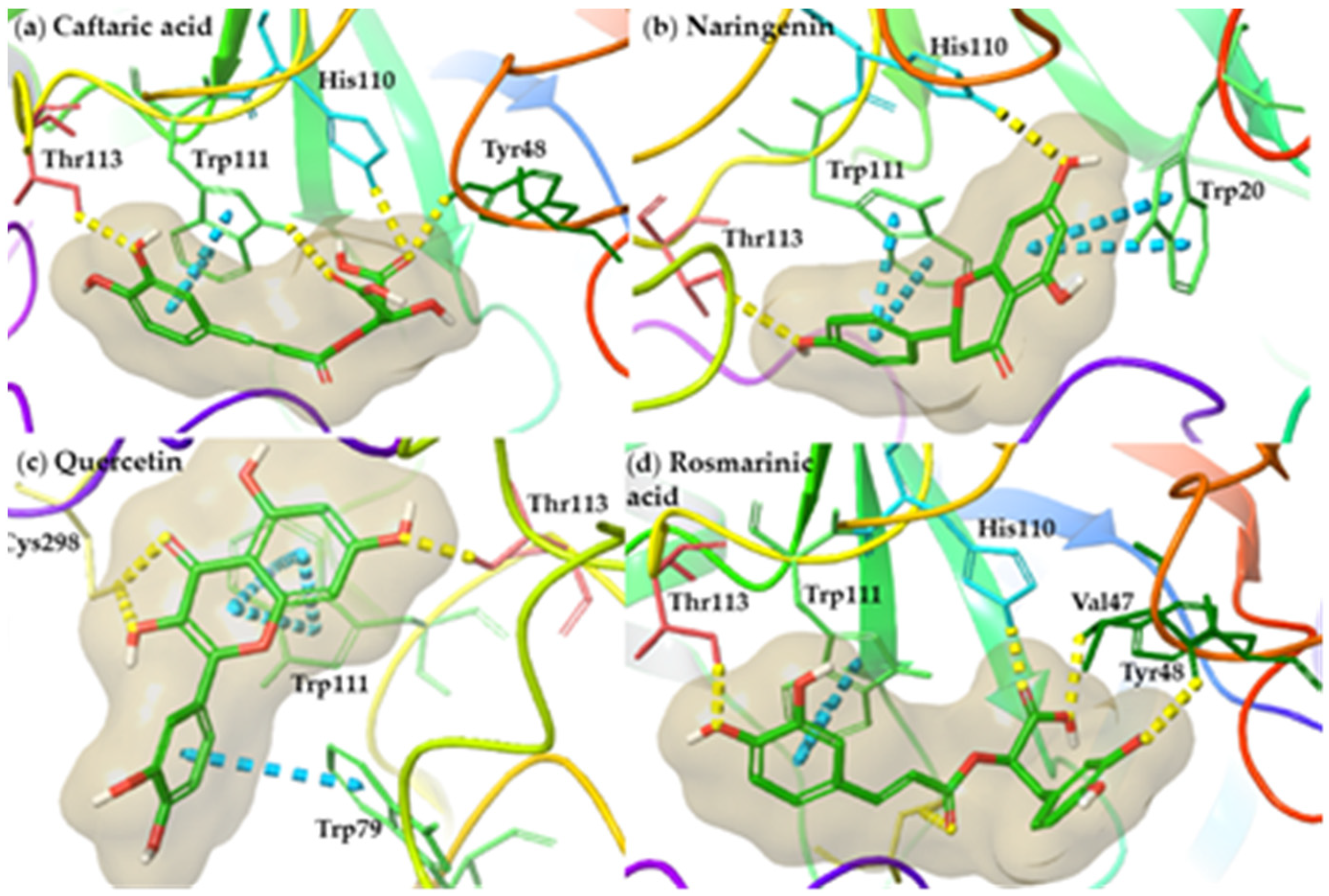
3.1. In silico Analysis

A set of 86 phenolic compounds (Table S1), comprising phenolic acids, flavanols, flavones, flavanonols, and flavanones, which was derived from an extensive literature review, was generated [30,31,32,33,34,35,36,37,38,39,40]. The mentioned phenolic compounds contain the characteristic phytochemicals of the studied herb species.
In continuation, the molecular targets selection for docking studies was based on the results of the open web server TargetNet (http://targetnet.scbdd.com (accessed on 1 July 2022). An ensemble target prediction was performed in this study, including a combination of fingerprint types (such as FP2, FP4, Daylight, MACCS, ECFP2, ECFP4, and ECFP6) [41]. The results for all combinatiplons indicated aldose reductase (AR) as the most promising target. This finding is supported by the area under the receiver operating characteristic curve (AUC) values, which exhibited an acceptable values (greater than 0.5), highlighting the model probability of selecting active over inactive compounds [41]. Therefore, molecular docking simulations were performed for all phytochemicals in an effort to predict their potential inhibitory affinity against AR enzyme.
Aldose reductase (AR) is the principal enzyme of polyol pathway, which is related to hyperglycemic conditions and plays a critical role in the development of diabetic complications including cataract, retinopathy, nephropathy, and neuropathy [46,47]. As a result, AR represents an attractive therapeutic target for the prevention of and reduction in the effects of diabetic complications. Many studies have been performed targeting the discovery of novel molecules as potential AR inhibitors [48,49,50]. The already known synthetic AR inhibitors, such as sorbinil and tolrestat, suffer from drawbacks due to their poor permeation and safety concerns [46]. Therefore, compounds of natural origin have received considerable attention as an alternative. Specifically, among the phytochemicals, for instance, quercetin and other flavonoids or their derivatives, exhibit significant inhibitory activity [51,52].
The derived docking pose evaluation was based not only on docking scores, but also on the visual inspection focusing on the presence of interactions with crucial amino acids identified by the cocrystallized ligand of AR (Figure 1).
Particularly in the case of the AR enzyme, the criteria for visual inspection include hydrogen bond formation with the amino acids Tyr48, His110, and Trp111. The formation of halogen bonds with Val47 and Thr113, and the existence of pi–pi interactions with Trp111 are all characterized as crucial for binding (Figure 1).
Therefore, the molecular docking results analysis indicated caftaric acid, naringenin, quercetin, and rosmarinic acid (Figure 2), which are compounds bearing completely different chemical scaffolds from known synthetic AR inhibitors, as the most promising candidates. Notably, quercetin has already been reported as a strong AR inhibitor [46,52], confirming our docking results and methodology.
The carbonyl groups of caftaric acid (docking score (ds) = −8.05 kcal·mol−1) form hydrogen bonds with the crucial amino acids Tyr48, His110, and Trp111. Additionally, the phenolic ring of caftaric acid interacts via a pi–pi stacking with the aromatic ring of the crucial amino acid Trp111. The docked pose of naringenin indicated an interaction motif, including the formation of hydrogen bonds with His110 and Thr113 and a pi–pi stacking with Trp111, as the cocrystallized ligand. The binding is also reinforced by the development of pi–pi stacking with Trp20, an amino acid that is part of the AR active site. Quercetin interacts through the formation of a hydrogen bond and a pi–pi stacking with the crucial amino acids Thr113 and Trp111, respectively. In addition, the binding is enhanced by the creation of a hydrogen bond and a pi–pi stacking with Cys298 and Trp79 amino acids, which are involved in the active site of AR. In the case of quercetin the presented interaction pattern is in accordance with published studies [46]. Finally, the carboxyl acid group of rosmarinic acid creates hydrogen bonds with the crucial amino acids Tyr48 and His110. Moreover, the hydroxyl group of its phenolic rings form hydrogen bonds with the amino acids Val47 and Thr113, which are critical for binding. An additional pi–pi stacking with the crucial amino acid Trp111 and a hydrogen bond with Cys298 stabilize the binding into AR. The representative docking poses of the abovementioned compounds are illustrated in Figure 3.

3.2. Phytochemical Phenolic Profile of the Examined Infusion by LC-MS/MS Analysis

After assessing the results of the in silico studies, an LC-MS/MS analysis was performed in order to provide information regarding the phenolic profile of the investigated herbal infusions and to determine the preparation that contained the compounds from molecular docking (rosmarinic acid, caftaric acid, naringenin, and quercetin).
By applying the conditions of the developed method, 40 standard phenolic compounds were analyzed in order to build an in-house phenolic library. The spectral information of the phenolic standards is presented in Table 1.
After creating the LC-MS/MS library of the phenolic compounds, based on the standard solutions, the infusions samples were analyzed. The LC-MS/MS analysis of herbal preparations elucidated 36 tentative (poly)phenols, by using both the library of the phenolic standards and data from published references. The identified compounds are illustrated in Table 2. Rosmarinic acid, caftaric acid, naringenin, and quercetin, which were identified as molecules with putative inhibition activity against the AR enzyme, were detected in the infusions of specific herb families, as shown in Table 2.
The elucidated compounds in the ten herbal infusions were mainly organic acids (i.e., caffeic acid, ferulic acid, rosmarinic acid, etc.), flavonoids (catechin, apigenin, etc.), and glycosides of flavonoids (rutin, kaempherol-3-o-rutinoside, luteolin-7-o-glucoside, etc.) [61,62,63,64,65]. Based on results of the LC-MS/MS analysis, the preparations in which the most phenolic compounds were identified were T. capitatus and H. perforatum, while L. citriodora and O. vulgare were the two infusions with the poorest phenolic profile. For T. capitatus and L. citriodora, the rich phenolic fingerprint of these two preparations aligned with the results of our previously published work [12], where headed savory and St. John’s Wort presented higher total phenolic content values. Despite its not-so-rich phenolic fingerprint, because only three phenolic compounds were identified in its infusions, O. vulgare exhibited significant antioxidant activity [12]. However, the high content of rosmarinic acid may account for the significant antioxidant potential of oregano [63].
In support of the abovementioned outcomes, the results of in silico screening also highlighted the importance of rosmarinic acid, which presented the highest binding affinity for AR among the selected compounds [66]. Therefore, the herbs M. spicata, O. vulgare and T. capitatus, which contain high amounts of rosmarinic acid and belong to the Lamiaceae family [67,68,69], show promise as herbal preparations with considerable antihyperglycemic activity. Special focus should be paid to T. capitatus because its putative action against hyperglycemia-related conditions may be attributed to the synergistic effect of rosmarinic acid and naringenin, which also presented putative binding affinity against AR [70]. Based on the docking results, the infusion of O. majorana, another member of the Lamiaceae family, in which quercetin was detected [71], may also be an ideal candidate for the inhibition of AR [72]. In addition, caftaric acid, a phenolic acid of E. purpurea [73,74], exhibited important binding affinity toward AR [75], placing Echinacea among the herbs with possible action against hyperglycemia and metabolic syndrome induced by high blood sugar levels [76].
To summarize, in accordance with the findings of other studies [66,77,78], the results of docking studies combined with the phenolic profile of the studied preparations designated the species of Lamiaceae family and, principally, T. capitatus, as the lead source of phenolic compounds, which may act as inhibitors of AR.

4. Conclusions

Currently, the consumption of herbal infusions is rapidly increasing due to their wide array of biological properties and their possible links to human health, mainly attributed to compounds such as flavonoids and phenolic acids. Thus, in this study, we focused on identifying any possible biological activities of herbal phytochemicals against certain pathological conditions using in silico techniques and LC-MS/MS analysis. According to our results, the phenolic profile of ten common herbs (chamomile, purple coneflower, lemon verbena, pennyroyal, spearmint, marjoram, oregano, headed savory, sea buckthorn, and St. John’s wort) were assessed by LC-MS/MS, and 36 tentative phytoconstituents were identified in the studied herbal infusions. In parallel, 86 phenolic compounds, characteristic of these plants and as reported in the literature and in web libraries of natural products, were investigated against targets by applying molecular prediction tools, such as TargetNet. Four of the tested compounds (rosmarinic acid, caftaric acid, naringenin, and quercetin), which were also detected by LC-MS/MS mainly in the preparations of Lamiaceae species, showed a significant binding affinity against aldose reductase (AR), based on the molecular docking results. Therefore, the combination of in silico tools and high-throughput analytical methodologies identified the herbs of the Lamiaceae family and especially T. capitatus (headed savory), as the most ideal candidates against the AR enzyme, showcasing these infusions as herbal preparations with potent antihyperglycemic activity.

Supplementary Materials

The following supporting information can be downloaded at: https://www.mdpi.com/article/10.3390/app12168361/s1, Figure S1. The similarity in the overlapping between crystallographic (blue) and docked (green) poses, derived from aldose reductase (AR) enzyme; Table S1. Phenolic compounds contained to the examined herbs, derived from an extensive literature review; Table S2. Molecular target prediction results derived from all collected phenolic compounds, by applying Ensemble TargetNet (http://targetnet.scbdd.com/calcnet/index_ensemble/ (accessed on 1 July 2022)) calculations [12].

Author Contributions

Conceptualization, T.T., E.K., D.Z.L., V.J.S. and P.Z.; methodology, E.K., D.Z.L. and D.T.; software, E.K., D.Z.L., P.C. and D.T.; validation, T.T., E.K. and I.F.S.; formal analysis, T.T., E.K., D.Z.L. and D.T.; investigation, T.T., E.K., P.C. and I.F.S.; resources, V.J.S. and P.Z.; data curation, E.K., D.Z.L. and D.T.; writing—original draft preparation, T.T., E.K. and V.J.S.; writing—review and editing, T.T., E.K., I.F.S., V.J.S. and P.Z.; visualization, V.J.S. and P.Z.; supervision, E.K., V.J.S. and P.Z.; project administration, V.J.S. and P.Z.; funding acquisition, V.J.S. and P.Z. All authors have read and agreed to the published version of the manuscript.

Funding

This research received no external funding.

Institutional Review Board Statement

Not applicable.

Informed Consent Statement

Not applicable.

Data Availability Statement

Not applicable.

Acknowledgments

The authors gratefully acknowledge the biofarm ‘Bioagroktima-Menekos’ in Chalkidiki, Greece, for providing the herbs.

Conflicts of Interest

The authors declare no conflict of interest.

References

  1. Tejera, E.; Pérez-Castillo, Y.; Toscano, G.; Noboa, A.L.; Ochoa-Herrera, V.; Giampieri, F.; Álvarez-Suarez, J.M. Computational Modeling Predicts Potential Effects of the Herbal Infusion “Horchata” against COVID-19. Food Chem. 2022, 366, 130589. [Google Scholar] [CrossRef] [PubMed]
  2. Krishnasamy, R.; T, A.; Baba, M.; Bharath, M.V.; Phuntsho, J.; Arunachalam, D.; K, V.; Natarajan, K.; Ramasamy, M. In Silico Analysis of Active Compounds from Siddha Herbal Infusion of Ammaiyar Koondhal Kudineer (Akk) against SARS-CoV-2 Spike Protein and Its ACE2 Receptor Complex. 2020. Available online: https://www.scienceopen.com/document?vid=51068cee-c7d2-49c5-9f1e-00592e601e74 (accessed on 17 July 2022).
  3. Hussain, S.A.; Panjagari, N.R.; Singh, R.R.B.; Patil, G.R. Potential Herbs and Herbal Nutraceuticals: Food Applications and Their Interactions with Food Components. Crit. Rev. Food Sci. Nutr. 2015, 55, 94–112. [Google Scholar] [CrossRef] [PubMed]
  4. Caldeirão, L.; Sousa, J.; Nunes, L.C.G.; Godoy, H.T.; Fernandes, J.O.; Cunha, S.C. Herbs and Herbal Infusions: Determination of Natural Contaminants (Mycotoxins and Trace Elements) and Evaluation of Their Exposure. Food Res. Int. 2021, 144, 110322. [Google Scholar] [CrossRef]
  5. Williamson, E.M.; Liu, X.; Izzo, A.A. Trends in Use, Pharmacology, and Clinical Applications of Emerging Herbal Nutraceuticals. Br. J. Pharmacol. 2020, 177, 1227–1240. [Google Scholar] [CrossRef] [PubMed] [Green Version]
  6. Rocha, C.; Moura, A.P.; Cunha, L.M. Consumers’ Associations with Herbal Infusions and Home Preparation Practices. Food Qual. Prefer. 2020, 86, 104006. [Google Scholar] [CrossRef]
  7. Farzaneh, V.; Carvalho, I.S. A Review of the Health Benefit Potentials of Herbal Plant Infusions and Their Mechanism of Actions. Ind. Crops Prod. 2015, 65, 247–258. [Google Scholar] [CrossRef]
  8. Altemimi, A.; Lakhssassi, N.; Baharlouei, A.; Watson, D.G.; Lightfoot, D.A. Phytochemicals: Extraction, Isolation, and Identification of Bioactive Compounds from Plant Extracts. Plants 2017, 6, 42. [Google Scholar] [CrossRef]
  9. Li, Y.; Zidorn, C. Seasonal Variations of Natural Products in European Herbs. Phytochem. Rev. 2022, 1–27. [Google Scholar] [CrossRef]
  10. Nwosu, O.K.; Ubaoji, K.I. Nutraceuticals: History, Classification and Market Demand. In Functional Foods and Nutraceuticals: Bioactive Components, Formulations and Innovations; Egbuna, C., Dable Tupas, G., Eds.; Springer International Publishing: Cham, Switzerland, 2020; pp. 13–22. ISBN 978-3-030-42319-3. [Google Scholar]
  11. Çelik, G.; Kılıç, G.; Kanbolat, Ş.; Özlem Şener, S.; Karaköse, M.; Yaylı, N.; Karaoğlu, Ş.A. Biological Activity, and Volatile and Phenolic Compounds from Five Lamiaceae Species. Flavour Fragr. J. 2021, 36, 223–232. [Google Scholar] [CrossRef]
  12. Fotakis, C.; Tsigrimani, D.; Tsiaka, T.; Lantzouraki, D.Z.; Strati, I.F.; Makris, C.; Tagkouli, D.; Proestos, C.; Sinanoglou, V.J.; Zoumpoulakis, P. Metabolic and Antioxidant Profiles of Herbal Infusions and Decoctions. Food Chem. 2016, 211, 963–971. [Google Scholar] [CrossRef]
  13. Barral-Martinez, M.; Garcia-Oliveira, P.; Nuñez-Estevez, B.; Silva, A.; Finimundy, T.C.; Calhelha, R.; Nenadic, M.; Sokovic, M.; Barroso, F.; Simal-Gandara, J.; et al. Plants of the Family Asteraceae: Evaluation of Biological Properties and Identification of Phenolic Compounds. Chem. Proc. 2021, 5, 51. [Google Scholar] [CrossRef]
  14. Rezaeifar, M.; Mehdizadeh, T.; Mojaddar Langroodi, A.; Rezaei, F. Effect of Chitosan Edible Coating Enriched with Lemon Verbena Extract and Essential Oil on the Shelf Life of Vacuum Rainbow Trout (Oncorhynchus mykiss). J. Food Saf. 2020, 40, e12781. [Google Scholar] [CrossRef]
  15. Silva, M.; Cueva, C.; Alba, C.; Rodriguez, J.M.; de Pascual-Teresa, S.; Jones, J.; Caturla, N.; Victoria Moreno-Arribas, M.; Bartolomé, B. Gut Microbiome-Modulating Properties of a Polyphenol-Enriched Dietary Supplement Comprised of Hibiscus and Lemon Verbena Extracts. Monitoring of Phenolic Metabolites. J. Funct. Foods 2022, 91, 105016. [Google Scholar] [CrossRef]
  16. Hematian Sourki, A.; Ghani, A.; Kiani, F.; Alipour, A. Phytochemical Profiles of Lemon Verbena (Lippia citriodora H.B.K.) and Its Potential Application to Cookie Enrichment. Food Sci. Nutr. 2021, 9, 3100–3113. [Google Scholar] [CrossRef] [PubMed]
  17. Sánchez-Marzo, N.; Lozano-Sánchez, J.; de la Luz Cádiz-Gurrea, M.; Herranz-López, M.; Micol, V.; Segura-Carretero, A. Relationships between Chemical Structure and Antioxidant Activity of Isolated Phytocompounds from Lemon Verbena. Antioxidants 2019, 8, 324. [Google Scholar] [CrossRef] [PubMed] [Green Version]
  18. Gani, A.; Jan, R.; Ashwar, B.A.; Ashraf, Z.U.; Shah, A.; Gani, A. Encapsulation of Saffron and Sea Buckthorn Bioactives: Its Utilization for Development of Low Glycemic Baked Product for Growing Diabetic Population of the World. LWT 2021, 142, 111035. [Google Scholar] [CrossRef]
  19. Gâtlan, A.-M.; Gutt, G. Sea Buckthorn in Plant Based Diets. An Analytical Approach of Sea Buckthorn Fruits Composition: Nutritional Value, Applications, and Health Benefits. Int. J. Environ. Res. Public Health 2021, 18, 8986. [Google Scholar] [CrossRef]
  20. Zhao, X.; Guo, Y.; Xu, Q.; Shi, Z.; Xiang, M.; Li, H.; Wang, Y.; Qi, C.; Zhang, Y. (±)-Hyperpyran A: Terpenoid-Based Bicyclic Dihydropyran Enantiomers with Hypoglycemic Activity from Hypericum perforatum (St. John’s Wort). Fitoterapia 2022, 161, 105221. [Google Scholar] [CrossRef]
  21. Lou, H.; Ma, F.; Yi, P.; Hu, Z.; Gu, W.; Huang, L.; He, W.; Yuan, C.; Hao, X. Bioassay and UPLC-Q-Orbitrap-MS/MS Guided Isolation of Polycyclic Polyprenylated Acylphloroglucinols from St. John’s Wort and Their Neuroprotective Activity. Arab. J. Chem. 2022, 15, 104057. [Google Scholar] [CrossRef]
  22. Mullaicharam, A.R.; Halligudi, N. St John’s Wort (Hypericum perforatum L.): A Review of Its Chemistry, Pharmacology and Clinical Properties. Int. J. Res. Phytochem. Pharmacol. Sci. 2019, 1, 5–11. [Google Scholar] [CrossRef]
  23. Muyumba, N.W.; Mutombo, S.C.; Sheridan, H.; Nachtergael, A.; Duez, P. Quality Control of Herbal Drugs and Preparations: The Methods of Analysis, Their Relevance and Applications. Talanta Open 2021, 4, 100070. [Google Scholar] [CrossRef]
  24. Balekundri, A.; Mannur, V. Quality Control of the Traditional Herbs and Herbal Products: A Review. Future J. Pharm. Sci. 2020, 6, 67. [Google Scholar] [CrossRef]
  25. Tsiaka, T.; Kritsi, E.; Tsiantas, K.; Christodoulou, P.; Sinanoglou, V.J.; Zoumpoulakis, P. Design and Development of Novel Nutraceuticals: Current Trends and Methodologies. Nutraceuticals 2022, 2, 71–90. [Google Scholar] [CrossRef]
  26. Sharma, S.; Bhatia, V. Phytochemicals for Drug Discovery in Alzheimer’s Disease: In Silico Advances. Curr. Pharm. Des. 2021, 27, 2848–2860. [Google Scholar] [CrossRef]
  27. Sharanya, C.S.; Sabu, A.; Haridas, M. Potent Phytochemicals against COVID-19 Infection from Phyto-Materials Used as Antivirals in Complementary Medicines: A Review. Future J. Pharm. Sci. 2021, 7, 113. [Google Scholar] [CrossRef] [PubMed]
  28. Beltrán-Noboa, A.; Proaño-Ojeda, J.; Guevara, M.; Gallo, B.; Berrueta, L.A.; Giampieri, F.; Perez-Castillo, Y.; Battino, M.; Álvarez-Suarez, J.M.; Tejera, E. Metabolomic Profile and Computational Analysis for the Identification of the Potential Anti-Inflammatory Mechanisms of Action of the Traditional Medicinal Plants Ocimum basilicum and Ocimum tenuiflorum. Food Chem. Toxicol. 2022, 164, 113039. [Google Scholar] [CrossRef] [PubMed]
  29. Zainab, B.; Ayaz, Z.; Alwahibi, M.S.; Khan, S.; Rizwana, H.; Soliman, D.W.; Alawaad, A.; Mehmood Abbasi, A. In-Silico Elucidation of Moringa oleifera Phytochemicals against Diabetes Mellitus. Saudi J. Biol. Sci. 2020, 27, 2299–2307. [Google Scholar] [CrossRef] [PubMed]
  30. Taamalli, A.; Arráez-Román, D.; Abaza, L.; Iswaldi, I.; Fernández-Gutiérrez, A.; Zarrouk, M.; Segura-Carretero, A. LC-MS-Based Metabolite Profiling of Methanolic Extracts from the Medicinal and Aromatic Species Mentha pulegium and Origanum majorana. Phytochem. Anal. 2015, 26, 320–330. [Google Scholar] [CrossRef]
  31. Milevskaya, V.V.; Prasad, S.; Temerdashev, Z.A. Extraction and Chromatographic Determination of Phenolic Compounds from Medicinal Herbs in the Lamiaceae and Hypericaceae Families: A Review. Microchem. J. 2019, 145, 1036–1049. [Google Scholar] [CrossRef]
  32. Skendi, A.; Irakli, M.; Chatzopoulou, P. Analysis of Phenolic Compounds in Greek Plants of Lamiaceae Family by HPLC. J. Appl. Res. Med. Aromat. Plants 2017, 6, 62–69. [Google Scholar] [CrossRef]
  33. Jabri-Karoui, I.; Bettaie, I.; Msaada, K.; Hammami, M.; Marzouk, B. Research on the Phenolic Compounds and Antioxidant Activities of Tunisian Thymus capitatus. J. Funct. Foods 2012, 4, 661–669. [Google Scholar] [CrossRef]
  34. Cirlini, M.; Mena, P.; Tassotti, M.; Herrlinger, K.A.; Nieman, K.M.; Dall’Asta, C.; Del Rio, D. Phenolic and Volatile Composition of a Dry Spearmint (Mentha spicata L.) Extract. Molecules 2016, 21, 1007. [Google Scholar] [CrossRef] [PubMed] [Green Version]
  35. Coelho, J.; Barros, L.; Dias, M.I.; Finimundy, T.C.; Amaral, J.S.; Alves, M.J.; Calhelha, R.C.; Santos, P.F.; Ferreira, I.C.F.R. Echinacea purpurea (L.) Moench: Chemical Characterization and Bioactivity of Its Extracts and Fractions. Pharmaceuticals 2020, 13, 125. [Google Scholar] [CrossRef] [PubMed]
  36. Haghi, G.; Hatami, A.; Safaei, A.; Mehran, M. Analysis of Phenolic Compounds in Matricaria chamomilla and Its Extracts by UPLC-UV. Res. Pharm. Sci. 2014, 9, 31–37. [Google Scholar]
  37. Villegas-Aguilar, M.D.C.; Leyva-Jiménez, F.J.; Cádiz-Gurrea, M.D.L.L.; Segura-Carretero, A.; Arráez-Román, D. Comprehensive Analysis of Antioxidant Compounds from Lippia citriodora and Hibiscus sabdariffa Green Extracts Attained by Response Surface Methodology. Antioxidants 2020, 9, 1175. [Google Scholar] [CrossRef]
  38. Ivanović, M.; Alañón, M.E.; Arráez-Román, D.; Segura-Carretero, A. Enhanced and Green Extraction of Bioactive Compounds from Lippia citriodora by Tailor-Made Natural Deep Eutectic Solvents. Food Res. Int. 2018, 111, 67–76. [Google Scholar] [CrossRef]
  39. Zadernowski, R.; Naczk, M.; Czaplicki, S.; Rubinskiene, M.; Szałkiewicz, M. Composition of Phenolic Acids in Sea Buckthorn (Hippophae rhamnoides L.) Berries. J. Am. Oil Chem. Soc. 2005, 82, 175–179. [Google Scholar] [CrossRef]
  40. Tusevski, O.; Krstikj, M.; Stanoeva, J.P.; Stefova, M.; Gadzovska-Simic, S. Phenolic Profile and Biological Activity of Hypericum perforatum L.: Can Roots Be Considered as a New Source of Natural Compounds? S. Afr. J. Bot. 2018, 177, 301–310. [Google Scholar] [CrossRef]
  41. Yao, Z.J.; Dong, J.; Che, Y.J.; Zhu, M.F.; Wen, M.; Wang, N.N.; Wang, S.; Lu, A.P.; Cao, D.S. TargetNet: A Web Service for Predicting Potential Drug-Target Interaction Profiling via Multi-Target SAR Models. J. Comput.-Aided Mol. Des. 2016, 30, 413–424. [Google Scholar] [CrossRef]
  42. Schrödinger Release 2020-3: Protein Preparation Wizard; Schrödinger, LLC: New York, NY, USA, 2020.
  43. Schrödinger Release 2020-3: LigPrep; Schrödinger, LLC: New York, NY, USA, 2020.
  44. Schrödinger Release 2020-3: Maestro; Schrödinger, LLC: New York, NY, USA, 2020.
  45. Schrödinger Release 2020-3: Glide; Schrödinger, LLC: New York, NY, USA, 2020.
  46. Antony, P.; Vijayan, R. Identification of Novel Aldose Reductase Inhibitors from Spices: A Molecular Docking and Simulation Study. PLoS ONE 2015, 10, e0138186. [Google Scholar] [CrossRef]
  47. Grewal, A.S.; Bhardwaj, S.; Pandita, D.; Lather, V.; Sekhon, B.S. Updates on Aldose Reductase Inhibitors for Management of Diabetic Complications and Non-Diabetic Diseases. Mini Rev. Med. Chem. 2016, 16, 120–162. [Google Scholar] [CrossRef] [PubMed]
  48. Qin, X.; Hao, X.; Han, H.; Zhu, S.; Yang, Y.; Wu, B.; Hussain, S.; Parveen, S.; Jing, C.; Ma, B.; et al. Design and Synthesis of Potent and Multifunctional Aldose Reductase Inhibitors Based on Quinoxalinones. J. Med. Chem. 2015, 58, 1254–1267. [Google Scholar] [CrossRef] [PubMed]
  49. Digiacomo, M.; Sartini, S.; Nesi, G.; Sestito, S.; Coviello, V.; La Motta, C.; Rapposelli, S. Synthesis and Functional Evaluation of Novel Aldose Reductase Inhibitors Bearing a Spirobenzopyran Scaffold. Open Med. Chem. J. 2017, 11, 9–23. [Google Scholar] [CrossRef] [PubMed] [Green Version]
  50. Imran, A.; Shehzad, M.T.; Shah, S.J.A.; Adhami, T.; Laws, A.; Rahman, K.M.; Alharthy, R.D.; Khan, I.A.; Shafiq, Z.; Iqbal, J. Development and Exploration of Novel Substituted Thiosemicarbazones as Inhibitors of Aldose Reductase via in Vitro Analysis and Computational Study. Sci. Rep. 2022, 12, 5734. [Google Scholar] [CrossRef] [PubMed]
  51. Kim, H.M.; Lee, J.M.; Lee, K.H.; Ahn, Y.H.; Lee, S.H. Aldose Reductase Inhibition by Luteolin Derivatives from Parasenecio pseudotaimingasa. Nat. Prod. Sci. 2011, 17, 367–371. [Google Scholar]
  52. Veeresham, C.; Rama Rao, A.; Asres, K. Aldose Reductase Inhibitors of Plant Origin. Phytother. Res. 2014, 28, 317–333. [Google Scholar] [CrossRef]
  53. Koşar, M.; Dorman, H.J.D.; Can Başer, K.H.; Hiltunen, R. Screening of Free Radical Scavenging Compounds in Water Extracts of Mentha Samples Using a Postcolumn Derivatization Method. J. Agric. Food Chem. 2004, 52, 5004–5010. [Google Scholar] [CrossRef]
  54. Miron, T.L.; Plaza, M.; Bahrim, G.; Ibáñez, E.; Herrero, M. Chemical Composition of Bioactive Pressurized Extracts of Romanian Aromatic Plants. J. Chromatogr. A 2011, 1218, 4918–4927. [Google Scholar] [CrossRef] [Green Version]
  55. Raal, A.; Orav, A.; Püssa, T.; Valner, C.; Malmiste, B.; Arak, E. Content of Essential Oil, Terpenoids and Polyphenols in Commercial Chamomile (Chamomilla recutita L. Rauschert) Teas from Different Countries. Food Chem. 2012, 131, 632–638. [Google Scholar] [CrossRef]
  56. Pellati, F.; Orlandini, G.; Benvenuti, S. Simultaneous Metabolite Fingerprinting of Hydrophilic and Lipophilic Compounds in Echinacea Pallida by High-Performance Liquid Chromatography with Diode Array and Electrospray Ionization-Mass Spectrometry Detection. J. Chromatogr. A 2012, 1242, 43–58. [Google Scholar] [CrossRef]
  57. Rainha, N.; Koci, K.; Coelho, A.V.; Lima, E.; Baptista, J.; Fernandes-Ferreira, M. HPLC-UV-ESI-MS Analysis of Phenolic Compounds and Antioxidant Properties of Hypericum undulatum Shoot Cultures and Wild-Growing Plants. Phytochemistry 2013, 86, 83–91. [Google Scholar] [CrossRef] [PubMed]
  58. Fecka, I.; Turek, S. Determination of Polyphenolic Compounds in Commercial Herbal Drugs and Spices from Lamiaceae: Thyme, Wild Thyme and Sweet Marjoram by Chromatographic Techniques. Food Chem. 2008, 108, 1039–1053. [Google Scholar] [CrossRef] [PubMed]
  59. Stashenko, E.E.; Martínez, J.R.; Cala, M.P.; Durán, D.C.; Caballero, D. Chromatographic and Mass Spectrometric Characterization of Essential Oils and Extracts from Lippia (Verbenaceae) Aromatic Plants. J. Sep. Sci. 2013, 36, 192–202. [Google Scholar] [CrossRef] [PubMed]
  60. Chen, C.; Xu, X.-M.; Chen, Y.; Yu, M.-Y.; Wen, F.-Y.; Zhang, H. Identification, Quantification and Antioxidant Activity of Acylated Flavonol Glycosides from Sea Buckthorn (Hippophae rhamnoides Ssp. Sinensis). Food Chem. 2013, 141, 1573–1579. [Google Scholar] [CrossRef]
  61. Bouyahya, A.; Chamkhi, I.; Guaouguaou, F.-E.; Benali, T.; Balahbib, A.; El Omari, N.; Taha, D.; El-Shazly, M.; El Menyiy, N. Ethnomedicinal Use, Phytochemistry, Pharmacology, and Food Benefits of Thymus capitatus. J. Ethnopharmacol. 2020, 259, 112925. [Google Scholar] [CrossRef]
  62. Seyis, F.; Yurteri, E.; Özcan, A.; Cirak, C. Altitudinal Impacts on Chemical Content and Composition of Hypericum perforatum, a Prominent Medicinal Herb. S. Afr. J. Bot. 2020, 135, 391–403. [Google Scholar] [CrossRef]
  63. Jafari Khorsand, G.; Morshedloo, M.R.; Mumivand, H.; Emami Bistgani, Z.; Maggi, F.; Khademi, A. Natural Diversity in Phenolic Components and Antioxidant Properties of Oregano (Origanum vulgare L.) Accessions, Grown under the Same Conditions. Sci. Rep. 2022, 12, 5813. [Google Scholar] [CrossRef]
  64. Özer, Z. Investigation of Phenolic Compounds and Antioxidant Activity of Mentha spicata L. Subsp. Spicata and M. longifolia (L.) L. Subsp. Typhoides (Briq.) Harley Decoction and Infusion. J. Turk. Chem. Soc. Sect. A Chem. 2018, 5, 445–456. [Google Scholar] [CrossRef] [Green Version]
  65. Gutiérrez-Grijalva, E.P.; Picos-Salas, M.A.; Leyva-López, N.; Criollo-Mendoza, M.S.; Vazquez-Olivo, G.; Heredia, J.B. Flavonoids and Phenolic Acids from Oregano: Occurrence, Biological Activity and Health Benefits. Plants 2018, 7, 2. [Google Scholar] [CrossRef] [Green Version]
  66. Zuo, G.; Je, K.-H.; Guillen Quispe, Y.N.; Shin, K.-O.; Kim, H.Y.; Kim, K.H.; Arce, P.H.G.; Lim, S.S. Separation and Identification of Antioxidants and Aldose Reductase Inhibitors in Lepechinia meyenii (Walp.) Epling. Plants 2021, 10, 2773. [Google Scholar] [CrossRef]
  67. Duțu, L.E.; Popescu, M.L.; Purdel, C.N.; Ilie, E.I.; Luță, E.-A.; Costea, L.; Gîrd, C.E. Traditional Medicinal Plants—A Possible Source of Antibacterial Activity on Respiratory Diseases Induced by Chlamydia pneumoniae, Haemophilus influenzae, Klebsiella pneumoniae and Moraxella catarrhalis. Diversity 2022, 14, 145. [Google Scholar] [CrossRef]
  68. Marc, R.A.; Mureșan, V.; Mureșan, A.E.; Mureșan, C.C.; Tanislav, A.E.; Pușcaș, A.; Marţiș, G.S.; Ungur, R.A. Spicy and Aromatic Plants for Meat and Meat Analogues Applications. Plants 2022, 11, 960. [Google Scholar] [CrossRef] [PubMed]
  69. Mutlu-Ingok, A.; Devecioglu, D.; Dikmetas, D.N.; Karbancioglu-Guler, F.; Capanoglu, E. Antibacterial, Antifungal, Antimycotoxigenic, and Antioxidant Activities of Essential Oils: An Updated Review. Molecules 2020, 25, 4711. [Google Scholar] [CrossRef] [PubMed]
  70. Saija, A.; Speciale, A.; Trombetta, D.; Leto, C.; Tuttolomondo, T.; La Bella, S.; Licata, M.; Virga, G.; Bonsangue, G.; Gennero, M.C.; et al. Phytochemical, Ecological and Antioxidant Evaluation of Wild Sicilian Thyme: Thymbra capitata (L.) Cav. Chem. Biodivers. 2016, 13, 1641–1655. [Google Scholar] [CrossRef] [PubMed]
  71. Amaghnouje, A.; Mechchate, H.; Es-safi, I.; Boukhira, S.; Aliqahtani, A.S.; Noman, O.M.; Nasr, F.A.; Conte, R.; Calarco, A.; Bousta, D. Subacute Assessment of the Toxicity and Antidepressant-Like Effects of Origanum majorana L. Polyphenols in Swiss Albino Mice. Molecules 2020, 25, 5653. [Google Scholar] [CrossRef]
  72. Quilantang, N.G.; Ryu, S.H.; Park, S.H.; Byun, J.S.; Chun, J.S.; Lee, J.S.; Rodriguez, J.P.; Yun, Y.-S.; Jacinto, S.D.; Lee, S. Inhibitory Activity of Methanol Extracts from Different Colored Flowers on Aldose Reductase and HPLC-UV Analysis of Quercetin. Hortic. Environ. Biotechnol. 2018, 59, 899–907. [Google Scholar] [CrossRef]
  73. Waidyanatha, S.; Pierfelice, J.; Cristy, T.; Mutlu, E.; Burback, B.; Rider, C.V.; Ryan, K. A Strategy for Test Article Selection and Phytochemical Characterization of Echinacea purpurea Extract for Safety Testing. Food Chem. Toxicol. 2020, 137, 111125. [Google Scholar] [CrossRef]
  74. Senica, M.; Mlinsek, G.; Veberic, R.; Mikulic-Petkovsek, M. Which Plant Part of Purple Coneflower (Echinacea purpurea (L.) Moench) Should Be Used for Tea and Which for Tincture? J. Med. Food 2019, 22, 102–108. [Google Scholar] [CrossRef]
  75. Ajiboye, B.O.; Iwaloye, O.; Owolabi, O.V.; Ejeje, J.N.; Okerewa, A.; Johnson, O.O.; Udebor, A.E.; Oyinloye, B.E. Screening of Potential Antidiabetic Phytochemicals from Gongronema latifolium Leaf against Therapeutic Targets of Type 2 Diabetes Mellitus: Multi-Targets Drug Design. SN Appl. Sci. 2021, 4, 14. [Google Scholar] [CrossRef]
  76. Chiou, S.-Y.; Sung, J.-M.; Huang, P.-W.; Lin, S.-D. Antioxidant, Antidiabetic, and Antihypertensive Properties of Echinacea purpurea Flower Extract and Caffeic Acid Derivatives Using In Vitro Models. J. Med. Food 2017, 20, 171–179. [Google Scholar] [CrossRef]
  77. Wang, Z.; Hwang, S.H.; Guillen Quispe, Y.N.; Gonzales Arce, P.H.; Lim, S.S. Investigation of the Antioxidant and Aldose Reductase Inhibitory Activities of Extracts from Peruvian Tea Plant Infusions. Food Chem. 2017, 231, 222–230. [Google Scholar] [CrossRef] [PubMed]
  78. Bhatti, H.A.; Tehseen, Y.; Maryam, K.; Uroos, M.; Siddiqui, B.S.; Hameed, A.; Iqbal, J. Identification of New Potent Inhibitor of Aldose Reductase from Ocimum basilicum. Bioorg. Chem. 2017, 75, 62–70. [Google Scholar] [CrossRef] [PubMed]
Figure 1. The binding mode of the cocrystallized ligand ({2-[(4-bromobenzyl)carbamoyl]-5-chlorophenoxy}acetic acid-W8X) at the active site of AR (PDB ID: 4LAU). Halogen bonds, hydrogen bonds, and pi–pi interactions are depicted in purple, yellow, and blue dashed lines, respectively.
Applsci 12 08361 g001
Figure 2. Chemical structures of caftaric acid, naringenin, quercetin, and rosmarinic acid.
Applsci 12 08361 g002
Figure 3. Representative binding poses and the crucial interactions of (a) caftaric acid, (b) naringenin, (c) quercetin, and (d) rosmarinic acid derived from molecular docking studies in the active site of AR. Hydrogen bonds are depicted with dashed yellow lines and pi–pi interactions with dashed blue lines.
Applsci 12 08361 g003
Table 1. Spectral information of LC-MS/MS analysis of phenolic standards.
CompoundRetention Time (min)Parent Ion [M–H]Product Ion (MS/MS)
Gallic acid1.51169.33124
Protocatechuic acid2.66153.11108.79
Hydroxytyrosol2.67153.17122.84
Gentisic acid3.65153.02108.84
4-Hydroxybenzoic acid4.75136.9492.87
Chlorogenic acid4.91353.03191.02
(+)-Catechin5.08289.38245.05
2-4-Dihydroxy benzoic acid3.16153.01108.88
Vanillic acid5.81166.99151.96
Caffeic acid6.16179.21134.86
Syringic acid6.28197.02181.94
(−)-Catechin6.40289.38245.06
4-Hydroxybenzaldehyde6.63120.8458.78
Vanillin7.28151.00135.87
Syringaldehyde7.45181.07165.94
p-Coumaric acid7.52163.00118.84
Ferulic acid7.73193.06148.86
Rutin7.84609.37301.03
Ellagic acid8.12301.31257.10
Taxifolin8.12303.38285.04
Sinapic acid8.14223.10207.88
trans-3-Hydroxycinnamic acid8.27163.18118.90
Acetosyringone8.70195.14179.98
Salicylic acid9.09136.9192.89
Benzoic acid9.17121.0358.78
Naringin9.35579.41459.09
Hesperidin9.66609.27301.02
Rosmarinic acid9.68359.09160.88
o-Coumaric acid9.82163.10118.87
Oleuropein10.70539.37376.82
Lariciresinol11.36418.91329.12
Eriodictyol11.80287.70150.86
Luteolin12.16285.30174.97
Quercetin12.18301.23178.86
Resveratrol12.22227.40184.95
Cinnamic acid12.34147.05102.84
Naringenin12.87271.29150.88
Kaempferol12.94285.28285.04
Isorhamnetin13.22315.75300.08
Table 2. Detected phenolic compounds in the examined herbal infusions by using the in-house phenolic compound library and literature data.
Detected CompoundsM.ChamomillaEch.PurpureaL.CitriodoraM.PulegiumM.SpicataO.MajoranaO.VulgareT.CapitatusHyp.PerforatumHip.RhamnoidesReferences
(−) Catechin (s) 1 2
(+) Catechin (s)
4-Hydroxycinnamic acid (s)
Caffeic acid (s)
Chlorogenic acid (s)
p-Coumaric acid (s)
Ellagic acid (s)
Ferulic acid (s)
Gentisic acid (s)
Naringenin (s)
Quercetin (s)
Resveratrol (s)
Rosmarinic acid (s)
Rutin (s)
Apigenin [53]
Apigenin-c-hexoside-c-hexoside [54]
Apιgenin-7-o-glucoside [55]
Caftaric acid [56]
Catechin dimer [57]
Cichoric acid [56]
Citric acid [58]
Dihydrocaffeic acid [59]
Ellagic acid (p-coumaryl) hexoside [60]
Ellagic acid hexoside [60]
Ellagic acid pentoside [58,60]
Eriodictyol-7-o-glucuronide [58]
Ferulic acid glucoside [55]
Ferulic acid hexoside dimer [55]
Gallocatechin [58]
Isoquercitrin [57]
Kaempherol-3-o-rutinoside [56,57,58]
Laricitrin-3-o-glucoside [55]
Luteolin-7-o-glucoside [53,58,59]
Medioresinol [54]
Quercetin-3o-glucoside [57,58]
Rosmarinic acid-o-hexoside [58]
1 Standard phenolic compounds. 2 Indicates the presence of the phenolic compounds in the infusions.
Publisher’s Note: MDPI stays neutral with regard to jurisdictional claims in published maps and institutional affiliations.

