
Effective Framework for Pulmonary Nodule Classification from CT Images Using the Modified Gradient Boosting Method

 and
and
Abstract
:1. Introduction
1.1. Related Works
1.2. Research Contributions
- -
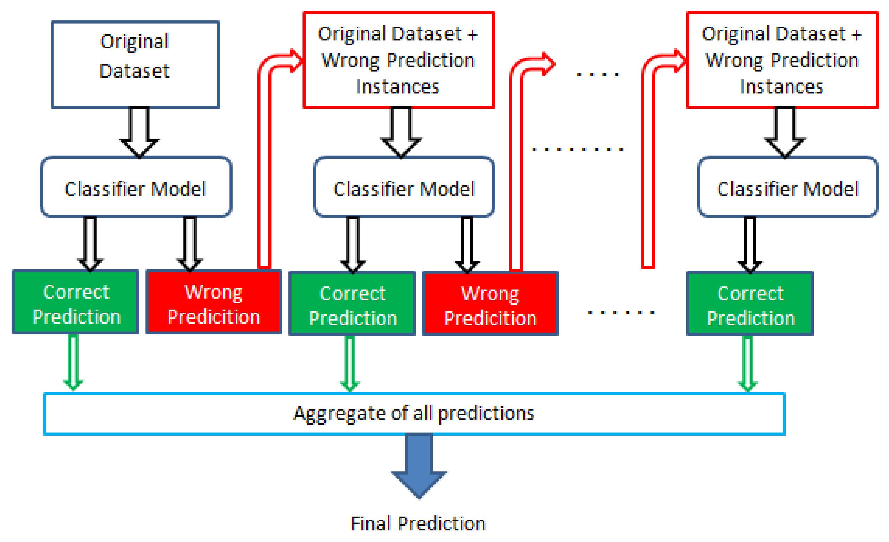
- The gradient boosting algorithm is modified in a sequential manner by feeding the wrong prediction as the input to the next classifier model, thus improving the efficacy of the classification.
- -
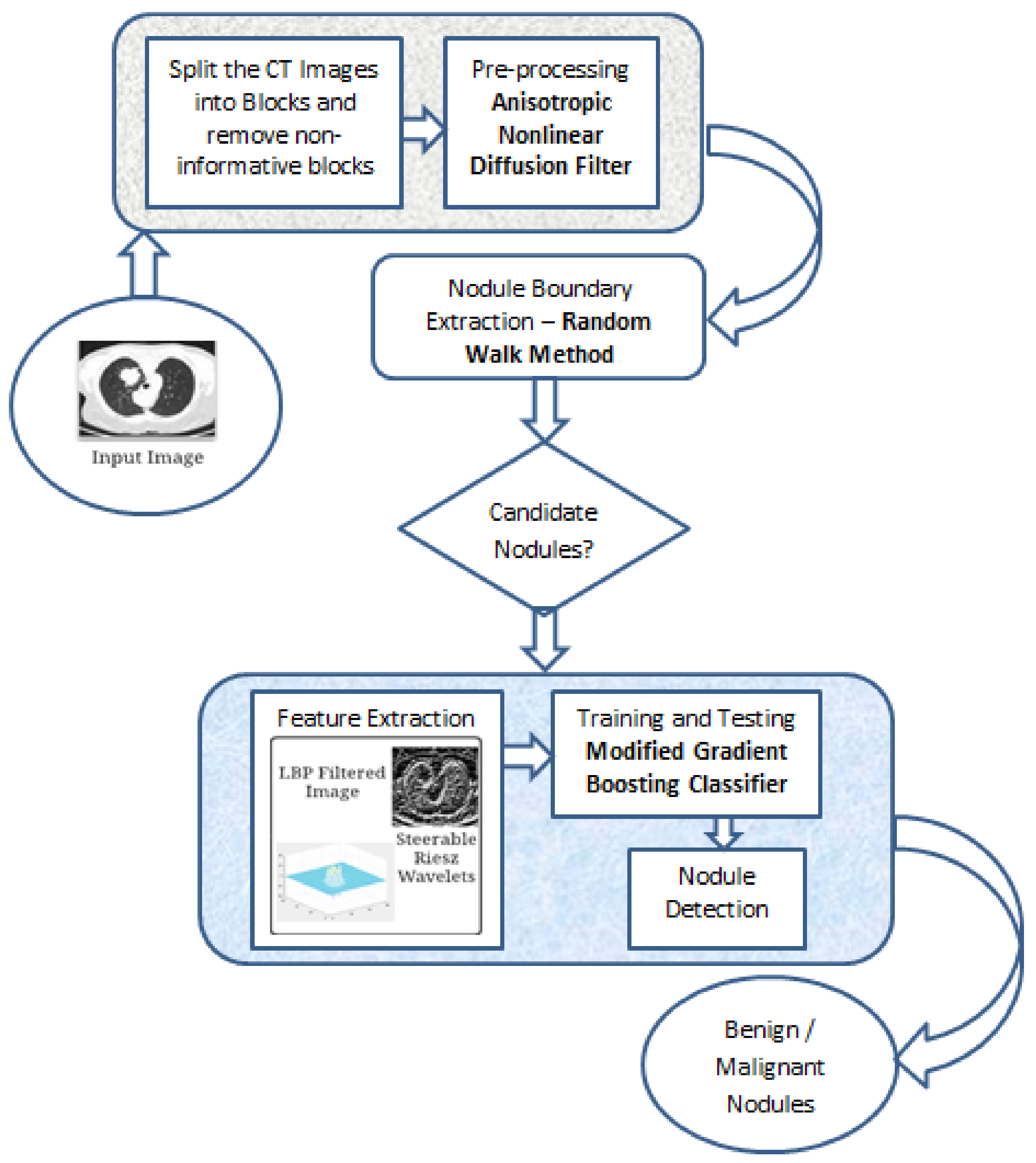
- An effective framework for pulmonary nodule classification from CT images using the modified gradient boosting method for the classification of benign (class 1 and 2) and malignant (class 4 and 5) lung nodules are proposed.
- -
- The random walker method is utilized as the boundary extraction method to segment the pulmonary nodule according to seeds governed by the user.
- -
- Features such as the intensity and texture are extracted using Reisz wavelet coefficients and an LBP filter.
- -
- The performance of the proposed segmentation and classifier model using the modified gradient boosting algorithm method is evaluated by comparing with the existing methods, and is found to be superior.
2. Materials and Methods
2.1. Dataset
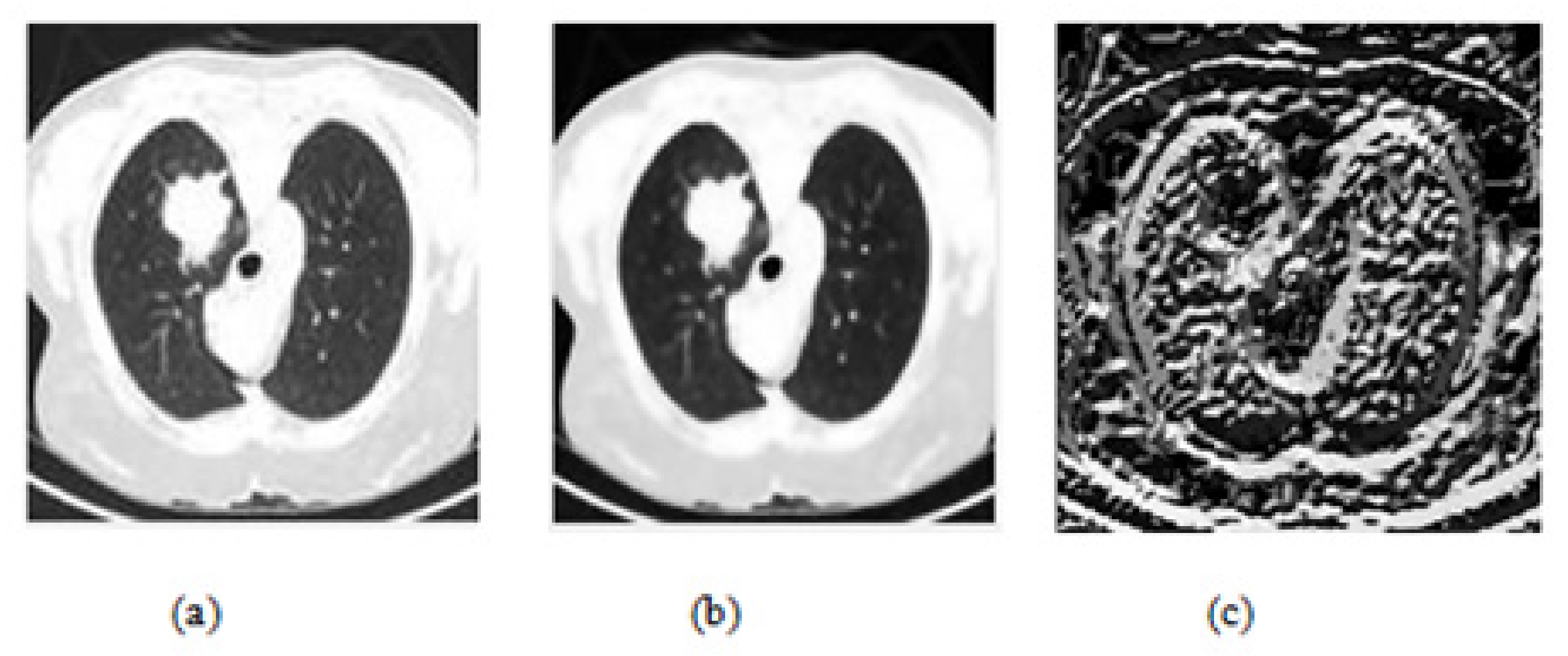
2.2. Preprocessing
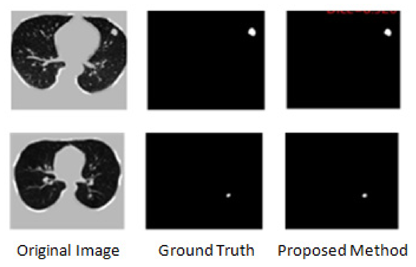
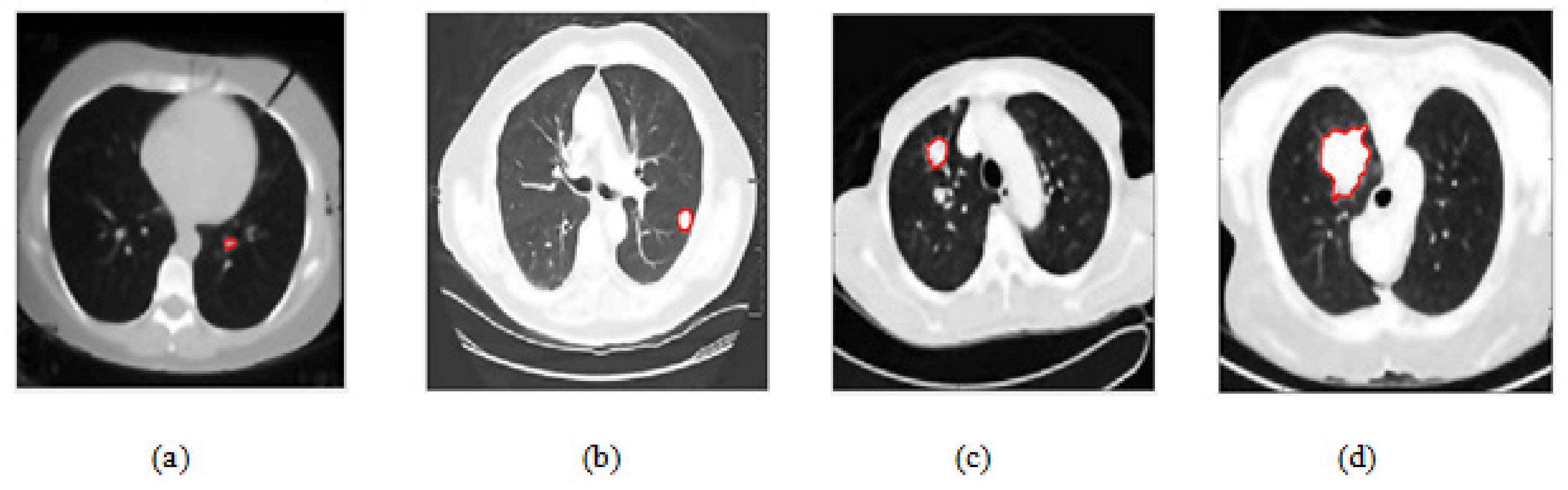
2.3. Random Walker Method for Nodule Boundary Extraction
- It can handle complex boundaries.
- As the model assumes the image as a graph where pixels are nodes or vertices, graph theory and popular graph algorithms can also be used.
- It can be easily employed to divide the image into multiple segments based on the number of seed labels provided.
2.4. Feature Extraction
2.4.1. Local Binary Pattern (LBP) Filter
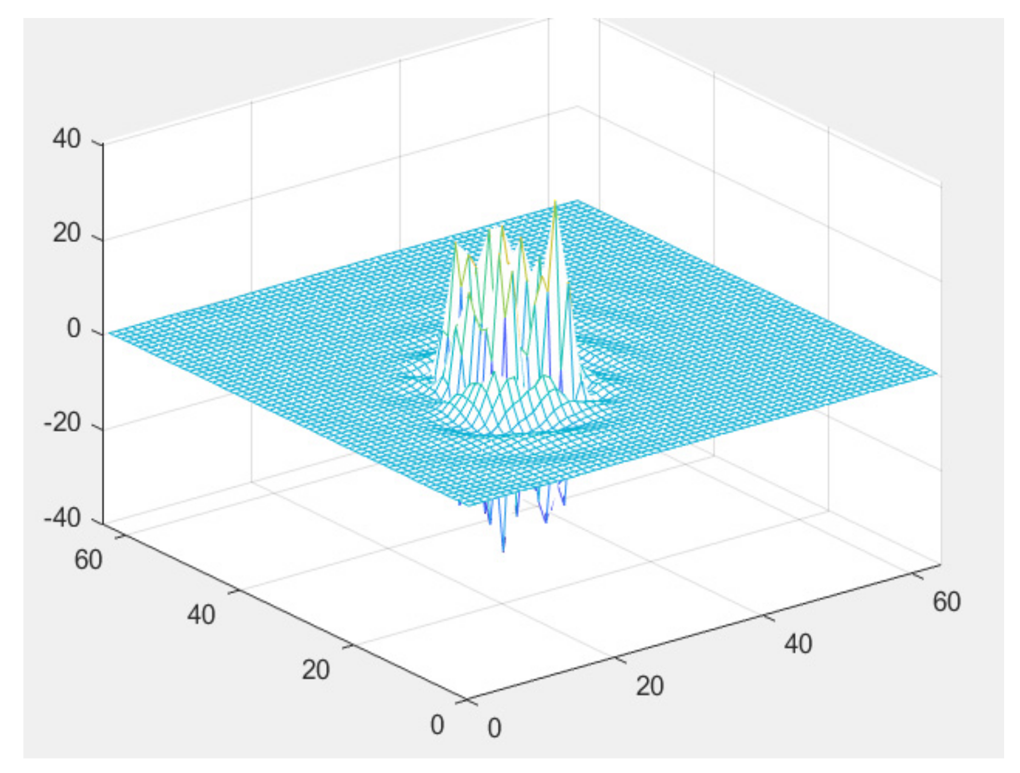
2.4.2. Riesz Wavelet Coefficients
2.5. Proposed Modified Gradient Boosting Classifier
| Algorithm 1. Proposed Modified Gradient Boosting Algorithm |
| Begin |
| Inputs |
| Dataset (xi y) |
| Number of iterations, M |
| Loss Functions, L(y, F(x)) |
| Choose the base-learner function, am |
| Initalize F0(x) |
| for m = 1 to M |
| Compute the negative gradient function gm(xi). |
| Generate the new base-learner function h(x; am). |
| , where h(x; a) is a weak learner |
| Evaluate the best gradient descent step-size . |
| Update the estimation/prediction function |
| Fm(x) = Fm−1(x) + h(x; am) = y |
| End for |
| Output prediction, y |
| End |
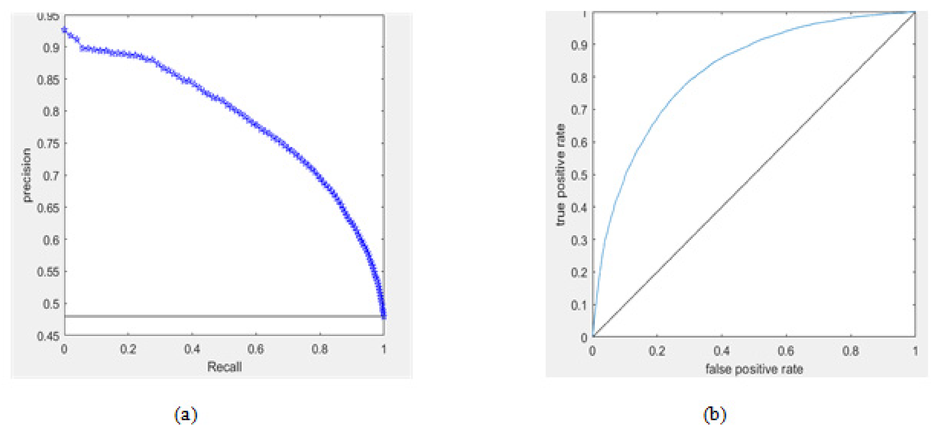
3. Performance Metrics
4. Experimental Results
4.1. Pre-Processing Result
4.2. Lung Nodule Segmentation Result
4.3. Feature Extraction Result
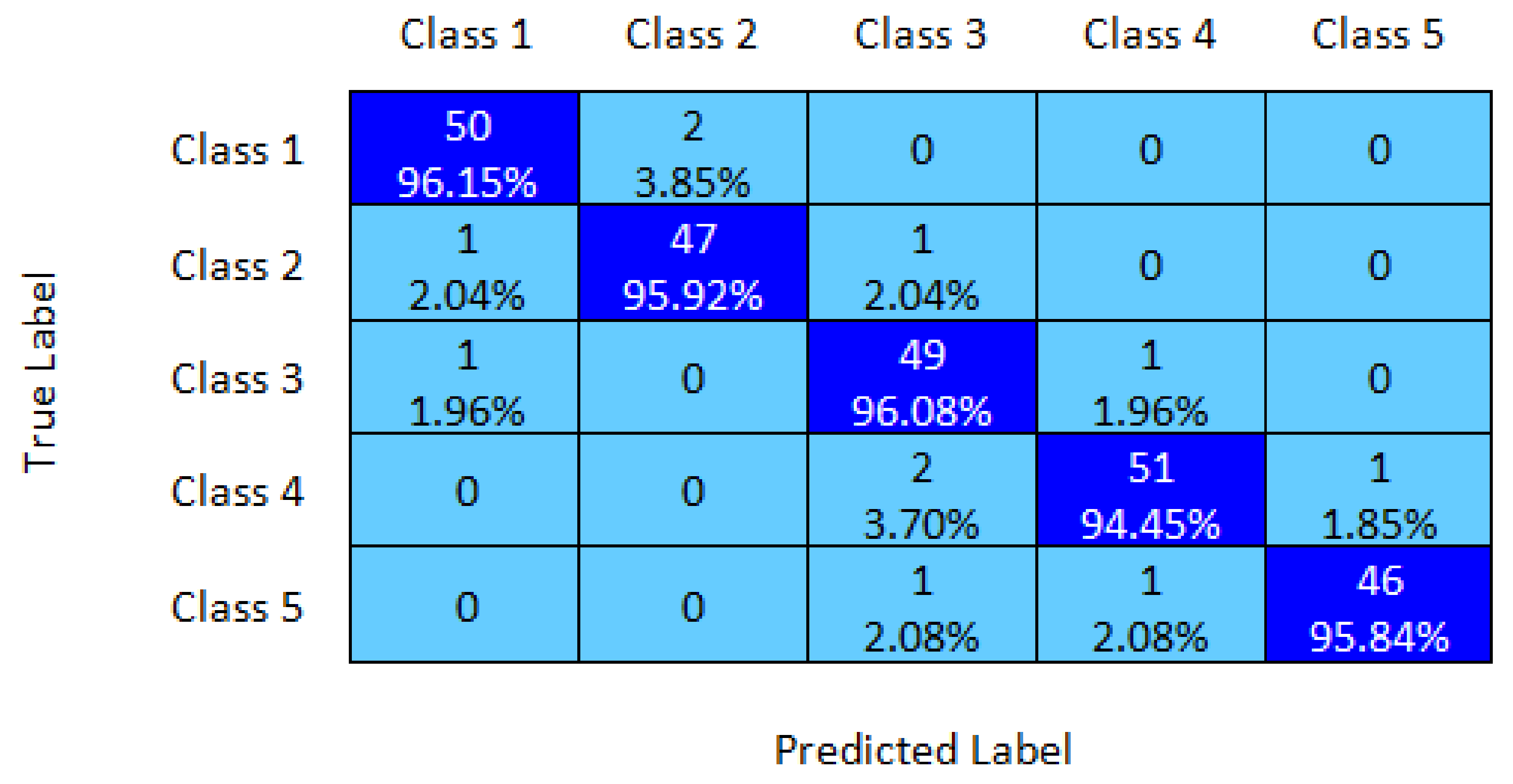
4.4. Lung Nodule Classification Result
5. Discussion
6. Conclusions
Author Contributions
Funding
Institutional Review Board Statement
Informed Consent Statement
Data Availability Statement
Conflicts of Interest
References
- World Health Organization. Cancer Report. Available online: https://www.who.int/news-room/fact-sheets/detail/cancer (accessed on 1 June 2022).
- Li, X.; Li, B.; Tian, L.; Zhang, L. Automatic benign and malignant classification of pulmonary nodules in thoracic computed tomography based on RF algorithm. IET Image Process. 2018, 12, 1253–1264. [Google Scholar] [CrossRef]
- Zhang, G.; Yang, Z.; Gong, L.; Jiang, S.; Wang, L. Classification of benign and malignant lung nodules from CT images based on hybrid features. Phys. Med. Biol. 2019, 64, 125011. [Google Scholar] [CrossRef] [PubMed]
- Abdillah, B.; Bustamam, A.; Sarwinda, D. Image processing based detection of lung cancer on CT scan images. J. Phys. Conf. Ser. 2017, 893, 012063. [Google Scholar] [CrossRef]
- Tanabe, Y.; Ishida, T. Development of a novel detection method for changes in lung conditions during radiotherapy using a temporal subtraction technique. Phys. Eng. Sci. Med. 2021, 44, 1341–1350. [Google Scholar] [CrossRef] [PubMed]
- Tartar, A.; Kilic, N.; Akan, A. Classification of Pulmonary Nodules by Using Hybrid Features. Comput. Math. Methods Med. 2013, 2013, 1–11. [Google Scholar] [CrossRef] [PubMed]
- Wang, X.; Mao, K.; Wang, L.; Yang, P.; Lu, D.; He, P. An Appraisal of Lung Nodules Automatic Classification Algorithms for CT Images. Sensors 2019, 19, 194. [Google Scholar] [CrossRef] [PubMed]
- Abaszade, M.; Effati, S. A New Method for Classifying Random Variables Based on Support Vector Machine. J. Classif. 2018, 36, 152–174. [Google Scholar] [CrossRef]
- Zhou, J.; Li, E.; Wei, H.; Li, C.; Qiao, Q.; Armaghani, D.J. Random Forests and Cubist Algorithms for Predicting Shear Strengths of Rockfill Materials. Appl. Sci. 2019, 9, 1621. [Google Scholar] [CrossRef]
- El-Askary, N.S.; Salem, M.A.M.; Roushdy, M.I. Lung Nodule Detection and Classification using Random Forest: A Review. In Proceedings of the 9th International Conference on Intelligent Computing and Information Systems (ICICIS), Cairo, Egypt, 8–10 December 2019; pp. 105–111. [Google Scholar] [CrossRef]
- Friedman, J.H. Greedy function approximation: A gradient boosting machine. Ann. Stat. 2001, 29, 1189–1232. [Google Scholar] [CrossRef]
- Jakimovski, G.; Davcev, D. Using Double Convolution Neural Network for Lung Cancer Stage Detection. Appl. Sci. 2019, 9, 427. [Google Scholar] [CrossRef]
- Song, Q.; Zhao, L.; Luo, X.; Dou, X. Using Deep Learning for Classification of Lung Nodules on Computed Tomography Images. J. Healthcare Eng. 2017, 2017, 1–7. [Google Scholar] [CrossRef] [PubMed]
- Khan, S.A.; Hussain, S.; Yang, S.; Iqbal, K. Effective and Reliable Framework for Lung Nodules Detection from CT Scan Images. Sci. Rep. 2019, 9, 4989. [Google Scholar] [CrossRef] [PubMed]
- Tomassini, S.; Falcionelli, N.; Sernani, P.; Burattini, L.; Dragoni, A.F. Lung nodule diagnosis and cancer histology classification from computed tomography data by convolutional neural networks: A survey. Comput. Biol. Med. 2022, 146, 105691. [Google Scholar] [CrossRef] [PubMed]
- Alves, A.F.F.; Souza, S.A.; Ruiz, R.L.; Reis, T.A.; Ximenes, A.M.G.; Hasimoto, E.N.; Lima, R.P.S.; Miranda, J.R.A.; Pina, D.R. Combining machine learning and texture analysis to differentiate mediastinal lymph nodes in lung cancer patients. Phys. Eng. Sci. Med. 2021, 44, 387–394. [Google Scholar] [CrossRef] [PubMed]
- Kumar, K.S.; Venkatalakshmi, K.; Karthikeyan, K. Lung Cancer Detection Using Image Segmentation by means of Various Evolutionary Algorithms. Comput. Math. Methods Med. 2019, 2019, 1–16. [Google Scholar] [CrossRef]
- Mukherjee, J.; Chakrabarti, A.; Shaikh, S.H.; Kar, M. Automatic Detection and Classification of Solitary Pulmonary Nodules from Lung CT Images. In Proceedings of the Fourth International Conference of Emerging Applications of Information Technology, Kolkata, India, 19–21 December 2014; pp. 294–299. [Google Scholar] [CrossRef]
- Sharma, K.; Soni, H.; Agarwal, K. Lung Cancer Detection in CT Scans of Patients Using Image Processing and Machine Learning Technique. In Advanced Computational and Communication Paradigms. Lecture Notes in Electrical Engineering; Bhattacharyya, S., Gandhi, T., Sharma, K., Dutta, P., Eds.; Springer: Singapore, 2018; Volume 475, pp. 336–344. [Google Scholar] [CrossRef]
- Veeraprathap, V.; Harish, G.S.; Narendra Kumar, G. Lung Cancer detection and multi-level classification using discrete Wavelet Transform approach. Int. J. Biomed. Biol. Eng. 2020, 14, 17–23. [Google Scholar] [CrossRef]
- Marentakis, P.; Karaiskos, P.; Kouloulias, V.; Kelekis, N.; Argentos, S.; Oikonomopoulos, N.; Loukas, C. Lung cancer histology classification from CT images based on radiomics and deep learning models. Med Biol. Eng. Comput. 2021, 59, 215–226. [Google Scholar] [CrossRef]
- Dhara, A.K.; Mukhopadhyay, S.; Dutta, A.; Garg, M.; Khandelwal, N. A Combination of Shape and Texture Features for Classification of Pulmonary Nodules in Lung CT Images. J. Digit. Imaging 2016, 29, 466–475. [Google Scholar] [CrossRef] [PubMed]
- Pabón, O.S.; Torrente, M.; Provencio, M.; Rodríguez-Gonzalez, A.; Menasalvas, E. Integrating Speculation Detection and Deep Learning to Extract Lung Cancer Diagnosis from Clinical Notes. Appl. Sci. 2021, 11, 865. [Google Scholar] [CrossRef]
- Maity, A.; Nair, T.R.; Mehta, S.; Prakasam, P. Automatic lung parenchyma segmentation using a deep convolutional neural network from chest X-rays. Biomed. Signal Process. Control 2021, 73, 103398. [Google Scholar] [CrossRef]
- Liu, D.; Liu, F.; Tie, Y.; Qi, L.; Wang, F. Res-trans networks for lung nodule classification. Int. J. Comput. Assist. Radiol. Surg. 2022, 17, 1059–1068. [Google Scholar] [CrossRef]
- Bruntha, P.M.; Pandian, S.I.; Anitha, J.; Abraham, S.S.; Kumar, S.N. A novel hybridized feature extraction approach for lung nodule classification based on transfer learning technique. J. Med. Phys. 2022, 47, 1–9. [Google Scholar] [CrossRef] [PubMed]
- Grady, L. Random Walks for Image Segmentation. IEEE Trans. Pattern Anal. Mach. Intell. 2006, 28, 1768–1783. [Google Scholar] [CrossRef]
- Eslami, A.; Karamalis, A.; Katouzian, A.; Navab, N. Segmentation by retrieval with guided random walks: Application to left ventricle segmentation in MRI. Med Image Anal. 2013, 17, 236–253. [Google Scholar] [CrossRef] [PubMed]
- Dogra, A.; Goyal, B.; Agrawal, S. Bone vessel image fusion via generalized reisz wavelet transform using averaging fusion rule. J. Comput. Sci. 2017, 21, 371–378. [Google Scholar] [CrossRef]
- Unser, M.; Chenouard, N. A Unifying Parametric Framework for 2D Steerable Wavelet Transforms. SIAM J. Imaging Sci. 2013, 6, 102–135. [Google Scholar] [CrossRef]
- Chenouard, N.; Unser, M. 3D Steerable Wavelets in Practice. IEEE Trans. Image Process. 2012, 21, 4522–4533. [Google Scholar] [CrossRef]
- Cirujeda, P.; Muller, H.; Rubin, D.; Aguilera, T.A.; Loo, B.W.; Diehn, M.; Binefa, X.; Depeursinge, A. 3D Riesz-wavelet based Covariance descriptors for texture classification of lung nodule tissue in CT. In Proceedings of the 37th Annual International Conference of the IEEE Engineering in Medicine and Biology Society (EMBC), Milan, Italy, 25–29 August 2015; pp. 7909–7912. [Google Scholar] [CrossRef]
- Bentéjac, C.; Csörgő, A.; Martínez-Muñoz, G. A comparative analysis of gradient boosting algorithms. Artif. Intell. Rev. 2020, 54, 1937–1967. [Google Scholar] [CrossRef]
- Birunda, S.S.; Devi, R.K. A Novel Score-Based Multi-Source Fake News Detection using Gradient Boosting Algorithm. In Proceedings of the International Conference on Artificial Intelligence and Smart Systems (ICAIS), Coimbatore, India, 25–27 March 2021; pp. 406–414. [Google Scholar] [CrossRef]
- Shin, Y. Application of Stochastic Gradient Boosting Approach to Early Prediction of Safety Accidents at Construction Site. Adv. Civ. Eng. 2019, 2019, 1–9. [Google Scholar] [CrossRef]
- Yu, H.; Li, J.; Zhang, L.; Cao, Y.; Yu, X.; Sun, J. Design of lung nodules segmentation and recognition algorithm based on deep learning. BMC Bioinform. 2021, 22, 1–21. [Google Scholar] [CrossRef]
- Shoji, K.; Shunske, K.; Yasushi, H.; Shingo, M.; Tohru, K.; Nobuyuki, T.; Yuki, S.; Masahiro, Y.; Noriyuki, T. Segmentation of Lung Nodules on CT Images Using a Nested Three-Dimensional Fully Connected Convolutional Network. Front. Artif. Intell. 2022, 5, 782225. Available online: https://www.frontiersin.org/articles/10.3389/frai.2022.782225 (accessed on 10 March 2022).
- Nada, S.; El-Askary, M.; Salem, A.-M.; Roushdy, M.I. Feature Extraction and Analysis for Lung Nodule Classification using Random Forest. In Proceedings of the 8th International Conference on Software and Information Engineering, Cairo, Egypt, 9–12 April 2019; pp. 248–252. [Google Scholar] [CrossRef]
- Wang, S.; Dong, L.; Wang, X.; Wang, X. Classification of pathological types of lung cancer from CT images by deep residual neural networks with transfer learning strategy. Open Med. 2020, 15, 190–197. [Google Scholar] [CrossRef] [PubMed]

| Methods/Model | DSC | JSC |
|---|---|---|
| Graph Cut Method | 0.566 ± 0.025 | 0.414 ± 0.021 |
| Watershed Method | 0.628 ± 0.027 | 0.494 ± 0.025 |
| 3D U-Net | 0.911 ± 0.009 | 0.811 ± 0.011 |
| 3D FCN | 0.845 ± 0.008 | 0.738 ± 0.011 |
| Proposed Method | 0.979 ± 0.011 | 0.877 ± 0.008 |
| Methods/Model | Dataset Used | Accuracy (%) | Precision | Recall | F1 Score |
|---|---|---|---|---|---|
| Random Forest [38] | LIDC-IDRI | 85.00 | 0.7962 | 0.7810 | 0.7918 |
| Deep RNN [39] | LIDC-IDRI/Luna 16 | 85.71 | 0.879 | 0.892 | 0.882 |
| SVM [16] | LIDC-IDRI | 93.0 | 0.87 | 0.86 | 0.84 |
| Res U-Net [36] | LIDC-IDRI | 90.70% | 0.916 | 0..902 | 0.911 |
| Proposed Modified Gradient Boosting Classifier | LIDC-IDRI | 95.67 | 0.957 | 0.91 | 0.941 |
Publisher’s Note: MDPI stays neutral with regard to jurisdictional claims in published maps and institutional affiliations. |
© 2022 by the authors. Licensee MDPI, Basel, Switzerland. This article is an open access article distributed under the terms and conditions of the Creative Commons Attribution (CC BY) license (https://creativecommons.org/licenses/by/4.0/).
Share and Cite
Donga, H.V.; Karlapati, J.S.A.N.; Desineedi, H.S.S.; Periasamy, P.; TR, S. Effective Framework for Pulmonary Nodule Classification from CT Images Using the Modified Gradient Boosting Method. Appl. Sci. 2022, 12, 8264. https://doi.org/10.3390/app12168264
Donga HV, Karlapati JSAN, Desineedi HSS, Periasamy P, TR S. Effective Framework for Pulmonary Nodule Classification from CT Images Using the Modified Gradient Boosting Method. Applied Sciences. 2022; 12(16):8264. https://doi.org/10.3390/app12168264
Chicago/Turabian StyleDonga, Harsha Vardhan, Jaya Sai Aditya Nandan Karlapati, Harsha Sri Sumanth Desineedi, Prakasam Periasamy, and Sureshkumar TR. 2022. "Effective Framework for Pulmonary Nodule Classification from CT Images Using the Modified Gradient Boosting Method" Applied Sciences 12, no. 16: 8264. https://doi.org/10.3390/app12168264
APA StyleDonga, H. V., Karlapati, J. S. A. N., Desineedi, H. S. S., Periasamy, P., & TR, S. (2022). Effective Framework for Pulmonary Nodule Classification from CT Images Using the Modified Gradient Boosting Method. Applied Sciences, 12(16), 8264. https://doi.org/10.3390/app12168264