Article

Aqueous Synthesis of the Tiopronin-Capped Gold Nanoclusters/Nanoparticles with Precise Size Control via Deprotonation of the Ligand

1
Department of Bio-Convergence Science and Technology, Kyungpook National University, Daegu 41566, Korea
2
Department of Electrical Engineering, Kyungpook National University, Daegu 41566, Korea
3
School of Electronic and Electric Engineering, Kyungpook National University, Daegu 41566, Korea
4
Department of Electrical Engineering and Smart Grid Research Center, Jeonbuk National University, Jeonju 54896, Korea
*
Author to whom correspondence should be addressed.
These authors contributed equally to this work.
Appl. Sci. 2022, 12(16), 8263; https://doi.org/10.3390/app12168263
Submission received: 13 June 2022 / Revised: 11 August 2022 / Accepted: 15 August 2022 / Published: 18 August 2022

Abstract

:
Gold nanoparticles have led to numerous advances in nanomaterial-based sensors and biomedical technologies owing to their chemical inertness and outstanding physiochemical and optical properties. Gold nanoparticles are still considered one of the most promising types of nanomaterials in various biomedical fields, including drug delivery, cancer therapy, biomolecule detection, and high-accuracy diagnosis. Surface functionalization of gold nanoparticles with various ligands modifies the physicochemical properties of the surface, thereby improving the biocompatibility and uptake efficiency of a living system. Tiopronin, one of the most commonly used ligands for gold nanoparticles, has both thiol and carboxyl functional groups that can be easily attached to various biomolecules. However, the conventional method of synthesizing tiopronin-capped gold nanoclusters using methanol and acetic acid as a solvent requires a laborious and time-consuming dialysis process to remove methanol and acetic acid. In this study, we demonstrate a novel and simple aqueous synthesis method to obtain tiopronin-capped gold nanoclusters/nanoparticles with precise size control in the sub-nanometer to nanometer range. The main advantage of our synthesis method is that it does not require a dialysis process because it uses water as a solvent. The boron byproduct produced during the synthesis can be removed with a simple volatilization process. Moreover, we characterized the physical morphologies, photoelectronic properties, hydrodynamic size, and crystal structure of the tiopronin-capped gold nanoclusters/nanoparticles using transmission electron microscopy, spectrophotometry, fluorescence spectrometry, dynamic light scattering, zeta potential, and X-ray diffraction.

1. Introduction

Gold nanoclusters/nanoparticles have revolutionized nanomaterial-based sensors and biomedical technologies owing to their outstanding photoelectronic properties, such as superior plasmonic characteristics and chemical–biological inertness. In biomedical nanotechnology, surface-functionalized gold nanoclusters/nanoparticles capable of binding to numerous biomolecules, including antibodies, proteins, and oligonucleotides, have been developed for biomedical imaging, biosensors, and drug delivery [1,2,3,4,5,6,7,8,9,10,11,12,13,14,15,16,17,18]. Recently, the plasmonic properties of gold nanoparticles have been utilized for the detection of cancer cell-derived exosomes [19,20,21]. In addition, the chemical inertness of gold nanoclusters/nanoparticles has driven the research of nanoparticle-mediated catalysis [21,22,23]. Moreover, the variation in photo-absorption characteristics of gold nanoparticles with size is being studied to aid in the development of gold nanoparticle sensors [24,25]. Furthermore, gold nanoclusters, which exhibit single-molecule-like behaviors owing to their extremely small size, have been widely studied as electron-trapping layers in photoelectronic sensors [26,27]. The size of gold nanoparticles is considered to depend on various factors, including temperature, pH, and reaction time [28,29].
The Brust–Schiffrin method [30,31], based on two-phase aqueous-organic medium and strong gold-alkanethiol ligand passivation, is a representative method for the synthesis of small-sized gold nanoparticles and nanoclusters (<5 nm) with a narrow size distribution. Templeton et al. first synthesized tiopronin-protected gold nanoclusters based on the Brust–Schiffrin method [32]. They synthesized tiopronin-protected gold nanoclusters in a methanol/acetic acid mixture (ratio of 6:1) and dispersed them in water through dialysis. As tiopronin has a carboxyl group at the active site that can bind with amines via the 1-ethyl-3-(3-dimethylamino)propyl carbodiimide/N-hydroxysuccinimide (EDC/NHS) reaction, tiopronin-capped gold nanoclusters/nanoparticles have been utilized in various biotechnologies. Most of the currently used tiopronin-capped gold nanoclusters/nanoparticles of sizes smaller than 5 nm have been synthesized using the method developed by Templeton et al. [33,34,35,36]. To the best of our knowledge, the method developed by Templeton et al. is the only approach to synthesize tiopronin-capped gold nanoclusters/nanoparticles with sizes smaller than 5 nm reported to date. However, the synthesis method requires laborious and time-consuming dialysis processes to substitute the organic solvent and water.
In this work, we present a novel and simple synthesis method to obtain tiopronin-capped gold nanoclusters/nanoparticles, with size control in the range from less than a nanometer to several nanometers. The advantages and highlights of this work are as follows. First, unlike the conventional Brust–Schiffrin method, the synthesis method proposed herein does not require dialysis processes because it uses water as a solvent during the entire synthesis. Second, our synthesis method uses sodium hydroxide to ensure the dispersion stability of the precursors, whereas the Brust-Schiffrin method uses organic solvents. Sodium hydroxide deprotonates the carboxyl groups in tiopronin and hinders drastic chemical reactions, resulting in stable nucleation and growth of the nanoclusters/nanoparticles. Finally, the boron byproduct was removed by converting it into volatile trimethyl borate using a small amount of methanol. The characteristics of the tiopronin-capped gold nanoclusters/nanoparticles with different sizes were investigated using Fourier-transformed infrared (FT-IR) spectroscopy, transmission electron microscopy (TEM), UV–Vis spectrophotometry, fluorescence spectrophotometry, dynamic light scattering (DLS), and X-ray diffraction (XRD).

2. Materials and Methods

2.1. Reagents

Hydrogen tetrachloroaurate tetrahydrate (chloroauric acid tetrahydrate, ≥99%) was purchased from Fujifilm Wako Chemicals (Osaka, Japan). N-(2-mercaptopropionyl)glycine (tiopronin, ≥98%) and sodium borohydride (99.99%) were purchased from Sigma-Aldrich, Inc. (Burlington, MA, USA). Double-distilled water (HPLC grade) and methyl alcohol (99.8%) were purchased from Daejung Chemicals and Metals Company, Ltd. (Siheung, Korea).

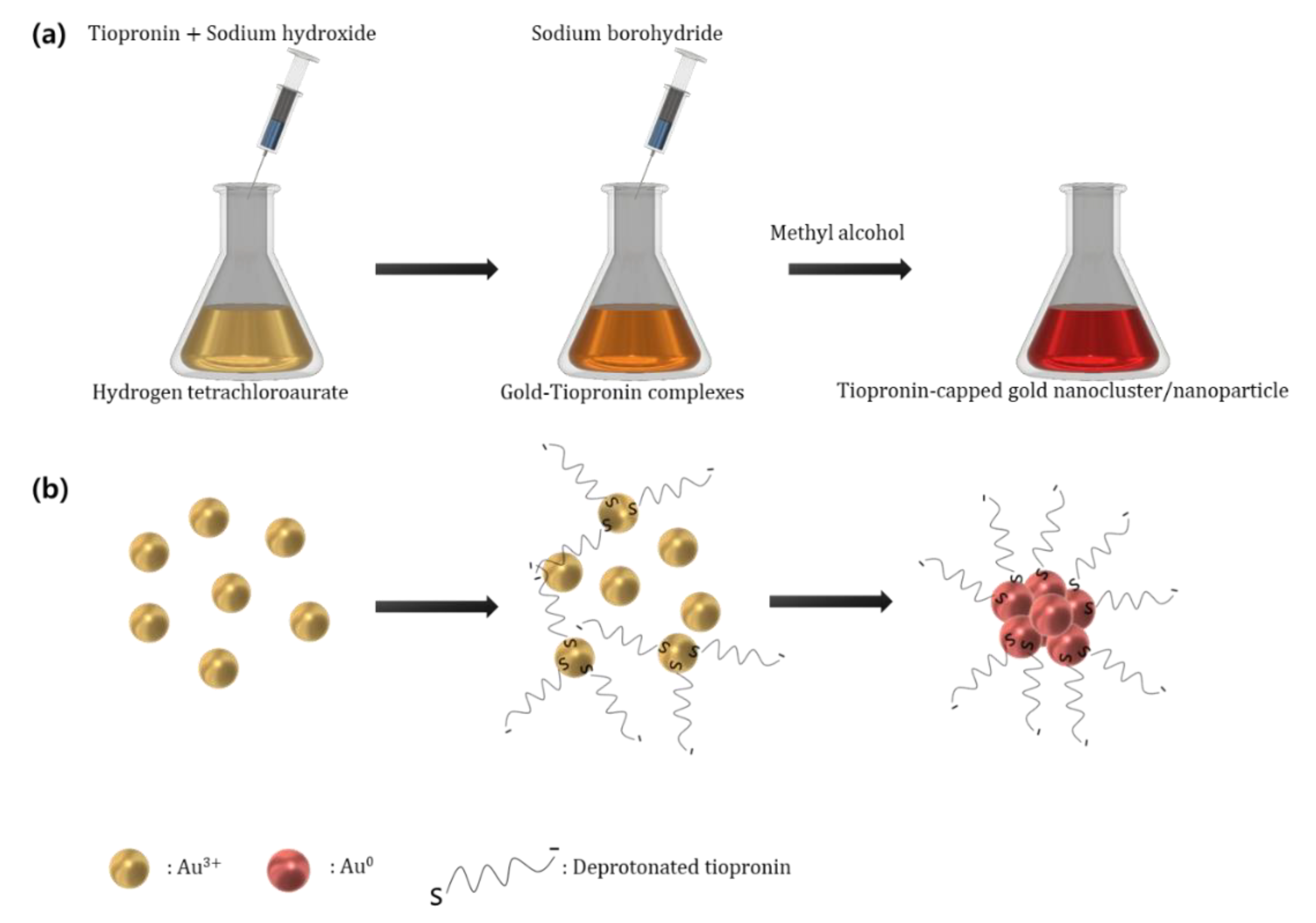
2.2. Synthesis of Tiopronin-Capped Gold Nanoclusters/Nanoparticles

The aqueous synthesis of tiopronin-capped gold nanoclusters/nanoparticles began with the formation of gold-tiopronin complexes. The gold precursor solution was prepared by dissolving 0.01 mmol of hydrogen tetrachloroaurate tetrahydrate in 25 mL of double-distilled water. A mixture of 0.01 M tiopronin and sodium hydroxide (1:1) aqueous solution was added to the gold precursor solution, and the reaction led to the formation of gold-tiopronin complex. The amounts of tiopronin and sodium hydroxide mixture solution used in this study were 3, 2, 1, 0.5, 0.2, 0.1, 0.05, and 0.02 mL. During this stage, sodium hydroxide deprotonated the carboxyl group of the tiopronin tail, enabling stable nucleation of the tiopronin-capped gold nanoclusters/nanoparticles during the subsequent reduction process. In the absence of the deprotonation of the carboxyl functional group, the gold nanoparticle seeds tended to aggregate via hydrogen-bonding interactions between the carboxyl groups, resulting in unstable gold nanoparticle growth during the subsequent reduction process. In contrast, an excessive reaction between gold precursors and a mixture of tiopronin and sodium hydroxide for more than an hour led to a spontaneous reduction of gold, resulting in the formation of bare gold microflakes that floated on the surface of the solution.
The gold ions were reduced by adding 0.1 mmol of fresh sodium borohydride dissolved in 1 mL double distilled water with rapid stirring. The reduction process lasted for 50 min with rapid stirring. This resulted in the formation of tiopronin-capped gold nanoclusters/nanoparticles via metallic bonding between the reduced gold and gold–tiopronin complex. The size of the tiopronin-capped gold nanocluster/nanoparticles was determined by the molar ratio between gold and tiopronin. Finally, boric acid, a byproduct, was removed by adding a small amount of methanol to the resulting colloid. According to Equations (1) and (2), this process yields volatile trimethyl borate, which can be degassed via ultrasonication. Figure 1 illustrates the synthesis method for tiopronin-capped gold nanoclusters/nanoparticles.
2Au3+ + 6NaBH4 + 18H2O → 2Au0 + 6H3BO3 + 21H2 ↑ + 6Na+
H3BO3 + 3CH3OH → B(OCH3)3 ↑ + 3H2O

2.3. Measurements

The presence of tiopronin in the tiopronin-capped gold nanoclusters/nanoparticles was confirmed via FT-IR using a Vertex 80/80v (Bruker Corporation, Billerica, MA, USA). Thin layers of the tiopronin-capped gold nanoclusters/nanoparticles were prepared on a silicon wafer (10 mm × 10 mm) by repeated droplet evaporation (~5 times) and measured under the attenuated total reflection mode.
The core size, lattice structure, and morphology of the tiopronin-capped gold nanoclusters/nanoparticles were investigated via TEM using a Titan G2 ChemiSTEM Cs Probe (FEI Company, Hillsboro, OR, USA). The size distribution of the tiopronin-capped gold nanoclusters/nanoparticles was analyzed using the image processing software ImageJ.
The photoelectronic properties of the tiopronin-capped gold nanoclusters/nanoparticles were examined via UV–Vis spectrophotometry and fluorescence spectrophotometry using a LAMBDA 950 spectrometer (Pelkin Elmer, Inc., Waltham, MA, USA) and Hitachi F-7000 (Hitachi, Ltd., Tokyo, Japan), respectively. The colloids of the tiopronin-capped gold nanoclusters/nanoparticles were diluted five times using double-distilled water to ensure accurate spectrophotometry results.
The hydrodynamic sizes of the tiopronin-capped gold nanoclusters/nanoparticles were determined via DLS measurements using a Nano ZS (Malvern Panalytical, Ltd., Malvern, UK). The samples used for the DLS size analysis were the same as those used for spectrophotometry.
The colloidal stability of the tiopronin-capped gold nanoclusters/nanoparticles was determined by measuring the zeta potential distributions using a Nano ZS (Malvern Panalytical, Ltd., Malvern, UK).
The crystal structure of the tiopronin-capped gold nanoclusters/nanoparticles was confirmed via XRD measurements conducted using an EMPYREAN (Malvern Panalytical, Ltd., Malvern, UK). The thin layers of the tiopronin-capped gold nanoclusters/nanoparticles were prepared on a silicon wafer (10 mm × 10 mm) by repeated droplet evaporation (~5 times).

3. Results and Discussions

Figure 2 shows the TEM images of the tiopronin-capped gold nanoclusters/nanoparticles. The numbers on the upper-left indicate the molar ratio of gold to tiopronin and the mean particle size. The insets on the upper-right show the size distribution obtained using ImageJ. Figure 2i shows the lattice structure of the tiopronin-capped gold nanoparticles synthesized with a gold-to-tiopronin molar ratio of 1:0.02. The size of the tiopronin-capped gold nanoclusters/nanoparticles increased with the decrease in the amount of tiopronin. The modal size, mean size, and standard deviation of the size distribution of the tiopronin-capped gold nanoclusters/nanoparticles with different gold-to-tiopronin molar ratios are presented in Table 1. The lattice plane spacings of the tiopronin-capped gold nanoparticle were measured to be 0.24 nm and 0.15 nm, which respectively correspond to the (111) and (220) planes of gold (Figure 2i).
Figure 3 shows the crystal structure of the tiopronin-capped gold nanocluster/nanoparticle measured using XRD. Owing to their small size, the characteristics of the tiopronin-capped gold nanoclusters observed herein were similar to those of a thin film of amorphous crystals. Therefore, only the XRD pattern of the silicon wafer used as the substrate was obtained. However, as the particle size increased, the inherent crystallinity of gold gradually began to appear. The gold nanoparticles exhibited broad XRD patterns at 38.1°, 44.3°, 64.5°, and 77.7°, corresponding to Au (111), Au (200), Au (220), and Au (311), respectively. The lattice spacings obtained from TEM agreed well with those obtained via XRD, which indicated that the synthesized particles were gold nanoclusters/nanoparticles. According to the Scherrer equation (Equation (3)—which explains the correlation between particle size and the full-width half-maximum (FWHM) of a signal peak in an XRD measurement—the smaller the particle is, the wider the peak.
τ = K λ β c o s τ θ
where τ denotes the mean crystallite size, K the shape factor, λ the wavelength of the X-ray, β the line broadening at the FWHM, and θ the Bragg angle.
Figure 4 shows the FT-IR results for the tiopronin and the tiopronin-capped gold nanocluster/nanoparticle; the results confirmed the binding of tiopronin to gold. The tiopronin-capped gold nanoclusters (Au:tiopronin = 1:3) showed characteristic peaks at 3419, 2954, 2925, 2852, 1662, 1635, and 1600 cm−1. These peaks correspond to the O-H stretching band of the carboxyl group (3419), the C-H stretching of the alkane compound (2954, 2925, 2852), C=C stretching (1662, 1600), and C=O (1635) and indicate the presence of alkane compound containing carboxylic groups. The tiopronin-capped gold nanoparticles (Au:tiopronin = 1:0.1) show characteristic peaks at 3350, 2954, 2925, 2854, 1662, 1635, and 1595 cm−1. These peaks correspond to N-H stretching of the secondary amines (3350), C-H stretching of the alkanes (2954, 2925, 2854), C=C stretching (1662, 1595), and C=O stretching (1635). In addition, the broad band corresponding to the O-H stretching of the carboxyl group existed near the 3350 cm−1 peak. Therefore, the FT-IR results confirmed the presence of an alkane compound containing a carboxyl group in both samples, indicating that tiopronin had bound to the gold nanocluster/nanoparticle as a ligand.
Figure 5a shows the absorption characteristics of the tiopronin-capped gold nanoclusters/nanoparticles. Unlike tiopronin-capped gold nanoclusters with sizes smaller than 2.21 nm, tiopronin-capped gold nanoparticles with sizes larger than 2.45 nm have distinct surface plasmon resonance peaks at 501 (2.45 nm), 507 (3.69 nm), 510 (4.15 nm), and 511 (4.38 nm) nm due to the strong plasmonic interaction of gold. As the size of the gold nanoparticles increases, the wavelength of the light absorbed by localized surface plasmon resonance increases. Therefore, the size of the gold nanoparticles was proportional to the wavelength of the peak surface plasmon resonance (SPR). This agreed well with the results obtained via TEM. Figure 5b shows the photoluminescence for the tiopronin-capped gold nanoclusters of sizes 0.58 and 0.81 nm with the respective peaks at 827.4 and 830 nm. The fluorescence property of the gold nanoclusters observed herein was attributed to the molecular-like behaviors of the gold nanoclusters with sizes smaller than 2 nm [37,38].
Figure 6a shows the sizes of the tiopronin-capped gold nanoclusters/nanoparticles measured using DLS. The hydrodynamic size of the tiopronin-capped gold nanoclusters/nanoparticles increased as the amount of tiopronin decreased. This trend was consistent with the results obtained via TEM. The modal hydrodynamic sizes of the tiopronin-capped nanoclusters/nanoparticles with different gold-to-tiopronin ratios were measured to be 1.74, 2.33, 3.62, 4.85, 5.61, 6.5, and 6.5 nm. Figure 6b shows a comparison of the sizes measured using TEM and DLS. The sizes measured by DLS were slightly larger than those obtained using TEM. The average size difference with different gold-to-tiopronin ratios was calculated to be 1.83 ± 0.4 nm. The disparity in the sizes obtained from the different measurements was attributed to the molecular length of tiopronin, which is 0.77 nm as calculated by density functional theory.
Figure 7 presents the zeta potential distribution of the tiopronin-capped gold nanoclusters/nanoparticles (Au:tiopronin = 1:3 and 1:0.1). In general, the colloidal stability of the particles can be considered excellent when the absolute amplitude of the zeta potential is greater than 30 mV. The zeta potentials of the synthesized tiopronin-capped gold nanoclusters/nanoparticles were measured to be −59.5 mV (Au:tiopronin = 1:3) and −30 mV (Au:tiopronin = 1:0.1), indicating excellent colloidal stability.

4. Conclusions

We demonstrated a dialysis-free synthesis method to obtain tiopronin-capped gold nanoclusters/nanoparticles with size control in the range of less than a nanometer to several nanometers. Unlike the conventional Brust–Schiffrin method, which utilizes a two-phase organic-aqueous environment for stable nucleation and growth and requires a laborious, time-consuming dialysis process, we synthesized the tiopronin-capped gold nanoclusters/nanoparticles in water, and stable nucleation and growth were achieved by deprotonating the carboxyl group of tiopronin in the gold–tiopronin complex solution before the reduction process. The characteristics of the tiopronin-capped gold nanoclusters/nanoparticles, including the morphology, size, absorbance, photoluminescence, hydrodynamic size, zeta potential distribution, and crystal structure, were investigated. This work describes a simple synthesis method for tiopronin-capped gold nanoclusters/nanoparticles, which have great potential to develop bio-nanotechnologies.

Author Contributions

Conceptualization, D.K.; methodology, D.K.; formal analysis, C.S.; investigation, D.K., C.S. and H.L.; writing—original draft preparation, D.K.; writing—review and editing, C.S., H.P. and J.P.; visualization, H.L. and T.K.; supervision, H.P., T.K. and J.P.; project administration, J.P.; funding acquisition, J.P. All authors have read and agreed to the published version of the manuscript.

Funding

This research was supported by a National Research Foundation of Korea (NRF) grant funded by the Korean government (MSIT) (NRF-2020R1F1A1076037 and NRF-2020R1A4A1019518).

Institutional Review Board Statement

Not applicable.

Informed Consent Statement

Not applicable.

Acknowledgments

The authors acknowledge NRF for financial support.

Conflicts of Interest

The authors declare no conflict of interest.

References

  1. Lee, J.H.; Cho, H.Y.; Choi, H.K.; Lee, J.Y.; Choi, J.W. Application of Gold Nanoparticle to Plasmonic Biosensors. Int. J. Mol. Sci. 2018, 19, 2021. [Google Scholar] [CrossRef] [PubMed]
  2. Zhang, L.; Mazouzi, Y.; Salmain, M.; Liedberg, B.; Boujday, S. Antibody-Gold Nanoparticle Bioconjugates for Biosensors: Synthesis, Characterization and Selected Applications. Biosens. Bioelectron. 2020, 165, 112370. [Google Scholar] [CrossRef] [PubMed]
  3. Yang, Y.; Zhong, S.; Wang, K.; Huang, J. Gold Nanoparticle Based Fluorescent Oligonucleotide Probes for Imaging and Therapy in Living Systems. Analyst 2019, 144, 1052–1072. [Google Scholar] [CrossRef]
  4. Wu, Y.; Ali, M.R.K.; Chen, K.C.; Fang, N.; El-Sayed, M.A. Gold Nanoparticles in Biological Optical Imaging. Nano Today 2019, 24, 120–140. [Google Scholar] [CrossRef]
  5. Siddique, S.; Chow, J.C.L. Gold Nanoparticles for Drug Delivery and Cancer Therapy. Appl. Sci. 2020, 10, 3824. [Google Scholar] [CrossRef]
  6. Sharifi, M.; Attar, F.; Saboury, A.A.; Akhtari, K.; Hooshmand, N.; Hasan, A.; El-Sayed, M.A.; Falahati, M. Plasmonic Gold Nanoparticles: Optical Manipulation, Imaging, Drug Delivery and Therapy. J. Control. Release 2019, 311–312, 170–189. [Google Scholar] [CrossRef]
  7. Pingarrón, J.M.; Yañez-Sedeño, P.; González-Cortés, A.J.E.A. Gold Nanoparticle-Based Electrochemical Biosensors. Electrochim. Acta 2008, 53, 5848–5866. [Google Scholar] [CrossRef]
  8. Zeng, S.W.; Yong, K.T.; Roy, I.; Dinh, X.Q.; Yu, X.; Luan, F. A Review on Functionalized Gold Nanoparticles for Biosensing Applications. Plasmonics 2011, 6, 491–506. [Google Scholar] [CrossRef]
  9. Sharma, P.; Brown, S.; Walter, G.; Santra, S.; Moudgil, B. Nanoparticles for Bioimaging. Adv. Colloid Interface Sci. 2006, 123–126, 471–485. [Google Scholar] [CrossRef]
  10. Yong, K.T.; Swihart, M.T.; Ding, H.; Prasad, P.N. Preparation of Gold Nanoparticles and Their Applications in Anisotropic Nanoparticle Synthesis and Bioimaging. Plasmonics 2009, 4, 79–93. [Google Scholar] [CrossRef]
  11. Cheng, Y.; Samia, A.C.; Meyers, J.D.; Panagopoulos, I.; Fei, B.; Burda, C. Highly Efficient Drug Delivery with Gold Nanoparticle Vectors for in Vivo Photodynamic Therapy of Cancer. J. Am. Chem. Soc. 2008, 130, 10643–10647. [Google Scholar] [CrossRef] [PubMed]
  12. Daraee, H.; Eatemadi, A.; Abbasi, E.; Fekri Aval, S.; Kouhi, M.; Akbarzadeh, A. Application of Gold Nanoparticles in Biomedical and Drug Delivery. Artif. Cells Nanomed. Biotechnol. 2016, 44, 410–422. [Google Scholar] [CrossRef] [PubMed]
  13. Han, G.; Ghosh, P.; Rotello, V.M. Functionalized Gold Nanoparticles for Drug Delivery. Nanomedicine 2007, 2, 113–123. [Google Scholar] [CrossRef] [PubMed]
  14. Duncan, B.; Kim, C.; Rotello, V.M. Gold Nanoparticle Platforms as Drug and Biomacromolecule Delivery Systems. J. Control. Release 2010, 148, 122–127. [Google Scholar] [CrossRef] [PubMed]
  15. Li, Y.; Zhai, T.; Chen, J.; Shi, J.; Wang, L.; Shen, J.; Liu, X. Water-dispersible Gold Nanoclusters: Synthesis Strategies, Optical Properties, and Biological Applications. Chem. Eur. J. 2022, 28, e202103736. [Google Scholar] [CrossRef]
  16. Chen, W.; Deng, H.H.; Hong, L.; Wu, Z.Q.; Wang, S.; Liu, A.L.; Lin, X.H.; Xia, X.H. Bare Gold Nanoparticles as Facile and Sensitive Colorimetric Probe for Melamine Detection. Analyst 2012, 137, 5382–5386. [Google Scholar] [CrossRef]
  17. Ijaz, M.; Zafar, M.; Iqbal, T. Green Synthesis of Silver Nanoparticles by Using Various Extracts: A review. Inorg. Nano-Met. Chem. 2020, 51, 744–755. [Google Scholar] [CrossRef]
  18. Iqbal, T.; Tufail, S.; Ghazal, S. Synthesis of Silver, Chromium, Manganese, Tin and Iron Nano Particles by Different Techniques. Int. J. Nanosci. Nanotechnol. 2017, 13, 19–52. [Google Scholar]
  19. Wang, Z.L.; Zong, S.F.; Wang, Y.J.; Li, N.; Li, L.; Lu, J.; Wang, Z.Y.; Chen, B.A.; Cui, Y.P. Screening and Multiple Detection of Cancer Exosomes Using an SERS-Based Method. Nanoscale 2018, 10, 9053–9062. [Google Scholar] [CrossRef]
  20. Pang, Y.; Wang, C.; Lu, L.; Wang, C.; Sun, Z.; Xiao, R. Dual-SERS Biosensor for One-Step Detection of MicroRNAs in Exosome and Residual Plasma of Blood Samples for Diagnosing Pancreatic Cancer. Biosens. Bioelectron. 2019, 130, 204–213. [Google Scholar] [CrossRef]
  21. Wang, Q.; Zou, L.; Yang, X.; Liu, X.; Nie, W.; Zheng, Y.; Cheng, Q.; Wang, K. Direct Quantification of Cancerous Exosomes via Surface Plasmon Resonance with Dual Gold Nanoparticle-Assisted Signal Amplification. Biosens. Bioelectron. 2019, 135, 129–136. [Google Scholar] [CrossRef] [PubMed]
  22. Gogoi, N.; Borah, G.; Gogoi, P.K.; Chetia, T.R. TiO2 Supported Gold Nanoparticles: An Efficient Photocatalyst for Oxidation of Alcohol to Aldehyde and Ketone in Presence of Visible Light Irradiation. Chem. Phys. Lett. 2018, 692, 224–231. [Google Scholar] [CrossRef]
  23. Deraedt, C.; Salmon, L.; Gatard, S.; Ciganda, R.; Hernandez, R.; Ruiz, J.; Astruc, D. Sodium Borohydride Stabilizes Very Active Gold Nanoparticle Catalysts. Chem. Commun. 2014, 50, 14194–14196. [Google Scholar] [CrossRef] [PubMed]
  24. Shellaiah, M.; Simon, T.; Sun, K.W.; Ko, F.H. Simple Bare Gold Nanoparticles for Rapid Colorimetric Detection of Cr3+ Ions in Aqueous Medium with Real Sample Applications. Sens. Actuators B Chem. 2016, 226, 44–51. [Google Scholar] [CrossRef]
  25. Shellaiah, M.; Thirumalaivasan, N.; Sun, K.W.; Wu, S.P. A pH Cooperative Strategy for Enhanced Colorimetric Sensing of Cr(III) Ions Using Biocompatible L-Glutamic Acid Stabilized Gold Nanoparticles. Microchem. J. 2021, 160, 105754. [Google Scholar] [CrossRef]
  26. Chu, Y.Z.; Schonbrun, E.; Yang, T.; Crozier, K.B. Experimental Observation of Narrow Surface Plasmon Resonances in Gold Nanoparticle Arrays. Appl. Phys. Lett. 2008, 93, 181108. [Google Scholar] [CrossRef]
  27. Zayats, M.; Kharitonov, A.B.; Pogorelova, S.P.; Lioubashevski, O.; Katz, E.; Willner, I. Probing Photoelectrochemical Processes in Au-CdS Nanoparticle Arrays by Surface Plasmon Resonance: Application for the Detection of Acetylcholine Esterase Inhibitors. J. Am. Chem. Soc. 2003, 125, 16006–16014. [Google Scholar] [CrossRef]
  28. Briñas, R.P.; Hu, M.; Qian, L.; Lymar, E.S.; Hainfeld, J.F. Gold Nanoparticle Size Controlled by Polymeric Au(I) Thiolate Precursor Size. J. Am. Chem. Soc. 2008, 130, 975–982. [Google Scholar] [CrossRef]
  29. Karimi, S.; Moshaii, A.; Nikkhah, M. Controlled Synthesis of Colloidal Monodisperse Gold Nanoparticles in a Wide Range of Sizes; Investigating the Effect of Reducing Agent. Mater. Res. Express 2019, 6, 1150f2. [Google Scholar] [CrossRef]
  30. Brust, M.; Walker, M.; Bethell, D.; Schiffrin, D.J.; Whyman, R. Synthesis of Thiol-Derivatised Gold Nanoparticles in a Two-Phase Liquid–Liquid System. J. Chem. Soc. Chem. Commun. 1994, 7, 801–802. [Google Scholar] [CrossRef]
  31. Brust, M.; Fink, J.; Bethell, D.; Schiffrin, D.; Kiely, C. Synthesis and Reactions of Functionalised Gold Nanoparticles. J. Chem. Soc. Chem. Commun. 1995, 16, 1655–1656. [Google Scholar] [CrossRef]
  32. Templeton, A.C.; Chen, S.W.; Gross, S.M.; Murray, R.W. Water-Soluble, Isolable Gold Clusters Protected by Tiopronin and Coenzyme A Monolayers. Langmuir 1999, 15, 66–76. [Google Scholar] [CrossRef]
  33. Daood, U.; Akram, Z.; Matinlinna, J.P.; Fawzy, A.S. Dentine Collagen Cross-Linking Using Tiopronin-Protected Au/EDC Nanoparticles Formulations. Dent. Mater. 2019, 35, 1017–1030. [Google Scholar] [CrossRef] [PubMed]
  34. Grueso, E.; Perez-Tejeda, P.; Giráldez-Pérez, R.M.; Prado-Gotor, R.; Muriel-Delgado, F. Ethanol Effect on Gold Nanoparticle Aggregation State and Its Implication in the Interaction Mechanism with DNA. J. Colloid Interface Sci. 2018, 529, 65–76. [Google Scholar] [CrossRef]
  35. Muraca, F.; Boselli, L.; Castagnola, V.; Dawson, K.A. Ultrasmall Gold Nanoparticle Cellular Uptake: Influence of Transient Bionano Interactions. ACS Appl. Bio Mater. 2020, 3, 3800–3808. [Google Scholar] [CrossRef]
  36. Dahan, I.; Sorrentino, S.; Boujemaa-Paterski, R.; Medalia, O. Tiopronin-Protected Gold Nanoparticles as a Potential Marker for Cryo-EM and Tomography. Structure 2018, 26, 1408–1413. [Google Scholar] [CrossRef]
  37. Ramakrishna, G.; Varnavski, O.; Kim, J.; Lee, D.; Goodson, T. Quantum-Sized Gold Clusters as Efficient Two-Photon Absorbers. J. Am. Chem. Soc. 2008, 130, 5032–5033. [Google Scholar] [CrossRef]
  38. Wilcoxon, J.P.; Martin, J.E.; Parsapour, F.; Wiedenman, B.; Kelley, D.F. Photoluminescence from Nanosize Gold Clusters. J. Chem. Phys. 1998, 108, 9137–9143. [Google Scholar] [CrossRef]
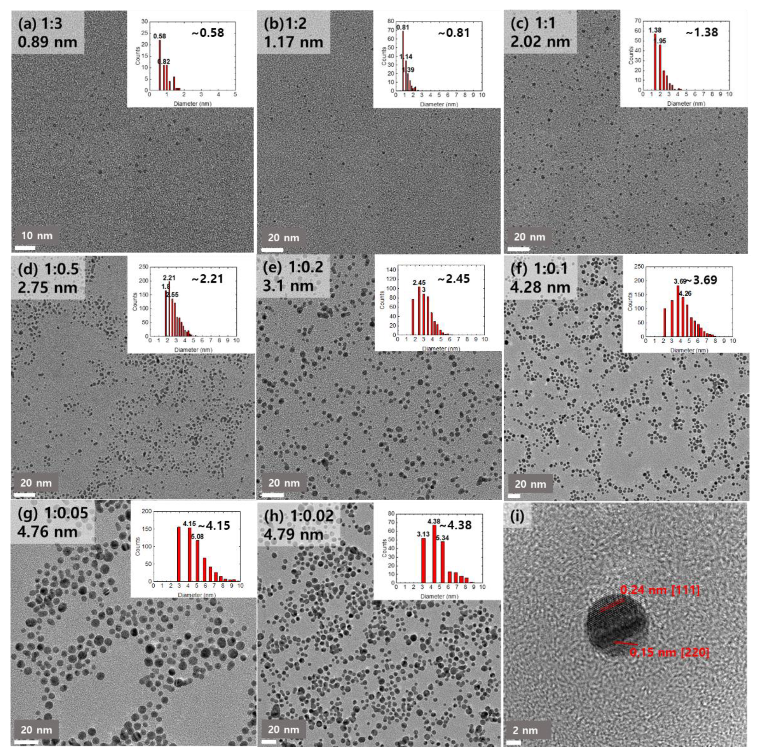
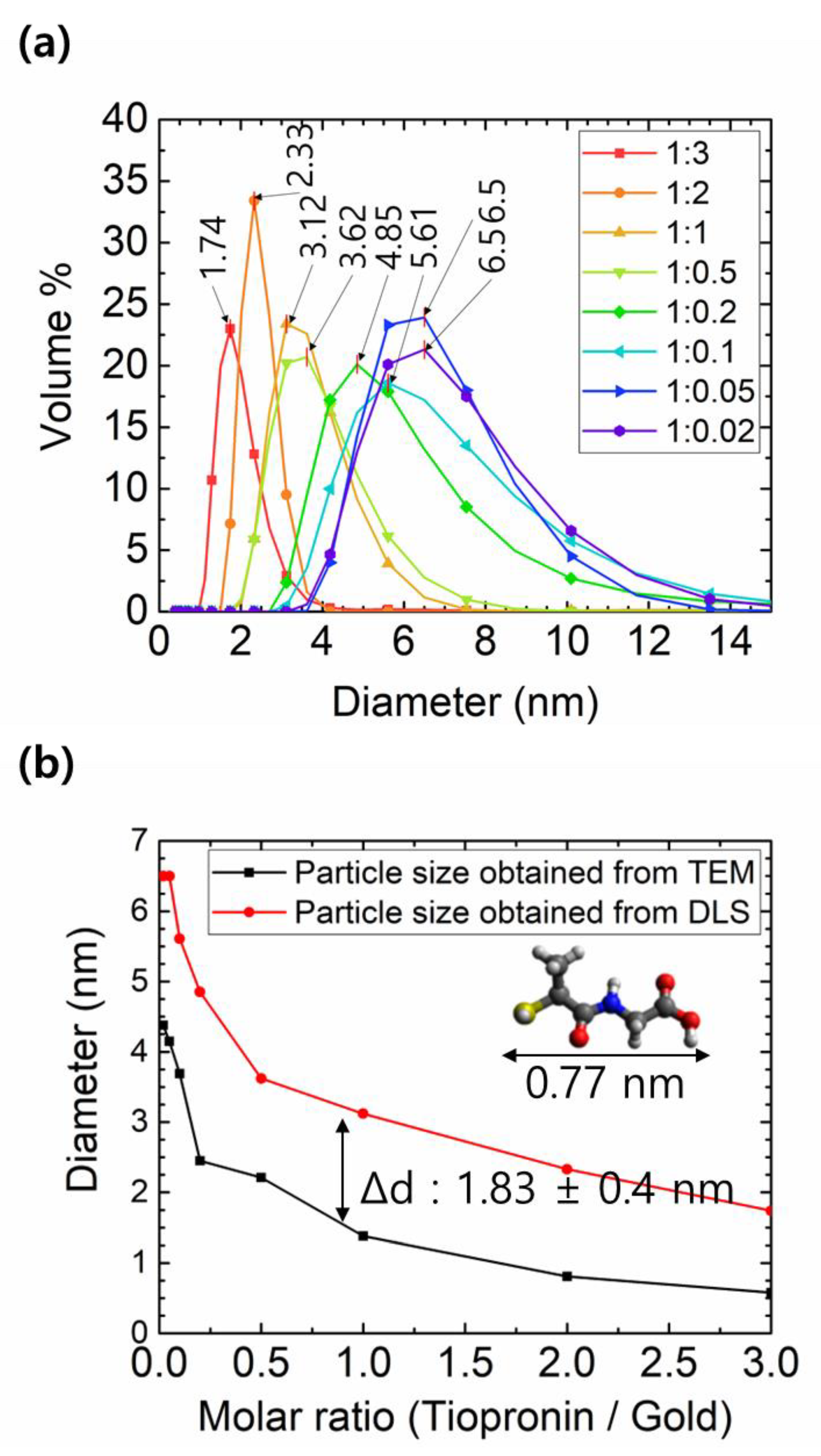
Figure 1. Schematic of the synthesis method for tiopronin-capped gold nanoclusters/nanoparticles. (a) The experimental process for the synthesis. (b) The chemical reaction flow for the process, showing the state of the gold ions, the tiopronin-gold complexes, and the tiopronin-capped gold nanoclusters/nanoparticles.
Applsci 12 08263 g001
Figure 2. TEM images of the tiopronin-capped gold nanoclusters/nanoparticles synthesized with different gold:tiopronin ratios: (a) 1:3, (b) 1:2, (c) 1:1, (d) 1:0.5, (e) 1:0.2, (f) 1:0.1, (g) 1:0.05, (h) 1:0.02. (i) HRTEM image of a tiopronin-capped gold nanocluster/nanoparticle showing the lattice structure. The insets show the size distribution of the tiopronin-capped gold nanoclusters/nanoparticles, estimated from the images using a particle size analysis tool.
Applsci 12 08263 g002
Figure 3. XRD results for the tiopronin-capped gold nanocluster/nanoparticle (black line: reference gold XRD pattern; red line: tiopronin-capped gold nanoparticle).
Applsci 12 08263 g003
Figure 4. FT-IR measurement results for the tiopronin-capped gold nanocluster (blue), tiopronin-capped gold nanoparticle (red), and tiopronin (black). An offset of 100 is given between each plot for readability.
Applsci 12 08263 g004
Figure 5. (a) Absorbance curves of the tiopronin-capped gold nanoclusters/nanoparticles obtained using a UV–Vis spectrophotometer. The numbers in the legend indicate the molar ratio of gold to tiopronin. (b) Photoluminescence of the tiopronin-capped gold nanoclusters synthesized with a gold-to-tiopronin ratio of 1:3 and 1:2.
Applsci 12 08263 g005
Figure 6. (a) Hydrodynamic particle size of the tiopronin-capped gold nanoclusters/nanoparticles obtained using DLS. The numbers in the legend indicate the molar ratio of gold to tiopronin. (b) The modal size discrepancies for the tiopronin-capped gold nanoclusters/nanoparticles obtained from TEM and DLS.
Applsci 12 08263 g006
Figure 7. Zeta potential distribution of the tiopronin-capped gold nanoclusters/nanoparticles. Au:tiopronin = 1:3 and 1:0.1, respectively.
Applsci 12 08263 g007
Table 1. The modal size (dmode), mean size (dmean), and standard deviation (σ) of the size distribution of the tiopronin-capped gold nanoclusters/nanoparticles with different gold-to-tiopronin molar ratios.
Au:Tiopronin1:31:21:11:0.51:0.21:0.11:0.051:0.02
dmode (nm)0.580.811.382.212.453.694.154.38
dmean (nm)0.891.172.022.753.14.284.764.79
σ (nm)±0.32±0.42±0.66±0.76±0.94±1.32±1.55±1.37
Publisher’s Note: MDPI stays neutral with regard to jurisdictional claims in published maps and institutional affiliations.

