Article

Constitutive High Expression Level of a Synthetic Deleted Encoding Gene of Talaromyces minioluteus Endodextranase Variant (rTmDEX49A–ΔSP–ΔN30) in Komagataella phaffii (Pichia pastoris)

by
Roberto Carlos Arísticas Ribalta
1,†,
Lisandra Martínez Valdés
1,†,
Meinardo Lafargue Gámez
1,
Sonia Rodríguez Davydenko
1,
Eric Dubreucq
2,
Veronique Perrier
2,
Benoît Moreau
3 and
Reinaldo Fraga Vidal
1,*
1
Cuban Research Institute on Sugarcane By-Products (ICIDCA), P.O. Box 4026, Havana 11000, Cuba
2
Agropolymer Engineering and Emerging Technologies (IATE), Institut Agro, French National Institute for Agriculture, Food and Environment (INRAE), University of Montpellier, 34060 Montpellier, France
3
Haute Ecole Province de Henaut–Condorcet, Rue Paul Pastur 11, 7800 Ath, Belgium
*
Author to whom correspondence should be addressed.
These authors contributed equally to this work.
Appl. Sci. 2022, 12(15), 7562; https://doi.org/10.3390/app12157562
Submission received: 21 June 2022 / Revised: 21 July 2022 / Accepted: 25 July 2022 / Published: 27 July 2022

Abstract

:

Featured Application

The dextranase variant (rTmDEX49A–ΔSP–ΔN30) can be used for dextran removal from sugar during the production process.

Abstract

In the sugar industry, dextran generates difficulties in the manufacturing process. Using crude dextranase (EC 3.2.1.11) to eliminate dextran in sugar is an effective practice. In this study, a synthetic dextranase-encoding gene of the filamentous fungus Talaromyces minioluteus, lacking its putative native signal peptide (1–20 amino acids) and the next 30 amino acids (rTmDEX49A–ΔSP–ΔN30), was fused to the Saccharomyces cerevisiae prepro α–factor (MFα–2) signal sequence and expressed in Komagataella phaffii under the constitutive GAP promoter. K. phaffii DEX49A–ΔSP–ΔN30, constitutively producing and secreting the truncated dextranase, was obtained. The specific activity of the truncated variant resulted in being nearly the same in relation to the full-length mature enzyme (900–1000 U·mg−1 of protein). At shaker scale (100 mL) in a YPG medium, the enzymatic activity was 273 U·mL−1. The highest production level was achieved in a fed-batch culture (30 h) at 5 L fermenter scale using the FM21–PTM1 culture medium. The enzymatic activity in the culture supernatant reached 1614 U·mL−1, and the productivity was 53,800 U·L−1·h−1 (53.8 mg·L−1·h−1), the highest reported thus far for a DEX49A variant. Dextran decreased rTmDEX49A–ΔSP–ΔN30 mobility in affinity gel electrophoresis, providing evidence of carbohydrate–protein interactions. K. phaffii DEX49A–ΔSP–ΔN30 shows great potential as a methanol-free, commercial dextranase production system.

1. Introduction

Dextran is a high molecular weight homo-polysaccharide composed of D-glucose units largely linked by type α (1→6) bonds and to a lesser extent α (1→3), α (1→4), and α (1→2) bonds [1]. The structure of a dextran, such as the type of bond and the degree of branching, depends on the dextransucrase responsible for its synthesis [2,3]. Dextransucrases (EC 2.4.1.5) are secreted by lactic acid bacteria (LAB) of the genera Leuconostoc, Lactobacillus, Streptococcus, Pediococcus, and Weissella [4,5]. Dextran is widely used in industrial applications. In the health area, it is used as a blood plasma extender, heparin substitute, and for anticoagulant therapy [3,6]. However, in the sugar industry this polymer has a negative effect. The formation of dextran results in a costly loss of sucrose and considerably decreases the yield in the sugar manufacturing process [7]. Accordingly, dextran is an undesirable compound in the sugar production process from sugarcane (Saccharum officinarum). Dextran formation is due to the presence of LAB producers of dextransucrases. Furthermore, dextran increases the viscosity of the sugarcane juices, decreasing the efficiency of clarification, filtration, and crystallization during sugar production. Increased dextran is a measure of sugarcane deterioration, affecting the quality of the final product, as well as production efficiency [8,9,10].
The use of dextranase (EC 3.2.1.11) preparations to eliminate dextran in sugar is an effective practice [8]. Dextranases called α–D–(1→6)–glucan–6–glucanohydrolases hydrolyze the glycosidic bond of dextran and release glucose or isomaltose molecules. They are found in the glucohydrolase families (GH) GH–49 and GH–66, and according to the catalytic specificity, they are divided into two groups: exodextranases that hydrolyze dextran at its ends and release glucose units, and endodextranases that cut the polymer within the chain to yield isomaltooligosaccharides [11]. These low molecular weight oligosaccharides do not influence the viscosity of the sugarcane juice. Thus, enzyme addition improves the performance of the cane sugar production process [12]. The enzyme source must be considered as one of the key factors when considering its use on an industrial scale, where it is essential to be cost competitive. These enzymes are found in different ecosystems and are produced by a variety of filamentous fungi, bacteria, and, to a lesser extent, yeasts [11,13]. Dextranases produced by fungi have received much attention because of their high specific enzyme activity and isomaltooligosaccharide products [14]. Recombinant dextranase production is a viable option to obtain enzyme preparations in large volumes [13,15].
The biochemical properties of the filamentous fungus Talaromyces minioluteus dextranase native and recombinant (TmDEX49A and rTmDEX49A, respectively) have been extensively studied [16,17]. The gene was cloned, sequenced (GenBank accession number L41562), and expressed in the yeast Pichia pastoris (currently Komagataella phaffii) using the methanol-regulated alcohol oxidase 1 (AOX1) promoter [15,18]. The 3D structure of rTmDEX49A was solved in the apo-enzyme and product-bound forms. The rTmDEX49A folds into two domains [19,20]. As an endodextranase, rTmDEX49A yielded isomaltose as the major product of dextran hydrolysis. This reaction occurs with net inversion at the anomeric carbon, implying a single displacement mechanism [19]. Fed–batch fermentation of K. phaffii expressing rTmDEX49A under the AOX1 and GAP promoter was reported [13,15]. The production was scaled up to 30 L under the AOX1 promoter [21].
K. phaffii seems to be a suitable heterologous host for the recombinant production of crude dextranase preparations to be used in the sugar industry, since it does not produce endogenous sucrose-transforming enzymes, neither dextranolytic activity nor endogenous dextranase enzymes. Constitutive promoter systems of K. phaffii have become a popular option in recombinant protein expression, due to the ease of process control and omission of potentially hazardous inducers, such as methanol [22]. The glyceraldehyde-3-phosphate dehydrogenase gene promoter (pGAP) of K. phaffii is a strong, constitutive promoter system that does not require strict optimization and monitoring of the methanol-induction step culture condition. It is more suited to industrial scale operations, as it avoids the potential risk and expense of storing and transporting large amounts of methanol. These advantages all contribute to the cost-effectiveness of the industrial production of non-toxic recombinant proteins using the GAP promoter system [23]. In our opinion, previous reports of rTmDEX49A production in K. phaffii still reflect enzyme yields that are too low to make those processes cost-effectively competitive.
In this study, we report the use of a synthetic gene encoding the mature T. minioluteus DEX49A with the first 30 amino acids downstream of the native signal peptide deleted, which resulted in a constitutive high–level expression of the N–terminal truncated dextranase (rTmDEX49A–ΔSP–ΔN30) in K. phaffii. Without any specific function, the 200–residue β–sandwich N–terminal domain in GH49 can be partially deleted to engineer DEX49A, searching for a more efficient dextranase variant to be used in the sugar industry. To the best of our knowledge, this research shows the highest reported productivity level of a rTmDEX49A dextranase variant in K. phaffii, thus making it suitable for large-scale production.

2. Materials and Methods

2.1. Strains, Vectors, and Media

The wild-type strain Komagataella phaffii PPS 9010 and pJexpress915 vector used for dextranase expression were obtained from ATUM (USA). Escherichia coli One Shot® Top10 (Invitrogen) was used as the host–vector system in the gene cloning procedure and plasmid maintenance. The E. coli was grown at 37 °C in low-salt Luria–Bertani medium (tryptone 1%, yeast extract 0.5%, NaCl 0.5%) containing 25 μg·mL−1 ZeocinTM, for selection of recombinant bacteria transformed with the expression vector. K. phaffii was routinely grown for maintaining cultures in yeast extract peptone dextrose YPD medium (yeast extract 1% w/v, peptone 2% w/v, and dextrose 2% w/v). The YPD medium was supplemented with sorbitol 1 mol·L−1 (YPDS) and T2000 dextran blue (1%) (YPDSDB) when needed. Long-term maintenance of the E. coli and K. phaffii strains was carried out at −80 °C in LB liquid medium with glycerol at 15% (v/v) and YPD liquid medium with glycerol at 30% (v/v), respectively.

2.2. Chemicals and Reagents

The chemical reagents used were from Sigma (USA), BioCen (Cuba), Merck (USA), Pharmacia (Sweden), Difco (USA), and Fluka (USA). For solid culture media, 20 g·L−1 of Bacto-Agar (Difco) was added. DNA purifications were done with the QiaPrep Spin Plasmid kit (Qiagen). DNA gel extraction was performed using QIAquick kit (Qiagen). DNA manipulation used standard methods [24]. Restriction and modification enzymes were purchased from New England Biolabs and used according to the manufacturer’s recommendations.

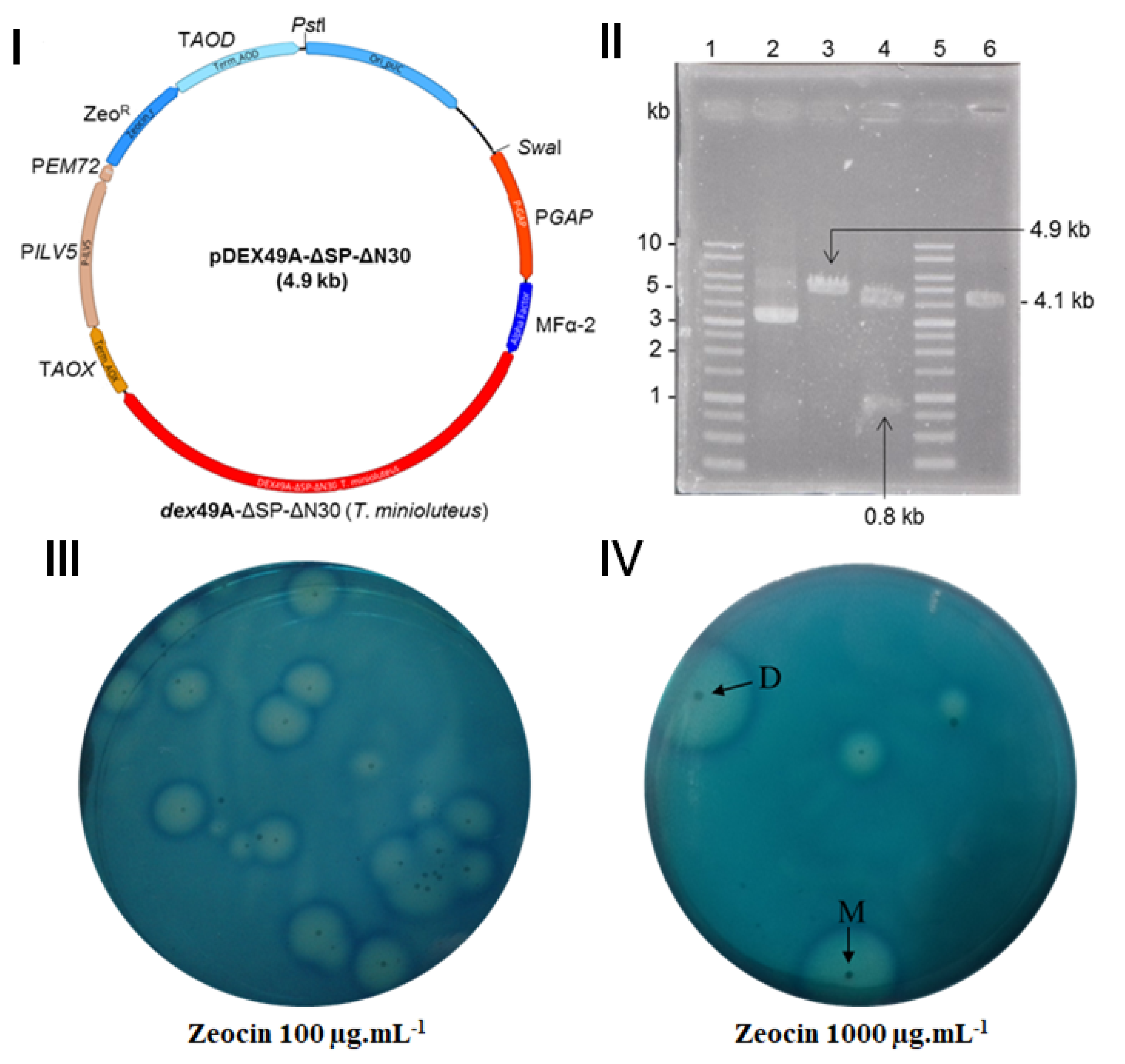
2.3. Vector Construction and Transformation

From the dextranase encoding gene (GenBank: L41562) of Talaromyces minioluteus MUCL 38929 (HI–4) (anamorph: Penicillium minioluteum), a coding region of 1827-bp is reported consisting of an ORF1 of 608 amino acids, including a putative secretion signal peptide of 20 amino acids (MATMLKLLALTLAISESAIG) based upon prediction using the SignalP–5.0 server (http://www.cbs.dtu.dk/services/SignalP, accessed on 7 June 2019). Nonetheless, the experimentally determined N–terminal sequence of the first 16 amino acids of mature dextranase protein (MGTTXNTXXGADFXTW) matches the predicted translation product starting at M35 (ORF4 of 574-aa) [15]. This ORF4 seems to be the real mature dextranase protein lacking the putative native secretion signal sequence (1–20 amino acids) plus the first 14-aa of the putative mature region of rTmDEX49A. To obtain rTmDEX49A–ΔSP–ΔN30, the first 16-aa of ORF4 were deleted. The synthetic DNA was fused to the optimized synthetic codon Saccharomyces cerevisiae prepro α–factor (MFα–2) signal sequence under the constitutive glyceraldehyde 3–phosphate dehydrogenase promoter (pGAP) of the K. phaffii expression vector pJexpress915. This strategy (fusion to MFα–1) is inspired by earlier work in budding yeast [25], which has been adapted to K. phaffii [22]. The recombinant construction of the pJexpress915 carrying the rTmDEX49A–ΔSP–ΔN30 synthetic gene, designated as pDEX49A–ΔSP–ΔN30, was ordered from the ATUM Company (USA). With this plasmid, E. coli TOP10 and K. phaffii PSS 9010 were transformed. Restriction analysis and DNA sequencing confirmed the recombinant pDEX49A–ΔSP–ΔN30 plasmid construction after amplification in E. coli TOP10. Finally, plasmid linearization was accomplished by SwaI enzyme (New England Biolabs), while subsequent transformation into K. phaffii PSS 9010 competent cells was achieved by electroporation using the Electroporator System Eporator® 2510 (Eppendorf, Germany), according to the condensed protocol for competent cell preparation and transformation of the methylotrophic yeast K. phaffii [26].

2.4. Screening of Recombinant K. phaffii Transformants and Production of the r–TmDEX49A–ΔSP–ΔN30

The K. phaffii transformants were screened from the YPDS Dextran blue plates supplemented with the antibiotic ZeocinTM at several final concentrations (100, 500, and 1000 µg·mL−1). The plates were incubated at 30 °C for 120 h, and the clones were selected by dex+ (dextranase positive) phenotype. After 24 h, the formation of a halo of transparency (dextran hydrolysis) around each of the enzyme-producing colonies was detected. Colonies with different dextran hydrolysis halo sizes were selected for extracellular dextranase activity quantification in shaking batch cultures.

2.5. Production of the r–TmDEX49A–ΔSP–ΔN30 in Shake Flasks

Small-scale cultures (25 mL) of YPGlycerol medium (yeast extract 1% w/v, peptone 2% w/v, and glycerol 2% w/v) in 250 mL shake flasks were used as inoculants to test the expression of the rTmDEX49A–ΔSP–ΔN30 enzyme in K. phaffii PSS 9010 grown overnight at 30 °C on a rotating shaker at 180 rpm. The following day, each culture was scaled up to a 1 L shake flask with 100 mL of YPGlycerol medium by diluting 1:10 and grown at 30 °C, 180 rpm, for 48 h. The cells were separated from the medium by centrifugation at 21,390× g, 4 °C, for 10 min (Eppendorf Centrifuge 5804 R) and suspended in NaAc buffer 20 mmol·L−1, pH 5.4. The samples (cleared supernatants and cells) were stored at −20 °C until they were used in other analyses. The clone with the highest enzyme activity was chosen for cultivation in a bioreactor.

2.6. Production of the r–TmDEX49A–ΔSP–ΔN30 in a 5 L Bioreactor (Fed-Batch Cultures)

Fed batch fermentation was performed from the pre-culture of the selected K. phaffii transformant inoculated in 10 mL of YPGlycerol medium in a 100 mL shake flask and grown overnight at 30 °C, 180 rpm, on a rotary shaker. The pre-culture was grown for 24 h at 30 °C, 180 rpm, at a 1:10 ratio in 100 mL of the synthetic medium FM21–PTM1 [27].
FM21–PTM1 composition: Glycerol 40 g·L−1; Salts FM21 (10×) [CaSO4 2·H2O 1.5 g·L−1, K2SO4 23.8 g·L−1, MgSO4 7·H2O 19.5 g·L−1, KOH 6.5 g·L−1, H3PO4 3.5% (v/v)]; Trace elements PTM1 (100×) [ZnCl2 2 g·L−1, Fe SO4 7·H2O 6.5 g·L−1, CuSO4 5·H2O 0.6 g·L−1, MnSO4 H2O 0.3 g·L−1, KI 0.01 g·L−1, H3BO3 0.002 g·L−1, H2SO4, 0.2% (v/v)]; Biotin 20 mg·L−1; Tartrate buffer solution (tartaric acid 0.1 mol·L−1, Na2HPO4 12·H2O 0.2 mol·L−1, pH 5.4). The FM21 solution was sterilized in an autoclave at 1.0 bar (121 °C) for 20 min, while PTM1 and biotin were sterilized by filtration. The pH in the medium was kept at 5.4 by adding NH4OH 15% (v/v).
Dextranase production was carried out in a 5 L fermenter (B.E. Marubishi, model MDL, Tokyo, Japan) with an initial working volume of 1.5 L at a 1:5 inoculum ratio. The synthetic medium was used: glycerol 40 g·L−1, distilled H2O 1029 mL, FM21 salts (10x) 150 mL, PTM1 trace elements (100×) 15 mL, and biotin (20 mg·L−1) 6 mL. The temperature was controlled at 28 °C, and the pH was adjusted to 5.0 by automatic addition of NH4OH 15% (v/v). The fermenter was stirred at 1000 rpm. Aeration started at 1 vvm in the batch phase and increased to 2 vvm in the feeding phase. A sharp increase in dissolved oxygen (approximately 14 h after inoculation) marked the beginning of the feeding phase; 100 mL of the feeding solution (glycerol 500 g·L−1) was added, repeating this feeding at intervals of 2 h until 5 increments were made, and the fermentation was stopped at 30 h. The antifoam (Glanapon 2000, Bussetti, Italy) was added to the fermentation medium in a 1:1 × 10⁴ ratio. Samples were taken at time intervals, and biomass and dextranase activity were analyzed.

2.7. Enzyme Assay and Protein Analysis

Dextranase activity was carried out at 40 °C in NaAc buffer 20 mmol·L−1 (pH 5.5) and dextran T110 (Pharmacia, Sweden) 2.5% (w/v) [28]. The levels of free reducing sugars were detected by the 3′,5′-dinitrosalicylic acid assay (DNSA colorimetric method) [29]. One unit (1 U) of dextranase activity was defined as the amount of enzyme that liberated 1 µmol of reducing sugar (glucose equivalent) per minute from T110 dextran under the conditions tested. The specific activity of rTmDEX49A–ΔSP–ΔN30 was calculated as the ratio of dextranase activity to enzyme concentration in the culture supernatant of fed batch cultures. All determinations were made in triplicate.
Dextranase was detected in SDS–PAGE (denaturing conditions) according to Laemmli et al. [30] and in native conditions for zymography analysis with 10% polyacrylamide gels. Approximately 10 µL of culture supernatant sample was applied to the gels. The proteins in SDS–PAGE were stained in Coomassie Brilliant Blue R–250 (CBB) 0.5% (w/v). The dextranolytic activity in the gel was detected in native conditions to which dextran blue T2000 (Pharmacia, Sweden) was added at a final concentration of 0.5% (w/v) [31]. Following electrophoresis, the gel was incubated in a renaturation solution containing Triton X-100 0.1% (v/v) for 30 min with slow shaking at 80 rpm at room temperature to remove SDS. Triton X-100 was replaced by NaAc 100 mmol·L−1 pH 5.5 solution and incubated for 30 min. Finally, it was changed to the same solution and was incubated with shaking at 37 °C overnight. Areas with dextranase activity appeared as clear bands on the dark background of the zymographic gel.
The affinity of the dextranase for the dextran substrate was determined in native polyacrylamide gels supplemented with increasing amounts of dextran [32]. The dextran was from Leuconostoc spp. Mr 150,000–200,000 Da (Sigma-Aldrich, USA) at concentrations from 0 to 1.0% (w/v). The PageRuler™ Prestained Protein Ladder molecular weight standard was used as a standard and as a negative control. The gels were stained with CBB to detect proteins.

2.8. Statistical Analysis of the Data

All determinations, shake flask and fermentation cultures, were made in triplicate, and the values subjected to an analysis of variance using Duncan’s multiple rank test to compare the corresponding means. These analyses were performed with a significance level of 99% using the statistical package Statgraphics Centurion XV version 15.2.05, Copyright by StatPoint, Inc. et al., 1982–2007.

3. Results and Discussion

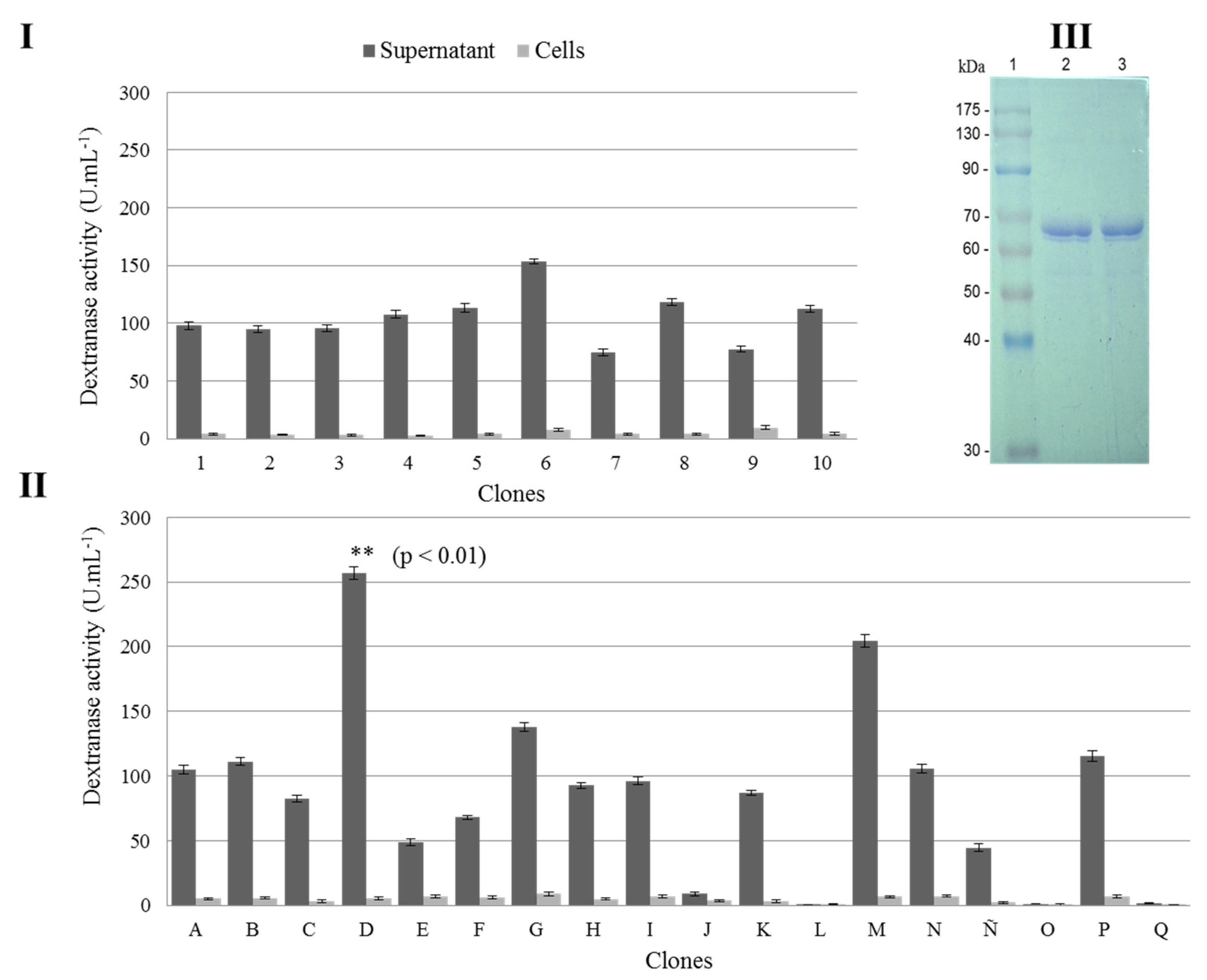
3.1. r–TmDEX49A–ΔSP–ΔN30 Variant and Histochemical Screening of Dextranolytic Activity in K. phaffii Clones

Figure 1I shows the rTmDEX49A–ΔSP–ΔN30 in pJexpress915 (ATUM, USA). E. coli TOP10 was transformed with the K. phaffii integrative expression vector pDEX49A–ΔSP–ΔN30. The expression module was detected in an agarose electrophoresis (Figure 1II). The size pattern obtained in the electrophoretic assay after restriction enzyme treatments corroborates the presence of the desired genetic construct (Figure 1II). The synthetic DNA fragment was verified by Sanger sequencing.
The native strain K. phaffii PSS 9010 and the integrative expression vector pDEX49A–ΔSP–ΔN30 were selected as the expression system to produce and secrete the biologically active truncated dextranase. The expression vector harbors a mature region comprising the amino acids 51–608 of rTmDEX49A–ΔSP–ΔN30 fused in frame with the Saccharomyces cerevisiae prepro α–factor (MFα–2) signal sequence under the constitutive pGAP and the AOX1 terminator. Transformation of PSS 9010 with the expression vector SwaI–linearized yielded numerous transformants on the solid medium with dextran blue (YPDSDB) plates containing various concentrations of ZeocinTM to screen for potential multiple inserts. This histochemical screening system has been previously reported [33,34,35,36]. Twenty-eight colonies showing the positive dextranase phenotype through the formation of degradation halos around the colony were selected for further analysis. The enzyme secreted by the producing clones diffuses into the solid medium and hydrolyzes the dextran blue, resulting in a radial halo of transparency (Figure 1III,IV). The transformants D and M showing a higher radial halo of transparency on the YPDSDB plate containing 1000 μg·mL−1 of ZeocinTM (Figure 1IV) also outperformed the dextranase activity level in the shake flask cultures compared with the rest of the transformants. This phenomenon of clonal variation due to a different copy number of expression cassettes is common in strains of K. phaffii genetically modified by inserting genes into the genome, as was reported for the expression of tetanus toxin fragment C [37].

3.2. Selection of the Best K. phaffii Clone Producer of r–TmDEX49A–ΔSP–ΔN30

Dextranase activity was determined from the supernatant of the selected clones. The levels of enzymatic activity obtained are shown in Figure 2I,II. The supernatant and cell fractions of 28 selected transformants were analyzed. The dextranase activity in the culture supernatant represented 93% of the total enzyme activity, while only 7% was associated with the cellular fraction. The highest value of enzymatic activity was obtained from the supernatant produced by clone D with 257 U·mL−1, followed by clone M with 204 U·mL−1 (Figure 2II). The specific activity of the truncated variant resulted in being nearly the same in relation to the full-length mature enzyme (900–1000 U·mg−1 of protein). The production of heterologous protein in the cell-free culture of the two best-producing clones was analyzed by SDS–PAGE (Figure 2III). A CBB-stained protein band was observed, which has an apparent MW of approximately 64–67 kDa, which (considering N-glycosylation) is expected for the rTmDEX49A–ΔSP–ΔN30 enzyme and very similar to the rTmDEX49A variant produced in K. phaffii [13,18]. Clone D (K. phaffii DEX49A–ΔSP–ΔN30) was selected for further characterization and protein production studies at shaker scale and in high-cell density fermentation at 5 L scale.

3.3. Dextranase Production at Shaker Scale by K. phaffii DEX49A–ΔSP–ΔN30

Growth and enzyme secretion for K. phaffii DEX49A–ΔSP–ΔN30 clone were studied in triplicate for 48 h in 100 mL of the YPGlycerol culture medium (Figure 3). The dried cell weight (DCW) reached 19 g·L−1 at 48 h.
The product formation (rTmDEX49A–ΔSP–ΔN30) curve measured as a function of enzymatic activity describes a continuous increase from the beginning to 48 h cultivation, which is typical of constitutive production [38]. The level of enzyme activity or amount of dextranase in the extracellular medium (273 U·mL−1 or 273 mg·L−1) of K. phaffii DEX49A–ΔSP–ΔN30 is higher than the 37–98 mg·L−1 reported by Roca et al. [15] for rTmDEX49A in a similar K. phaffii system at shaker scale. Although, in this case, the expression of the rTmdex49A gene was carried out under the transcriptional control of the methanol-induced promoter (pAOX1) for 120 h in a different culture medium. The only report found on the constitutive expression of rTmDEX49A in K. phaffii at shaker scale with a duration of 72 h of culture, but using the Saccharomyces cerevisiae SUC2 signal peptide for secretion, yielded 13.8 U·mL−1 or 13.8 mg·L−1 (considering a specific activity of ≈1000 U·mg−1) [13]. All examples explored at shaker scale were inferior to the enzyme activity of the clone in this study.

3.4. Detection in Polyacrylamide Gels of the Shaker Produced r–TmDEX49A–ΔSP–ΔN30

The accumulation of dextranase in the supernatant fraction at different times of the culture was analyzed through SDS–PAGE (Figure 4I), and the enzymatic activity was detected by zymogram (Figure 4II). A progressive increase in the intensity of a CBB-stained protein band was observed, which corresponds to the molecular size of the rTmDEX49A–ΔSP–ΔN30 enzyme (~64–67 kDa, considering N–glycosylation) [13,15,39]. The product present in the cell-free culture predominates in the analyzed samples (Figure 4I), coinciding with that reported by Roca et al. [15] and Martínez et al. [13]. However, when a higher cell biomass is achieved, the secreted protein could suffer some proteolytic degradation, as at least two smeared protein bands with lower MW were detected (Figure 4I, lanes 4–8). Proteolytic action is due to environmental stress or nutrient deficiency [40].
The zymography (Figure 4II) confirms the rTmDEX49A–ΔSP–ΔN30 activity in the culture supernatant. The clear bands where there was hydrolysis of the dextran blue have a size close to 130 kDa, and the thickness of the gel zone with transparency increases in the same way that the culture time increases. This is similar to the CBB-stained gel, but with the difference being that the enzyme migrated less. Other authors report the same MW (CBB staining and dextran blue zymogram) for recombinant dextranases expressed in Escherichia coli [31,41]. Similar results were obtained from the analysis in gels for the DEX protein produced by Aspergillus allahabadii X26 and Catenovulum agarivorans MNH15 [35,42]. The difference in apparent MW due to a delay in the rTmDEX49A migration in polyacrylamide gels containing dextran blue was recently suggested as the result of a tendency of this protein to form aggregates in the culture supernatant [13]. In this study, the phenomenon has been revisited, but the results point to other causes (see below, Section 3.7 and Figure 7). On the other hand, bands lower than 64–67 kDa (Figure 4I) did not appear to show dextranase activity (Figure 4II), so they might be native proteins or inactive rTmDEX49A–ΔSP–ΔN30 degradation products.

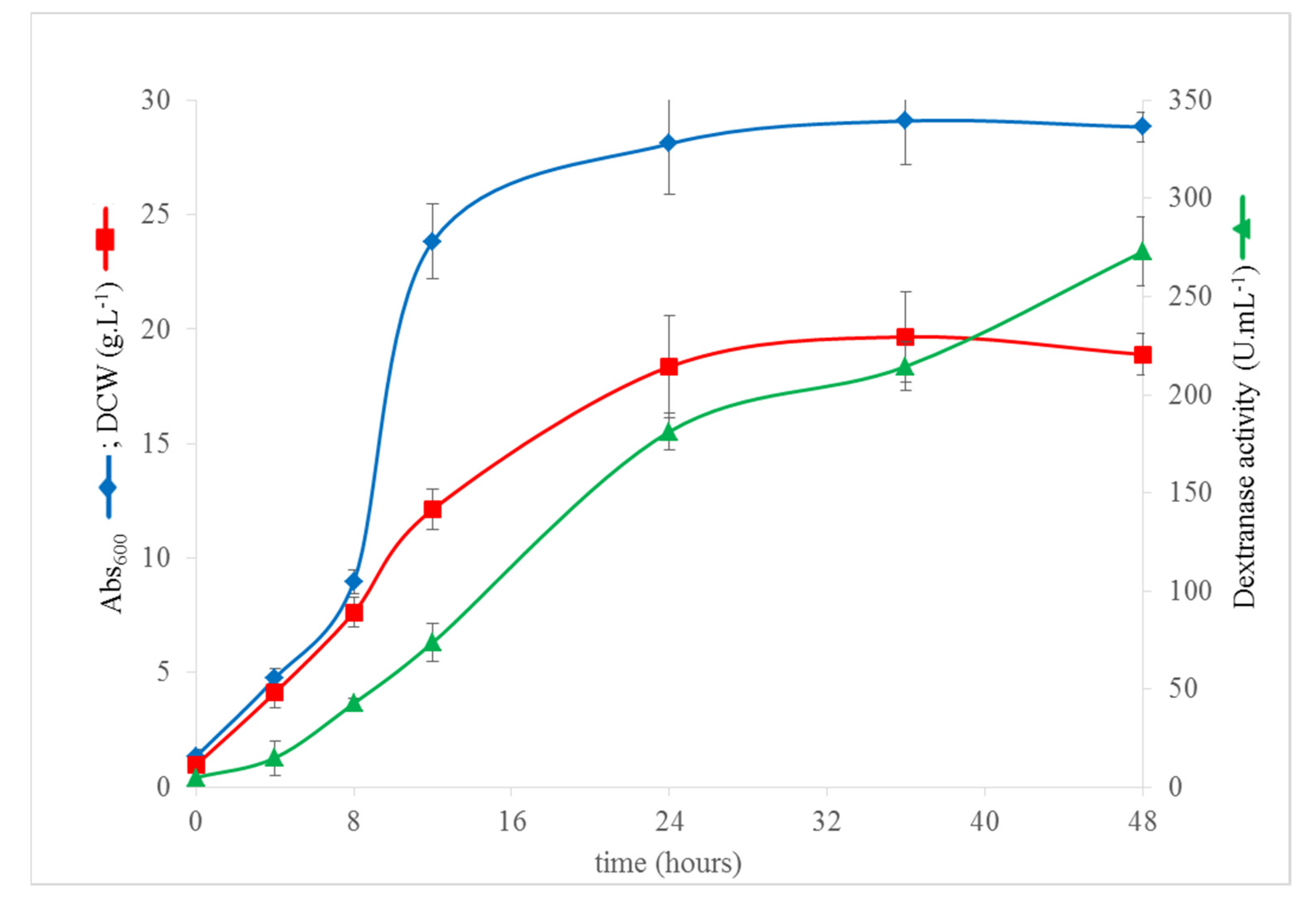
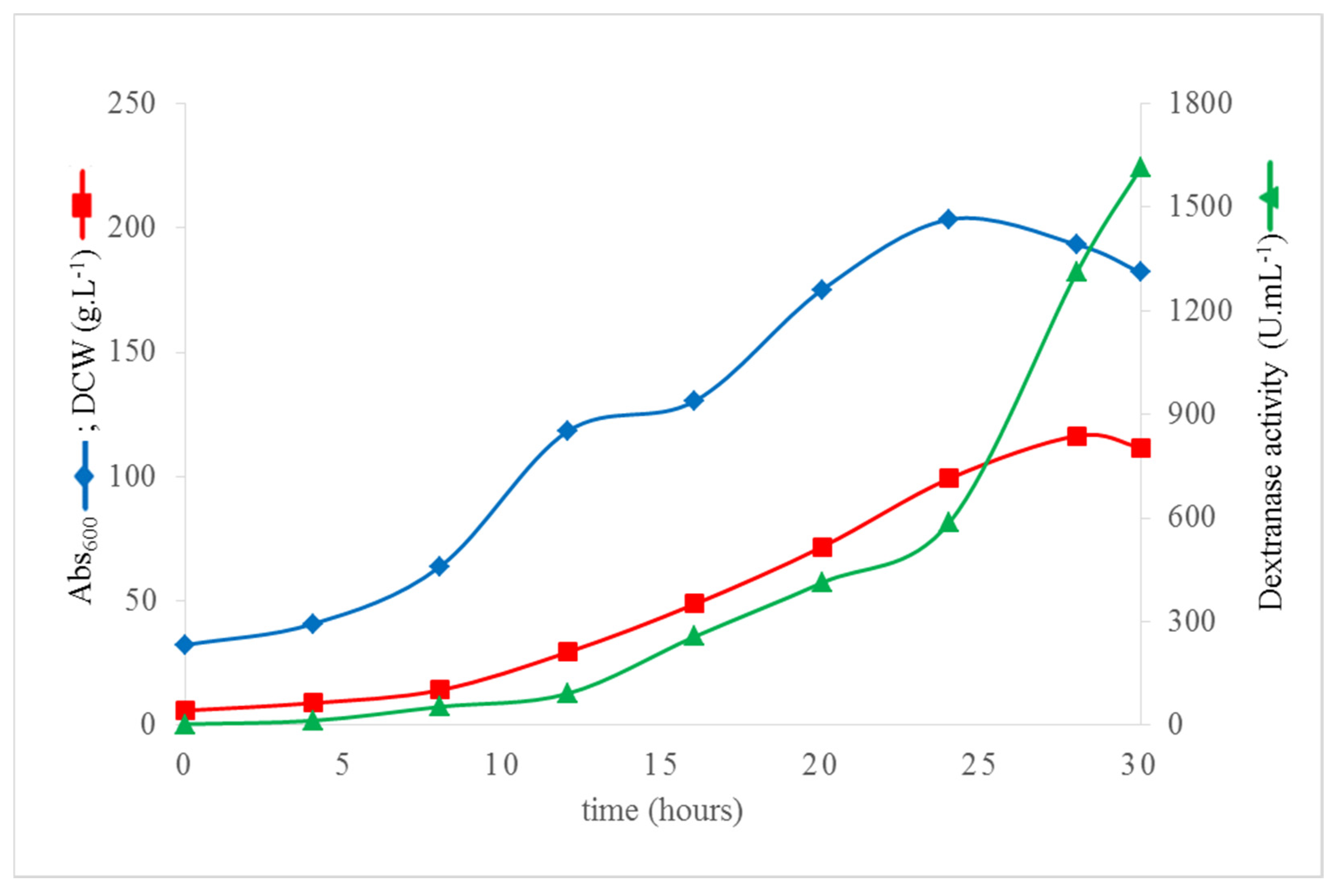
3.5. High Cell Density Fermentation of K. phaffii DEX49A–ΔSP–ΔN30 for High-Level Extracellular Production of r–TmDEX49A–ΔSP–ΔN30

The production of the rTmDEX49A–ΔSP–ΔN30 was carried out as a fed-batch culture in a 5 L bioreactor with the synthetic medium FM21-PTM1 [26]. The use of chemically defined or synthetic culture media in a large-scale fermentation strategy generally has the advantage of its greater batch-to-batch consistency compared to complex media, in addition to allowing for easy recovery of secreted protein in some cases [43], as well as the ability to reach up to 130 g·L−1 of biomass in high cell density cultures [44]. In Figure 5, the cell growth and the secreted dextranase activity are shown. The fermentation strategy used in this study allowed for reaching DCW values above 100 g·L−1 in 30 h of culture, without inhibiting cell growth or compromising the enzyme secretion. The enzyme activity values were compared with reports of dextranase production (rTmDEX49A) in K. phaffii on a 5 L scale. In the first report of the recombinant production secreted dextranase (rTmDEX49A) in K. phaffii, it was possible to increase from 98 U·mL−1 (98 mg·L−1) in the shaker (100 mL) to 3.2 g·L−1 (~3200 U·mL−1) in a 5 L scale fermenter in 120 h and repeat methanol feedings for the induction of the AOX1 promoter, obtaining an enzymatic productivity of 26.6 mg·L−1·h−1 [15].
Subsequently, a new feeding strategy with methanol was proposed, where the best values of enzyme secretion were achieved for this system, reaching up to 5135 g·L−1 (~5135 U·mL−1) in the culture supernatant, and a productivity of 42.7 mg·L−1·h−1 in a 2 L fermenter [45]. The values of the enzyme titer (3.2 g·L−1 and 5135 g·L−1) exceed the ones reached in this study by two and three times, respectively, but in terms of the bioprocess system productivity, the outcome reported here for rTmDEX49A–ΔSP–ΔN30 is higher than for rTmDEX49A. This study also exceeds the recently reported values in a fermenter of 2 L (97.34 U·mL−1 or 97.34 mg·L−1) and 30 L (62.64 U·mL−1 or 62.64 mg·L−1) of rTmDEX49A produced in K. phaffii in the optimized fermentation (100 h) methanol-induced manner with a productivity of 0.62 mg·L−1·h−1 [21]. Despite the wide use of the AOX1 promoter, it has some limitations, as methanol is a highly dangerous compound and even more so when working in large volumes, and storage can also be risky in industry [46,47]. Additionally, being a derivative from petrochemical sources, it may require purification steps to produce food and food additive products [48]. Such evaluations must be considered to design a technology in bioreactors and choose the most convenient system (pAOX1–inducible/pGAP–constitutive) that suits the needs.
As opposed to the induced systems, in this study, the product formation (rTmDEX49A–ΔSP–ΔN30) describes a continuous increase from the beginning to 30 h of cultivation, which is typical of constitutive production [49]. In the fed-batch culture, the feeding phase began at hour 14, taking as feeding criterion the carbon source depletion, judged by a sharp increase of dissolved oxygen as suggested by Potvin et al. [50]. In total, five increments were made between hour 14 and hour 24. The level of enzyme activity produced by K. phaffii DEX49A–ΔSP–ΔN30 was 1614 U·mL−1 (1.614 g·L−1), which is the highest production level reported so far for a dextranase constitutively produced in K. phaffii. Recently, using a similar constitutive system, the dextranase rTmDEX49A was produced at 69 U·mL−1 (69 mg·L−1) in the culture supernatant using glycerol as a carbon source and 87 U·mL−1 (87 mg·L−1) and 110 U·mL−1 (110 mg·L−1) with molasses and glucose, respectively [13]. This same study reported a productivity of the constitutive K. phaffii elite clone PpDEX(4x) grown with glucose (2157 U·L−1·h−1 or 2.15 mg·L−1·h−1), molasses (1706 U·L−1·h−1 or 1.70 mg·L−1·h−1), and glycerol (1353 U·L−1·h−1 or 1.35 mg·L−1·h−1) after 51 h of culture [13]. However, with the level of enzyme activity reported for K. phaffii DEX49A–ΔSP–ΔN30 in this study, a productivity of 53,800 U·L−1·h−1 (53.8 mg·L−1·h−1) in 5 L fed-batch fermentation after 30 h using FM21–PTM1 culture media was reached. This outperformed at least 25-fold the productivity reported by Martínez et al. [13] and is, to the best of our knowledge, the highest productivity reported so far for a glycoside hydrolase family 49 dextranase.
The results reported in this study confirm the efficiency of the constitutive pGAP to produce recombinant dextranase rTmDEX49A–ΔSP–ΔN30 using the K. phaffii expression system. High yields of secreted enzyme in just 30 h of culture were obtained. Compared with the shake flasks batch cultures, the high cell density fermentation of K. phaffii, favorable dissolved oxygen levels, and nutrient balance in FM21–PTM1 culture medium in a 5 L fermenter all contributed to a significantly higher level of recombinant expression of the truncated dextranase variant rTmDEX49A–ΔSP–ΔN30.
From the results of this study, it seems that the deletion of the first 30 amino acids in the dextranase rTmDEX49A–ΔSP–ΔN30 does not affect, and even improves, the production levels of the enzyme in the culture supernatant of K. phaffii DEX49A–ΔSP–ΔN30. Therefore, further studies are needed to corroborate this in more detail since the genetic constructions harbored by the clones used for the constitutive expression comparison were designed with different signal peptides. However, in addition, different strains of K. phaffii (GS115 and PPS 9010) were used in both studies, and differences in the expression efficiency of dextranase genes could be associated with differences between the genetic background of the two strains [39].

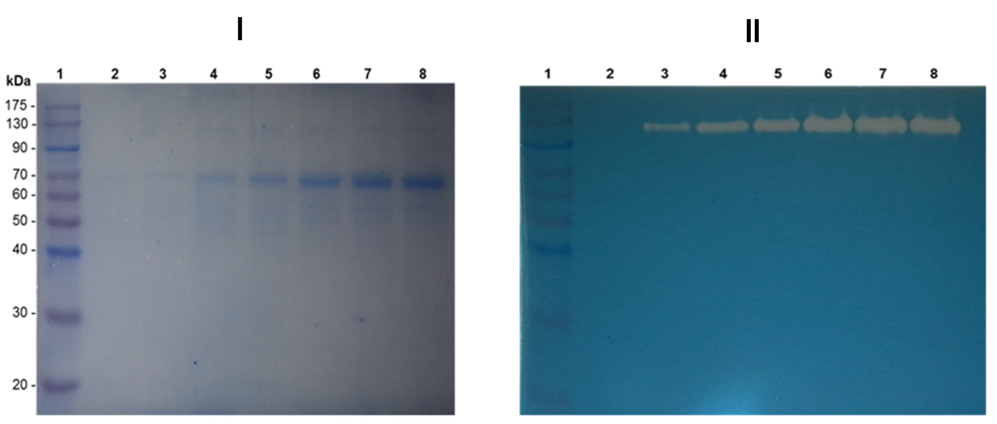
3.6. Detection of the Proteins Secreted to the Culture Supernatant Using the Synthetic Medium FM21–PTM1

The results of the 10% SDS-PAGE of the K. phaffii DEX49A–ΔSP–ΔN30 clone supernatant proteins stained with CBB are shown (Figure 6). The intense smeared protein bands corroborate the presence of rTmDEX49A–ΔSP–ΔN30 close to the molecular size described by Roca et al. [15] of 67 kDa (considering N–glycosylation). However, fewer additional stained protein bands are detected: one below 63 kDa and the other slightly above 25 kDa (Figure 6, lanes 14–15). According to the K. phaffii secretome, those proteins could be native host proteins [51].
This result indicates that the FM21-PTM1 synthetic medium has the potential to produce rTmDEX49A–ΔSP–ΔN30 in high concentrations. This synthetic medium permits the formation of a high biomass concentration (100–120 g·L−1) in a relatively short time (30 h), which translates into a greater productivity of recombinant protein production, as described [52].

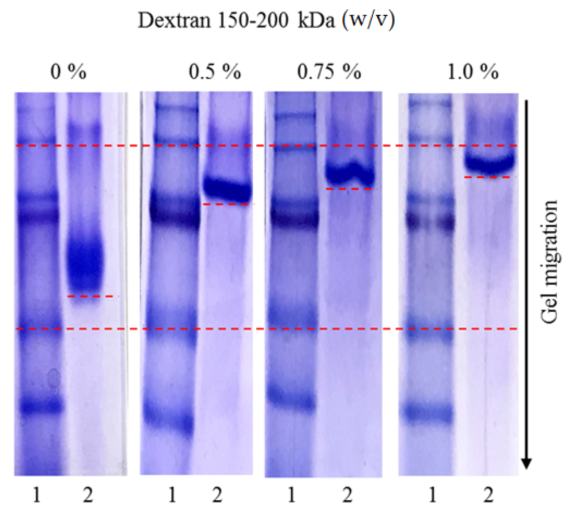
3.7. Affinity Study in Acrylamide Gels under Native Conditions

The affinity of rTmDEX49A–ΔSP–ΔN30 for the substrate (dextran) was studied by electrophoresis in 10% acrylamide gels under native conditions (Figure 7, lanes 2). The distance of the rTmDEX49A–ΔSP–ΔN30 migration in each gel was marked with dashed short lines. Dashed long lines correspond to the same molecular weight in the different gels presented (Figure 7, lanes 1), used as a negative control. A delay in the electrophoretic mobility of the dextranase enzyme is observed as the concentration of dextran in the gel increases.
The result of this affinity study for dextran explains the difference in the mobility of the rTmDEX49A–ΔSP–ΔN30 enzyme when it was analyzed by SDS–PAGE stained with CBB and dextranase zymogram (Figure 4I,II). The presence of dextran blue in the zymographic technique when using non-denaturing conditions allows the protein in its native state to interact with the polymer. Its retarded mobility provides evidence of carbohydrate–protein interactions probably by the presence of a carbohydrate (dextran)-binding region or a surface binding site with dextran affinity. A similar phenomenon has been described for dextransucrases of the GH-70 family [32,53].

4. Conclusions

The synthetic deleted gene construct encoding a truncated DEX49A variant of Talaromyces minioluteus dextranase (rTmDEX49A–ΔSP–ΔN30) was constitutively expressed to high levels in the yeast K. phaffii. The histochemical screening method permitted the selection of promising producer clones with high levels of dextranase activity. This is the first report of the constitutive, high-level expression of an N–terminal truncated dextranase in K. phaffii. The enhanced transgene expression in the K. phaffii DEX49A–ΔSP–ΔN30 clone neither limited cell growth nor compromised the secretion of the recombinant dextranase (rTmDEX49A–ΔSP–ΔN30). The deletion of the mature DEX49A first 30 amino acids downstream of the native signal peptide does not affect the enzymatic activity of r–TmDEX49A–ΔSP–ΔN30. The constitutive K. phaffii DEX49A–ΔSP–ΔN30 clone provides an interesting alternative for the large-scale production of rTmDEX49A–ΔSP–ΔN30 and its use for dextran removal at sugar mills, but also for other uses in the sugarcane derivative and food industries.

Author Contributions

R.C.A.R. and L.M.V.: Investigation, Methodology, and Data curation. R.F.V.: Conceptualization, Supervision, Investigation, Methodology, Data curation, Funding acquisition, Project administration, Roles/Writing—Original draft, and Writing—Review and editing. M.L.G., S.R.D., E.D. and V.P.: Conceptualization, Formal analysis, Resources, and Validation. B.M.: Funding acquisition, Project administration, Resources, and Formal analysis. All authors have read and agreed to the published version of the manuscript.

Funding

This work was supported by a Development Research Project financed by the Development Cooperation Program of ARES (Académie de Recherche et d’Enseigment Supérieur) from Belgium (2017–2022).

Institutional Review Board Statement

Not applicable.

Informed Consent Statement

Not applicable.

Data Availability Statement

Not applicable.

Acknowledgments

Reinaldo Fraga Vidal, C. Aríticas Ribalta, and Lisandra Martínez Valdés were supported by a Development Research Project financed by the Development Cooperation Program of ARES (Académie de Recherche et d’Enseigment Supérieur) from Belgium (2017–2022). Reinaldo Fraga Vidal thanks William David Rau (MS Rau Antiques, New Orleans, USA) for his kind support. We thank Miguel A. Peña and José A. Vela for their technical assistance with the bioreactors. We also thank Joan Combie for the style correction of the manuscript.

Conflicts of Interest

The authors declare no conflict of interest. The funders had no role in the design of the study; in the collection, analyses, or interpretation of data; in the writing of the manuscript; or in the decision to publish the results.

References

  1. Robyt, J.F.; Eklund, S.H. Stereochemistry involved in the mechanism of action of dextransucrase in the synthesis of dextran and the formation of acceptor products. Bioorganic Chem. 1982, 11, 115–132. [Google Scholar] [CrossRef]
  2. Bashari, M.; Tounkara, F.; Abdelhai, M.H.; Lagnika, C.; Xu, X.; Jin, Z. Impact of Dextranase on Sugar Manufacturing and its Kinetic on the Molecular Weights of Remaining Dextran. Sugar Tech 2013, 15, 84–93. [Google Scholar] [CrossRef]
  3. Monchois, V.; Willemot, R.M.; Monsan, P. Glucansucrases: Mechanism of action and structure-function relationships. FEMS Microbiol. Rev. 1999, 23, 131–151. [Google Scholar] [CrossRef]
  4. Bounaix, M.S.; Robert, H.; Gabriel, V.; Morel, S.; Remaud-Siméon, M.; Gabriel, B.; Fontagné-Faucher, C. Characterization of dextran-producing Weissella strains isolated from sourdoughs and evidence of constitutive dextransucrase expression. FEMS Microbiol. Lett. 2010, 311, 18–26. [Google Scholar] [CrossRef] [PubMed] [Green Version]
  5. Hector, S.; Willard, K.; Bauer, R.; Mulako, I.; Slabbert, E.; Kossmann, J.; George, G.M. Diverse exopolysaccharide producing bacteria isolated from milled sugarcane: Implications for cane spoilage and sucrose yield. PLoS ONE 2015, 10, e0145487. [Google Scholar] [CrossRef] [PubMed]
  6. Leemhuis, H.; Pijning, T.; Dobruchowskaa, J.M.; van Leeuwen, S.S.; Kralj, S.; Dijkstra, B.W.; Dijkhuizen, L. Glucansucrases: Three-dimensional structures, reactions, mechanism, alpha-glucan analysis and their implications in biotechnology and food applications. J. Biotechnol. 2013, 163, 250–272. [Google Scholar] [CrossRef] [Green Version]
  7. Ninchan, B.; Vanichsriratana, W.; Sriroth, K. Investigation of the optimized dextran-degrading enzyme conditions on the decomposition of different molecular weights of pure dextran using response surface methodology. Arch. Ind. Biotechnol. 2017, 1, 8–19. [Google Scholar]
  8. Eggleston, G.; Monge, A.; Montes, B.; Stewart, D. Application of dextranases in sugarcane factory: Overcoming practical problems. Sugar Tech 2009, 11, 135–141. [Google Scholar] [CrossRef]
  9. Rodríguez, E. Dextranase in sugar industry: A review. Sugar Tech 2009, 11, 124–134. [Google Scholar] [CrossRef]
  10. Xiao, Z.; Liao, X.; Guo, S. Analysis of Sugarcane Juice Quality Indexes. J. Food Qual. 2017, 2017, 1746982. [Google Scholar] [CrossRef] [Green Version]
  11. Khalikova, E.; Susi, P.; Korpela, T.; Khalikova, E.; Susi, P.; Korpela, T. Microbial Dextran-Hydrolyzing Enzymes: Fundamentals and Applications. MMBR 2005, 69, 306–325. [Google Scholar] [CrossRef] [Green Version]
  12. Jiao, Y.L.; Wang, S.J.; Lv, M.S.; Jiao, B.H.; Li, W.J.; Fang, Y.W.; Liu, S. Characterization of a marine-derived dextranase and its application to the prevention of dental caries. Ind. Microbiol. Biotechnol. 2014, 41, 17–26. [Google Scholar] [CrossRef] [PubMed]
  13. Martínez, D.; Menéndez, C.; Chacón, O.; Fuentes, A.D.; Borges, D.; Sobrino, A.; Ramírez, R.; Pérez, E.R.; Hernández, L. Removal of bacterial dextran in sugarcane juice by Talaromyces minioluteus dextranase expressed constitutively in Pichia pastoris. J. Biotechnol. 2021, 333, 10–20. [Google Scholar] [CrossRef]
  14. Kim, D.; Robyt, J.F.; Lee, S.Y.; Lee, J.H.; Kim, Y.M. Dextran molecular size and degree of branching as a function of sucrose concentration, pH, and temperature of reaction of Leuconostoc mesenteroides B-512FMCM dextransucrase. Carbohydr. Res. 2003, 338, 1183–1189. [Google Scholar] [CrossRef]
  15. Roca, H.; García, B.; Rodríguez, E.; Mateu, D.; Coroas, L.; Cremata, J.; García, R.; Pons, T.; Delgado, J. Cloning of the Penicillium minioluteum gene encoding dextranase and its expression in Pichia pastoris. Yeast 1996, 12, 1187–1200. [Google Scholar] [CrossRef]
  16. Raices, M.; Molerio, M.C.; Li, I.M.; Morera, V.; Roca, H.; Delgado, J.; Cremata, J.; Herrera, L. Purificación parcial de una enzima dextranasa a partir de una cepa de hongo del género Penicillium. Biotecnol. Apl. 1991, 8, 248–255. [Google Scholar]
  17. Betancourt, L.H.; García, R.; González, J.; Montesino, R.; Quintero, O.; Takao, T.; Shimonishi, Y.; Cremata, J.A. Dextranase (α-1,6 glucan-6-glucanohydrolase) from Penicillium minioluteum expressed in Pichia pastoris: Two host cells with minor differences in N-glycosylation. FEMS Yeast Res. 2001, 1, 151–160. [Google Scholar] [CrossRef] [PubMed]
  18. García, B.; Margolles, E.; Roca, H.; Mateu, D.; Raices, M.; Gonzáles, M.E.; Herrera, L.; Delgado, J. Cloning and sequencing of a dextranase-encoding cDNA from Penicillium minioluteum. FEMS Microbiol. Lett. 1996, 143, 175–183. [Google Scholar] [CrossRef] [PubMed]
  19. Larsson, A.M.; Andersson, R.; Stahlberg, J.; Kenne, L.; Jones, T.A. Dextranase from Penicillium minioluteum: Reaction course, crystal structure, and product complex. Structure 2003, 11, 1111–1121. [Google Scholar] [CrossRef]
  20. Yoder, M.D.; Lietzke, S.E.; Jurnak, F. Unusual structural features in the parallel beta-helix in pectate lyases. Structure 1993, 1, 241–251. [Google Scholar] [CrossRef]
  21. Marín, A.M.; Jáuregui, J.; Carreón, L.; Chávez, N.A. Dextranase production by recombinant Pichia pastoris under operational volumetric mass transfer coefficient (kLa) and volumetric gassed power input (Pg/V) attainable at commercial large scale. Prep. Biochem. Biotechnol. 2019, 49, 606–615. [Google Scholar] [CrossRef]
  22. Ahmad, M.; Hirz, M.; Pichler, H.; Schwab, H. Protein expression in Pichia pastoris: Recent achievements and perspectives for heterologous protein production. Appl. Microbiol. Biotechnol. 2014, 98, 5301–5317. [Google Scholar] [CrossRef] [PubMed] [Green Version]
  23. Cos, O.; Ramon, R.; Montesinos, J.L.; Valero, F. Operational strategies, monitoring and control of heterologous protein production in the methylotrophic yeast Pichia pastoris under different promoters: A review. Microb. Cell Factories 2006, 5, 17. [Google Scholar] [CrossRef] [PubMed] [Green Version]
  24. Sambrook, J.; Russell, D.W. Molecular Cloning: A Laboratory Manual, 3rd ed.; Cold Spring Harbor Laboratory Press: Cold Spring Harbor, NY, USA, 2001. [Google Scholar]
  25. Emr, S.; Schekman, R.; Flessel, M.; Thorner, J. An MFαl-SUC2 (α-factor-invertase) gene fusion for study of protein localization and gene expression in yeast. Proc. Natl. Acad. Sci. USA 1983, 80, 7080–7084. [Google Scholar] [CrossRef] [PubMed] [Green Version]
  26. Lin-Cereghino, J.; Wong, W.W.; Xiong, S.; Giang, W.; Luong, L.T.; Vu, J.; Johnson, S.D.; Lin-Cereghino, G.P. Condensed protocol for competent cell preparation and transformation of the methylotrophic yeast Pichia pastoris. BioTechniques 2005, 38, 44–48. [Google Scholar] [CrossRef] [PubMed] [Green Version]
  27. Wegner, E.H. Biochemical Conversions by Yeast Fermentation at High Cell Densities. U.S. Patent 4,414,329, 8 November 1983. [Google Scholar]
  28. Kosaric, N.; Yu, K.; Zajic, J.E.; Rozanis, J. Dextranase production from Penicillium funiculosum. Biotech. Bioeng. 1973, 15, 729–741. [Google Scholar] [CrossRef]
  29. Miller, G.L. Use of Dinitrosalicylic Acid Reagent for Determination of Reducing Sugar. Anal. Chem. 1959, 31, 426–428. [Google Scholar] [CrossRef]
  30. Laemmli, U.K. Cleavage of Structural Proteins during the Assembly of the Head of Bacteriophage T-4. Nature 1970, 227, 680–685. [Google Scholar] [CrossRef] [PubMed]
  31. Kim, J.K.; Shin, S.Y.; Moon, J.S.; Li, L.; Cho, S.K.; Kim, T.J.; Han, N.S. Isolation of dextran-hydrolyzing intestinal bacteria and characterization of their dextranolytic activities. Biopolymers 2015, 103, 321–327. [Google Scholar] [CrossRef] [PubMed]
  32. Brison, Y.; Malbert, Y.; Czaplicki, G.; Mourey, L.; Remaud-Simeón, M.; Tranier, S. Structural Insights into the Carbohydrate Binding Ability of an α-(1→2) Branching Sucrase from Glycoside Hydrolase Family 70. J. Biol. Chem. 2016, 291, 7527–7540. [Google Scholar] [CrossRef] [PubMed] [Green Version]
  33. Kang, H.K.; Kim, S.H.; Park, J.Y.; Jin, X.J.; Oh, D.K.; Kang, S.S.; Kim, D. Cloning and characterization of a dextranase gene from Lipomyces starkeyi and its expression in Saccharomyces cerevisiae. Yeast 2005, 22, 1239–1248. [Google Scholar] [CrossRef] [PubMed] [Green Version]
  34. Kang, H.K.; Park, J.Y.; Ahn, J.S.; Kim, S.H.; Kim, D. Cloning of a gene encoding dextranase from Lipomyces starkeyi and its expression in Pichia pastoris. J. Microbiol. Biotechnol. 2009, 19, 172–177. [Google Scholar] [CrossRef] [PubMed] [Green Version]
  35. Lai, X.; Liu, X.; Liu, X.; Deng, T.; Feng, Y.; Tian, X.; Lyu, M. The Marine Catenovulum agarivorans MNH15 and Dextranase: Removing Dental Plaque. Mar. Drugs 2019, 17, 592. [Google Scholar] [CrossRef] [PubMed] [Green Version]
  36. Menéndez, J.; Valdés, I.; Cabrera, N. The ICL1 gene of Pichia pastoris, transcriptional regulation and use of its promoter. Yeast 2003, 20, 1097–1108. [Google Scholar] [CrossRef] [Green Version]
  37. Clare, J.J.; Rayment, F.B.; Ballantine, S.P.; Sreekrishna, K.; Romanos, M.A. High-level expression of tetanus toxin fragment C in Pichia pastoris strains containing multiple tandem integrations of the gene. Bio/Technology 1991, 9, 455–460. [Google Scholar] [CrossRef] [PubMed]
  38. Parashar, D.; Satyanarayana, T. Enhancing the production of recombinant acidic α-amylase and phytase in Pichia pastoris under dual promoters [constitutive (GAP) and inducible (AOX)] in mixed fed batch high cell density cultivation. Process. Biochem. 2016, 51, 1315–1322. [Google Scholar] [CrossRef]
  39. García, B.M.; Rodríguez, E.; Rivero, T.; Hidalgo, Y.; Menéndez, J.J. Dextranase Expression in two Different Host-vector Systems of the Methylotrophic Yeast Pichia pastoris. Biotecnol. Apl. 2000, 17, 11–15. [Google Scholar]
  40. Safder, I.; Khan, S.; Islam, I.; Kazim, M.; Bibi, Z.; Waqas, M. Pichia pastoris expression system: A potential candidate to express protein in industrial and biopharmaceutical domains. Biomed. Lett. 2018, 4, 1–14. [Google Scholar]
  41. Rerngsamran, P.; Temjitpukdee, P.; Assavasirijinda, N.; Chareonpornwattana, S.; Thaniyavarn, S. Cloning, characterization, and heterologous expression of a dextranase gene from Penicillium pinophilum SMCU3-14. ScienceAsia 2014, 40, 405–413. [Google Scholar] [CrossRef]
  42. Netsopa, S.; Niamsanit, S.; Araki, T.; Kongkeitkajorn, M.B.; Milintawisamai, N. Purification and Characterization Including Dextran Hydrolysis of Dextranase from Aspergillus allahabadii X26. Sugar Tech 2019, 21, 329–340. [Google Scholar] [CrossRef]
  43. Matthews, C.B.; Kuo, A.; Love, K.R.; Love, J.C. Development of a general defined medium for Pichia pastoris. Biotechnol. Bioeng. 2017, 115, 103–113. [Google Scholar] [CrossRef] [PubMed]
  44. Barrero, J.J.; Casler, J.C.; Valero, F.; Ferrer, P.; Glick, B.S. An improved secretion signal enhances the secretion of model proteins from Pichia pastoris. Microb. Cell Factories 2018, 17, 161. [Google Scholar] [CrossRef] [PubMed]
  45. Rodríguez, E.; Sánchez, K.; Roca, H.; Delgado, J.M. Different methanol feeding strategies to recombinant Pichia pastoris cultures producing high level of dextranase. Biotechnol. Tech. 1997, 11, 461–466. [Google Scholar] [CrossRef]
  46. Goodrick, J.C.; Xu, M.; Finnegan, R.; Schilling, B.M.; Schiavi, S.; Hoppe, H.; Wan, N.C. High- level expression and stabilization of recombinant human chitinase produced in a continuous constitutive Pichia pastoris expression system. Biotechnol. Bioeng. 2001, 74, 492–497. [Google Scholar] [CrossRef] [PubMed]
  47. Piva, L.C.; Bentacur, M.O.; Castelo, V.; Reis, B.; De Marco, J.L.; de Moraes, L.M.P.; Gonçalves, F.A. Molecular strategies to increase the levels of heterologous transcripts in Komagataella phaffii for protein production. Bioengineered 2017, 8, 441–445. [Google Scholar] [CrossRef] [PubMed] [Green Version]
  48. Misra, V.; Mall, A.K.; Pathak, A.D.; Solomon, S.; Kishor, R. Microorganisms affecting Post-Harvest Sucrose Losses in Sugarcane. Int. J. Curr. Microbiol. Appl. Sci. 2017, 6, 2554–2566. [Google Scholar] [CrossRef] [Green Version]
  49. Zepeda, A.B.; Pessoa, A.; Farías, J.G. Carbon metabolism influenced for promoters and temperature used in the heterologous protein production using Pichia pastoris yeast. Braz. J. Microbiol. 2018, 49, 119–127. [Google Scholar] [CrossRef] [PubMed]
  50. Potvin, G.; Ahmad, A.; Zhang, Z. Bioprocess engineering aspects of heterologous protein production in Pichia pastoris: A review. Biochem. Eng. J. 2012, 64, 91–105. [Google Scholar] [CrossRef]
  51. Mattanovich, D.; Graf, A.; Stadlmann, J.; Dragosits, M.; Redl, A.; Maurer, M.; Kleinheinz, M.; Sauer, M.; Altmann, F.; Gasser, B. Genome, secretome and glucose transport highlight unique features of the protein production host Pichia pastoris. Microb. Cell Factories 2009, 8, 29. [Google Scholar] [CrossRef] [PubMed] [Green Version]
  52. Joseph, J.A.; Akkermans, S.; Nimmegeers, P.; Van Impe, J.F.M. Bioproduction of the Recombinant Sweet Protein Thaumatin: Current State of the Art and Perspectives. Front. Microbiol. 2019, 10, 695. [Google Scholar] [CrossRef] [PubMed] [Green Version]
  53. Molina, M.; Moulis, C.; Monties, N.; Guieysse, D.; Morel, S.; Cioci, G.; Remaud-Siméon, M. A specific oligosaccharide-binding site in the alternansucrase catalytic domain mediates alternan elongation. J. Biol. Chem. 2020, 295, 9474–9489. [Google Scholar] [CrossRef] [PubMed]
Figure 1. General cloning and screening strategy to isolate dextranolytic K. phaffii clones. Detection of the DNA fragment encoding rTmDEX49A–ΔSP–ΔN30. (I)—Schematic representation of plasmid pDEX49A–ΔSP–ΔN30. (II)—DNA electrophoresis; lane 2: undigested plasmid pDEX49A–ΔSP–ΔN30, lane 3: pDEX49A–ΔSP–ΔN30/SwaI, lane 4: pDEX49A–ΔSP–ΔN30/SwaI–PstI, and lane 6: purified DNA fragment of approximately 4.2 kb from sample of lane 4. Lanes 1 and 5: 1Kb DNA Ladder RTU (Genetics, Japan). (III,IV)—Qualitative histochemical detection of dextranase activity on YPDSDB plates through the formation of a degradation halo around the colonies of the positive clones. YPDSDB plates supplemented with Zeocin 100 µg·mL−1 (III) and 1000 µg·mL−1 (IV).
Applsci 12 07562 g001
Figure 2. Detection of rTmDEX49A–ΔSP–ΔN30 production in recombinant K. phaffii clones. Secreted and cell-associated dextranase levels (I and II, respectively) of 28 different clones (1–10) and (A–Q). Dextranase levels of different clones are normalized to the biomass (OD). (III): SDS–PAGE of the culture supernatant of clones D (2) and M (3) stained with Coomassie Brilliant Blue R-250, (1) molecular weight standard PiNK Prestained Protein Ladder (NIPPON Genetics Europe, Germany). (**) refers to highly significant differences (p < 0.001) between clone D and the other clones. Significance (or p value) was obtained by applying the Student’s test.
Applsci 12 07562 g002
Figure 3. Cell growth and dextranase enzyme activity of K. phaffii DEX49A–ΔSP–ΔN30. Abs600, (DCW) values in g·L−1 and dextranase activity (U·mL−1) of the supernatant in 100 mL of YPG medium.
Applsci 12 07562 g003
Figure 4. Analysis of the production of recombinant rTmDEX49A–ΔSP–ΔN30 in K. phaffii at shaker level in 100 mL of YPGlycerol medium. (I) SDS-PAGE of the fermented supernatant stained with CBB. 1—Molecular weight standard, PiNK Prestained Protein Ladder (NIPPON Genetics Europe, Germany); (2 to 8)—culture supernatants at hours 0, 4, 8, 12, 24, 36, 48. (II) Zymogram to detect dextranase activity; samples were analyzed in the same order from gel (A).
Applsci 12 07562 g004
Figure 5. Cell growth and dextranase enzyme production of K. phaffii DEX49A–ΔSP–ΔN30 in a fed-batch culture. Abs600, DCW values in g·L−1 and dextranase activity (U·mL−1) from the study in a 5 L fermenter using FM21–PTM1 medium.
Applsci 12 07562 g005
Figure 6. Polyacrylamide gel electrophoresis under denaturing conditions (SDS–PAGE) of the K. phaffii DEX49A–ΔSP–ΔN30 secreted proteins. In lanes 2–15, culture supernatants hours 0, 2, 4, 6, 8, 10, 12, 14, 16, 20, 24, 26, 28, and 30 of the fermentation. Lane 1 and 16: Molecular weight marker (BLUeye Prestained Protein Ladder, GeneDireX, Inc. Germany).
Applsci 12 07562 g006
Figure 7. Affinity gel electrophoresis of rTmDEX49A–ΔSP–ΔN30 in polyacrylamide gels (PAGE under native conditions) with different concentrations of dextran. The dextran concentration is expressed in % (w/v). Lanes 1: molecular weight marker (PageRuler™ Prestained Protein Ladder, Thermo Scientific™, USA); lanes 2: K. phaffii DEX49A–ΔSP–ΔN30 supernatant (end time of fermentation in FM21–PTM1 medium).
Applsci 12 07562 g007
Publisher’s Note: MDPI stays neutral with regard to jurisdictional claims in published maps and institutional affiliations.

