Article

Metagenomic Research of Infectious Diseases in Archaeological Contexts: Evidence from the Hospital Real de Todos-os-Santos (Portugal)

by
Francisca Alves-Cardoso
1,2,*,
Sara Palomo-Díez
3,4,
Alejandro Alonso Conde
4,5,
Cláudia Gomes
3,4,
Silvia Casimiro
1,6,
Rodrigo Banha da Silva
7,8 and
Eduardo Arroyo-Pardo
3,4
1
LABOH—Laboratory of Biological Anthropology and Human Osteology, CRIA—Centro em Rede de Investigação em Antropologia, Faculdade de Ciências Sociais e Humanas, Universidade NOVA de Lisboa, Av. Berna, 26, 1069-061 Lisboa, Portugal
2
Cranfield Defense & Security, Cranfield University, Bedford MK43 0AL, UK
3
Laboratory of Forensic and Population Genetics, Legal Medicine, Psychiatry and Pathology Department, Medicine School, Complutense University of Madrid, 28040 Madrid, Spain
4
Forensic Sciences Group, Forensic Genetics and Toxicology, Health Research Institute of the Hospital Clínico San Carlos (IdISSC), 28040 Madrid, Spain
5
Department of Genetics, Physiology and Microbiology, Complutense University of Madrid, 28040 Madrid, Spain
6
Institute of Medieval Studies IEM, NOVA-FCSH, Colégio Almada Negreiros (CAN), Campus de Campolide Universidade NOVA de Lisboa, 1099-032 Lisboa, Portugal
7
Centro de Humanidades—CHAM, NOVA-FCSH, Colégio Almada Negreiros (CAN), Campus de Campolide Universidade NOVA de Lisboa, 1099-032 Lisboa, Portugal
8
Cal -Centro de Arqueologia de Lisboa, Câmara Municipal de Lisboa, Avenida da Índia 166, 1400-038 Lisboa, Portugal
*
Author to whom correspondence should be addressed.
Appl. Sci. 2022, 12(12), 6096; https://doi.org/10.3390/app12126096
Submission received: 23 April 2022 / Revised: 9 June 2022 / Accepted: 13 June 2022 / Published: 15 June 2022
(This article belongs to the Section Applied Microbiology)

Abstract

:
Syphilis is one of the most exciting diseases explored in paleopathology and, therefore, tracing back its origin and development has provided a prolific debate. The combination of paleopathological data with historical sources, iconography, and archaeological contexts were the primary sources used to reconstruct its historical path. However, there are some limitations to paleopathological diagnosis due to the nature of bone reaction to stimuli. In addition, historical sources are subjected to a bias of social and cultural nature and the knowledge of those who wrote them. Hence, ancient DNA analysis offers the possibility of acquiring proof of cause by identifying pathogens in an organism. We undertook a metagenomic study of a skeleton exhumed from the Royal Hospital of All Saints (Portugal), renowned for treating syphilis from the 16th century onwards. The skeleton had previously been diagnosed with syphilis according to paleopathological analysis. However, the metagenomics analysis showed no presence of the pathogen associated with syphilis (i.e., Treponema pallidum) but revealed pathogenic microorganisms related to respiratory diseases (pneumonia), nonspecific bone infections (osteomyelitis), and oral bacterial pathologies as well as Hansen’s disease (also known as leprosy). The results are exciting and demand a reappraisal of the observed bone changes, recontextualizing their characterization as syphilis related. They prove that past reconstruction of health and disease diagnoses based on assessing human osteological remains of known context (such as a syphilitic hospital) may bias interpretations and, therefore, caution is recommended, not forgetting that the absence of evidence is not evidence of absence (in this case of syphilis) in life.

1. Introduction

Palaeopathological studies reconstruct past health patterns and identify diseases, such as Hansen’s disease (leprosy), tuberculosis, and syphilis, by analyzing human osteological remains [1,2]. However, this discipline has limitations since it relies mainly on the macroscopic observation of bones and teeth. Moreover, identifying a disease based on bone changes alone is not without interpretative error [3,4]. For this reason, paleopathology has embraced a multidisciplinary approach when exploring diseases in the past in later years. Unfortunately, this multidisciplinary approach is often dependent on institutional resources and, for this reason, palaeopathological studies continue to rely heavily on osteological remains (e.g., bone) macroscopic analysis.
In recent years, advanced paleopathological research has been complemented with ancient DNA (aDNA) analysis, alongside other biochemical and biomolecular techniques, furthering our understanding of disease at a microscopic and evolutionary level, allowing the identification of disease-related pathogens. Pathological cases with a conditioned diagnosis can be reassessed, and a positive diagnosis reached. The integration of molecular analysis in palaeopathological studies has already yielded exciting data, for example, on Mycobacterium tuberculosis, leprosy, plague, and syphilis [5,6,7,8,9].
Within paleopathology, syphilis is one of the few infectious diseases inferred by analyzing skeletonized human remains with a certain degree of certainty. Other diseases include Hansen’s disease (leprosy) and tuberculosis. These diseases may leave bone changes easily observable, identified as disease-related, and tagged as pathognomonic of specific diseases, be it syphilis, leprosy, or tuberculosis. For example, destruction of the maxillary alveolar bone and atrophy of the anterior spine may be viewed as pathognomonic of leprosy. Furthermore, cranial typical crater-like lesions with stellate radiating lines are pathognomonic of skeletal treponematosis (as detailed below) [1,10,11]. However, these need to be interpreted with care, and their presence does not exclude the need for a differential diagnosis when assessing bone changes and addressing their probable etiology. Bone reaction to disease tends to be monotonous because bone tissue reactions are essentially destructive or reactive, with variations in degree and form. This diversification, allied to its distribution in the various bone elements of a skeleton and/or within a bone itself, is the basis for building a paleopathological diagnosis. Hence, a disease’s pathognomonic trait is a significant asset in the paleopathological interpretation and diagnosis of diseases in human remains. However, it must be contextualized.
In syphilis, the skeleton is affected only in its tertiary stage, and bone lesions may take between 2 and 10 years to develop after the infection [10]. Although any bone from the skeleton may exhibit changes, these tend to be bilateral, with a preference towards the tibias, the nasal cavity bones, and the cranial vault, followed by other long bones of the extremities, and cancellous bone, such as the ribs and sternum, rich in hematopoietic marrow [10]. The bone-change pattern results from chronic non-granulomatous inflammation and/or a granulomatous (gummatous) processes involving bones’ outer surfaces, cortex, and, sometimes, the medullary cavity. Syphilitic bone lesions are primarily characterized as gummatous destruction, with perifocal osteosclerotic reactions involving both the periosteum and underlying bone [10,11]. The diagnostic feature (pathognomonic feature) is observable in the skull and named “caries sicca”, corresponding to healed superficial gummatous osteitis of the calvaria, described as “...nodules coming together to form star-shaped depressions, which may be deep, or wide and flat...”, i.e., cranial crater-like lesions with stellate radiating lines, forming a consistent pattern of changes [11]. Hackett [11] (p. 233) described the continuous lesion sequence from bone destruction (serpiginous cavitation) to bone formation (nodular cavitation) and bone remodeling (caries sicca).
The reconstruction of the origin of syphilis is ongoing. Whether it was brought to Europe from the Americas or disseminated by Europeans is a debate that has been enriched by paleopathological analysis of specimens and aDNA studies (for details on this debate, see [12,13,14,15,16,17]). The history and origins of syphilis in Europe are intimately related to the late 15th century syphilis epidemic, which coincided with Columbus’s “discovery” and early voyages to the Americas [12,13,14,15,16,17]. Like many European countries at the time, Portugal was also profoundly affected by syphilis. Between the years 1511 and 1532, a total of 20,000 syphilitic patients were treated in Portugal at the Hospital Real de Todos-os-Santos (Royal Hospital of All Saints—RHAS) (Lisboa, Portugal) [18].
The RHAS was a European reference in the past treatment of infectious disease, with an emphasis on syphilis. The hospital was built between 1492 and 1504, coinciding with the first historical records of the syphilis epidemic of 1496, which struck large parts of Europe. Affected by a large fire in 1750, in 1755, it suffered significant damage due to the Lisbon earthquake, but its activity carried on until 1773–1775, when the institution was transferred to the former Jesuit College of Santo Antão-o-Novo building and renamed the Royal Hospital of São José (Lisboa, Portugal). This later hospital is still in activity today [19,20].

The Royal Hospital of All Saints Bioanthropological Collection

The Praça da Figueira’s archaeological excavation (Lisbon, Portugal), in 1999–2000, partially unveiled archaeological structures associated with the RHAS, including those associated with a significant structural reform during the early 18th century. This reform transformed a corridor formerly intended for circulation into a small burial ground where 17 skeletons were exhumed, comprising 15 adults and 2 juveniles, alongside an ossuary with the human remains of at least four adults and two juveniles. It was only possible to assess the biological sex of 13 skeletons, eight males and five females. This diagnosis was made based on the morphological assessment of the hip bones and skull [19]. The individual skeletons were exhumed from primary graves, with single and multiple burials. The archaeothanatological interpretation of the context suggested that the bodies were buried with no coffins, promoting the maintenance of all anatomical skeletal connections during soft-tissue decomposition, suggesting that individuals in multiple graves were buried simultaneously or with limited time apart. The graves were reused, as proven by the presence in at least three graves of partially articulated bones not belonging to any of the primary burials. In addition, bodies were fitted to the available space, as multiple graves contained skeletons in opposite directions (feet of one, over the skull of another). Overall, most of the skeletons had some sort of disease-related bone changes, but these were found to be discreet, except for one individual: Sk.1310.
The Sk.1310, a skeleton of an adult female, was buried with three adult males and is the only skeleton in this collection exhibiting syphilitic-related bone changes [21]. Sk.1310’s disease-related bone changes were composed of a matrix of new bone formation, affecting both the left and right upper limbs and distal ends of the femurs and tibias, and fibula diaphysis. The tibias also had a bowing deformity, with a “saber shape” morphology described in paleopathology [10]. In addition, the frontal bone had several bone lesions described as healed nodular cavitations, akin to “caries sicca”, a biological indicator of syphilis in the paleopathological analysis. A multimethodological approach was used to study this case. The methods included the macroscopic visual examination of the remains, an imagological assessment of the bone changes, and a paleohistological study. All these supported a diagnosis of acquired syphilis [21].
This finding is relevant due to the fact of its historical context, which is intimately associated with treating syphilis and other infectious diseases such as leprosy. Since both pathologies may share similar bone changes [10] and because macro- and microscopic visualizations of bone are not without limitations, a molecular analysis (which was the focus of this research paper) was undertaken to complete further the original bioanthropological and paleopathological assessment and diagnosis of syphilis [21]. However, this option was not without challenges since previous studies showed no evidence of syphilitic-related pathogens in the archaeological remains. In contrast, others highlighted its presence [22,23,24,25] and the need to understand better how the pathogen may survive in human osteological remains. Moreover, since in the tertiary stages of the treponemal disease, organisms in the host have been described as reduced, and inferences about syphilis when assessing human osteological remains will always be limited [26].
With the abovementioned challenges in mind, the aim of this research was two-fold: to confirm or discard the presence of Treponema pallidum, the leading infectious agent of acquired syphilis, in the human remains; to assess the presence of other pathogens and/or microorganisms that could explain the lesions observed, offering a comprehensive assessment of the case at hands. In the present case, the individual, Sk.1310, exhibited a mosaic of remodeled and active lesions; hence, microorganisms’ presence was expected. In addition, whether the bone changes observed were syphilis related or not is addressed. Hence, we used a metagenomic analysis of the 16S ribosomal subunit to determine and identify the variety of microorganisms present in the samples rather than targeting a specific microorganism.

2. Materials and Methods

The bioanthropological analysis of the skeleton SK1310 has already been described by Assis et al. [21]. The skeleton was that of an adult female. Sexual diagnosis and age at death estimation followed conventional methodological analysis [27,28,29]. The bone lesions found in the skeletons comprised a mosaic of new bone deposition, some remodeled, others not, distributed in the upper limbs, the distal end of femurs and tibias and fibulas diaphysis, with the frontal bone exhibiting cranial crater-like lesions with stellate radiating lines [21] (p. 429). The remains were macroscopically assessed via the naked eye and with magnifying lenses and conventional X-ray and CT-scan techniques, allied with histological analyses of some samples [21] (p. 429).
For the metagenomic analysis, three independent samples were selected from the skeleton SK1310. The region sequenced in the samples was the 16S fourth hypervariable (V4) region (V4 16S rRNA). Samples were primarily extracted from two teeth—the upper left and right canines (references: HRTS1310.1 and HRTS1310.2). The canines were fully formed and erupted without significant damage to the roots and crowns, except for minor enamel wear with dentine exposition. The third sample was collected on a fragment of bone with evidence of disease-related bone changes (reference: HRTS1310.3)—a fragment from the left humerus, exhibiting a mosaic of remodeled and non-healed bone lesions, was used.
All sample analysis was undertaken at the Forensic Genetics and Population Genetics Laboratory (FGPGL) at the University Complutense of Madrid. The FGPGL is a restricted-access, isolated laboratory used only to analyze critical DNA samples; the laboratory is equipped with UV lights and possesses exclusive material (i.e., to be used only in this laboratory). All equipment was cleaned before and after use with 70% (v/v) diluted bleach. Furthermore, the researchers only accessed the laboratory wearing sterilized clothes, glasses, masks, and shoe covers. Moreover, all the laboratory staff were genotyped to control contamination issues and detect contamination. In addition, negative controls were analyzed during DNA extraction and DNA amplification to detect exogenic contaminant DNA. All these measures were intended to preserve the DNA samples, avoid contamination with exogenic DNA, and, if this was not possible, detect any DNA contamination that may have occurred.
The DNA extracts were prepared in an isolated room not used for handling modern DNA or PCR products. Sample preparation and ancient DNA analysis were as follows: first, all samples were documented according to the normalized FGPGL work protocol [30]. Secondly, sample surfaces were cleaned with a Sand-Blaster (Base 1 Plus, Dentalfarm), using aluminum oxide and UV radiation for 30 min [30].
After this step, two methods were performed to prepare samples for DNA extraction. The first procedure consisted of pulverizing samples HRTS1310.1 and HRTS1310.3 using a freezer mill with liquid nitrogen (Spex 6700) [30]. The other method was to maintain HRTS1310.2 intact after the surface cleaning, according to Gomes et al. [30], a nondestructive aDNA extraction method. Then, DNA extraction was performed using the abovementioned samples, pulverized and intact, following the recommendations and procedures as described in Rohland and Hofreiter [31,32], Rohland et al. [33], and Gomes et al. [30]. In addition, all DNA analyses followed the ancient DNA authenticity criteria, accredited by international guidelines, and established by the FGPGL laboratory [34,35]. Therefore, negative controls were analyzed during DNA extraction and amplification to detect any exogenic DNA contaminant: this process did not detect DNA molecule contamination.
The metagenomic study undertaken focused on the 16S rRNA V4 region. This region was amplified and sequenced using WineSeq® specific primers (patent: WO2017096385) [36]. The library was performed using the two-step PCR protocol described by Feld et al. [37] and Albers et al. [38]. The PCR conditions (i.e., number of cycles, temperatures, thermal cycler, and composition of the master mix) were carried out following the WineSeq® technical procedure [39]. Sequencing was performed in an Illumina® MiSeq instrument (Illumina®, San Diego, CA, USA). The sequences were then compared with information available in various databases using the Basic Local Assignment Search Tool (BLAST) of the NCBI (National Center of Biotechnology Information) and CLUSTAL-OMEGA [40,41,42]. Finally, the sequences were analyzed on the MG-RAST platform using the Greengenes database [43,44] with the following parameters: e-value = 0.05; %-ident = 60; length = 15; min. abundance = 1. Considering the paleopathological diagnosis of this individual and the wide range of bone lesions observed (some still active at the time of death), an e-value (0.05) was deemed sufficient to establish a plausible relationship between the bone changes and their etiological agent extracted from the bacterial DNA.
The research sequences used in this study are available on the NCBI server, with the BioProject accession: PRJNA522260 (ID: 522260). The information includes data for all three samples: HRTS1310.1, HRTS1310.2, and HRTS1310.3 (SRX5372162; SRX5372161; SRX5372160) (https://www.ncbi.nlm.nih.gov/bioproject/PRJNA522260 registration on 13 February 2019).
The interpretation of the results acknowledges the limitations that sequencing platforms may have, as highlighted by Kircher and Kelson [45], and because the technique used depends on the PCR amplification of the V4 16S rRNA region. In such cases, the polymerase may have a higher or lower affinity with this region. In the analysis, the amplification was assumed to be homogeneous for all DNA fragments of the V4 region, not disregarding the possibility that the bacterial population may have been overestimated and/or underestimated [45].

3. Results and Discussion

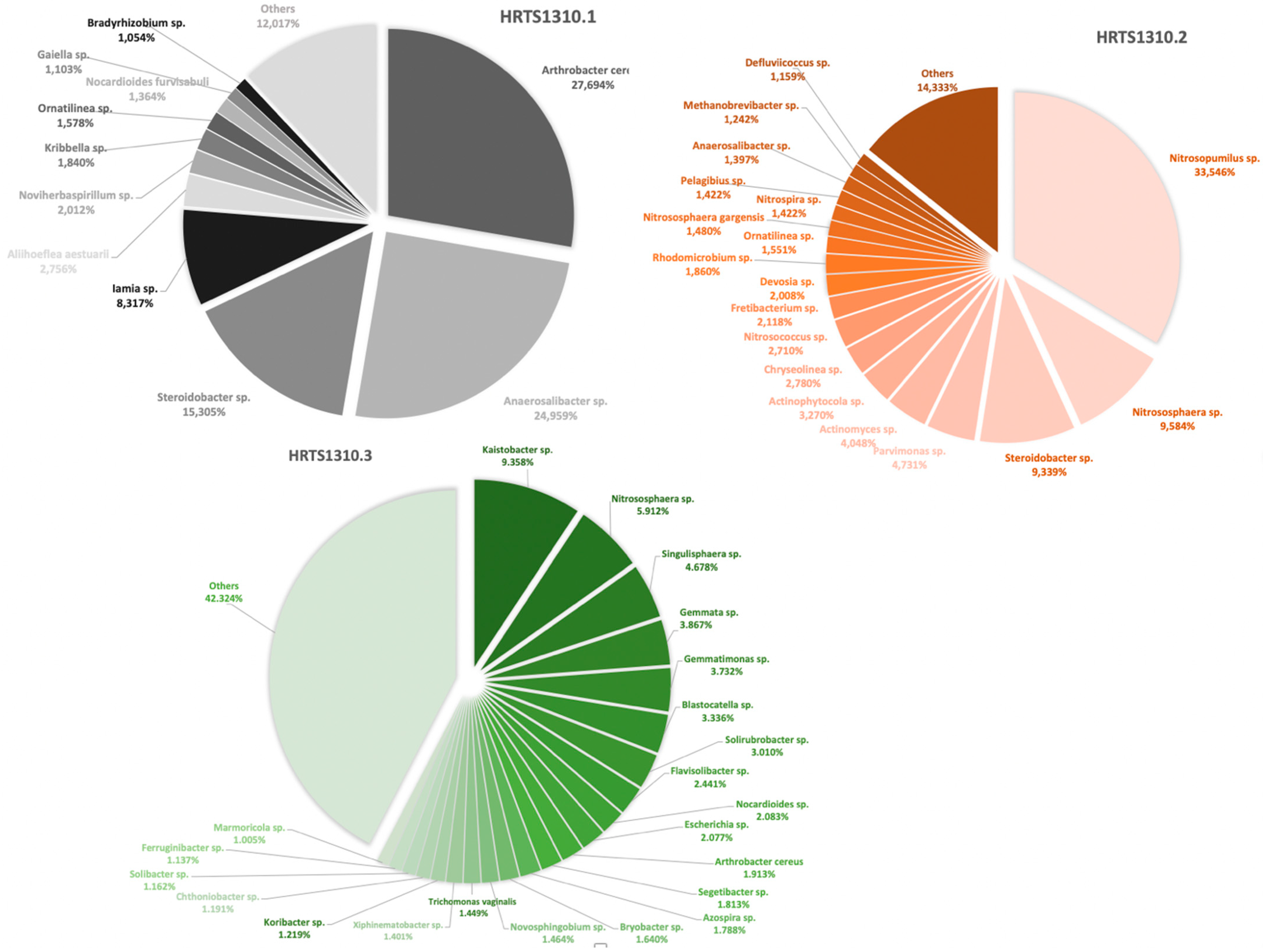
3.1. Overall Profile of the Microbiomes

All three samples showed various pathogenic organisms, i.e., there was no clear association between the presence of specific microorganisms and the fact that the samples were extracted from teeth or bone. Most microorganisms detected were related to soil bacteria and marine microorganisms (Figure 1; a detailed list of the microorganisms detected is provided in Supplementary Materials Table S1).
Some of the soil bacteria microorganisms with the highest frequencies per sample included Arthrobacter cereus, Nitrosopumilus sp., and Nitrososphaera sp. (Figure 1) [46,47,48]. It is noteworthy that some of the microorganisms detected in the samples were related to the human gut microbiota such as Ruminococcus sp. and Odoribacter sp. [48,49,50,51]. These are associated with inflammatory bowel disease, Crohn’s disease, and pancolonic ulcerative colitis, although they are also known to exist in the human body without causing any harm [49,50,51].
The presence of soil bacteria and marine microorganisms is not surprising, since we are dealing with human remains exhumed from downtown Lisbon, an area flooded in the 1755 tsunami [52,53] and a site of recurrent permeability by the Tagus River. Furthermore, the archaeological site, and surrounding area, were of a wet and swampy nature: characteristics provided by the confluence of several watercourses that flow into the Tagus River [54]. Additionally, the archaeological site was excessively contaminated during the excavation process, which must be considered [55]. In addition, gut bacteria in the soil should not be regarded as a surprise considering the chronology of the sample (i.e., late 18th century), since the Lisbon sewer system became a concern mainly during and after the 19th century. Before that, accounts of foreign visitors to Lisbon clearly expressed their disgust regarding the inefficiency of the city’s sewerage system and the filthiness scattered in the streets [56]. Hence, sample contamination via the soil was considered when interpreting the data.
Several human pathogens were found in the three samples, although the percentages varied, along with the soil bacteria and marine microorganisms. These included bacteria related to diseases such as pneumonia (i.e., Pseudomonas aeruginosa and Staphylococcus aureus) and osteomyelitis (i.e., Staphylococcus aureus) [57,58,59,60,61].
Pseudomonas aeruginosa (HRTS1310.1 = 0.848%; HRTS1310.2 = 0.103%; HRTS1310.3 = 0.019%—the percentual values corresponded to those found on the samples’ microbiome) is a benign bacterium and a typical inhabitant of healthy human skin. It plays a vital role as an opportunistic pathogen. It can colonize and infect a wide range of tissues and organs, such as lungs, airways, and the urinary tract. Such infections tend to occur in immunosuppressed individuals, many in hospital contexts. The bacterium enters the skin commonly via compromised skin due to the presence of wounds from burnt cases. Its impact may go from mild (i.e., dermatitis) to more severe clinical cases as the bacterium enters the bold stream causing gastrointestinal, joint, bone, respiratory, and even systemic infections. If affecting the airways, it may cause pneumonia, and it is a very threatening bacterium to individuals with highly compromised immune systems such as individuals with cystic fibrosis or AIDS [46,62,63,64,65].
Staphylococcus aureus (HRTS1310.1 = 0.055%; HRTS1310.2 = 0.032%) is one of the most virulent species of Staphylococcus, although it does not usually cause infection on healthy skin. However, complications may arise if it is permitted to enter internal tissues or the bloodstream. This species can be found in the skin and anterior part of the nostrils of adults. If circumstances allow, it may produce a wide range of diseases. These may include harmless cutaneous and mucous conditions and relatively benign infections (such as folliculitis, furunculosis, or conjunctivitis) or life-threatening diseases such as deep abscesses, osteomyelitis, meningitis, sepsis, endocarditis, or pneumonia (to name but a few examples) [62,66]. The fact that Staphylococcus aureus may be related to osteomyelitis is fascinating since, in the original bioanthropological assessment, the presence of osteomyelitis as a cause of the bone changes observed was excluded. This exclusion was argued based on the widespread distribution of the bone lesions and the absence of a sequestrum, bone surface formation of an involucrum, and the absence of a cloaca perforating the involucrum [10,21]. All these features (i.e., bone changes) are used in the bioanthropological diagnosis of osteomyelitis [10,21]. Furthermore, none of the remains’ radiological and histological features credited to osteomyelitis were observed [21] (p. 432). The same was valid for tuberculosis or pneumonia-related bone changes: none of the abnormal bone lesions and distribution patterns allowed for the identification of those diseases in the skeleton [21] (p. 430).
Other pathogenic microorganisms found are related to commonly known oral pathology bacteria. These included Treponema lecithinolyticum, Actinomyces naeslundii, and Treponema socranskii [53,54,55,56,57,58,59,60,61,62,63,64,65]. These fit the profile of the oral pathology found associated with this individual: they suffered from severe bilateral mandibular abscesses, associated with the second incisor and canine and additional antemortem tooth loss. Moreover, the results also revealed the presence of two other bacterial genera: Actinomyces and Escherichia. The Actinomyces sp. Are typically environmental bacteria. However, many actinomyces are opportunistic pathogens of humans and other mammals, particularly in the oral cavity. In rare cases, they can cause actinomycosis, a disease characterized by abscesses in the mouth, lungs, gastrointestinal tract, and female genital tracts [40,59]. Due to the manner of its impacts on the organs and its features, actinomycosis is a pathology frequently misdiagnosed and confused with neoplasia [67,68,69].
One of the major limitations of interpreting the microorganism detected relates to sample preservation and contamination. In some cases, it was not possible to specify the species of many of the pathogens found. In addition, many of the pathogens found were environmental/soil bacteria; hence, sample contamination via the diagenesis process, both due to the burial and taphonomy processes and/or proximity with other remains, need to be considered.

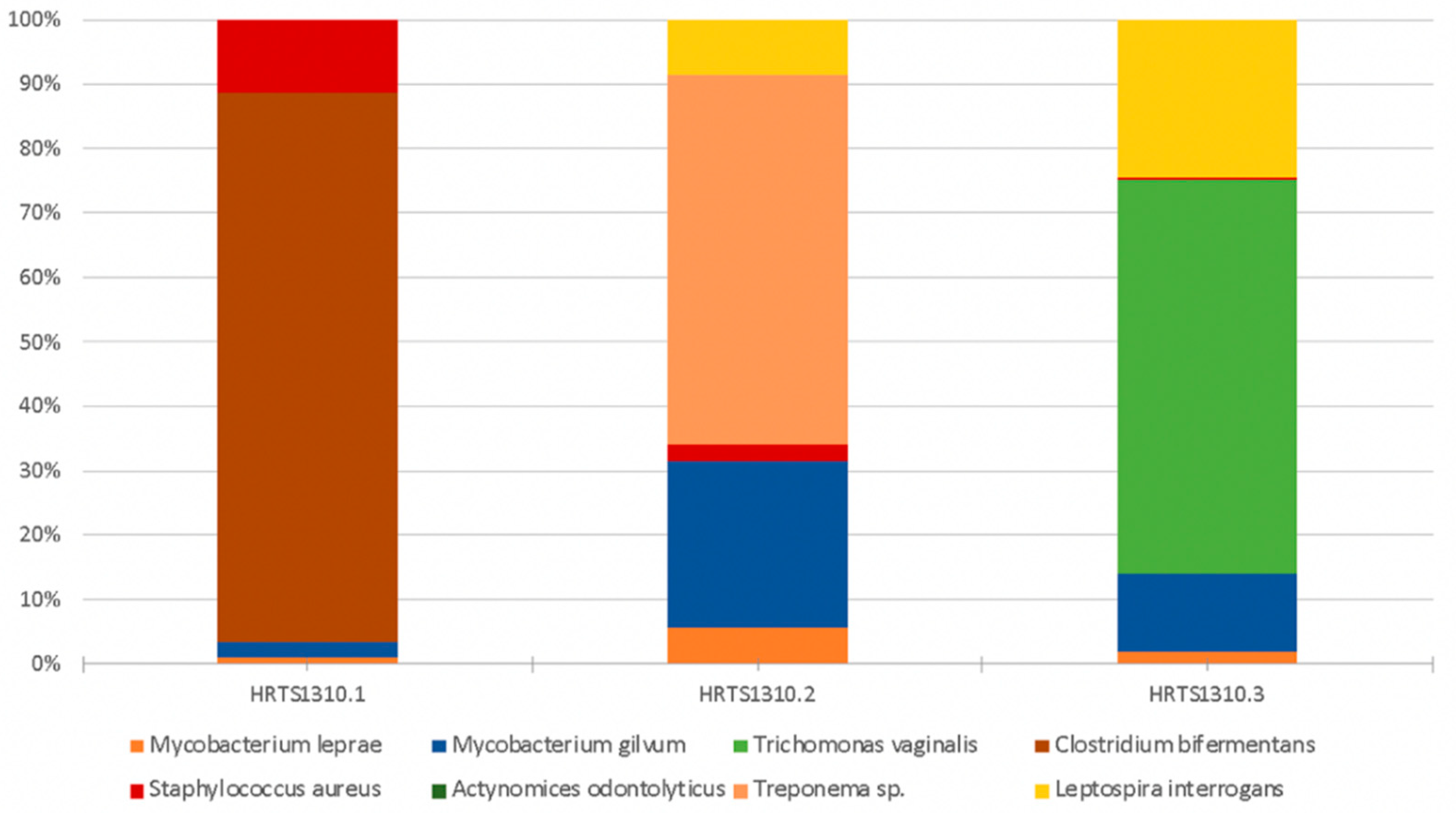
3.2. Human Pathogenic Microorganisms

A closer assessment of the samples identified different human pathogenic microorganisms. These are summarized in Table 1, complemented with their allocated percentage per sample (Figure 2). The expression of the pathogens per sample varied, and no specific pattern of microorganism per type of sample (i.e., teeth versus bone sample) was observed. Staphylococcus aureus was the microorganism with the lowest frequency observed in all three samples. The highest frequency recorded was that of the Trichomonas vaginalis in sample HRTS1310.3, corresponding to the bone sample. The bone fragment came from the humerus. It exhibited a mosaic of remodeled and non-healed bone lesions, indicative that the cause of the lesions and bone remodeling was still active at the time of death. The fragment healing stage may account for the microorganism being identified, as an active infection may have facilitated its detection, and it may be embedded in the bone matrix during the remodeling process.
Although some of the microorganisms identified continue to be environmentally related (e.g., Mycobacterium gilvum), they have also been found to be opportunistic and able to cause harm to humans. Overall, there is some overlapping of the diseases that may be caused by the pathogens identified. The diseases include sexually transmitted diseases, septic arthritis, osteomyelitis, soft tissue infection, meningitis, sepsis, endocarditis, pneumonia, and renal failure [69]. Bone changes in human remains, exhumed from archaeological contexts, have already been described in association with septic arthritis, osteomyelitis, sepsis, pneumonia, and Hansen’s disease [1,10,11,21,70,76,77], to name a few. In addition, soft tissue infection and meningitis can leave marks on bone, since they compromise the macro- and microstructure of the cortical, compact, and cancellous bone tissue [10].
The bone changes observed in the remains of the individual female, HRTS1310, could be discussed as early osteomyelitis (less expressive in bone) and prolonged infection and/or inflammatory process. These three scenarios fit with the changes observed and their distribution within the skeleton: multiple bone lesions composed of a mosaic of new bone deposition, some of which were active at the time of death, others not, of a disseminated pattern, some bilateral, localized in several bones of the upper limbs, the distal end of the femurs, tibias, and fibulas shaft, as well as the frontal and right parietal bones (for details see [21] (pp. 434–439). Unfortunately, and as stated above, the absence of osteological features used in the diagnosis of osteomyelitis (i.e., sequestrum, bone surface forming an involucrum, and several cloacae perforating the involucrum), excluded osteomyelitis as the cause of the bone changes observed [10,21].
The most relevant find was the presence of the pathogen related to leprosy (Hansen’s disease)—Mycobacterium leprae—in all three samples analyzed. This pathogen’s presence indicates that the individual may have been exposed to leprosy, having probably been infected, and was a disease carrier after contamination due to the close contact with infected people [78], not excluding the possibility of zoonotic leprosy, infection via contact with environmental reservoirs, and trauma-related transmission [78,79,80,81]. Regardless, the presence of Mycobacterium leprae in the samples raises the possibility of its presence, and it is necessary to discuss that the bone lesions observed were not necessarily related to syphilis but to other diseases, namely, leprae (alongside osteomyelitis). However, the comprehensive differential diagnosis provided by Assis et al. [21] and the detailed skeletal analysis undertaken (inclusively by two coauthors of this paper) excluded leprae and osteomyelitis as probable causes of the bone changes observed. Overall, no macroscopic, radiological, or histological features related to leprae were found in the human remains [21] (p. 431). For example, no primary and secondary bone involvements of the hand and foot with bone resorption and “penciling” of the metatarsals and osteolysis of the hands and feet distal phalanges were recorded, which are essential to building a leprae diagnosis [10,70]. Unfortunately, it was not possible to assess the facial and rhinomaxillary changes due to the fragmentation of the human remains; thus, the assessment of the osteological remains could be considered compromised as well as the final diagnosis presented by Assis et al. [21].

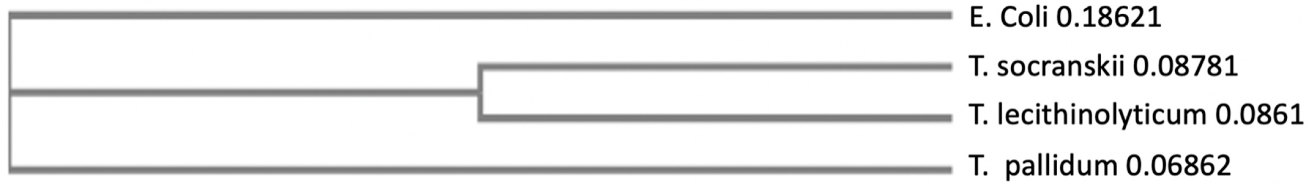
3.3. The Absence of Treponema Pallidum

One of the aims of this research was to confirm or discard the presence of Treponema pallidum, being aware of the limitations of such an endeavor [22,23,24,25]. However, the metagenomic analysis did not detect the presence of Treponema pallidum. Nevertheless, Treponema pallidum absence does not mean that the individual did not suffer from the disease. A combination of low pathogen concentration and poor sample preservation may have contributed to its nondetection.
Despite the absence of Treponema pallidum, two different species of the Treponema genus (i.e., T. socranskii and T. lecithinolyticum) were identified in sample HRTS1310.2. These were both found in the human oral microbiota associated with periodontitis and gingivitis [82,83,84]. Gingivitis and periodontitis are widespread inflammatory gum diseases. However, while gingivitis is reversible if treated, periodontitis has major implications in the maxilla and mandibula, involving the erosion of the dental ligament, which attaches the teeth to the supporting bone at the base of the periodontal pocket [46]. Consequently, if not treated, it will revert to tooth loss, which was observed in the skeletal HRTS1310.
To ascertain that T. socranskii and T. lecithinolyticum were adequality classified, a follow-up assessment was undertaken using the Basic Local Alignment Search Tool (BLAST). The aim was to discard the possibility of a wrong assignation of the pathogen species, since the analysis was based on the 16S rDNA, and we considered the possibility that this DNA region could be similar in different species of the same genus. This follow-up analysis showed that both T. socranskii and T. lecithinolyticum were correctly allocated (see Supplementary Materials Figures S1 and S2); this result was also supported by an analysis of the phylogenetic relationship and alignment sequencing of T. pallidum with the other Treponema species found. This analysis was performed using the CLUSTAL-OMEGA multiple sequence alignment program (Supplementary Materials Figure S3), creating a phylogenetic tree (Figure 3). For this last analysis, we included the 16S sequences of T. pallidum, T. socranskii, T. lecithinolyticum, and the 16S sequence of other unrelated bacteria species (E. coli) used as a control.
The phylogenetic tree (Figure 3) clearly shows that E. coli was different from the other species analyzed. In contrast, in the case of the Treponema species, the T. pallidum was positioned in a distinct branch compared to T. socranskii and T. lecithinolyticum, showing that although they shared the same genus, they were distinct from T. pallidum. The results are further supported by the CLUSTAL-OMEGA sequences alignment (Supplementary Materials Figure S3). The two species located in the three analyzed samples were: Pseudomonas aeruginosa and Escherichia sp.

4. Conclusions

Despite the paleopathological assessment of a probable diagnosis of syphilis, the metagenomic analysis did not identify the presence of T. pallidum in any of the analyzed samples. However, many other different pathogenic microorganisms were detected. The majority were related to environmental soil and water, highlighting the high degree of sample contamination and diagenesis processes. Nevertheless, others were found to be human pathogens able to cause significant harm to people. These included Trichomonas vaginalis; Clostridium bifermentans; Staphylococcus aureus; Leptospira interrogans; Treponema lecithinolyticum; T. socranskii, responsible for a significant number of conditions that range from sexually transmitted diseases, periodontitis, septic arthritis, osteomyelitis, soft tissue infection, meningitis, sepsis, endocarditis to pneumonia—some of which have severe impact on bones. Noteworthy was the detection of the Mycobacterium leprae, even though the paleopathological diagnosis had discarded this possibility.
As to the presence of T. pallidum, the pathogen responsible for syphilis, which was the primary diagnosis of the bone changes observed in the skeleton, it was not detected in the samples. Two microorganisms belonging to the Treponema genus were found, but these were correctly allocated to species other than T. pallidum, i.e., T. socranskii and T. lecithinolyticum. However, it is also possible that the absence of T. pallidum was simply related to the inability to detect the pathogen in human remains as described by additional studies [12,13,14,15,16,17]. The absence of a positive result does not exclude the presence of the pathogen during the individual’s life. It is only indicative that it may no longer be possible to detect this bacterial genetic material after approximately two centuries, either by a poor pathogen concentration in that specific human organism or by degradation of that genetic material, therefore, not being detected.
Despite the absence of the targeted pathogen of this study (i.e., T. pallidum), many other microorganisms that may have had a significant impact on the life and death of the SK1310 female were identified. These included microorganisms associated with respiratory diseases, oral pathology, osteomyelitis, and Trichomonas vaginalis associated with sexually transmitted infections. The presence of evidence of sexually transmitted infections fits with the history of syphilis, known to also be sexually transmitted with the added negative social and cultural connotations. Overall, all these microorganisms could have contributed to some of the bone lesions found that were extensive and possessed various degrees of healing, suggesting that these were chronic.
An intriguing surprise was the identification of Mycobacterium leprae, even in the absence of credited bone-related changes. Identifying Mycobacterium leprae raises the question of the paleopathological approach to leprae when assessing human remains. It urges the need for complementary analyses of human remains when exploring diseases based on the macroscopic analysis of human remains. Paleopathological analysis has made significant progress since its debut as a discipline, but it has much to gain with the ongoing development of methodological approaches, not forgetting the essential requirement to balance the samples’ preservation, aiming for a more ethical approach to human remains in scientific development.

Supplementary Materials

The following supporting information can be downloaded at: www.mdpi.com/article/10.3390/app12126096/s1: Figure S1: BLAST search of the T. socranskii sequence; Figure S2: BLAST search of the T. lecithinolyticum sequence; Figure S3: CLUSTAL sequences alignment; Table S1: Detailed list of the microorganisms detected. In addition, the sequences used in this research are available on the NCBI server, BioProject accession: PRJNA522260 (ID: 522260) (https://www.ncbi.nlm.nih.gov/bioproject/PRJNA522260, registration on 13 February 2019).

Author Contributions

Conceptualization, F.A.-C.; methodology, F.A.-C., C.G., S.P.-D., A.A.C. and S.C.; aDNA analysis, C.G. and S.P.-D.; formal data analysis and interpretation, F.A.-C., S.P.-D. and A.A.C.; bioanthropological, archaeology, and archeothanatology data analysis, F.A.-C., S.C. and R.B.d.S.; data curation, F.A.-C., A.A.C., S.P.-D. and S.C.; writing—original draft preparation, F.A.-C., S.P.-D., A.A.C., C.G., S.C., R.B.d.S. and E.A.-P.; writing—review and editing, F.A.-C., C.G., S.P.-D., A.A.C. and S.C.; visualization, F.A.-C.; project administration, F.A.-C.; funding acquisition, F.A.-C. All authors have read and agreed to the published version of the manuscript.

Funding

This research was funded by the research projects: Bone Matters/Matérias Ósseas (reference: IF/00127/2014/CP1233/CT0003/) and Life After Death: Rethinking Human Remains and Human Osteological Collections as Cultural Heritage and Biobanks (reference: 2020.01014.CEECIND)—both projects are funded both by Fundação para a Ciência e Tecnologia (Portugal) and Coordinated by Francisca Alves Cardoso. F.A.-C.’s research was also funded by Universidade NOVA FCSH 6ª Edição do Financiamento Exploratório para Projetos. This research was within the scope of CRIA—Centro em Rede de Investigação em Antropologia (reference UIDB/04038/2020) Strategic Plan and development of CRIA’s laboratory profile via its Laboratory of Biological Anthropology and Human Osteology—LABOH, funded by Fundação para a Ciência e Tecnologia (Portugal).

Institutional Review Board Statement

The study on human remains was conducted according to the American Association of Biological Anthropology ethical guidelines and the British Association for Biological Anthropology and Osteoarchaeology (BABAO) Code of Ethics and Code of Ethics Practice for the analysis and handling of human remains. Only limited samples were used and targeted specific analyses that were impossible to conduct in any other way. All data will be made available to prevent unnecessary resampling of the remains.

Informed Consent Statement

Not applicable.

Data Availability Statement

The sequences used in this research are available on the NCBI server, BioProject: PRJNA522260 (ID: 522260) (https://www.ncbi.nlm.nih.gov/bioproject/PRJNA522260, registration on 13 February 2019).

Acknowledgments

We would like to acknowledge the privilege to have had the opportunity to study the human remains that led to this research, further recognizing their contribution to the scientific development of the disciplines involved in this study as well as the historical context of the Hospital Real de Todos-os-Santos, in Lisbon, Portugal. We would also like to highlight the significance of human remains as a legacy from past populations and their importance as cultural heritage.

Conflicts of Interest

The authors declare to have no conflict of interest.

References

  1. Buikstra, J.E. (Ed.) Ortner’s Identification of Pathological Conditions in Human Skeletal Remains; Academic Press: London, UK, 2019. [Google Scholar]
  2. Alves-Cardoso, F. Palaeopathology. In The SAS Encyclopaedia of Archaeological Sciences; Sandra, L., López Varela, Eds.; John Wiley & Sons, Inc.: London, UK, 2019. [Google Scholar]
  3. Buikstra, J.E.; Cook, D.C.; Bolhofner, K.L. Introduction: Scientific rigor in paleopathology. Int. J. Paleopathol. 2017, 19, 80–87. [Google Scholar] [CrossRef] [PubMed]
  4. Snoddy, A.M.E.; Beaumont, J.; Buckley, H.R.; Colombo, A.; Halcrow, S.E.; Kinaston, R.L.; Vlok, M. Sensationalism and speaking to the public: Scientific rigour and interdisciplinary collaborations in palaeopathology. Int. J. Paleopathol. 2020, 28, 88–91. [Google Scholar] [CrossRef] [PubMed]
  5. Blevins, K.E.; Crane, A.E.; Lum, C.; Furuta, K.; Fox, K.; Stone, A.C. Evolutionary history of Mycobacterium leprae in the Pacific Islands. Philos. Trans. R. Soc. B 2020, 375, 20190582. [Google Scholar] [CrossRef] [PubMed]
  6. Bos, K.I.; Harkins, K.M.; Herbig, A.; Coscolla, M.; We, N.; Comas, I.; Forrest, S.A.; Bryant, J.M.; Harris, S.R.; Schuenemann, V.J.; et al. Pre-Columbian mycobacterial genomes reveal seals as a source of New World human tuberculosis. Nature 2014, 514, 494–497. [Google Scholar] [CrossRef]
  7. Bos, K.I.; Herbig, A.; Sahl, J.; Waglechner, N.; Fourment, M.; Forrest, S.A.; Klunk, J.; Schuenemann, V.J.; Poinar, D.; Kuch, M.; et al. Eighteenth century Yersinia pestis genomes reveal the long-term persistence of an historical plague focus. Elife 2016, 5, e12994. [Google Scholar] [CrossRef]
  8. Schuenemann, V.J.; Kumar Lankapalli, A.; Barquera, R.; Nelson, E.A.; Hernández, D.I.; Alonzo, V.A.; Boss, K.I.; Morfín, L.M.; Herbig, A.; Krause, J. Historic Treponema pallidum genomes from Colonial Mexico retrieved from archaeological remains. PLoS Negl. Trop. Dis. 2018, 12, e0006447. [Google Scholar] [CrossRef] [Green Version]
  9. Schuenemann, V.J.; Singh, P.; Mendum, T.A.; Krause-Kyora, B.; Jäger, G.; Bos, K.I.; Herbig, A.; Economou, C.; Benjak, A.; Busso, P.; et al. Genome-wide comparison of medieval and modern Mycobacterium leprae. Science 2013, 341, 179–183. [Google Scholar] [CrossRef]
  10. Ortner, D. Identification of Pathological Conditions in Human Skeletal Remains; Academic Press: Amsterdam, The Netherlands, 2003. [Google Scholar]
  11. Hackett, C.J. Diagnosis Criteria of Syphilis Yaws and Treponarid (Treponematosis) and Some other Diseases in Dry Bones; Springer: Heidelberg, Germany, 1976. [Google Scholar]
  12. Baker, B.; Armelagos, G.; Becker, M.; Brothwell, D.; Drusini, A.; Geise, M.; Kelley, M.; Moritoto, I.; Morris, A.; Nurse, G.; et al. The origin and antiquity of syphilis. Curr. Anthopol. 1988, 29, 703–737. [Google Scholar] [CrossRef]
  13. Rothschild, B. History of syphilis. Clin. Infec. Dis. 2005, 40, 1454–1463. [Google Scholar] [CrossRef]
  14. Rothschild, B.; Calderon, F.; Coppa, A.; Rothschild, C. First European exposure to syphilis: The Dominican Republic at the time of Columbian contact. Clin. Infect. Dis. 2000, 31, 936–941. [Google Scholar] [CrossRef] [Green Version]
  15. Saunders, L.; Rothschild, B.; Rothschild, C. Old world origins of syphilis in New York. Chungara 2000, 32, 179–184. [Google Scholar] [CrossRef] [Green Version]
  16. Harper, K.N.; Ocampo, P.S.; Steiner, B.M.; George, R.W.; Silverman, M.S.; Bolotin, S.; Pillay, A.; Saunders, N.J.; Armelagos, G.J. On the origin of the treponematoses: A phylogenetic approach. PLoS Negl. Trop. Dis. 2008, 2, e148. [Google Scholar] [CrossRef] [PubMed] [Green Version]
  17. Harper, K.N.; Zuckerman, M.K.; Armelagos, G.J. Syphilis: Then and now. Scientist Magazine, 1 February 2014; 1–11. [Google Scholar]
  18. Sousa, J. Impacto social da sífilis: Alguns aspectos historicos. Med. Interna 1996, 3, 184–192. [Google Scholar]
  19. Casimiro, S.; Alves-Cardoso, F. Death in the Royal Hospital: An 18th Century Testimony. In Hospital Real de Todos-os-Santos/All Saints Royal Hospital: Lisbon and Public Health; Alberto, E.M., Silva, R.B., Teixeira, A., Eds.; Câmara Municipal de Lisboa/Santa Casa da Misericórdia: Lisboa, Portugal, 2021; pp. 515–517. [Google Scholar]
  20. Alberto, E.M.; Silva, R.B.; Teixeira, A. (Eds.) All Saints Royal Hospital: Lisbon and Public Health; Câmara Municipal de Lisboa/Santa Casa da Misericórdia: Lisboa, Portugal, 2021. [Google Scholar]
  21. Assis, S.; Casimiro, S.; Alves-Cardoso, F. A possible case of acquired syphilis at the former Royal Hospital of All-Saints (RHAS) in Lisbon, Portugal (18th century): A comparative methodological approach to differential diagnosis. Anthropol. Anz. 2015, 72, 427–449. [Google Scholar] [CrossRef] [Green Version]
  22. de Melo, F.L.; de Mello, J.C.M.; Fraga, A.M.; Nunes, K.; Eggers, S. Syphilis at the crossroad of phylogenetics and paleopathology. PLoS Negl. Trop. Dis. 2010, 4, e575. [Google Scholar] [CrossRef]
  23. Gaul, J.S.; Winter, E.; Grossschmidt, K. Ancient pathogens in museal dry bone specimens: Analysis of paleocytology and aDNA. Wien. Med. Wochenschr. 2015, 165, 133–139. [Google Scholar] [CrossRef]
  24. Majander, K.; Pfrengle, S.; Kocher, A.; Neukamm, J.; Plessis, L.; Pla-Díaz, M.; Arora, N.; Akgül, G.; Salo, K.; Schats, R. Ancient bacterial genomes reveal a high diversity of Treponema pallidum strains in early modern Europe. Curr. Biol. 2020, 30, 3788–3803. [Google Scholar] [CrossRef]
  25. Guedes, L.; Dias, O.; Neto, J.; Ribeiro da Silva, L.D.P.; Mendonça de Souza, S.M.F.; Iñiguez, A.M. First Paleogenetic Evidence of Probable Syphilis and Treponematoses Cases in the Brazilian Colonial Period. BioMed Res. Int. 2018, 2018, 8304129. [Google Scholar] [CrossRef] [Green Version]
  26. Ortner, D. Differential diagnosis and issues in disease classification. In A Companion to Paleopathology; Grauer, A., Ed.; Blackwell Publishing Ltd.: Chichester, UK, 2012; pp. 250–267. [Google Scholar]
  27. Bruzek, J. A method for visual determination of sex using the human hip bone. Am. J. Phys. Anthropol. 2002, 117, 157–168. [Google Scholar] [CrossRef]
  28. Buikstra, J.E.; Ubelaker, D.H. Standards for Data Collection from Human Skeletal Remains; Arkansas Archaeological Survey: Fayetteville, AR, USA, 1994. [Google Scholar]
  29. White, T.; Folkens, P. The Human Bone Manual; Academic Press: London, UK, 2005. [Google Scholar]
  30. Gomes, C.; Palomo-Díez, S.; Roig, J.; López-Parra, A.M.; Baeza-Richer, C.; Esparza-Arroyo, A.; Gibaja, J.; Arroyo-Pardo, E. Nondestructive extraction DNA method from bones or teeth, true or false? Forensic Sci. Int. Forensic Sci. Int. Genet. Suppl. Ser. 2015, 5, e279–e282. [Google Scholar] [CrossRef] [Green Version]
  31. Rohland, N.; Hofreiter, M. Ancient DNA extraction from bones and teeth. Nat. Protoc. 2007, 2, 1756–1762. [Google Scholar] [CrossRef] [PubMed] [Green Version]
  32. Rohland, N.; Hofreiter, M. Comparison and optimization of ancient DNA extraction. Biotechniques 2007, 42, 343–352. [Google Scholar] [CrossRef] [Green Version]
  33. Rohland, N.; Siedel, H.; Hofreiter, M. A rapid column-based ancient DNA extraction method for increased sample throughput. Mol. Ecol. Resour. 2010, 10, 677–683. [Google Scholar] [CrossRef] [PubMed]
  34. Pääbo, S.; Poinar, H.; Serre, D.; Jaenicke-Despres, V.; Hebler, J.; Rohland, N.; Kuch, M.; Krause, J.; Vigilant, L.; Hofreiter, M. Genetic analyses from ancient DNA. Annu. Rev. Genet. 2004, 38, 645–679. [Google Scholar] [CrossRef] [PubMed] [Green Version]
  35. Palomo-Díez, S. Caracterización Genética de las Poblaciones de las Edades del Cobre y del Bronce en la Submeseta Norte de la Península Ibérica. Ph.D. Thesis, Universidad Complutense de Madrid, Madrid, Spain, 2015. [Google Scholar]
  36. Becares, A.A.; Fernandez, A.F. Microbiome Based Identification, Monitoring and Enhancement of Fermentation Processes and Products. U.S. Patent Application 15/779,531, 20 December 2018. [Google Scholar]
  37. Feld, L.; Nielsen, T.K.; Hansen, L.H.; Aamand, J.; Albers, C.N. Establishment of bacterial herbicide degraders in a rapid sand filter for bioremediation of phenoxypropionate-polluted groundwater. Appl. Environ. Microbiol. 2016, 82, 878–887. [Google Scholar] [CrossRef] [PubMed] [Green Version]
  38. Albers, C.N.; Ellegaard-Jensen, L.; Hansen, L.H.; Sørensen, S.R. Bioaugmentation of rapid sand filters by microbiome priming with a nitrifying consortium will optimize production of drinking water from groundwater. Water Res. 2018, 129, 1–10. [Google Scholar] [CrossRef]
  39. Belda, I.; Zarraonaindia, I.; Perisin, M.; Palacios, A.; Acedo, A. From Vineyard Soil to Wine Fermentation: Microbiome Approximations to Explain the “terroir”. Concept. Front. Microbiol. 2017, 8, 821. [Google Scholar] [CrossRef] [Green Version]
  40. Altschul, S.F.; Gish, W.; Miller, W.; Myers, E.W.; Lipman, D.J. Basic Local Alignment Search Tool. J. Mol. Biol. 1990, 215, 403–410. [Google Scholar] [CrossRef]
  41. McWilliam, H.; Li, W.; Uludag, M.; Squizzato, S.; Park, Y.M.; Buso, N.; Cowley, A.P.; Lopez, R. Analysis Tool Web Services from the EMBL-EBI. Nucleic Acids Res. 2013, 41, W597–W600. [Google Scholar] [CrossRef] [PubMed] [Green Version]
  42. Wheeler, D.; Bhagwat, M. BLAST quickstart. In Comparative Genomics; Bergman, N.H., Ed.; Human Press: Totowa, NJ, USA, 2007; Volume 1 and 2, pp. 149–175. [Google Scholar]
  43. Meyer, F.; Paarmann, D.; D’Souza, M.; Olson, R.; Glass, E.M.; Kubal, M.; Paczian, T.; Rodriguez, A.; Stevens, R.; Wilke, A.; et al. The metagenomics RAST server—A public resource for the automatic phylogenetic and functional analysis of metagenomes. BMC Bioinform. 2008, 386, 9. [Google Scholar] [CrossRef] [Green Version]
  44. Keegan, K.P.; Glass, E.M.; Meyer, F. MG-RAST, a metagenomics service for analysis of microbial community structure and function. In Microbial Environmental Genomics (MEG); Martin, F., Uroz, S., Eds.; Humana Press: New York, NY, USA, 2016; pp. 207–233. [Google Scholar]
  45. Kircher, M.; Kelso, J. High-throughput DNA sequencing-concepts and limitations. Bioessays 2010, 326, 524–536. [Google Scholar] [CrossRef] [PubMed]
  46. Schmidt, T.M. Encyclopaedia of Microbiology; Academic Press: Amsterdam, The Netherlands, 2019. [Google Scholar]
  47. Schaechter, M. Encyclopaedia of Microbiology; Academic Press: Amsterdam, The Netherlands, 2009. [Google Scholar]
  48. Madigan, M.T.; Bender, K.S.; Buckley, D.H.; Sattley, W.M.; Stahl, D.A. Brock Biology of Microorganisms, 16th ed.; Pearson Education: Hoboken, NJ, USA, 2021. [Google Scholar]
  49. Hardham, J.M.; King, K.W.; Dreier, K.; Wong, J.; Strietzel, C.; Eversole, R.R.; Sfintescu, C.; Evans, R.T. Transfer of Bacteroides splanchnicus to Odoribacter gen. nov. as Odoribacter splanchnicus comb. nov., and description of Odoribacter denticanis sp. nov., isolated from the crevicular spaces of canine periodontitis patients. Int. J. Syst. Evol. Microbiol. 2008, 58 Pt 1, 103–109. [Google Scholar] [CrossRef] [Green Version]
  50. Morgan, X.C.; Tickle, T.; Sokol, H.; Gevers, D.; Devaney, K.L.; Ward, D.V.; Reyes, J.A.; Shah, S.A.; LeLeiko, N.; Snapper, S.B.; et al. Dysfunction of the intestinal microbiome in inflammatory bowel disease and treatment. Genome Biol. 2012, 13, R79. [Google Scholar] [CrossRef]
  51. Werner, H.; Rintelen, G.; Kunstek-Santos, H. A new butyric acid-producing Bacteroides species: B. splanchnicus n. sp. (author’s transl). Zentralbl. Bakteriol. Orig. A 1975, 231, 133–144. [Google Scholar] [PubMed]
  52. Heitor, S.; Miranda, J.M.; Miranda, P.; Mendes-Victor, L. The 1755 Lisbon tsunami; evaluation of the tsunami parameters. J. Geodyn. 1998, 25, 143–157. [Google Scholar]
  53. Mendes-Victor, L. (Ed.) The 1755 Lisbon Earthquake: Revisited; Springer Science & Business Media: Lisbon, Portugal, 2008. [Google Scholar]
  54. Oliveira, E.F. Elementos Para a História do Município de Lisboa; Typographia Universal: Lisboa, Portugal, 1882. [Google Scholar]
  55. Bargão, A. Vivências do Quotidiano do Hospital Real de Todos-os-Santos (Lisboa): Os contextos do poço SE do claustro NE.; Tese de Mestrado em Arqueologia, Universidade Nova de Lisboa Faculdade de Ciências Sociais e Humanas: Lisboa, Portugal, 2015. [Google Scholar]
  56. Cardoso, J.L. Água, iluminação e esgotos em Lisboa nos finais do século XVIII. Análise Soc. 2000, 35, 495–509. [Google Scholar]
  57. Diekema, D.J.; Pfaller, M.A.; Jones, R.N.; Doern, G.V.; Winokur, P.L.; Gales, A.C.; Sader, H.S.; Kugler, K.; Beach, M. Survey of bloodstream infections due to gram-negative bacilli: Frequency of occurrence and antimicrobial susceptibility of isolates collected in the United States, Canada, and Latin America for the SENTRY Antimicrobial Surveillance Program, 1997. Clin. Infect. Dis. 1999, 29, 595–607. [Google Scholar]
  58. Kidd, J.M.; Kuti, J.L.; Nicolau, D.P. Novel pharmacotherapy for the treatment of hospital-acquired and ventilator-associated pneumonia caused by resistant gram-negative bacteria. Expert Opin. Pharmacother. 2018, 19, 397–408. [Google Scholar] [CrossRef]
  59. Tordecilla Echenique, Y.; Salamanca Bautista, M.P.; Arias Jiménez, J.L.; Guisado Espartero, E.; Ortega Calvo, M.; Pérez Cano, R. Hematogenous sternal osteomyelitis and community acquired pneumonia in a methicillin-susceptible Staphilococcus aureus sepsis. An. De Med. Interna 2005, 22, 91–193. [Google Scholar]
  60. Akinkugbe, O.; Stewart, C.; Mckenna, C. Presentation and investigation of pediatric bone and joint infections in the pediatric emergency department. Pediatr. Emerg. Care 2019, 35, 700–704. [Google Scholar] [CrossRef]
  61. Suligoy, C.M.; Lattar, S.M.; Llana, M.N.; González, C.D.; Alvarez, L.P.; Ashley Robinson, D.; Gómez, M.I.; Buzzola, F.R.; Sordelli, D.O. Mutation of Agr is associated with the adaptation of Staphylococcus aureus to the host during chronic osteomyelitis. Front. Cell Infect. Microbiol. 2018, 8, 18. [Google Scholar] [CrossRef] [PubMed]
  62. Romani Vestman, N.; Chen, T.; Lif Holgerson, P.; Öhman, C.; Johansson, I. Oral microbiota shift after 12-week supplementation with Lactobacillus reuteri DSM 17938 and PTA 5289; a randomized control trial. PLoS ONE 2015, 10, e0125812. [Google Scholar] [CrossRef] [PubMed]
  63. Scapoli, L.; Girardi, A.; Palmieri, A.; Martinelli, M.; Cura, F.; Lauritano, D.; Carinci, F. Quantitative analysis of periodontal pathogens in periodontitis and gingivitis. J. Biol. Regul. Homeost. Agents 2015, 29 (Suppl. 1), 101–110. [Google Scholar] [PubMed]
  64. Wasiela, M.; Brzezińska-Błaszczyk, E. The influence of anaerobic bacteria and mycoplasma isolated from the genital tract on mast cell activation. Med. Dosw. Mikrobiol. 2000, 52, 389–396. [Google Scholar] [PubMed]
  65. Okamoto-Shibayama, K.; Sekino, J.; Yoshikawa, K.; Saito, A.; Ishihara, K. Antimicrobial susceptibility profiles of oral Treponema species. Anaerobe 2017, 48, 242–248. [Google Scholar] [CrossRef] [PubMed]
  66. Taylor, T.A.; Unakal, C.G. Staphylococcus Aureus; StatPearls: Treasure Island, FL, USA, 2022. [Google Scholar]
  67. Chandranesan, A. Actinomycosis in Cancer patients: Dilemma in Differentiating Colonization Versus True Disease. Infect. Dis. Clin. Pract. 2018, 26, 303. [Google Scholar] [CrossRef]
  68. Farazmand, C.; Klinkova, O.; Denham, J.; Nanjappa, S.; Greene, J. Actinomycosis in cancer patients: A single-center chart review study. Infect. Dis. Clin. Pract. 2018, 26, 339–343. [Google Scholar] [CrossRef]
  69. Lamrabet, O.; Drancourt, M. Mycobacterium gilvum illustrates size-correlated relationships between mycobacteria and Acanthamoeba polyphaga. Appl. Environ. Microbiol. 2013, 79, 1606–1611. [Google Scholar] [CrossRef] [Green Version]
  70. Matos, V. O diagnóstico retrospectivo da lepra: Complementaridade clínica e paleopatológica no arquivo médico do Hospital-colónia Rovisco Pais (Século XX, Tocha, Portugal) e na colecção de Esqueletos da Leprosaria Medieval De St. Jørgen’s (Odense, Dinamarca). Ph.D. Thesis, University of Coimbra, Coimbra, Portugal, 2009. [Google Scholar]
  71. Monde, N.; Munyeme, M.; Muwonge, A.; Muma, J.B.; Malama, S. Characterization of non-tuberculous mycobacterium from humans and water in an Agropastoral area in Zambia. BMC Infect. Dis. 2018, 18, 20. [Google Scholar] [CrossRef] [Green Version]
  72. Lavania, M.; Turankar, R.; Singh, I.; Nigam, A.; Sengupta, U. Detection of Mycobacterium gilvum first time from the bathing water of leprosy patient from Purulia, West Bengal. Int. J. Mycobacteriol. 2014, 3, 286–289. [Google Scholar] [CrossRef] [Green Version]
  73. Feleke, D.G.; Yemanebrhane, N. Trichomonas vaginalis infection in Ethiopia: A systematic review and meta-analysis. Int. J. STD AIDS 2022, 33, 232–241. [Google Scholar] [CrossRef] [PubMed]
  74. Barrett, L.F.; Saragadam, S.D.; DiMaria, C.N.; Delgado-Daza, A. Infection of a prosthetic knee joint with Clostridium bifermentans. Oxf. Med. Case Rep. 2020, 8, omaa057. [Google Scholar] [CrossRef] [PubMed]
  75. Edagiz, S.; Lagace-Wiens, P.; Embil, J.; Karlowsky, J.; Walkty, A. Empyema caused by Clostridium bifermentans: A case report. Can. J. Infect. Dis. Med. Microbiol. 2015, 26, 105–107. [Google Scholar] [CrossRef] [PubMed] [Green Version]
  76. Scanlan, D.R.; Smith, M.A.; Isenberg, H.D.; Engrassia, S.; Hilton, E. Clostridium bifermentans bacteremia with metastatic osteomyelitis. J. Clin. Microbiol. 1994, 32, 2867–2868. [Google Scholar] [CrossRef] [Green Version]
  77. Henneberg, M.; Holloway-Kew, K.; Lucas, T. Human major infections: Tuberculosis, treponematoses, leprosy—A paleopathological perspective of their evolution. PLoS ONE 2021, 16, e0243687. [Google Scholar] [CrossRef]
  78. Tió-Coma, M.; Wijnands, T.; Pierneef, L.; Schilling, A.K.; Alam, K.; Roy, J.C.; Faber, W.R.; Menke, H.; Pieters, T.; Stevenson, K.; et al. Detection of Mycobacterium leprae DNA in soil: Multiple needles in the haystack. Sci. Rep. 2019, 9, 3165. [Google Scholar] [CrossRef] [Green Version]
  79. Hagge, D.; Parajuli, P.; Kunwar, C.; Rana, D.; Thapa, R.; Neupane, K.; Nicholls, P.; Adams, L.; Geluk, A.; Shah, M.; et al. Opening a Can of Worms: Leprosy Reactions and Complicit Soil-Transmitted Helminths. EBioMedicine 2017, 23, 119–124. [Google Scholar] [CrossRef] [Green Version]
  80. Bratschi, M.W.; Steinmann, P.; Wickenden, A.; Gillis, T.P. Current knowledge on Mycobacterium leprae transmission: A systematic literature review. Lepr. Rev. 2015, 86, 142–155. [Google Scholar] [CrossRef]
  81. da Silva, M.B.; Portela, J.M.; Li, W.; Jackson, M.; Gonzalez-Juarrero, M.; Hidalgo, A.S.; Belisle, J.T.; Bouth, R.C.; Gobbo, A.R.; Barreto, J.G.; et al. Evidence of zoonotic leprosy in Pará, Brazilian Amazon, and risks associated with human contact or consumption of armadillos. PLoS Negl. Trop. Dis. 2018, 12, e0006532. [Google Scholar] [CrossRef]
  82. Sakamoto, M.; Takeuchi, Y.; Umeda, M.; Ishikawa, I.; Benno, Y.; Nakase, T. Detection of Treponema socranskii associated with human periodontitis by PCR. Microbiol. Immunol. 1999, 43, 485–490. [Google Scholar] [CrossRef]
  83. Takeuchi, Y.; Umeda, M.; Sakamoto, M.; Benno, Y.; Huang, Y.; Ishikawa, I. Treponema socranskii, Treponema denticola, and Porphyromonas gingivalis are associated with severity of periodontal tissue destruction. J. Periodontol. 2001, 72, 1354–1363. [Google Scholar] [CrossRef] [PubMed]
  84. Choi, B.K.; Ohk, S.H.; Lee, H.J.; Kang, J.H.; Jeong, G.J.; Yoo, Y.J. Effects of whole cell sonicates of Treponema lecithinolyticum on osteoclast differentiation. J. Periodontol. 2001, 72, 1172–1177. [Google Scholar] [CrossRef] [PubMed]
Figure 1. Graphical representation of the most prevalent (% > 1) microorganisms detected in the three samples. The percentages of the valid reads using the QC MG-RAST server were as follows: SRX5372162: 24.942/23.333 (93.55%) sequences; mean sequence length of 290 ± 20 bp; SRX5372161: 33.035/28.225 (85.44%) sequences; mean sequence length of 289 ± 21 bp; finally, SRX5372160: 89.422/76.325 (85.35%) sequences; mean sequence length of 294 ± 21 bp. Sequences are available on the NCBI server, BioProject accession: PRJNA522260 (SRX5372162; SRX5372161; SRX5372160).
Applsci 12 06096 g001
Figure 2. Percentual distribution of the most abundant pathogenic microorganisms (100% = all pathogen readings per sample). The color breakdown represents the pathogens found per sample. The figure is based on the data in Table 1 data.
Applsci 12 06096 g002
Figure 3. CLUSTAL-OMEGA phylogenetic tree.
Applsci 12 06096 g003
Table 1. List of the most abundant human pathogenic microorganisms found in the various samples of the HRTS SK1310 individual and associated frequency.
MicroorganismObservationsHRTS1310.1 aHRTS1310.2 bHRTS1310.3 c
Mycobacterium lepraeMycobacterium leprae causes leprosy (Hansen’s disease). This disease can leave its signature in bones if untreated. It primarily leads to the destruction of peripheral nerves and muscles and their atrophy, and the motor function is impaired. The loss of sensation in the extremities causes accidental injuries with subsequent infections. In addition, loss of bone calcium contributes to a slow shrinking of the digits and their transition to claw/pencil-like forms in late-stage leprosy [10,48,70].0.0050.0330.043
Mycobacterium gilvumThis is an environmental mycobacterium isolated from river sediments. M. gilvum has rarely been isolated as an opportunistic pathogen [69] and is viewed as a nontuberculous mycobacterium and as nonpathogenic and found in water and humans [71]. Its presence has been detected in accumulated water associated with a bathing place for leprosy patients [72].0.0110.1480.290
Trichomonas vaginalisMay cause vaginitis or sexually transmitted infections [48,73].001.449
Clostridium bifermentansIt is a commensal in the gut, oral cavity, and female genital tract and can be found in soil, sewage, feces, and marine sedimentations. It was once considered to be nonpathogenic; however, it has been recently associated with septic arthritis, osteomyelitis, soft tissue infection, brain abscess, bacteremia, and endocarditis cases [74,75].0.40500
Staphylococcus aureusStaphylococcus aureus is a highly virulent, life-threatening pathogen that can cause diseases such as deep abscesses, osteomyelitis, meningitis, sepsis, endocarditis, or pneumonia (to name but a few examples) [62,66].0.0550.0320.007
Treponema sp. The genus Treponema includes a wide diversity of commensal and pathogen species, most of which exist in complex bacterial communities [47,48].00.3280
Leptospira interrogansThis pathogen evolves from environmental organisms and a human parasite [47,48]. The most common leptospiral syndrome in humans is leptospirosis, a disorder that localizes in the kidneys and can cause renal failure and ultimately death [48].00.0490.580
The various colors correspond to the percentage gradient: darker colors = higher percentages. The values represent the ratio of the analyses shown as the number of specific readings divided by the number of total readings. The readings have been debugged to remove errors/artifacts using the MG-RAST server. The reads obtained per sample: a 24.942 spots, 15M bases (tooth sample); b 33.035 spots, 19.9M bases (tooth sample); c 89.422 spots, 53.8M bases (bone sample).
Publisher’s Note: MDPI stays neutral with regard to jurisdictional claims in published maps and institutional affiliations.

