
Spectroscopic and Microscopic Characterization of Flashed Glasses from Stained Glass Windows

 , ,
, ,  , ,
, ,  and
and
Abstract
1. Introduction
2. Materials and Methods
3. Results and Discussion
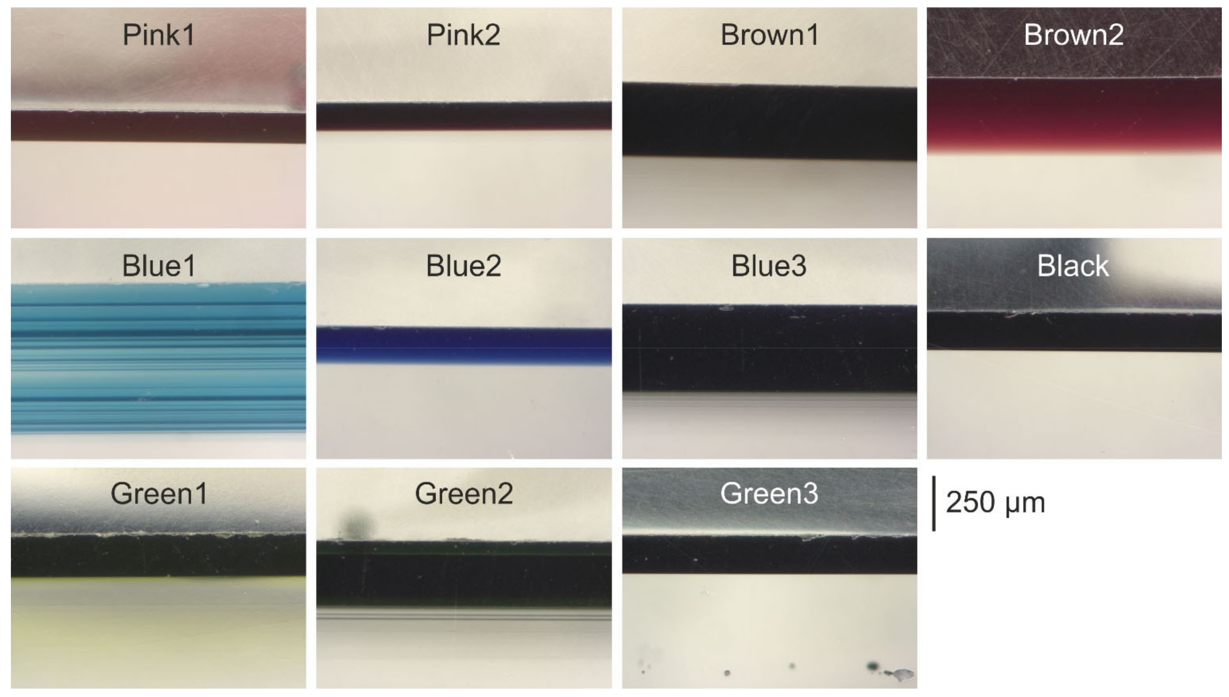
3.1. Thickness Measurements
3.2. Chemical Characterization
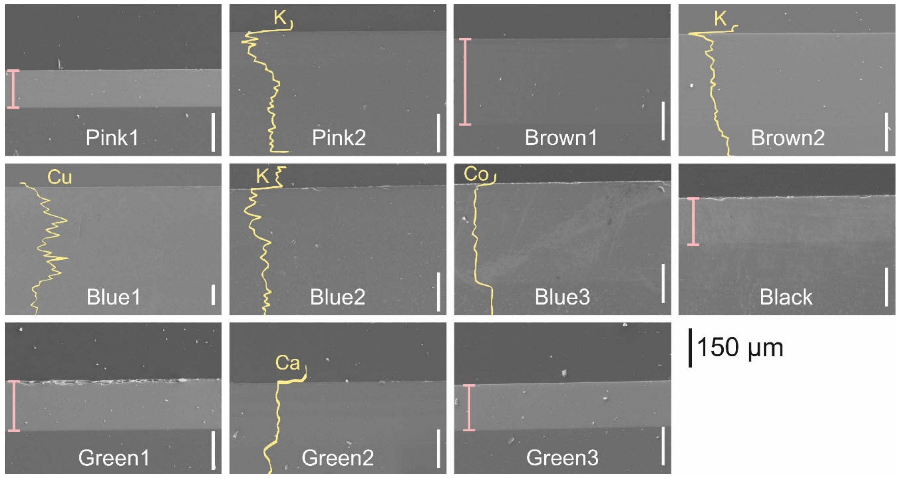
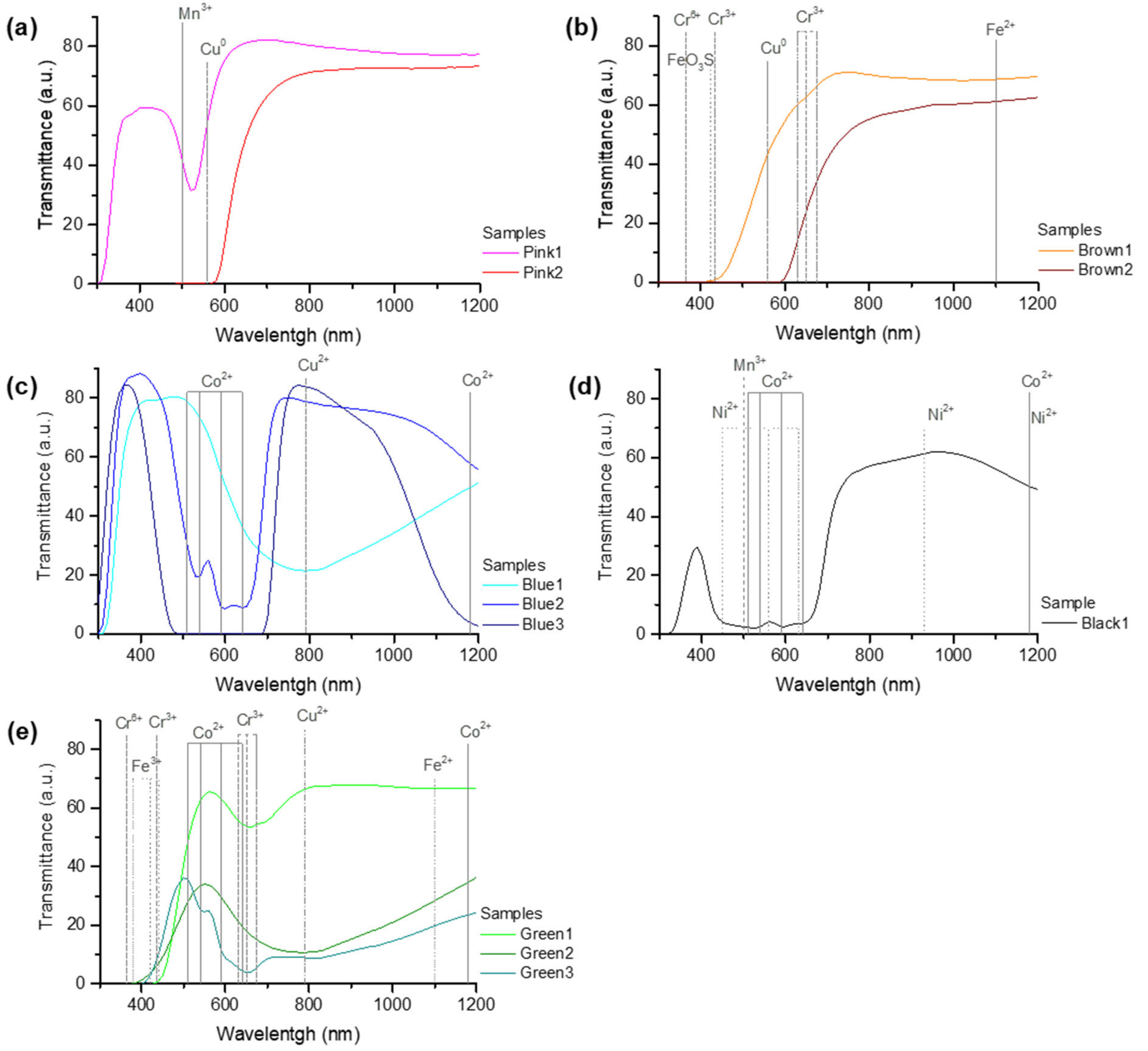
3.2.1. Field Emission Scanning Electron Microscopy/Energy-Dispersive X-ray Spectroscopy and UV-Vis-IR Spectroscopy
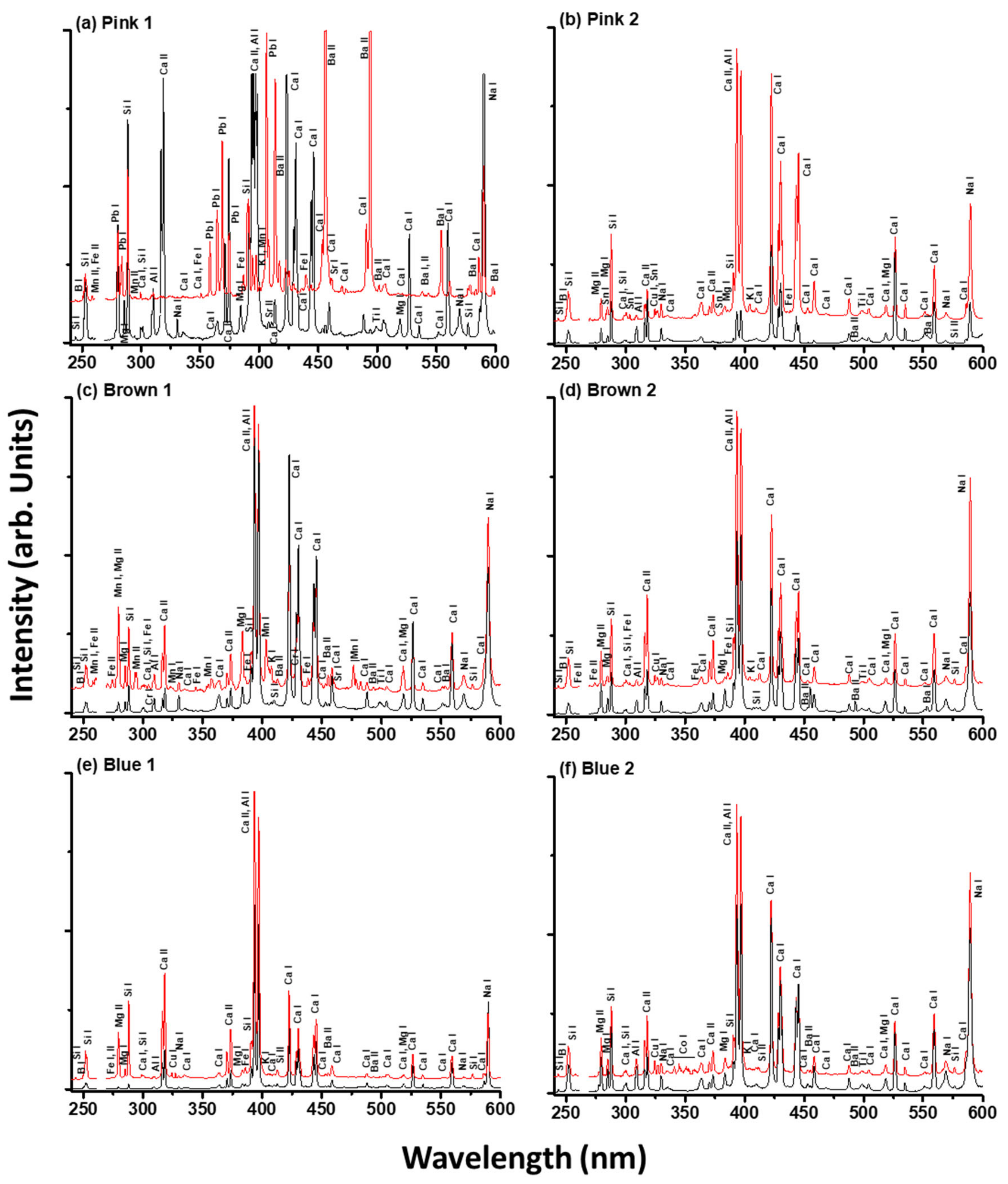
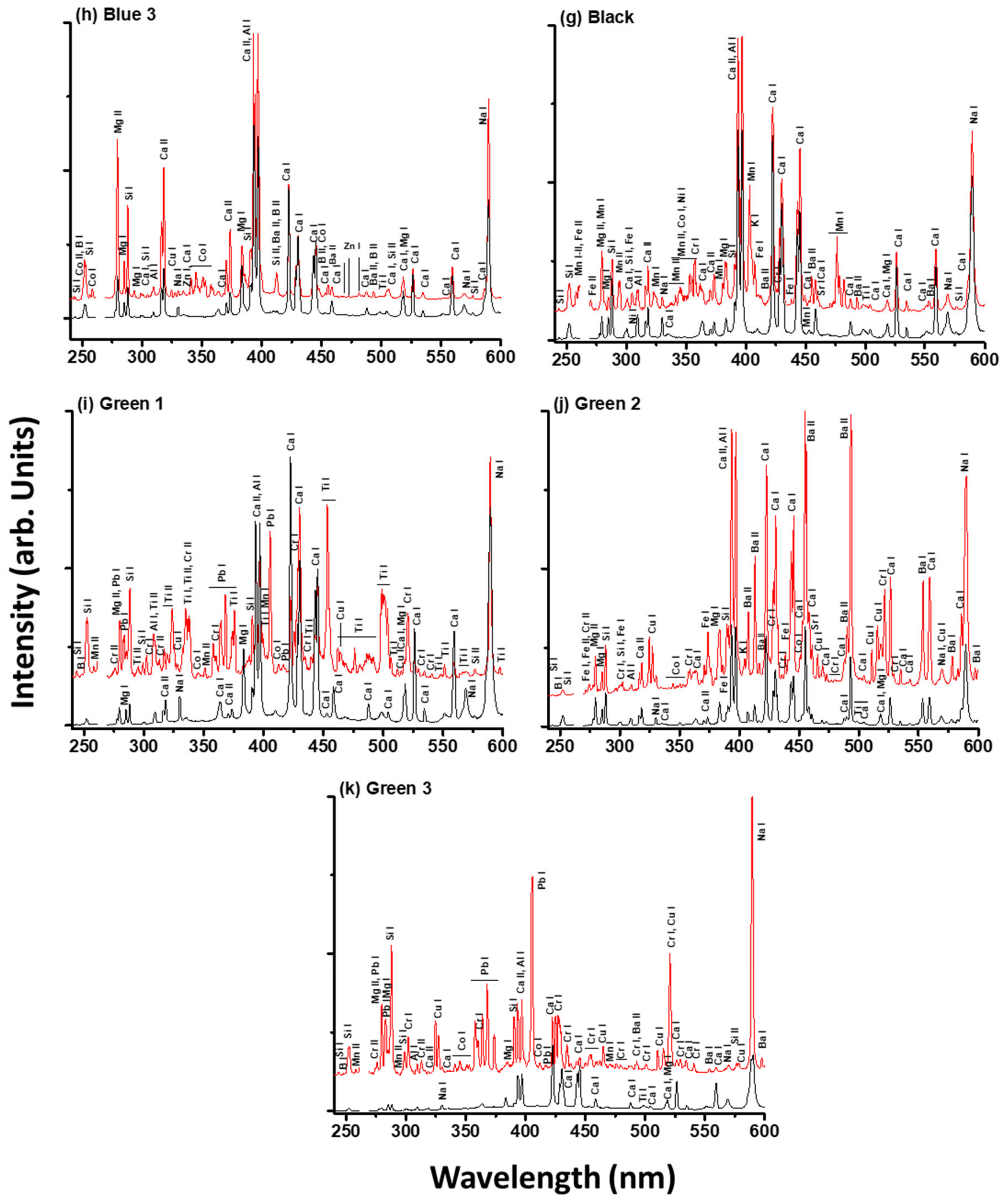
3.2.2. Laser-Induced Breakdown Spectroscopy
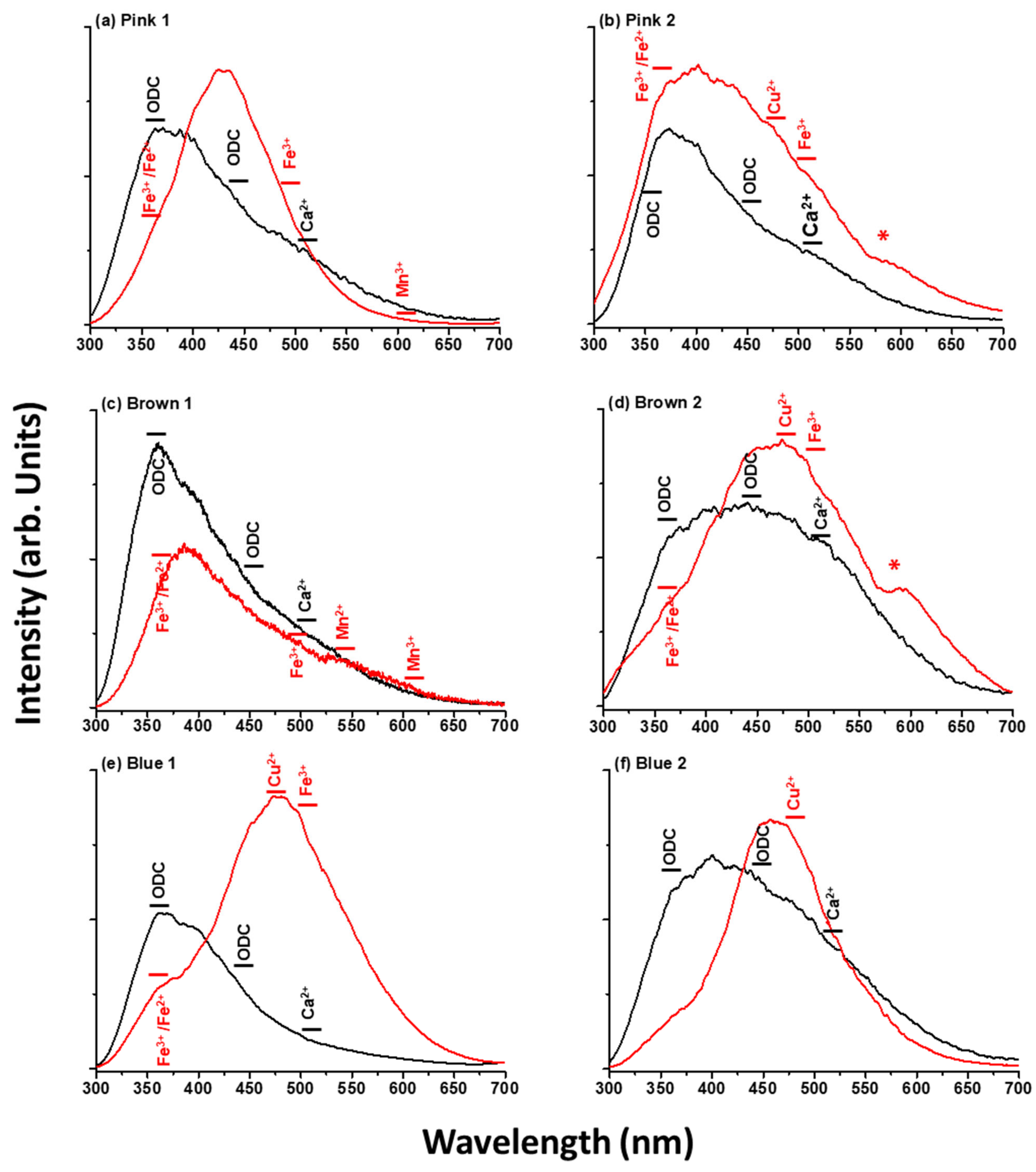
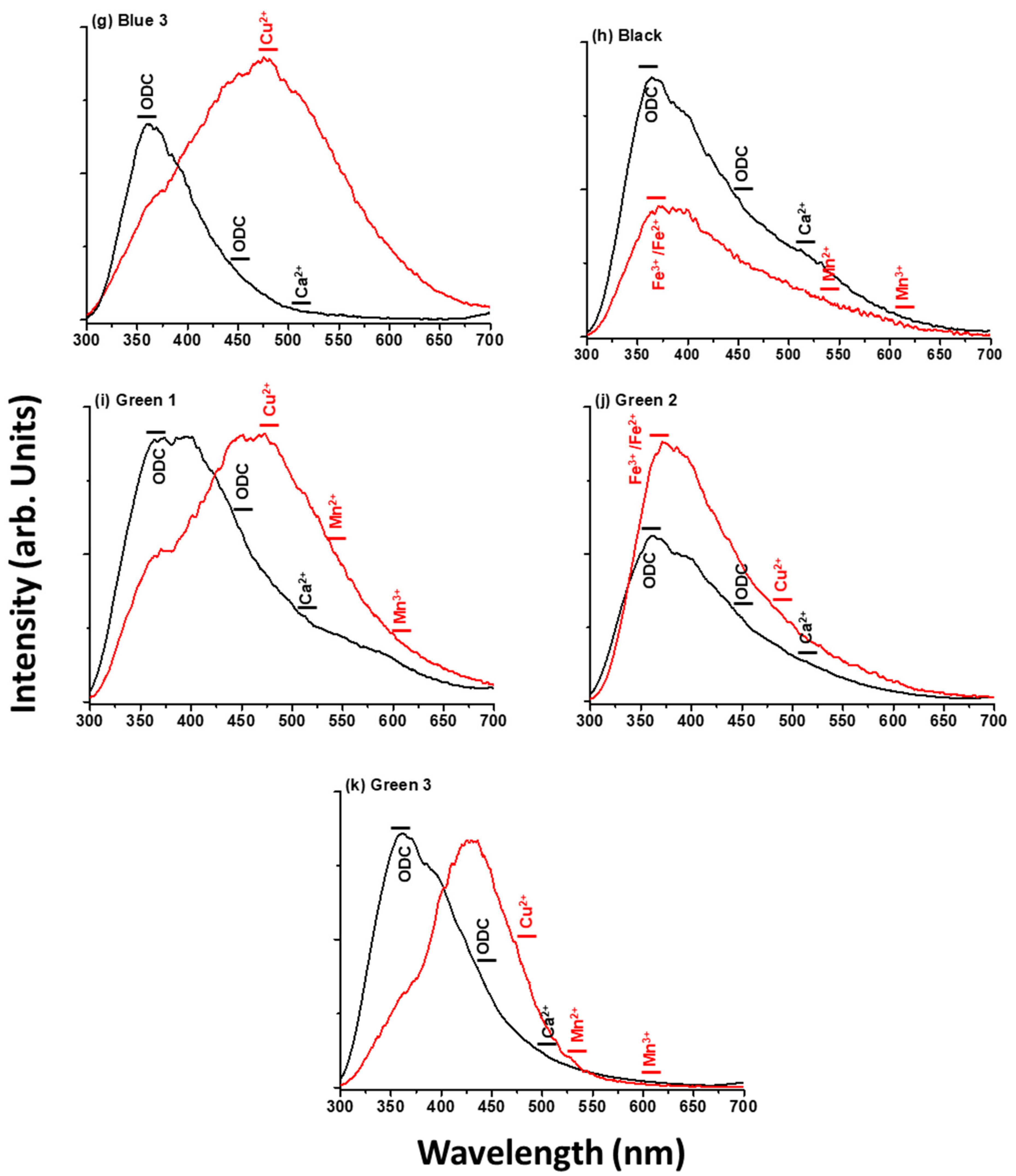
3.2.3. Laser-Induced Fluorescence
4. Conclusions
Author Contributions
Funding
Institutional Review Board Statement
Informed Consent Statement
Data Availability Statement
Acknowledgments
Conflicts of Interest
References
- Ceglia, A.; Meulebroeck, W.; Wouters, H.; Baert, K.; Nys, K.; Terryn, H.; Thienpont, H. Using Optical Spectroscopy to Characterize the Material of a 16th c. Stained Glass Window; Thienpont, H., Meulebroeck, W., Nys, K., Vanclooster, D., Eds.; Routledge: Kent, UK, 2012; p. 84220A. [Google Scholar]
- Rich, C.; Mitchell, M.; Ward, R. Stained Glass Basics: Techniques, Tools & Projects; Sterling Publishing Company, Inc.: New York, NY, USA, 1997. [Google Scholar]
- Weyl, W.A. Coloured Glasses; Society of Glass Technology: Sheffield, UK, 1951; ISBN 9780900682797. [Google Scholar]
- Drünert, F.; Blanz, M.; Pollok, K.; Pan, Z.; Wondraczek, L.; Möncke, D. Copper-Based Opaque Red Glasses—Understanding the Colouring Mechanism of Copper Nanoparticles in Archaeological Glass Samples. Opt. Mater. 2018, 76, 375–381. [Google Scholar] [CrossRef]
- Meulebroeck, W.; Wouters, H.; Nys, K.; Thienpont, H. Authenticity Screening of Stained Glass Windows Using Optical Spectroscopy. Sci. Rep. 2016, 6, 37726. [Google Scholar] [CrossRef] [PubMed]
- Adlington, L.W.; Freestone, I.C. Using Handheld PXRF to Study Medieval Stained Glass: A Methodology Using Trace Elements. MRS Adv. 2017, 2, 1785–1800. [Google Scholar] [CrossRef]
- Marchesi, V.; Negri, E.; Messiga, B.; Riccardi, M.P. Medieval Stained Glass Windows from Pavia Carthusian Monastery (Northern Italy). Geol. Soc. Lond. Spec. Publ. 2006, 257, 217–227. [Google Scholar] [CrossRef]
- Alonso Abad, M.P.; Capel, F.; Valle Fuentes, F.J.; de Pablos Pérez, Á.; Ortega-Feliú, I.; Gómez-Tubio, B.M.; Respaldiza Galisteo, M.Á. Caracterización de Un Vidrio Rojo Medieval Procedente de Las Vidrieras Del Monasterio de Las Huelgas de Burgos. Bol. Soc. Esp. Ceram. Vidr. 2009, 48, 179–186. [Google Scholar]
- Farges, F.; Etcheverry, M.P.; Scheidegger, A.; Grolimund, D. Speciation and Weathering of Copper in “Copper Red Ruby” Medieval Flashed Glasses from the Tours Cathedral (XIII Century). Appl. Geochem. 2006, 21, 1715–1731. [Google Scholar] [CrossRef]
- Kunicki-Goldfinger, J.J.; Freestone, I.C.; McDonald, I.; Hobot, J.A.; Gilderdale-Scott, H.; Ayers, T. Technology, Production and Chronology of Red Window Glass in the Medieval Period—Rediscovery of a Lost Technology. J. Archaeol. Sci. 2014, 41, 89–105. [Google Scholar] [CrossRef]
- Palomar, T. Chemical Composition and Alteration Processes of Glasses from the Cathedral of León (Spain). Bol. Soc. Esp. Ceram. Vidr. 2018, 57, 101–111. [Google Scholar] [CrossRef]
- Machado, A.; Wolf, S.; Alves, L.C.; Katona-Serneels, I.; Serneels, V.; Trümpler, S.; Vilarigues, M. Swiss Stained-Glass Panels: An Analytical Study. Microsc. Microanal. 2017, 23, 878–890. [Google Scholar] [CrossRef]
- Atrei, A.; Scala, A.; Giamello, M.; Uva, M.; Pulselli, R.M.; Marchettini, N. Chemical Composition and Micro Morphology of Golden Laminae in the Wall Painting “La Maestà” by Simone Martini: A Study by Optical Microscopy, XRD, FESEM-EDS and ToF-SIMS. Appl. Sci. 2019, 9, 3452. [Google Scholar] [CrossRef]
- Legrand, S.; Van der Snickt, G.; Cagno, S.; Caen, J.; Janssens, K. MA-XRF Imaging as a Tool to Characterize the 16th Century Heraldic Stained-Glass Panels in Ghent Saint Bavo Cathedral. J. Cult. Herit. 2019, 40, 163–168. [Google Scholar] [CrossRef]
- Bernady, E.; Goryl, M.; Walczak, M. XRF Imaging (MA-XRF) as a Valuable Method in the Analysis of Nonhomogeneous Structures of Grisaille Paint Layers. Heritage 2021, 4, 3193–3207. [Google Scholar] [CrossRef]
- Van der Snickt, G.; Legrand, S.; Caen, J.; Vanmeert, F.; Alfeld, M.; Janssens, K. Chemical Imaging of Stained-Glass Windows by Means of Macro X-Ray Fluorescence (MA-XRF) Scanning. Microchem. J. 2016, 124, 615–622. [Google Scholar] [CrossRef]
- Capobianco, N.; Hunault, M.O.J.Y.; Balcon-Berry, S.; Galoisy, L.; Sandron, D.; Calas, G. The Grande Rose of the Reims Cathedral: An Eight-Century Perspective on the Colour Management of Medieval Stained Glass. Sci. Rep. 2019, 9, 3287. [Google Scholar] [CrossRef] [PubMed]
- Hunault, M.O.J.Y.; Bauchau, F.; Boulanger, K.; Hérold, M.; Calas, G.; Lemasson, Q.; Pichon, L.; Pacheco, C.; Loisel, C. Thirteenth-Century Stained Glass Windows of the Sainte-Chapelle in Paris: An Insight into Medieval Glazing Work Practices. J. Archaeol. Sci. Rep. 2021, 35, 102753. [Google Scholar] [CrossRef]
- Palomar, T.; Grazia, C.; Pombo Cardoso, I.; Vilarigues, M.; Miliani, C.; Romani, A. Analysis of Chromophores in Stained-Glass Windows Using Visible Hyperspectral Imaging in-Situ. Spectrochim. Acta Part A Mol. Biomol. Spectrosc. 2019, 223, 117378. [Google Scholar] [CrossRef]
- Rahrig, M.; Torge, M. 3D Inspection of the Restoration and Conservation of Stained Glass Windows Using High Resolution Structured Light Scanning. In International Archives of the Photogrammetry, Remote Sensing and Spatial Information Sciences—ISPRS Archives; International Society of Photogrammetry and Remote Sensing (ISPRS): Hanover, Germany, 2019; Volume 42, pp. 965–972. [Google Scholar]
- Targowski, P.; Góra, M.; Wojtkowski, M. Optical Coherence Tomography for Artwork Diagnostics. Laser Chem. 2007, 2006, 35373. [Google Scholar] [CrossRef][Green Version]
- Liang, H.; Cid, M.G.; Cucu, R.G.; Dobre, G.M.; Podoleanu, A.G.; Pedro, J.; Saunders, D. En-Face Optical Coherence Tomography—A Novel Application of Non-Invasive Imaging to Art Conservation. Opt. Express 2005, 13, 6133. [Google Scholar] [CrossRef] [PubMed]
- Filippidis, G.; Massaouti, M.; Selimis, A.; Gualda, E.J.; Manceau, J.M.; Tzortzakis, S. Nonlinear Imagig and THz Diagnostic Tools in the Service of Cultural Heritage. Appl. Phys. A Mater. Sci. Process. 2012, 106, 257–263. [Google Scholar] [CrossRef]
- Anglos, D. Laser-Induced Breakdown Spectroscopy in Art and Archaeology. Appl. Spectrosc. 2001, 55, 186A–205A. [Google Scholar] [CrossRef]
- Anglos, D.; Detalle, V. Cultural Heritage Applications of LIBS. In Springer Series in Optical Sciences; Springer: Berlin/Heidelberg, Germany, 2014; Volume 182, pp. 531–554. ISBN 9783642450846. [Google Scholar]
- Nevin, A.; Spoto, G.; Anglos, D. Laser Spectroscopies for Elemental and Molecular Analysis in Art and Archaeology. Appl. Phys. A Mater. Sci. Process. 2012, 106, 339–361. [Google Scholar] [CrossRef]
- Tognoni, E.; Palleschi, V.; Corsi, M.; Cristoforetti, G. Quantitative Micro-Analysis by Laser-Induced Breakdown Spectroscopy: A Review of the Experimental Approaches. Spectrochim. Acta-Part B At. Spectrosc. 2002, 57, 1115–1130. [Google Scholar] [CrossRef]
- Melessanaki, K.; Mateo, M.; Ferrence, S.C.; Betancourt, P.P.; Anglos, D. The Application of LIBS for the Analysis of Archaeological Ceramic and Metal Artifacts. Appl. Surf. Sci. 2002, 197–198, 156–163. [Google Scholar] [CrossRef]
- Giakoumaki, A.; Melessanaki, K.; Anglos, D. Laser-Induced Breakdown Spectroscopy (LIBS) in Archaeological Science-Applications and Prospects. Anal. Bioanal. Chem. 2007, 387, 749–760. [Google Scholar] [CrossRef]
- Oujja, M.; Sanz, M.; Agua, F.; Conde, J.F.; García-Heras, M.; Dávila, A.; Oñate, P.; Sanguino, J.; Vázquez De Aldana, J.R.; Moreno, P.; et al. Multianalytical Characterization of Late Roman Glasses Including Nanosecond and Femtosecond Laser Induced Breakdown Spectroscopy. J. Anal. At. Spectrom. 2015, 30, 1590–1599. [Google Scholar] [CrossRef]
- Martínez-Hernández, A.; Oujja, M.; Sanz, M.; Carrasco, E.; Detalle, V.; Castillejo, M. Analysis of Heritage Stones and Model Wall Paintings by Pulsed Laser Excitation of Raman, Laser-Induced Fluorescence and Laser-Induced Breakdown Spectroscopy Signals with a Hybrid System. J. Cult. Herit. 2018, 32, 1–8. [Google Scholar] [CrossRef]
- Bai, X.; Syvilay, D.; Wilkie-Chancellier, N.; Texier, A.; Martinez, L.; Serfaty, S.; Martos-Levif, D.; Detalle, V. Influence of Ns-Laser Wavelength in Laser-Induced Breakdown Spectroscopy for Discrimination of Painting Techniques. Spectrochim. Acta—Part B At. Spectrosc. 2017, 134, 81–90. [Google Scholar] [CrossRef][Green Version]
- Muller, K.; Stege, H. Evaluation of the Analytical Potential of Laser-Induced Breakdown Spectrometry (LIBS) for the Analysis of Historical Glasses. Archaeometry 2003, 45, 421–433. [Google Scholar] [CrossRef]
- Carmona, N.; Oujja, M.; Gaspard, S.; García-Heras, M.; Villegas, M.A.; Castillejo, M. Lead Determination in Glasses by Laser-Induced Breakdown Spectroscopy. Spectrochim. Acta—Part B At. Spectrosc. 2007, 62, 94–100. [Google Scholar] [CrossRef]
- Carmona, N.; Oujja, M.; Rebollar, E.; Römich, H.; Castillejo, M. Analysis of Corroded Glasses by Laser Induced Breakdown Spectroscopy. Spectrochim. Acta—Part B At. Spectrosc. 2005, 60, 1155–1162. [Google Scholar] [CrossRef]
- Palomar, T.; Oujja, M.; García-Heras, M.; Villegas, M.A.; Castillejo, M. Laser Induced Breakdown Spectroscopy for Analysis and Characterization of Degradation Pathologies of Roman Glasses. Spectrochim. Acta—Part B At. Spectrosc. 2013, 87, 114–120. [Google Scholar] [CrossRef]
- Klein, S.; Stratoudaki, T.; Zafiropulos, V.; Hildenhagen, J.; Dickmann, K.; Lehmkuhl, T. Laser-Induced Breakdown Spectroscopy for on-Line Control of Laser Cleaning of Sandstone and Stained Glass. Appl. Phys. A Mater. Sci. Process. 1999, 69, 441–444. [Google Scholar] [CrossRef]
- Gerhard, C.; Hermann, J.; Mercadier, L.; Loewenthal, L.; Axente, E.; Luculescu, C.R.; Sarnet, T.; Sentis, M.; Viöl, W. Quantitative Analyses of Glass via Laser-Induced Breakdown Spectroscopy in Argon. Spectrochim. Acta—Part B At. Spectrosc. 2014, 101, 32–45. [Google Scholar] [CrossRef]
- Oujja, M.; Palomar, T.; Martínez-Weinbaum, M.; Martínez-Ramírez, S.; Castillejo, M. Characterization of Medieval-like Glass Alteration Layers by Laser Spectroscopy and Nonlinear Optical Microscopy. Eur. Phys. J. Plus 2021, 136, 859. [Google Scholar] [CrossRef]
- Oujja, M.; Agua, F.; Sanz, M.; Morales-Martin, D.; García-Heras, M.; Villegas, M.A.; Castillejo, M. Multiphoton Excitation Fluorescence Microscopy and Spectroscopic Multianalytical Approach for Characterization of Historical Glass Grisailles. Talanta 2021, 230, 122314. [Google Scholar] [CrossRef]
- Romani, A.; Clementi, C.; Miliani, C.; Favaro, G. Fluorescence Spectroscopy: A Powerful Technique for the Noninvasive Characterization of Artwork. Acc. Chem. Res. 2010, 43, 837–846. [Google Scholar] [CrossRef] [PubMed]
- Fournier, J.; Néauport, J.; Grua, P.; Jubera, V.; Fargin, E.; Talaga, D.; Jouannigot, S. Luminescence Study of Defects in Silica Glasses under Near-UV Excitation. Phys. Procedia 2010, 8, 39–43. [Google Scholar] [CrossRef][Green Version]
- Reisfeld, R. Inorganic Ions in Glasses and Polycrystalline Pellets as Fluorescence Standard Reference Materials. J. Res. Notionol Bureou Stondords A Phys. Chem. 1972, 76, 613–635. [Google Scholar] [CrossRef]
- Skuja, L. Optically Active Oxygen-Deficiency-Related Centers in Amorphous Silicon Dioxide. J. Non. Cryst. Solids 1998, 239, 16–48. [Google Scholar] [CrossRef]
- Fernández Navarro, J.M. El Vidrio, 3rd ed.; Consejo Superior de Investigaciones Científicas; Madrid, Spain; Sociedad Española de Cerámica y Vidrio: Madrid, Spain, 2003; ISBN 8400081587. [Google Scholar]
- Palomar, T.; Enríquez, E. Evaluation of the Interaction of Solar Radiation with Colored Glasses and Its Thermal Behavior. J. Non. Cryst. Solids 2022, 579, 121376. [Google Scholar] [CrossRef]
- NIST Atomic Spectra Database. Available online: http://physics.nist.gov/asd (accessed on 15 January 2022).
- Stevens-Kalceff, M.A. Cathodoluminescence Microcharacterization of Point Defects in α-Quartz. Mineral. Mag. 2009, 73, 585–605. [Google Scholar] [CrossRef]
- Chen, H.; Peng, J.; Yu, L.; Chen, H.; Sun, M.; Sun, Z.; Ni, R.; Alamry, K.A.; Marwani, H.M.; Wang, S. Calcium Ions Turn on the Fluorescence of Oxytetracycline for Sensitive and Selective Detection. J. Fluoresc. 2020, 30, 463–470. [Google Scholar] [CrossRef] [PubMed]


| Sample | Thickness (µm) | ||
|---|---|---|---|
| OM | FESEM | Linear FESEM-EDS Analyses | |
| Pink1 | 137 ± 1 | 147 ± 1 | - |
| Pink2 | 127 ± 1 | - | 106–206 |
| Brown1 | 331 ± 2 | 334 ± 2 | - |
| Brown2 | 351 ± 1 | - | 306–403 |
| Blue1 | 675 ± 1 | 652–747 | |
| Blue2 | 173 ± 2 | - | 176–226 |
| Blue3 | 386 ± 2 | - | 373–391 |
| Black | 175 ± 1 | 177 ± 2 | - |
| Green1 | 193 ± 2 | 195 ± 1 | - |
| Green2 | 352 ± 1 | - | 235–314 |
| Green3 | 175 ± 1 | 180 ± 2 | - |
| Na2O | MgO | SiO2 | K2O | CaO | TiO2 | Cr2O3 | MnO | Fe2O3 | CoO | NiO | CuO | ZnO | BaO | PbO | ||
|---|---|---|---|---|---|---|---|---|---|---|---|---|---|---|---|---|
| Pink1 | CL | 0.9 | - | 29.1 | 5.5 | - | - | - | - | - | - | - | - | - | 3.7 | 60.8 |
| BG | 14.5 | - | 71.3 | 1.1 | 13.1 | - | - | - | - | - | - | - | - | - | - | |
| Pink2 | CL | 10.7 | - | 71.6 | 5.1 | 11.5 | - | - | - | - | - | - | 1.0 | - | - | - |
| BG | 15.0 | - | 69.6 | 1.4 | 14.0 | - | - | - | - | - | - | - | - | - | - | |
| Brown1 | CL | 8.7 | - | 48.9 | 2.3 | 9.3 | - | 16.7 | - | 14.1 | - | - | - | - | - | - |
| BG | 15.3 | - | 70.4 | 0.8 | 13.5 | - | - | - | - | - | - | - | - | - | - | |
| Brown2 | CL | 11.8 | - | 72.4 | 5.4 | 9.7 | - | - | - | - | - | - | 0.7 | - | - | - |
| BG | 15.3 | 0.8 | 70.4 | 0.7 | 12.6 | - | - | - | - | - | - | - | - | - | - | |
| Blue1 | CL | 14.9 | - | 70.7 | 1.6 | 10.9 | - | - | - | - | - | - | 1.9 | - | - | - |
| BG | 17.0 | - | 70.3 | 1.2 | 11.5 | - | - | - | - | - | - | - | - | - | - | |
| Blue2 | CL | 15.0 | 0.6 | 69.8 | 2.1 | 10.6 | - | - | - | - | 1.8 | - | - | - | - | - |
| BG | 17.6 | 0.6 | 70.4 | 0.6 | 10.8 | - | - | - | - | - | - | - | - | - | - | |
| Blue3 | CL | 16.6 | 1.2 | 64.3 | 1.1 | 8.9 | - | - | - | - | 6.7 | - | - | 1.1 | - | - |
| BG | 19.0 | 1.6 | 69.0 | 0.4 | 9.9 | - | - | - | - | - | - | - | - | - | - | |
| Black | CL | 12.9 | 0.6 | 60.0 | 1.6 | 8.9 | - | - | 12.8 | - | 1.2 | 1.9 | - | - | - | - |
| BG | 17.1 | - | 71.9 | 0.5 | 10.5 | - | - | - | - | - | - | - | - | - | - | |
| Green1 | CL | 2.5 | - | 46.6 | 9.4 | - | 4.4 | 1.5 | - | - | - | - | - | - | - | 35.6 |
| BG | 18.3 | 1.8 | 70.4 | - | 9.5 | - | - | - | - | - | - | - | - | - | - | |
| Green2 | CL | 11.7 | 1.2 | 55.5 | 0.4 | 6.6 | - | - | - | 6.3 | - | - | 6.7 | - | 11.5 | - |
| BG | 16.7 | 1.4 | 63.1 | 0.4 | 9.1 | - | - | - | - | - | - | - | - | 9.3 | - | |
| Green3 | CL | 2.3 | - | 44.4 | 10.5 | - | - | 1.4 | - | - | - | - | 3.9 | - | - | 37.4 |
| BG | 19.3 | 1.9 | 68.2 | 0.4 | 10.2 | - | - | - | - | - | - | - | - | - | - |
| Samples | Elemental Composition as Determined by LIBS | |
|---|---|---|
| Pink1 | CL | Si, B, Mn, Fe, Pb, Al, Ca, K, Ba, Sr, Ti, Na |
| BG | Si, Mg, Al, Ca, Na, K, Ba, Sr, Ti | |
| Pink2 | CL | Si, B, Mg, Sn, Al, Ca, Cu, Na, K, Fe, Ti |
| BG | Si, Mg, Al, Ca, Na, K, Ba, Sr, Ti | |
| Brown1 | CL | Si, B, Fe, Mn, Mg, Ca, Al, Cr, K, Ba, Sr, Ti, Na |
| BG | Si, Mg, Al, Ca, Na, K, Ba, Sr, Ti | |
| Brown2 | CL | Si, B, Fe, Mg, Ca, Al, Cu, Na, K, Ti |
| BG | Si, Mg, Al, Ca, Na, K, Ba, Ti | |
| Blue1 | CL | Si, B, Mg, Ca, Al, Cu, Na, Fe, K, Ba, Ti |
| BG | Si, Mg, Al, Ca, Na, Fe, K, Ba, Ti | |
| Blue2 | CL | Si, B, Mg, Ca, Al, Cu, Na, Co, K, Ba, Ti |
| BG | Si, Mg, Al, Ca, Na, K, Ba, Ti | |
| Blue3 | CL | Si, B, Co, Mg, Ca, Al, Cu, Na, Ba, Ti, Zn |
| BG | Si, Mg, Al, Ca, Na, Ba, Ti | |
| Black | CL | Si, Mg, Fe, Mn, Ca, Al, Ni, Na, Co, K, Ba, Cr, Sr, Ti |
| BG | Si, Mg, Al, Ca, Na, K, Ti | |
| Green1 | CL | Si, B, Mg, Mn, Ca, Al, Pb, Cr, Ti, Cu, Co, K, Na |
| BG | Si, Mg, Al, Ca, Na, K, Ti | |
| Green2 | CL | Si, B, Fe, Cr, Mg, Al, Ca, Cu, Co, K, Ba, Sr, Na |
| BG | Si, Mg, Al, Ca, Na, Ba, Sr, Ti | |
| Green3 | CL | Si, B, Mn, Cr, Mg, Pb, Al, Ca, Cu, Co, Ba, Na |
| BG | Si, Mg, Al, Ca, Na, Ti | |
Publisher’s Note: MDPI stays neutral with regard to jurisdictional claims in published maps and institutional affiliations. |
© 2022 by the authors. Licensee MDPI, Basel, Switzerland. This article is an open access article distributed under the terms and conditions of the Creative Commons Attribution (CC BY) license (https://creativecommons.org/licenses/by/4.0/).
Share and Cite
Palomar, T.; Martínez-Weinbaum, M.; Aparicio, M.; Maestro-Guijarro, L.; Castillejo, M.; Oujja, M. Spectroscopic and Microscopic Characterization of Flashed Glasses from Stained Glass Windows. Appl. Sci. 2022, 12, 5760. https://doi.org/10.3390/app12115760
Palomar T, Martínez-Weinbaum M, Aparicio M, Maestro-Guijarro L, Castillejo M, Oujja M. Spectroscopic and Microscopic Characterization of Flashed Glasses from Stained Glass Windows. Applied Sciences. 2022; 12(11):5760. https://doi.org/10.3390/app12115760
Chicago/Turabian StylePalomar, Teresa, Marina Martínez-Weinbaum, Mario Aparicio, Laura Maestro-Guijarro, Marta Castillejo, and Mohamed Oujja. 2022. "Spectroscopic and Microscopic Characterization of Flashed Glasses from Stained Glass Windows" Applied Sciences 12, no. 11: 5760. https://doi.org/10.3390/app12115760
APA StylePalomar, T., Martínez-Weinbaum, M., Aparicio, M., Maestro-Guijarro, L., Castillejo, M., & Oujja, M. (2022). Spectroscopic and Microscopic Characterization of Flashed Glasses from Stained Glass Windows. Applied Sciences, 12(11), 5760. https://doi.org/10.3390/app12115760