Article

Effects of Laser Irradiation at 488, 514, 532, 552, 660, and 785 nm on the Aqueous Extracts of Plantago lanceolata L.: A Comparison on Chemical Content, Antioxidant Activity and Caco-2 Viability

1
National Institute of Chemical Pharmaceutical R&D, ICCF, 112 Vitan, 031299 Bucharest, Romania
2
Apel Laser SRL, 15 Vintila Mihailescu, 060394 Bucharest, Romania
*
Authors to whom correspondence should be addressed.
Appl. Sci. 2022, 12(11), 5517; https://doi.org/10.3390/app12115517
Submission received: 6 May 2022 / Revised: 25 May 2022 / Accepted: 27 May 2022 / Published: 29 May 2022

Abstract

:

Featured Application

The use of monochrome laser radiation in extractive biotechnology to enhance antioxidant activity.

Abstract

In this study, six laser radiation (488 nm/40 mW, 514 nm/15 mW, 532 nm/20 mW, 552 nm/15 mW, 660 nm/75 mW, and at 785 nm/70 mW) were tested on the aqueous extracts of leaves of Plantago lanceolata L. to compare extraction efficacy and antioxidant and cell viability effects in vitro. Briefly, in comparison with the control extract, laser extracts at 488, 514, 532, and 552 nm revealed small acquisitions of total extractible compounds in samples (up to 6.52%; laser extracts at 488 and 532 nm also revealed minerals and micro-elements increases (up to 6.49%); the most prominent results were obtained upon Fe (up to 38%, 488 nm), Cr (up to 307%, 660 nm), and Zn (up to 465%, 532 nm). Laser extracts at 488, 514, 552, and 785 nm proved more intense antioxidant capacity than the control sample, while laser extract at 660 nm indicated clear pro-oxidant effects. Caco-2 cells study indicated stimulatory activity for the extracts at 488 nm, no effects at 532 nm, and the decrease of the cell viability in the case of extracts at 660 nm respectively. Further studies are necessary to understand the pro-oxidant effects observed in the case of extracts exposed to laser radiation at 660 nm.

1. Introduction

There are two main categories of extraction methods used for the isolation of active compounds from vegetal sources: classic methods using solvents (by maceration, percolation, and reflux techniques) including steam and hydro distillation variants, and green methods comprising microwave-assisted extraction (MAE), ultrasound-assisted extraction (UAE), super-critical fluid extraction (SFC), and pressurized liquid extraction (PLE) method [1,2]. Other, green, less-conventional extraction methods use negative pressure to produce cavitation or vacuum decompression, infrared, pulsed electric field, and high-voltage electrical discharges techniques, and mechano-chemical and high hydrostatic pressure techniques [3]; enzyme-assisted extraction (EAE) method combines biochemical process with classic or green extraction methods, therefore it requires more studying steps and parameters to obtain the final extract.
Generally, green methods offer the advantages of a shorter extraction time, higher selectivity, and lower organic solvent expenditure; they also allow more effective extraction and concentration of analogues compounds in one single extraction stage. In the case of classic approach, the polarity of the first solvent assures the overall character of the extract (the compounds are mostly polar, or mostly non-polar); therefore, more solvents and processing steps are necessary in the purpose of focusing the analogues compounds in a final selective extract.
The general recommendation is to use Snyder’s polarity scale, meaning consecutive extraction stages from non-polar to polar solvents: e.g., hexane-chloroform—ethyl acetate-acetone-methanol and water. Yet, in current practice, hydro-alcoholic solutions are preferred to other polar and non-polar solvents since they are less expensive and, by varying temperature and the ethanol content in hydro-alcoholic solutions, there can be extracted most of the secondary metabolites found in vegetal materials; pure compounds isolation from whole and selective extracts requires advanced methods, frequent membrane filtration and chromatography-based methods [1,2,3].
Laser irradiation also gained an important practical value in the extractive biotechnology, and food and waste recovery industries too.
According to data [4], the effects of laser radiation at the interaction with organic matter can be grouped as: thermal effects, photochemical effects, and mechanical effects. Briefly, (a) thermal effects are the result of conversion of light into heat, followed by heat transfer into the irradiated tissue leading to melting or liquid vaporization in organic matter, function of the power density used (usual range of 106–109 W/cm2); (b) photo-chemical effects depend on the punctual wavelength used (nm), being therefore linked with the perturbation of specific atoms in irradiated matter; (c) mechanical effects were evidenced when the intense plasma is emitted, punctually when the pulse duration is in the order of nanoseconds and the power density at least 0.1 MV/cm2, causing pressure, deformation, and expansion at the surface exposed [4].
In the specific case of extractive biotechnology, laser irradiation is used in the purpose of intensifying (bio)chemical reactions and biomass accumulation in medium, of intensifying heat and mass-exchange processes in medium, of modifying the structure of macromolecules in organic matter, of increasing the amount of specific compounds in the final extract (e.g., polysaccharides, proteins, enzymes, phenolics, minerals and micro-elements, etc.), as well as to obtain antimicrobial effects [4].
Food and waste recovery industries also are two major beneficiaries of laser effects. According to the data reported, laser pre-treatment of (citrus) fruits results in “an increase in pectin yield, gel strength and purity, at an insignificant reduction of molecular weight and degree of esterification”; also, mechanical, laser ablation effect allows the preservation of the naturalness, freshness, textures, but mostly the organoleptic and nutritive properties of the food [5]; processing waste from fish and seafood [4] are other examples of laser usefulness to obtain circular bio-economy.
Furthermore, some recent studies [6] have demonstrated that laser radiation at 532 nm induces chemical changes in the structure of phenylpropanoids from lignin. The punctual changes in lignin structure have been proved by Raman spectroscopy on sections of spruce trees (wood sections from Picea abies L. Karst., Pinus sylvestris L., Arabidopsis thaliana L., and Populus alba L.). As known, lignin is a polymeric structure (Figure 1, [6]) composed of several repetitive phenolic units in the category of phenylpropanoids (also known as magnolignols), precisely para-coumaryl alcohol, coniferyl alcohol, and synapilic alcohol. By binding the second component in the vegetal cell wall namely hemicellulose, lignin polymer allows impermeability, but water circulation in green plants, the shape of the vegetal cell wall, cell resistance, and plant indigestibility, thus allowing their protection against fungus, insects, and herbivores attack [7]. Specifically, laser studies on lignin polymer [6] have been done for three laser powers (10, 20, and 30 mW) and three exposure times (0.04, 0.13, and 1.03 s/s); the Raman changes in the spectrum (1600/1600 cm−1) of lignin monolignols (e.g., coniferyl alcohol and coniferyl aldehyde, sinapyl alcohol and sinapyl aldehyde, cinnamyl alcohol, isoeugenol and abietin) were then monitored, by time series experiments with an accumulative exposure time of 0.04 s for the three laser powers used.
The obtained results [6] indicated that laser radiation at 532 nm acts on the aromatic rings with two OH-groups in lignin polymer; punctually, laser irradiation at 532 nm resulted in double bonds stretching (C=O and C=C) of aldehydes and ethenyl moieties in lignin monolignols, particularly reflected on the OH-groups in coniferyl alcohol. Moreover, studies have proved that laser effects occur in aqueous solvent only, and not in alcoholic or acetone solvents, and the laser power of 35 mW had no oxidative effects on the wood.
This way, a weakening of forces between the components in the vegetal cell wall is to be expected, in the same time the increase of the accessibility of secondary metabolites in the aqueous extracts exposed to the laser radiation at 532 nm. The applications of potential disintegrative effects of laser radiation upon lignin are tremendous since cellulose and lignin are two bio-products of huge importance in the worldwide economy, being the main sources of paper, viscose, and numerous lignin bio-based products [8].
As known, lignin is an undesirable product in the processing stages of the wood material to obtain paper and viscose fibers, being the subject of so-called recalcitrance of the lignocellulosic biomass [9] caused by the heterogeneous multi-scale structure of the plant cell walls [10]. At the same time, lignin is an important bio-product for biotechnology and pharmaceutical chemistry, being the basis of many chemicals and plastic substitutes from renewable sources [8]. The interest on the lignin valorization started a few decades ago [11,12,13]. In the year 2001, two classes of bacterial oxidative enzymes (DyP peroxidase and Sphingobacterium Mn superoxide dismutase) were used to decompose lignin, and to generate vanillin and pyridine-dicarboxylic acids; both compounds were of high interest for chemical industry and synthesis of new compounds and natural medicines [14].
Regarding the species in Plantago genus, they are a natural source of a tremendous number of human health products, food ingredients, cosmetic products, and pharmaceutical medicines too [15,16,17].
Food ingredient usefulness is basically owing to the high content of fibers, in fact water soluble polysaccharides in the mucilage’s class, mainly found in the seed part, namely psyllium; essential amino acids, proteins, Vitamins A, C, K, minerals and trace elements in Plantaginaceae also are of interest for food industry and functional food field.
Pharmaceutical usefulness is basically due to the secondary metabolites; the most valuable secondary metabolites in Plantaginaceae are phenylpropanoid compounds (caffeic acid esters of plantamajoside and verbascoside type), flavonoids (luteolin and apigenin derivates), iridoid glycosides (e.g., aucubin, catalpol), and allantoin, mostly found in the plant leaf part [18].
Given their special bio-adhesion properties, the best pharmacological effects are expected by the combination of secondary metabolites with polysaccharides fraction, plantain mucilages being some of the most effective natural remedies in healing wounds, particularly at the level of gastro-intestinal mucosa; healing effects are explained exactly through polysaccharides bio-adhesive properties, more precisely by their ability to bind to the glycosyde chains in the mucin structure, leading to the strengthening of the protective mucopolysaccharide layer along the digestive system [19]. Based on the multiple benefits of the soluble polysaccharides (fibers) in humans [20,21], the products derived from seeds and seed chaff from plantain are placed in the category of food-medicines [22,23,24,25,26]. Thus, they are currently used as active ingredients for special diets, and dietary and food supplements too, being attributed to a plethora of digestive benefits: e.g., constipation and diarrhea benefits, healing gastric and intestinal mucosa damages, managing inflammatory bowel diseases, food intolerance, and disbiosis. Extended digestive benefits of seed and chaff plantain-derived products are also explained by mucilages ability of extracting water from medium, therefore they collect water, toxins, bacteria, and many other compounds in the feces, explaining all, constipation and diarrhea benefits and usefulness, detoxifying activity [23,24,25,26], cholesterol, glucose, fatty acids and bile caption activity as well [27,28,29,30].
In support, in vivo studies [22] on Zucher fatty rats and rats with diabetes type 2 receiving a standard control diet, and lean rats and rats with diabetes type 2 receiving a diet supplemented with seed chaff from Plantago ovata (3 and 5%, 25 weeks total time) with the purpose of measuring the plasma concentrations of triglycerides, total cholesterol, free fatty acids (FFAs), glucose, insulin, adiponectin, tumor necrosis factor alpha (TNF-α), and other vascular function parameters’ indicated that the diet supplemented with Plantago ovata fibers (3.5%) prevented the endothelial dysfunction, hypertension, and the development of the obesity, also improving dyslipidemia and abnormal plasma concentrations of adiponectin and TNF-α in Zucker fatty rats.
Furthermore, Plantago fibers have the effect of increasing the feeling of satiety, thus promoting maintenance and weight loss too [31], while it does not increase the level of the intestinal gas, thus avoiding the intestinal discomfort. Psyllium treatment for four months helped reduce digestive symptoms by 69% in patients with ulcerative colitis (UC), while a combination of psyllium with probiotics has been shown to be effective in treating ulcerative colitis and Crohn’s disease [32,33,34]. In terms of human use safety, clinical data have shown that psyllium fibers are well tolerated by most people and patients; doses from 5 to 10 g psyllium three times a day have shown no notable side effects [32,33,34]; on the other side, it was proved that psyllium-derived products may delay the absorption of certain drugs in humans, therefore it is not recommended to be taken along with chemical drugs. Above all, polysaccharides from Plantago are used as natural excipients in obtaining tablets and emulsions preparation in pharmaceutical industry [35,36,37].
Moreover, Plantago leaf plant part contains important quantities of active phenolics (from 2 to 5%, mg/d dry matter, [38]), the dominant species being high esterified caffeic acid derivates (also known as phenylpropanoids) and luteolin derivates, which proved to augment antioxidant properties as well as antispasmodic activity, therefore showing additional benefits to the digestive system in humans [39].
Summarizing, the products derived from plantain are assigned with extended anti-inflammatory effects (through the action of polar antioxidants such as flavonoids and caffeic acid esters aside non-polar antioxidants such as fatty acids, carotenoids pro-Vitamin A, and Vitamin E) [40,41,42,43,44,45], choleretic-collagogue effects (also by high esterified caffeic acid derivates) [46,47], wound healing and hemostatic effects (by combination of polysaccharides, allantoin, proteolytic enzymes, flavonoids, and Vitamin K) [23,24,48,49], spamolitic effects (by luteolin, apigenin and acteoside action) [50,51], and also with antimicrobial effects (by total phenolics) [52,53,54,55]. Plantago products also are good diuretics, specifically contributing to the elimination of chlorides, uric acid, and urea [56], thus being one of the most notorious vegetal species worldwide [57].
The present study aims to assess the performance of classic extraction in comparison with extraction assisted with monochrome laser radiation in visible range, specifically, the wavelengths 488 nm (40 mW), 514 nm (15 mW), 532 nm (20 mW), 552 nm (15 mW), 660 nm (75 mW), and 785 nm (70 mW). Studies have been carried out on the Plantago lanceolata L. folium, because of its high usefulness in food and chemical-pharmaceutical fields, due to its high content of phenylpropanoid compounds, and as being impacted by laser irradiation at 532 nm. Studies have been designed to compare: the extraction efficacy (by measuring the total content of the extractible compounds, and total content of phenolics and mineral and microelements in extracts), the antioxidant efficacy (by measuring antioxidant activity in chemical system), and the effects of the irradiated extracts on the viability of the cells in culture (Caco-2 line).

2. Materials and Methods

2.1. Plant Material Description

Plantago lanceolata L. folium (narrow-leaf plantain) was purchased from a specialized plant distributor in Romania (Fares, Romania); specifically, 25 packs of narrow-leaf plantain, medium size powder. About 50 g per unit (with identical series number) were homogenized for 15 min. A voucher specimen of the homogenized raw material (Plal-S2-2021) was deposited in the National Institute for Chemical Pharmaceutical R&D, ICCF Bucharest Plant Material Storing Room.

2.2. Chemicals, Reagents and References

Chemicals (e.g., sodium carbonate), reagents (e.g., Folin-Ciocalteau, Natural Product-NP/PEG), solvents (e.g., ethanol, ethyl acetate, formic acid, and glacial acetic acid), the reference compounds used for quantitative and qualitative studies namely rutin (min. 95%), chlorogenic acid (>95%), quercetin-3-O-galactoside/hyperoside (>97%), luteolin-7-O-glucoside/cynaroside (>98%), apigenin-8-C-glucoside/vitexin (analytical standard), caffeic acid (99%), and umbelliferone (analytical standard), as well as the cell culture reagents in the pharmacological study, Dulbecco’s Modified Essential Media, and Fetal Bovine Serum (FBS), were purchased from Sigma-Aldrich (Bucharest, Romania).

2.3. Plant Extracts Preparation

Fifty (50) grams of plant powder (three charges consecutively) was extracted with 1000 mL of distilled water in a continuous stirring mode (300 rpm.), two hours at 40 °C, without laser irradiation, to be used as control extract/sample (codified PlalC). Subsequently, other six series (three charges each) of 50 g of plant powder were separately extracted with 1000 mL of distilled water in continuously stirring mode (300 rpm.), one hour at 40 °C without laser irradiation, and one hour at 40 °C assisted with laser radiation at each 488, 514, 532, 552, 660, and 785 nm respectively. Similar to control experiment for each laser experiment, there were taken test sample (measuring 150 mL) at 1 h (and PlalC488, PlalC514, PlalC532, PlalC552, PlalC660 and PlalC785), and 2 h (codified Plal488, Plal514, Plal532, Plal552, Plal660 and Plal785) extraction time respectively, to assess the progress of the extraction process and optimum extraction time parameter; all test samples were analyzed for total extractible compounds, and total phenolics content (expressed as gallic acid derivates, GAE) in extracts; the samples at 2 h were assessed as minerals and microelements content too. Further, all the three aqueous extracts (control extracts at 2 h, control extract at 1 h, and test irradiated extracts at 2 h) were mixed together and aliquots of 250 mL aqueous extract were concentrated to a sicc product. The resulted sicc products were (each one) dissolved into 40% ethanol solution (v/v) to achieve seven standardized extracts with a content of 5 mg total phenols (GAE) per 1 mL sample; the standardized extracts were codified PlalSC and PlalS488, PlalS514, PlalS532, PlalS552, PlalS660 and PlalS785, and they were used for further studies.

2.4. Laser-Assisted Extraction Installation Description

Technological studies were done on an extraction installation (made by the SMI project partner in the study) following the principles of the classical extraction at reflux temperature, adapted for laser irradiation (Figure 2). Specifically, a classic steel bio-reactor of 2 L total capacity, and 1.5 L effective, extraction capacity was built (1). On the cover of the bio-reactor there were placed two connections, two windows of 3.50 cm diameter each (2); one ZnSe window with observation and sample collection purpose, and one quartz window through which the laser irradiation was done. The laser kit device (3), assembling the (six) monochrome laser beam in the study (4), was designed in a manner to be connected to a telescope (5). The telescopic system collects the radiation from the lasers through an optical fiber connector and delivers an uniform, circular, 15 mm diameter laser beam almost similar for every wavelength. The telescope was placed at about 16 cm from the liquid surface. Since the beam is collimated, this distance does not influence the power density (W/m2), providing a large area of irradiation (approximately 176 mm2) with perfect uniform and stable laser beams. The radiation was continuous, with 360 s total time of irradiation. Briefly, the bio-reactor (1) was charged with 50 g plant powder and 1000 mL distilled water, after that the vegetal mixture was homogenized with a glass rod for about 5 min. After closing the cover of the bio-reactor, the telescope (5) was placed in front of the quartz window, after that the other devices, specific to the classic reflux extraction, were gradually put into operation: specifically, the cooling system (6), the agitation system (7), the heating system (8,9). In the end, the punctual laser radiation in the study was fixed (4). In the end of the extraction process, the extraction devices were closed in the reverse order of starting, after that the cover was removed, and the vegetal mixture was filtered through a double cotton cloth filter. The extracts were processed as described in plant extraction preparation section.

2.5. Analytical Qualitative and Quantitative Studies

Qualitative analyses have been designed with the purpose of comparison of polyphenols fingerprint in the seven standardized extracts (control extract versus the six laser irradiated extracts); they were done using (HP)TLC method, solvent system ethyl acetate-glacial acetic acid-formic acid-water (100:12:12:26) for polyphenols assessment in plant-derived products [58,59]. Quantitative analyses assessed the total extractible compounds, total phenolics, and total minerals and microelements in the test extracts, control extract versus the six laser irradiated extracts respectively. Total extractible compounds in the extracts were assessed using standard Romanian Pharmacopoeia (F.R.X) method [60]; 25 mL of test extract (triplicate sample) was evaporated in a vacuum device (Buchi rotavapor) to obtain the residue, after which the residues were dehydrated till a constant mass is achieved in the desiccator; the results are expressed as mean value, mg% (d.w.). Total phenolics in the extracts were estimated by Folin-Ciocalteau method, F.R.X method [60] (Helios γ, Thermo Electron Corporation UV-Vis spectrophotometer); the results are expressed as mg gallic acid equivalents [GAE] per 1 mL sample (R2 = 0.995). Minerals and micro-elements content in the extracts were assessed by ICP-MS method (PerkinElmer® ELAN DRC-e ICP-MS model); specifically, the microwave digestion of the test samples was carried out using a model Multiwave™ 3000 microwave system (Anton Paar, Graz, Austria) equipped with a Pressure/Temperature (P/T) Sensor Accessory, while the detailed digestion and analytical conditions in the study are available in the authors work [61].

2.6. Antioxidant Activity Studies

Scavenger properties of the aqueous extracts from Plantago lanceolata L. folium were appraised by chemiluminescence (CL) method, luminol—H2O2 system, pH = 8.6 [62]. Tests were done on the non-irradiated extracts versus the six laser irradiated extracts in comparison with control negative sample, made as triplicate sample (n = 3). Briefly, each series of (three) aliquots of 50 μL aqueous extract were mixed with 200 μL of 10−5 M luminol (prepared in DMSO), 700 μL 0.2 M TRIS-HCl pH 8.6 (prepared in bi-distilled water), and 50 μL 10−3 M H2O2 (prepared in bi-distilled water); the control negative sample was prepared by replacing the test extract with 50 μL of distilled water. After the addition of the reactive oxygen radical in the medium (H2O2), CL reaction intensity, arbitrary units (a.u.) at each 5 s, 60 s total time are registered. Antioxidant activity (AA%) is calculated using a.u. values at 5 s (see formula bellow), while the comparison of the a.u. along the n dilution series allows the estimation of IC50 value; IC50 values are compared with punctual relevant reference compounds, e.g., gallic acid.
A A % = a . u .   c o n t r o l   s a m p l e     a . u .   t e s t   s a m p l e a . u .   c o n t r o l   s a m p l e × 100

2.7. Pharmacological Studies In Vitro

Studies were carried out using MTS test (CellTiter 96 AQueous One Solution Cell Proliferation Assay, Promega Corporation, Madison, WI, USA [63]), and human cancer colon cell line Caco-2 (ATCC-HTB-37); the potential cytotoxic effects of three irradiated standardized extracts were evaluated in the study, particularly PlalS488, PlalS532, and PlalS660 extracts, in comparison with the control non-irradiated extract (PlalSC). Briefly, the three test extracts and the control sample were applied at the time when approximately 70% of Caco-2 cell culture had occurred, namely “semi-confluent” culture. After one and two division cell cycles respectively (meaning 24 and 48 h Caco-2 cell exposing to the test/control samples), the culture medium was removed, after that Caco-2 cells were incubated with MTS solution for another 2 h. The viability of the adherent cells was determined [63], by measuring the absorbance of the test sample and control samples at 490 nm (Chameleon V Plate Reader, LKB Instruments); the recorded values were used for the estimation of Caco-2 cell viability in vitro (see formula below). The results are expressed as percent of cell viability (mean values ± SD, n = 3).
C e l l   v i a b i l i t y   ( % )   =   O . D .   ( 490   nm )   t e s t   s a m p l e O . D .   ( 490   nm )   c o n t r o l   s a m p l e × 100

3. Results

3.1. Analytical Results

3.1.1. Polyphenols Content in Extracts

Figure 3 shows the polyphenols fingerprint (HPTLC method) of the standardized extracts from leaves of P. lanceolata, the non-irradiated extract (PlalSC) versus the six irradiated extracts (PlalS488, PlalS514, PlalS532, PlalS552, PlalS660, and PlalS785) in comparison with several reference substances in the category of plant polyphenols.
There can be observed identical qualitative contents, therefore, the potential effects of the laser irradiation at 488, 514, 532, 552, 660, and 785 nm were not visible using the standardized extracts and HPTLC method. Moreover, there can be observed the presence of the two main polyphenols sub-classes in Plantago species, caffeic and chlorogenic acid esters known as phenylpropanoids compounds, platamajoside or verbascoside type (blue, fluorescent/fl. spots, visible in the bottom part of the chromatogram), and luteolin derivates (yellow fluorescent/fl. spots at Rf ~ 0.19 and Rf ~ 0.58).
It must be noted that high molecular caffeic/chlorogenic acid esters in Plantago genus have been shown to have a chemotaxonomic value [64,65]; particularly, phenylpropanoid glycoside namely plantamajoside (Figure 4a) is found in P. asiatica, P. major, P. japonica, and P. hostifolia, while phenylpropanoid glycoside namely acteoside (Figure 4b) also known as verbascoside is found in P. lanceolata, P. camischatia, P. depressa, and P. Virginica.

3.1.2. Total Extractible Compounds and Total Phenolics in Extracts

Table 1 presents total extractible compounds (dry matter) and total phenolics (expressed as gallic acid derivates) in the aqueous extracts from leaves of Plantago lanceolata, the non-irradiated extract (PlalC) versus the six irradiated extracts (Plal488, Plal514, Plal532, Plal552, Plal660, Plal785) at 2-h extraction; numerical values and rate (%) in comparison with the control extract, for each laser extract.
The comparison of the total extractible values in the samples indicates the extracts at 488, 514, 532, and 552 nm (Plal488, Plal532, Plal532, Plal552) with some small acquisitions of compounds in the extracts, up to 6.52% respectively, while the laser-assisted extracts at 660 and 785 nm (Plal 660 and Plal785) show lower values, till 2.14% respectively.
More visible variations were observed in the case of total phenolics content in the test extracts; for example, compared to the control non-irradiated sample, there were measured decreased total phenolics content in the case of extracts at 488 nm (−3.47%) and 514 nm (−15.50%), similar quantities in the case of extract at 532 nm (+0.31%), and significant increases, that exceed the potential variation of the batches, in the case of total phenolics in extracts at 552 nm (+12.5%), 660 nm (+15.50%), and 785 nm (+15.50).

3.1.3. Minerals and Micro-Elements Content in Extracts

Table 2 presents the content of minerals and micro-elements in the aqueous extracts from Plantaginis folium: also, the non-irradiated extract (PlalC) versus the six irradiated extracts (Plal488, Plal514, Plal532, Plal552, Plal660, and Plal785) at 2 h of extraction; numerical values and rate (%) in comparison with the control extract, for each extract.
Table 2 indicates increased contents of minerals and trace elements in the extracts at 488 nm (+5.60%) and 532 nm (+6.49%), while their depletion in the extracts at 514 nm (−30.29%), 552 nm (−12.68), 660 nm (−12.11), and 785 nm (−52.39); relative to 1 g plant powder, the variations of minerals and micro-elements in the test extracts are in fact of ±3%. Moreover, the punctual analysis of the trace-elements in the extracts indicated laser irradiation at 488 nm and 532 nm with general stimulatory activity, the most prominent results at 488 nm being upon Fe (+38.27%) and Cr (+31.70%), while the irradiation with laser at 532 nm resulted in high increases upon Zn element (+465%).
These results can be added to other reports with the purpose of studying Plantago genus, specifically through monitoring the content of minerals and trace-elements in relation to the place of cultivation, punctual biotopes, and air and soil pollution aspects too [66,67,68,69]; for example, Plantago species are known to interact with various mycorrhizal fungi leading to a higher capacity for nutrient and water appropriation from soil [66], in the same time they were proved very sensitive to air and soil pollutants, therefore they are currently used for bio-monitoring environmental issues, exactly through measuring the changes of minerals and trace elements levels in different plant parts [67,68,69].
Overall, the raw material used in the present study has proved a content in trace elements closer to those reported for Plantago lanceolata harvested from Russia [66].

3.2. Antioxidant Activity Results

Studies were done on the six laser irradiated extracts (Plal488, Plal514, Plal532, Plal552, Plal660, and Plal785) versus the corresponding control samples at 1 h (PlalC488, PlalC514, PlalC532, PlalC552, PlalC660, and PlalC785), and 2 h extraction time (PlalC) respectively, in comparison with a control negative sample (Control) in which the vegetal extract was replaced with distilled water. Figure 5a–f presents the results on the six test series in the study: Figure 5a—series at 488 nm; Figure 5b—series at 514 nm; Figure 5c—series at 532 nm; Figure 5d—series at 552 nm; Figure 5e—series at 660 nm; Figure 5f—series at 785 nm.
According to the data obtained, the control samples at 1 h (magenta line in series a. PlalC/488, b. PlalC/514, c. PlalC/532, d. PlalC/552, e. Plal/C660, f. PlalC/785), present similar antioxidant potency (±2% variation between the six series, see Table 3), and superior to that emphasized of the control sample at 2 h extraction time (red line, PlalC), suggesting 1 h extraction time as the best technological option.
By comparison with the control extracts at 1 h, and 2 h extraction time, laser-assisted extracts at 488, 514, 552, and 785 nm (yellow line in series a.Plal488, b.Plal514, d.Plal552, and f.Plal785) emphasized the increase of the antioxidant potency at the beginning of the chemiluminescence reaction compared to both control series; yet, till the end of the reaction (60 s) the effects were similar to those of the control sample at 1 h, confirming activatory, but transitory effects of laser irradiation on the test extracts; the activation effects, measured as an increase of the antioxidant activity, were estimated from 18 to 27% in comparison with control negative sample (which do not contain vegetal extract), and from 6 to 17% in comparison with control positive sample at 1 h.
Laser-assisted extract at 532 nm (yellow line in series c. Plal532) indicated a pro-oxidant activity in comparison with both control extracts (at 1 h and 2 h, respectively), but similar results in comparison with control negative sample (PlalC). These results suggest more augmented, but also reversible electronic transitions of laser irradiation at 532 nm upon the vegetal material, as resulted from the previous studies [6].
Differently, laser-assisted extract at 660 nm (yellow line in series e. Plal660) clearly revealed the presence of new radicalic species in the environment, suggesting the ability of laser radiation at 660 and 75 mW of producing strong electronic transitions in vegetal matter; the pro-oxidant effects were evaluated at (−)37% in comparison with control negative sample (PlalC), and at (−)54% in comparison with control positive sample at 1 h (PlalC660) extraction time, therefore indicating irreversible oxidation processes.
Table 3 summarizes antioxidant activities (AA%) of the extracts in the study, irradiated extracts versus control positive extracts at 1 h, in comparison with control negative sample (PlalC) in which the test sample was replaced with distilled water.
In summary, the results in Figure 5 and Table 3 raise the question if the pro-oxidant effects observed at 532 nm and, much more augmented at 660 nm, are related to effects of laser radiation upon phenylpropanoid compounds (very abundant in Plantaginis extracts), or comprises other chemical species. These results must be seen in the context in which laser extracts at 660 nm (Plal660) were computed with increased amounts of total phenolics in samples, in comparison with own control non-irradiated sample (PlalC660), and the lasers’ powers used in the study were 20 mW for 532 nm, and 75 mW for 660 nm. Otherwise, the present antioxidant activity screening study indicated that the exposure of vegetal biomass in aqueous solvent for 1 h at laser radiation in visible, specifically to laser beam of 488 nm/40 mW, 514 nm/15 mW, 532 nm/20 mW, 552 nm/15 mW, 660 nm/75 mW and 785 nm/70 mW increased the antioxidant potency of the aqueous extracts resulted, the effects likely being explained by the acquisition of increased amounts of phenolics, minerals, and micro-elements in the irradiated extracts.
In completion, some recent studies [70] on the antioxidant capacity of the freeze dried samples from Plantago lanceolata in Italy (Sardinia), specifically methanol/water extracts (8:2 v/v) from leaf, peduncle, and inflorescence plant parts, indicated antioxidant activity as significantly related by season and plant location. Moreover, an “increasing accumulation pattern of phenolics from vegetative stage to flowering, followed by a decrement towards the seed ripening and plant senescence” was evidenced, the differences being related to environmental conditions; e.g., there were computed up to 25% variations in terms of the leaf contents of total phenolics, non-tannic phenolics, tannic phenolics, and total flavonoids. Studies also concluded that verbascosides (caffeic acid esters) are the main antioxidants compounds in Plantago species.

3.3. Pharmacological Results

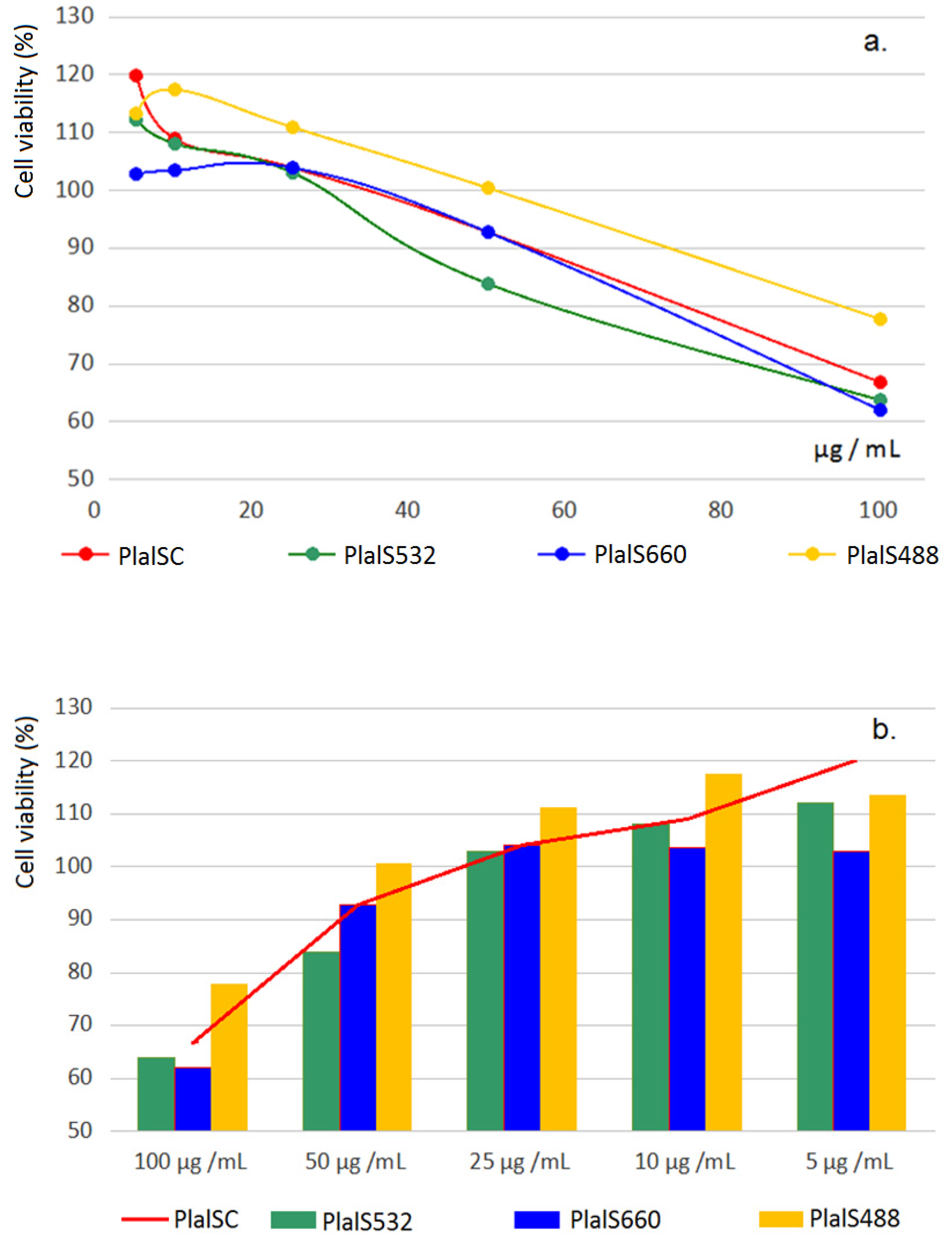
Effects on the Viability of Caco-2 Cells

Pharmacological studies in vitro were done using MTS test and human colon cancer cell line Caco-2 (ATCC-HTB-37). Basically, studies intended the comparison of the effects on Caco-2 cell viability of control standardized extract (PlalSC) at 2 h extraction time with the effects of the three types of laser-assisted (standardized) extracts (PlalS488, PlaSl532, and PlalS660), also at 2 h extraction time, portraying the three types of behavior noticed in antioxidant activity assay: laser-assisted extract at 488 nm (Plal488) proved the best chemical quantitative acquisition, at the same time increased antioxidant activity in chemiluminescence study; laser-assisted extract at 532 nm (Plal532) proved potential pro-oxidative effects on the vegetal compounds in the extract, and laser-assisted extract at 660 nm (Plal660) proved certain pro-oxidant effects.
The results are shown in Figure 6a,b and Table 4.
As general comments, Figure 6a shows that, by comparison with the control extract (PlalSC), laser-assisted extract at 488 nm (PlalS488) induced an increase in the viability of Caco-2 cells, laser-assisted extract at 532 nm (PlalS532) induced a decrease in the viability of Caco-2 cells, while laser-assisted extract at 660 nm (PlalS660) had no effects on the Caco-2 cells viability. Particularity, the analysis in the area of concentrations with real practical value (more clear in Figure 6b), meaning concentration range from 5 to 10 μg GAE/mL sample, indicates a similitude with the behavior observed in antioxidant activity study: an increase of the cell viability using Plal488 extract, identical effects on the cell viability using Plal532 extract, and the decrease of the cell viability using Plal660 extract respectively; inhibitory effects of laser extract at 660 nm also was consistent, along the entire concentration range in the study, 5–100 μg GAE/mL sample.
Another observation is that while at sample concentrations between 5 and 25 μg GAE/mL, both irradiated (Plal488, Plal532, Plal660) and control (PlalC) extracts at 2 h from Plantaginis folium have proved protective effects on Caco-2 cells (Caco-2 cell viability is over 100%); the protective interval being larger in the case of extract with the highest antioxidant activity (Plal488). Starting with the sample concentration over 25 μg GAE/mL, Plantaginis folium extracts have revealed cytotoxic effects, in fact the extracts proved an antiproliferative activity pharmacological testing being done on human colon cancer cell line (Caco-2). These effects are explained by luteolin and caffeic acid derivatives present in extracts, two effective anti-tumor natural compounds sub-classes.
The punctual IC50 values of the four tests extracts are presented in Table 4. There can be observed IC50 values over 100 μg GAE/mL sample, therefore a weak antitumor potency of the whole aqueous extracts from P. lanceolata species; new purification stages to increase the concentration of the active compounds in the extracts are imposed, with the purpose of using P. lanceolata extracts as an effective anti-tumor remedy.

4. Discussion

The present studies have evaluated potential effects of six laser radiation from the visible spectrum upon chemical composition and biological activity (cytotoxicity, in vitro) of aqueous extract from Plantago lanceolata, dried plant. The selection of particular laser wavelengths under study (488, 514, 532, 552, 660, and 785 nm) is based on several scientific reports, both on vegetal and animal test matter, from which the interest for identical, or close wavelengths was found.
Based on the observation that if the wavelength is short and the exposure time is long, burning (oxidation), or increase in thermal energy of the sample may be due [71,72], there were selected lower powers at higher frequencies (shorter wavelengths) and, inverse, higher powers at lower frequencies (longer wavelengths): 488 nm/40 mW, 514 nm/15 mW, 532 nm/20 mW, 552 nm/15 mW, 660 nm/75 mW, and at 785 nm/70 mW. Furthermore, laser experiments have proved that powers higher than 5 mW have an effect on the intensity of the aromatic bands [73]. This observation is clearly demonstrated in some laser studies at 532 nm, by Raman measurements of the lignin polymer in spruce trees, which proved punctual modifications at the main aromatic band at 1600/1600 cm−1 related to C=C extent of the phenol ring, finally reflected on coniferyl alcohol monomer [6]; studies also reported that wavelengths at 514 nm, 633 nm, and 785 nm have not produced subsequent changes in the Raman spectrum up to 220 mW and 200 s total irradiation time, even if the power used was sufficient to produce the activation of the coniferyl alcohol [6].
This way, in the case of living matter, in vitro studies on animal, murine lung epithelial line (MLE-12), cells irradiated with wavelengths of 488 and 514 nm (at power laser of mW) indicated clear harmful effects, while wavelength of 785 nm (115 mW for 40 min) indicated positive effects on the exposed cells [74]; similarly, laser irradiation at the wavelength 514.5 nm, but not at 660 nm (20 mW), indicated a clear spectral degradation (by Raman measurements) of the single human lymphocytes and chromosomes in the test [75].
In the specific case of living vegetal matter, the effects of the interaction of visible light (the wavelengths from 400–700 nm) with the green plants represents one of the essential conditions of life, and the final effects basically depend on its color (Figure 7) [76]. For example, radiation from 440–500 nm (blue light) was proved to play a major role in plant quality and plant development, by increasing chlorophyll absorption, therefore blue light has major beneficial effects on the leaf part; radiation from 510–610 nm (green light) results in photosynthesis stimulation, therefore the major benefits are on plant size, plant weight and synthesis of growth factors; radiation from 610–700 nm (yellow-red light) was related with stimulation of chlorophyll function, with germination and flower, and bud development; radiation from 700–800 nm (far red light) have revealed stimulatory effects on the plant growth by extension and general benefits [77].
Regarding the particular effects of light in visible on the secondary metabolites in plants, polyphenol compounds have been proved to be highly responsive to particular wavelengths in the spectrum. Thus, it was proved that blue and red light generally induces the accumulation of the bioactive compounds in medicinal plants [78]. Specifically, blue light increased the efficacy of accumulation of total phenols and total flavonoids in Prunella vulgaris calli [79]; blue and red light significantly increased coumarins accumulation in Sarcandra glabra seedlings [80], and callus culture of Eclipta alba L. [81], as well as dry mass, rosmarinic and caffeic acid contents of seedlings in cultured Ocimum basilicum [82], and biomass, anthocyanins, and 20-hydroxyecdisone in Pfaffia glomerata [83]. Phenylpropanoid compounds, specifically verbascoside compound in Verbena officinalis, also were stimulated by blue and red light [84], by regulating the transcript levels of phenylpropanoid biosynthetic genes [85,86]. Finally, studies have revealed that green light added to blue and red light led to positive effects on phenolics, and generally bioactive phytochemicals accumulation in flower microgreen species [87].
Summarizing, the expected effect at the interaction of radiation in visible range (1.6–3.1 eV) with lifeless vegetal material, dried matter, or aqueous extracts, is of energy transfer to those compounds that contain either pi bonds, or atoms with non-bonding orbitals (meaning lone pair of oxygen, nitrogen and halogen atoms). The highest energy transfer will occur for the compounds which have maximum absorption peaks identical with the wavelength used for irradiation. Function of the laser power used, the sensitive compounds, containing double, conjugated bonds, matching with the wavelength used in the study will suffer small to moderate electronic delocalization, leading to so called activatory effects (likely measured as an increase in the antioxidant activity). Depending on the ratio of frequency/power used, at higher energy there can be obtained massive delocalization of electrons in the sensitive compounds, leading to reactive (oxygen) species generation, likely measured as a decrease of the antioxidant activity.
Relative to the present study, antioxidant activity results suggest that excepting the wavelengths at 532 nm, and 660 nm, which indicated a clear pro-oxidant activity (evaluated as −14% to −54%, respectively), all other wavelengths under study have resulted in an increase of antioxidant activity of the aqueous extracts of P. lanceolata; activatory effects were 785 nm/70 mW > 488 nm/40 mW > 514 nm/15 mW > 552 nm/15 mW.
It must be reminded that all the experiments have been fulfilled in identical extraction conditions and technological parameters, therefore there are no other influences on the results apart from the laser wavelength and their powers.
Regarding the most probably explanation of certain pro-oxidant effects observed at the irradiation of aqueous extracts from leaves of P. lanceolata with laser at 660 nm, it must be noted that this wavelength is close to the maximum absorption peak of several phenolic acids frequently found in vegetable products. They are: cinnamic acid/666 nm; caffeic acid/670 nm; ferulic acid/672 nm; siringic acid/670 nm; gentisic acid/676 nm; gallic acid/684 nm and naringenin/676 nm. Caffeic acid and cinnamic acid are the main compounds in chlorogenic acid isomers and derivates such as phenylpropanoid compounds, the most abundant phenolic species in Plantago lanceolata-derived products.
Summarizing, the obtained results suggest that no valid conclusions can be drawn for both plant and animal raw materials, or even for plant materials only, as the response to irradiation with a certain wavelength depends on the specific content of the sensitive compounds, and the relationship between laser wavelength and power used.

5. Conclusions

The findings in the current paper are in the context of the high value of products from Plantaginaceae sp., polysaccharides, minerals and trace elements, polyphenols, and generally plantain-derived products, all of high interest for human health.
In this context, laser experiments at 488, 514, 532, 552, 660, and 785 nm on the aqueous extracts from leaves of Plantago lanceolata L. indicated interesting results and potential application in the current practice.
Concerning the total extractible compounds in irradiated samples, laser-assisted extracts at 488, 514, 532, and 552 nm have revealed up to 6.52% dry matter increases in the aqueous extracts; laser-assisted extracts at 488 and 532 nm also revealed minerals and trace element acquisitions (up to 6.49%), the most prominent results being upon Fe (+38.27%) and Cr (+31.70%) at 488 nm, and Zn (+465%) at 532 nm. Significant increases in total phenolics, that exceed the potential variation of the batches, were noticed in extracts at 552 nm (+12.5%), 660 nm (+15.50%), and 785 nm (+15.50).
Antioxidant activity measurements indicated laser-assisted extracts at 488, 514, 552, and 785 nm with more augmented antioxidant potency than the control extracts; the most prominent effects were noticed in the case of laser extract at 785 nm (27% increase in scavenger activity in comparison with control extract at 2 h, and 17% in comparison with control extract at 1 h), and at 488 nm (25% increase in scavenger activity in comparison with control extract at 2 h and 13% in comparison with control extract at 1 h); on the contrary, laser-assisted extract at 660 nm indicated the occurrence of radicalic species in the aqueous extracts from leaves of P. lanceolata, computed as (−)37% pro-oxidant effect; laser-assisted extract at 532 nm indicated pro-oxidative effects by comparison with control sample at 1 h, and no effects by comparison with control sample at 2 h, together suggesting some reversible oxidative modifications in the vegetal matter and aqueous extracts at 532 nm, as the literature data have suggested [6].
Regarding the effects on the viability of Caco-2 cells, laser-assisted extract at 488 nm generally induced the increase of the viability of Caco-2 cells, laser-assisted extract at 660 nm overall induced the decrease of the Caco-2 cell viability, while laser-assisted extract at 532 nm induced stimulatory effects bellow 25 μg GAE/mL sample, after that induced the decrease of the Caco-2 cell viability, likely by increasing the content of the pro-oxidant species in medium; also, it was observed that cytotoxic effects of the pro-oxidant compounds at 532 nm are stronger, but reversible, than that at 660 nm. The punctual analysis in the area of concentrations with practical value, from 5 to 10 μg GAE/mL sample respectively, indicated a similitude with the behavior observed in antioxidant activity assay; stimulatory activity of the extracts at 488 nm, no effects of extracts at 532 nm, and the decrease of the cell viability in the case of the extracts at 660 nm.
Together, the present study reveals laser radiation in visible range as a feasible method to improve the extraction efficacy of minerals, trace elements, and phenolics in aqueous extracts from Plantago lanceolata, and at the same time to augment their antioxidant capacity.
Further studies by spectroscopy methods (NIR, FTIR, Raman) are necessary, to explain the effects of laser radiation at 660 nm, proved with clear pro-oxidant effects.

6. Patents

Patent application no. A/00790 of 16.12.2021; Process for obtaining active plant products enriched in minerals and micro-elements by laser-assisted extraction; Authors: Pirvu Lucia Camelia 1, Niță Sultana 1, Bazdoaca Cristina 1, Rusu Nicoleta 1, Neagu Georgeta 1, Enache Alin 2, Udrea Mircea 2, Udrea Radu 2; Affiliation: 1 ICCF Bucharest, Romania, 2 Apel Laser SRL Bucharest, Romania; POC-G-2015, SMIS 105542, Contract D no. 35/08.11.2019.

Author Contributions

Conceptualization, L.C.P., A.E., S.N. and M.U.; methodology, L.C.P., A.E. and M.U.; formal analysis, L.C.P., N.R., C.B. (Cristina Bazdoaca); investigation, L.C.P., C.B. (Corina Bubueanu), and G.N.; resources, L.C.P. and A.E.; writing—original draft preparation, L.C.P.; writing—review and editing, L.C.P. and R.U.; visualization, L.C.P.; supervision, M.U. and S.N.; project administration, L.C.P. and A.E.; funding acquisition, L.C.P. All authors have read and agreed to the published version of the manuscript.

Funding

This paper has been financed through the POC-A1-A1.2.3-G-2015, SMIS 105542, Contract D no. 35/08.11. 2019, ICCF Bucharest and APEL LASER SRL partnership.

Institutional Review Board Statement

Not applicable.

Informed Consent Statement

Not applicable.

Data Availability Statement

Not applicable.

Conflicts of Interest

The authors declare no conflict of interest.

References

  1. Zhang, Q.W.; Lin, L.G.; Ye, W.C. Techniques for extraction and isolation of natural products: A comprehensive review. Chin. Med. 2018, 13, 20. [Google Scholar] [CrossRef] [PubMed] [Green Version]
  2. Altemimi, A.; Lakhssassi, N.; Baharlouei, A.; Watson, D.G.; Lighfoot, D.A. Phytochemicals: Extraction, Isolation, and Identification of Bioactive Compounds from Plant Extracts. Plants 2017, 6, 42. [Google Scholar] [CrossRef] [PubMed]
  3. Rodriguez de Luna, S.L.; Ramirez-Garza, R.E.; Serna Saldivar, S.O. Environmentally Friendly Methods for Flavonoid Extraction from Plant Material: Impact of Their Operating Conditions on Yield and Antioxidant Properties. Sci. World J. 2020, 2020, 6792069. [Google Scholar] [CrossRef] [PubMed]
  4. Galanakis, C. Food Waste Recovery: Processing Technologies, Industrial Techniques, and Applications, 2nd ed.; Elsevier: Vienna, Austria, 2020. [Google Scholar]
  5. Panchev, I.; Kirtchev, N.A.; Dimitrov, D.D. Possibilities for application of laser ablation in food technologies. Innov. Food Sci. Emerg. Technol. 2011, 12, 369–374. [Google Scholar] [CrossRef]
  6. Batirtze, P.M.; Bock, P.; Schroffenegger, M.; Toca-Herrera, J.L.; Gierlinger, N. Following laser induced changes of plant phenylpropanoids by Raman microscopy. Sci. Rep. 2018, 8, 1804. [Google Scholar]
  7. Monties, B. Plant-Cell Walls as Fibrous Lignocellulosic Composites—Relations with Lignin Structure and Function. Anim. Feed Sci. Technol. 1991, 32, 159–175. [Google Scholar] [CrossRef]
  8. Lora, J.H.; Glasser, W.G. Recent industrial applications of lignin: A sustainable alternative to nonrenewable materials. J. Polym. Environ. 2002, 10, 39–48. [Google Scholar] [CrossRef]
  9. Holwerda, E.K.; Worthen, R.S.; Kothari, N.; Lasky, R.C.; Davison, B.H.; Fu, C.; Wang, Z.-Y.; Dixon, R.A.; Biswal, A.K.; Mohnen, D.; et al. Multiple levers for overcoming the recalcitrance of lignocellulosic biomass. Biotechnol. Biofuels 2019, 12, 15. [Google Scholar] [CrossRef]
  10. Zoghlami, A.; Paes, G. Lignocellulosic Biomass: Understanding Recalcitrance and Predicting Hydrolysis. Front. Chem. 2019, 7, 874. [Google Scholar] [CrossRef] [Green Version]
  11. Chen, F.; Dixon, R.A. Lignin modification improves fermentable sugar yields for biofuel production. Nat. Biotechnol. 2007, 25, 759–761. [Google Scholar] [CrossRef]
  12. Zakzeski, J.; Bruijnincx, P.C.; Jongerius, A.L.; Weckhuysen, B.M. The catalytic valorization of lignin for the production of renewable chemicals. Chem. Rev. 2010, 110, 3552–3599. [Google Scholar] [CrossRef] [PubMed]
  13. Ragauskas, A.J.; Beckham, G.T.; Biddy, M.J.; Chandra, R.; Chen, F.; Davis, M.F.; Davison, B.H.; Dixon, R.A.; Gilna, P.; Keller, M.; et al. Lignin Valorization: Improving Lignin Processing in the Biorefinery. Science 2014, 344, 1246843. [Google Scholar] [CrossRef] [PubMed]
  14. Rashid, G.M.M.; Bugg, T.D.H. Enhanced biocatalytic degradation of lignin using combinations of lignin-degrading enzymes and accessory enzymes. Catal. Sci. Technol. 2021, 11, 10. [Google Scholar] [CrossRef]
  15. Ji, X.; Hou, C.; Guo, X. Physicochemical properties, structures, bioactivities and future prospective for polysaccharides from Plantago L. (Plantaginaceae): A review. Int. J. Biol. Macromol. 2019, 135, 637–646. [Google Scholar] [CrossRef] [PubMed]
  16. Nazario, F.E.A.; Sanches-Silva, A.; Ribeiro-Santos, R.; Ramos de Melo, N. Psyllium (Plantago ovata Forsk): From evidence of health benefits to its food application. Trends Food Sci. Technol. 2020, 96, 166–175. [Google Scholar]
  17. Fierascu, R.C.; Fierascu, I.; Ortan, A.; Paunescu, A. Plantago media L.-Explored and Potential Applications of an Underutilized Plant. Plants 2021, 10, 265. [Google Scholar] [CrossRef]
  18. Available online: https://www.ema.europa.eu/en/documents/herbal-report/final-assessment-report-plantago-lanceolata-l-folium_en.pdf (accessed on 25 January 2022).
  19. Schmidgall, J.; Schnetz, E.; Hensel, A. Evidence for bioadhesive effects of polysaccharides and polysaccharide-containing herbs in an ex vivo bioadhesion assay on buccal membranes. Planta Med. 2000, 66, 48–53. [Google Scholar] [CrossRef]
  20. McRorie, J.W. Evidence-based approach to fiber supplements and clinically meaningful health benefits, Part 1. Nutrition. Today 2015, 50, 82–89. [Google Scholar] [CrossRef] [Green Version]
  21. McRorie, J.W.; McKeown, N.M. Understanding the physics of functional fibers in the gastrointestinal tract: An evidence-based approach to resolving enduring misconceptions about insoluble and soluble fiber. J. Acad. Nutr. Diet. 2017, 117, 251–264. [Google Scholar] [CrossRef] [Green Version]
  22. Galisteo, M.; Sanchez, M.; Vera, R.; Gonzalez, M.; Anguera, A.; Duarte, J.; Zarzuelo, A. A diet supplemented with husks of Plantago ovata reduces the development of endothelial dysfunction, hypertension, and obesity by affecting adiponectin and TNF-alpha in obese Zucker rats. J. Nutr. 2005, 10, 2399–2404. [Google Scholar] [CrossRef] [Green Version]
  23. Ghiasian, M.; Niroomandi, Z.; Dastan, D.; Poorolajal, J.; Zare, F.; Ataei, S. Clinical and phytochemical studies of Plantago major in pressure ulcer treatment: A randomized controlled trial. Complement. Ther. Clin. Pract. 2021, 43, 101325. [Google Scholar] [CrossRef]
  24. Kartini, K.; Wati, N.; Gustav, R.; Wahyuni, R.; Angada, Y.F.; Hidayani, R.; Raharjo, A.; Islamie, R.; Dwi Putra, S.E. Wound healing effects of Plantago major extract and its chemical compounds in hyperglycemic rats. Food Biosci. 2021, 41, 100937. [Google Scholar] [CrossRef]
  25. Haddadian, K.; Haddadian, K.; Zahmatkash, M. A review of Plantago plant. Indian J. Tradit. Know 2014, 13, 681–685. [Google Scholar]
  26. Adom, M.B.; Taher, M.; Mutalabisin, M.F.; Amri, M.S.; Kudos, M.B.A.; Sulaiman, M.W.A.W.; Sengupta, P.; Susanti, D. Chemical constituents and medical benefits of Plantago major. Biomed. Pharmacother. 2017, 96, 348–360. [Google Scholar] [CrossRef] [PubMed]
  27. Cicero, A.G.; Derosa, G.; Bove, M.; Imola, F.; Borghi, C.; Gaddi, A. Psyllium improves dyslipidaemia, hyperglycaemia and hypertension, while guar gum reduces body weight more rapidly in patients affected by metabolic syndrome following an AHA Step 2 diet. Mediterr. J. Nutr. Metab. 2010, 3, 47–54. [Google Scholar] [CrossRef]
  28. Anderson, J.W.; Davidson, M.H.; Blonde, L.; Brown, W.V.; Howard, W.J.; Ginsberg, H.; Allgood, L.D.; Weingand, K.W. Long-term cholesterol-lowering effects of psyllium as an adjunct to diet therapy in the treatment of hypercholesterolemia. Am. J. Clin. Nutr. 2000, 71, 1433–1438. [Google Scholar] [CrossRef]
  29. Story, J.A.; Donkin, S.S.; Furumoto, E.J.; Buhman, K.K. Dietary psyllium increases fecal bile acid excretion, total steroid excretion and bile acid biosynthesis in rats. J. Nutr. 1998, 128, 1199–1203. [Google Scholar] [CrossRef]
  30. Turley, S.D.; Daggy, B.P.; Dietschy, J.M. Psyllium augments the cholesterol-lowering action of cholestyramine in hamsters by enhancing sterol loss from the liver. Gastroenterology 1994, 107, 444–452. [Google Scholar] [CrossRef]
  31. Brum, J.M.; Gibb, R.D.; Peters, J.C.; Matte, R.D. Satiety effects of psyllium in healthy volunteers. Appetite 2016, 105, 27–36. [Google Scholar] [CrossRef] [Green Version]
  32. Wong, C.; Harris, P.J.; Ferguson, L.R. Potential Benefits of Dietary Fibre Intervention in Inflammatory Bowel Disease. Int. J. Mol. Sci. 2016, 17, 919. [Google Scholar] [CrossRef] [Green Version]
  33. Ogata, M.; Ogita, T.; Tari, H.; Arakawa, T.; Suzuki, T. Supplemental psyllium fibre regulates the intestinal barrier and inflammation in normal and colitic mice. Br. J. Nutr. 2017, 118, 661–672. [Google Scholar] [CrossRef] [PubMed] [Green Version]
  34. Available online: https://www.cambridge.org/core/journals/british-journal-of-nutrition/article/supplemental-psyllium-fibre-regulates-the-intestinal-barrier-and-inflammation-in-normal-and-colitic-mice/B03DA9C101028A6167873660B5555D3C (accessed on 25 January 2022).
  35. Amiri, M.S.; Mohammadzadeh, V. Plant-Based Gums and Mucilages Applications in Pharmacology and Nanomedicine: A Review. Molecules 2021, 26, 1770. [Google Scholar] [CrossRef] [PubMed]
  36. Bashir, S.; Erum, A.; Saghir, S.; Tulain, U.R.; Rashid, A. Physicochemical characterization and evaluation of suspending properties of arabinoxylan from Ispaghula (Plantago ovata) husk. Pak. J. Pharm. Sci. 2014, 27, 1761–1766. [Google Scholar] [PubMed]
  37. Abbas, S.; Sherazi, M.; Khan, A.; Alyami, H.S.; Latif, M.; Qureshi, Z.U.R.; Majeedullah; Hassan Bin Asad, M.H. Investigation of Plantago ovata Husk as Pharmaceutical Excipient for Solid Dosage Form (Orodispersible Tablets). Biomed. Res. Int. 2021, 14, 5538075. [Google Scholar] [CrossRef]
  38. Zubair, M.; Nybom, H.; Lindholm, C.; Rumpunen, K. Major polyphenols in aerial organs of greater plantain (Plantago major L.), and effects of drying temperature on polyphenol contents in the leaves. Sci. Hortic. 2011, 128, 523–529. [Google Scholar] [CrossRef]
  39. Beara, I.N.; Lesjak, M.M.; Jovin, E.D.; Balog, K.J.; Anackov, G.T.; Orcic, D.Z.; Mimica-Dukic, N.M. Plantain (Plantago L.) species as novel sources of flavonoid antioxidants. J. Agric. Food Chem. 2009, 57, 9268–9273. [Google Scholar] [CrossRef]
  40. Samuelsen, A.B. The traditional uses, chemical constituents and biological activities of Plantago major. J. Ethnopharmacol. 2000, 71, 1–21. [Google Scholar] [CrossRef]
  41. Nazaizadeh, A.; Mikaili, P.; Moloudizargari, M.; Aghajanshakeri, S.; Javaherypour, S. Therapeutic uses and pharmacological properties of Plantago major L. and its active constituents. J. Basic Appl. Sci. Res. 2013, 3, 212–221. [Google Scholar]
  42. Carrillo-Ocampo, D.; Bazaldúa-Gómez, S.; Bonilla-Barbo, J.R.; Aburto-Amar, R.; Rodríguez-López, V. Anti-Inflammatory Activity of Iridoids and Verbascoside Isolated from Castilleja tenuiflora. Molecules 2013, 18, 12109–12118. [Google Scholar] [CrossRef]
  43. González, L.; Pereira-Crespo, S.; Dagnac, T.; Resch-Zafra, C.; Fernández-Lorenzo, B.; Botana, A.; Veiga, M.; Aguión, A.; Flores-Calvete, G. Fatty acid, carotenoid and vitamin-E contents of Plantago lanceolata at different maturity stages. In Conference Paper: The Multiple Roles of Grassland in the European Bioeconomy. Proceedings of the 26th General Meeting of the European Grassland Federation, Trondheim, Norway, 4–8 September 2016; NIBIO: Ås, Norway, 2016; pp. 302–304. [Google Scholar]
  44. Dzomeku, B.M.; Wald, J.P.; Wünsche, J.N.; Nohr, D.; Biesalski, H.K. Climate Change Enhanced Carotenoid Pro-Vitamin A Levels of Selected Plantain Cultivars. Plants 2020, 9, 541. [Google Scholar] [CrossRef]
  45. Hussan, F.; Mansor, A.S.; Hassan, S.N.; Effendy Kamaruddin, N.T.T.; Budin, S.B.; Othman, F. Anti-Inflammatory Property of Plantago major Leaf Extract Reduces the Inflammatory Reaction in Experimental Acetaminophen-Induced Liver Injury. Evid. Based Complement. Alternat Med. 2015, 347861. [Google Scholar] [CrossRef] [Green Version]
  46. Ahmed, S.; Imran, M.Z.; Hasan, M. Choleretic and cholagogic effects of anti- cholelithiatic plants. J. Pharmacogn. Phytochem. 2021, 10, 1–7. [Google Scholar] [CrossRef]
  47. Jazayeri, S.F.; Ghods, R.; Dabaghian, F.H.; Shojaii, A.; Al-Hadi Moravej, S.A.; Khadem, E.; Seyedian, S. The Efficacy of Plantago major Seed on Liver Enzymes in Nonalcoholic Fatty Liver Disease: A Randomized Double-Blind Clinical Trial. Evid. Based Complement Alternat Med. 2021, 2021, 6693887. [Google Scholar] [CrossRef] [PubMed]
  48. Israel Cardoso, F.C.; Peruzzo Apolinário, P.; Cunha Breder, J.S.; Paranhos, T.; Oliveira, H.C.; Dini Polidoro, A.; Souza Oliveira Kumakura, A.R.; Melo Lima, M.H. A protocol for systematic review of Plantago major L. effectiveness in accelerating wound-healing in animal models. Syst. Rev. 2019, 8, 337. [Google Scholar] [CrossRef] [Green Version]
  49. Najafian, Y.; Hamedi, S.S.; Farshchi, M.K.; Feyzabadi, Z. Plantago major in Traditional Persian Medicine and modern phytotherapy: A narrative review. Electron. Physician 2018, 10, 6390–6399. [Google Scholar] [CrossRef] [Green Version]
  50. Fleer, H.; Verspohl, E.J. Antispasmodic activity of an extract from Plantago lanceolata L. and some isolated compounds. Phytomedicine 2007, 14, 409–415. [Google Scholar] [CrossRef]
  51. Sadraei, H.; Ghanadian, M.; Asghari, G.; Sekhavati, N. Antispasmodic activity of apigenin and luteolin, two components of Dracocephalum kotschyi extract, on rat ileum contractions. J. Herbmed Pharmacol. 2018, 7, 100–105. [Google Scholar] [CrossRef] [Green Version]
  52. Fabrizio Ferrazzano, G.; Cantile, T.; Roberto, L.; Ingenito, A.; Catania, M.R.; Roscetto, E.; Palumbo, G.; Zarrelli, A.; Pollio, A. Determination of the In Vitro and In Vivo Antimicrobial Activity on Salivary Streptococci and Lactobacilli and Chemical Characterisation of the Phenolic Content of a Plantago lanceolata Infusion. Biomed. Res. Int. 2015, 2015, 286817. [Google Scholar] [CrossRef] [Green Version]
  53. Reddy, P.R.T.; Vandana, K.V.; Prakash, S. Antibacterial and anti-inflammatory properties of Plantago ovata Forssk. leaves and seeds against periodontal pathogens: An in vitro study. Ayu 2018, 39, 226–229. [Google Scholar] [CrossRef]
  54. Sharifa, A.A.; Neoh, Y.L.; Iswadi, M.I.; Khairul, O.; Abdul Halim, M.; Jamaludin, M.; MohamedAzman, A.B.; Hing, H.L. Effects of methanol, ethanol and aqueous extract of Plantago major on gram positive bacteria, gram negative bacteria and yeast. Ann. Microsc. 2008, 8, 42–44. [Google Scholar]
  55. Tsai, T.H.; Chien, Y.C.; Lee, C.W.; Tsai, P.J. In vitro antimicrobial activities against cariogenic streptococci and their antioxidant capacities: A comparative study of green tea versus different herbs. Food Chem. 2008, 110, 859–864. [Google Scholar] [CrossRef] [PubMed]
  56. Aziz, S.A.; See, T.L.; Khuay, L.Y.; Osman, K.; Bakar, M.A.A. In vitro effects of Plantago major on urolithiasis. Malays. J. Med. Sci. 2005, 12, 22–26. [Google Scholar] [PubMed]
  57. Committee on Herbal Medicinal Products (HMPC) Community Herbal Monograph on Plantago lanceolata L., Folium. 2011. 28 January 2014 EMA/HMPC/437858/2010, Corrigendum 28 January 2014. Available online: https://www.ema.europa.eu/en/documents/herbal-monograph/final-community-herbal-monograph-plantago-lanceolata-l-folium_en.pdf (accessed on 25 January 2022).
  58. Wagner, H.; Bladt, S. Plant. Drug Analysis. A Thin Layer Chromatography Atlas, 2nd ed.; Springer: Berlin/Heidelberg, Germany, 1996. [Google Scholar]
  59. Reikh, E.; Schibli, A. HPTLC for the Analysis of Medicinal Plants; Thieme: New York, NY, USA; Stuttgart, Germany, 2008. [Google Scholar]
  60. Collective Work. Romanian Pharmacopoeia, Xth ed.; Medicala: Bucharest, Romania, 1993. [Google Scholar]
  61. Rusu, N.; Meghea, A. Bioacumulation of toxic metals in fish oils capsules. Ecosystems 2015, 77, 131–140. [Google Scholar]
  62. Iftimie, N.; Giurginca, M.; Meghea, A. The estimation of the antioxidant effect of plants extracts by chemiluminescence. Rev. Chem. 2003, 55, 1025–1028. [Google Scholar]
  63. Protocols & Applications Guide. Available online: www.promega.com (accessed on 12 August 2021).
  64. Ronsted, N.; Franzyk, H.; Molgaard, P.; Jaroszevski, W.; Jensen, S.R. Chemotaxonomy and evolution of Plantago L. Plant. Syst. Evol. 2003, 242, 63–82. [Google Scholar] [CrossRef]
  65. Li, Y.; Gan, L.; Li, G.Q.; Deng, L.; Zhang, X. Pharmacokinetics of plantamajoside and acteoside from Plantago asiatica in rats by liquid chromatography-mass spectrometry. J. Pharm. Biomed. Anal. 2014, 89, 251–256. [Google Scholar] [CrossRef]
  66. Pol, M.; Schmidtke, K.; Lewandowska, S. Plantago lanceolata—An overview of its agronomically and healing valuable features. Open Agric. 2021, 6, 479–488. [Google Scholar] [CrossRef]
  67. Petrova, S.; Velcheva, I.; Yurukova, L.D.; Berova, M. Plantago lanceolata L. as a Biomonitor of Trace Elements in an Urban Area. Bulg. J. Agric. Sci. 2014, 20, 325–329. [Google Scholar]
  68. Drava, G.; Cornara, L.; Giordani, P.; Minganti, V. Trace elements in Plantago lanceolata L., a plant used for herbal and food preparations: New data and literature review. Environ. Sci. Pollut. Res. Int. 2019, 26, 2305–2313. [Google Scholar] [CrossRef]
  69. Skrynetska, I.; Karcz, J.; Barczyk, G.; Kandziora-Ciupa, M.; Ciepa, R.; Socha, N. Using Plantago major and Plantago lanceolata in environmental pollution research in an urban area of Southern Poland. Environ. Sci. Pollut. Res. Int. 2019, 26, 23359–23371. [Google Scholar] [CrossRef] [Green Version]
  70. Sanna, F.; Piluzza, G.; Campesi, G.; Molinu, M.G.; Re, G.A.; Sulas, L. Antioxidant Contents in a Mediterranean Population of Plantago lanceolata L. Exploited for Quarry Reclamation Interventions. Plants 2022, 11, 791. [Google Scholar] [CrossRef]
  71. Jirasek, A.; Georg Schulze, H.; Hughesman, C.H.; Creagh, A.L.; Haynes, C.A.; Blades, M.W.; Turner, R.F.B. Discrimination between UV radiation-induced and thermally induced spectral changes in AT-paired DNA oligomers using UV resonance Raman spectroscopy. J. Raman Spectrosc. 2006, 37, 1368–1380. [Google Scholar] [CrossRef]
  72. La Plant, F. Lasers, spectrographs, and detectors. In Emerging Raman Applications and Techniques in Biomedical and Pharmaceutical Fields; Springer: Berlin/Heidelberg, Germany, 2010. [Google Scholar] [CrossRef]
  73. Agarwal, U.P.; Atalla, R.H. In-situ Raman microprobe studies of plant cell walls: Macromolecular organization and compositional variability in the secondary wall of Picea mariana (Mill.) B.S.P. Planta 1986, 169, 325–332. [Google Scholar] [CrossRef] [PubMed]
  74. Notingher, I.; Verrier, S.; Romanska, H.; Bishop, A.E.; Polak, J.M.; Hench, L.L. In situ characterisation of living cells by Raman spectroscopy. Spectroscopy 2002, 16, 43–5143. [Google Scholar] [CrossRef] [Green Version]
  75. Segers-Nolten, G.M.J.; Otto, C.; de Mul, F.F.M.; Greve, J. Laser irradiation and Raman spectroscopy of single living cells and chromosomes: Sample degradation occurs with 514.5 nm but not with 660 nm laser light. Exp. Cell Res. 1991, 192, 361–367. [Google Scholar]
  76. Britannica. The Editors of Encyclopaedia. “Light Summary”. Encyclopedia Britannica. Available online: https://www.britannica.com/science/light (accessed on 18 May 2022).
  77. Ligh Science Technologies. The Visible Wavelength Range and Its Impact on Plant Growth. Available online: https://lightsciencetech.com/visible-wavelength-range-plant-growth/ (accessed on 18 May 2022).
  78. Zhang, S.; Zhang, L.; Zou, H.; Qui, L.; Zheng, Y.; Yang, D.; Wang, Y. Effects of Light on Secondary Metabolite Biosynthesis in Medicinal Plants. Front. Plant. Sci. 2021, 11, 497. [Google Scholar] [CrossRef]
  79. Fazal, H.; Abbasi, B.H.; Ahmad, N.; Ali, S.S.; Akbar, F.; Kanwal, F. Correlation of different spectral lights with biomass accumulation and production of antioxidant secondary metabolites in callus cultures of medicinally important Prunella vulgaris L. J. Photochem. Photobiol. B 2016, 159, 1–7. [Google Scholar] [CrossRef]
  80. Xie, D.J.; Tarin, M.W.K.; Chen, L.Y.; Ren, K.; Yang, D.M.; Zhou, C.C.; Wan, J.; He, T.; Rong, J.; Zheng, Y. Consequences of LED lights on root morphological traits and compounds accumulation in Sarcandra glabra seedlings. Int. J. Mol. Sci. 2021, 22, 7179. [Google Scholar] [CrossRef]
  81. Khurshid, R.; Ullah, M.A.; Tungmunnithum, D.; Drouet, S.; Shah, M.; Zaeem, A.; Hameed, S.; Hano, C.; Haider Abbasi, B. Lights triggered differential accumulation of antioxidant and antidiabetic secondary metabolites in callus culture of Eclipta alba L. PLoS ONE 2020, 15, e0233963. [Google Scholar] [CrossRef]
  82. Lobiuc, A.; Vasilache, V.; Pintilie, O.; Stoleru, T.; Burducea, M.; Oroian, M.; Zamfirache, M.M. Blue and red LED illumination improves growth and bioactive compounds contents in acyanic and cyanic Ocimum basilicum L. microgreens. Molecules 2017, 22, 2111. [Google Scholar] [CrossRef] [Green Version]
  83. Silva, T.D.; Batista, D.S.; Fortini, E.A.; de Castro, K.M.; Sousa Felipe, S.H.; Fernandes, A.M.; JesusSousa, K.C.; da Silva, J.V.S.; Freitas Correia, L.N.; Monteiro Farias, L.; et al. Blue and red light affects morphogenesis and 20-hydroxyecdisone content of in vitro Pfaffia glomerata accessions. J. Photochem. Photobiol. B 2020, 203, 111761. [Google Scholar] [CrossRef] [PubMed]
  84. Kubica, P.; Szopa, A.; Prokopiuk, B.; Komsta, L.; Pawlowska, B.; Ekiert, H. The influence of light quality on the production of bioactive metabolites-verbascoside, isoverbascoside and phenolic acids and the content of photosynthetic pigments in biomass of Verbena officinalis L. cultured in vitro. J. Photochem. Photobiol. B 2020, 203, 111768. [Google Scholar] [CrossRef] [PubMed]
  85. Hao, X.Y.; Li, L.T.; Hu, Y.R.; Zhou, C.; Wang, X.C.; Wang, L.; Zeng, J.; Yang, Y. Transcriptomic analysis of the effects of three different light treatments on the biosynthesis of characteristic compounds in the tea plant by RNA-Seq. Tree Genet. Genomes 2016, 12, 118. [Google Scholar] [CrossRef]
  86. Alrifai, O.; Hao, X.M.; Marcone, M.F.; Tsao, R. Current review of the modulatory effects of LED lights on photosynthesis of secondary metabolites and future perspectives of microgreen vegetables. J. Agric. Food Chem. 2019, 67, 6075–6090. [Google Scholar] [CrossRef] [PubMed]
  87. Orlando, M.; Trivellini, A.; Incrocci, L.; Ferrante, A.; Mensuali, A. The Inclusion of Green Light in a Red and Blue Light Background Impact the Growth and Functional Quality of Vegetable and Flower Microgreen Species. Horticulturae 2022, 8, 217. [Google Scholar] [CrossRef]
Figure 1. Chemical structure of lignin (a), and the main phenolic monomers (b).
Applsci 12 05517 g001
Figure 2. Laser-assisted extraction installation in the study. (1) Bio-reactor; (2) ZnSe observation and collecting sample window; (3) laser kit (488, 514, 532, 552, 660, and 785 nm); (4) laser controller (gathering the six laser beam in the study); (5) telescope (placed in front of the second quartz irradiation window); (6) cooling system (using current water); (7) agitation system (vertical stirrer); (8) heating system (tape heating); (9) heating controller.
Applsci 12 05517 g002
Figure 3. Polyphenols profile in the aqueous extracts from Plantago lanceolata L. Folium. Track T1, laser extract at 785 nm (PlalS785); Track T2, laser extract at 660 nm (PlalS660); Track T3, laser extract at 552 nm (PlalS552); Track T4, laser extract at 532 nm (PlalS532); Track T5, laser extract at 514 nm laser (PlalS514); Track T6, laser extract at 488 nm (PlalS488); Track T7, control, non-irradiated extract (PlalSC); Track T8, reference compounds series (ref.): rutin (Rf~0.42), chlorogenic acid (Rf~0.49), hyperoside (Rf~0.64), cynaroside (Rf~0.69), vitexin (Rf~0.75), caffeic acid (Rf~0.93), and umbeliferone Rf~(0.98).
Applsci 12 05517 g003
Figure 4. (a) Plantamajoside; (b) Acteoside.
Applsci 12 05517 g004
Figure 5. (af) Antioxidant activity of the test irradiated extracts vs. control extracts at 1 h and 2 h extraction time, in comparison with control negative sample: (a) Series at 488 nm; (b) Series at 514 nm; (c) Series at 532 nm; (d). Series at 552 nm; (e) Series at 660 nm; (f) Series at 785 nm (mean values, n = 3).
Applsci 12 05517 g005aApplsci 12 05517 g005b
Figure 6. (a,b) Caco-2 cell viability at 24 h., non-irradiated control extracts (PlalSC) versus laser-assisted extracts at 488 nm (PlalS488), 532 nm (PlaSl532), and 660 nm (PlaSl660).
Applsci 12 05517 g006
Figure 7. Light, the visible spectrum.
Applsci 12 05517 g007
Table 1. Total extractible compounds and total phenolic compounds (μg/g plant, d.w., n = 3, mean value) in the aqueous extracts, non-irradiated extracts (PlalC) versus irradiated extracts (Plal488, Plal514, Plal532, Plal552, Plal660, and Plal785).
Test Extract/(μg/g Plant)Control Extract (PlalC)Laser Extract Plal488PlalC/Plal488 (%)Laser Extract Plal514PlalC/Plal514 (%)Laser Extract Plal532PlalC/Plal532 (%)
Total extractible/dry matter331,204334,564+1.01352,802+6.52333,516+0.69
Total phenolics/[GAE]62015986−3.475240−15.506220+0.31
Test Extract/(μg/g Plant)Control Extract (PlalC)Laser Extract Plal552PlalC/Plal552 (%)Laser Extract Plal660PlalC/Plal660 (%)Laser Extract Plal785PlalC/Plal785 (%)
Total extractible/dry matter331,204352,793+6.52331,195−0.01324,110−2.14
Total phenolics/[GAE]62016948+12.057162+15.507162+15.50
Table 2. Minerals and micro-elements content (μg/g plant, d.w., n = 3, mean value) in the aqueous extracts; non-irradiated extracts (PlalC) versus irradiated extracts (Plal488, Plal514, Plal532, Plal552, Plal660, and Plal785).
Ref./(µg/g)Control Extract (PlalC)Laser Extract (Plal488)PlalC/Plal488 (%)Laser Extract (Plal514)PlalC/Plal514 (%)Laser Extract (Plal532)PlalC/Plal532 (%)
K111,305117,027+5.1476,032−31.69117,912+5.93
Ca31,98033,200+3.8121,537−32.6533,104+3.51
Mg67337122+5.775760−14.457412+9.15
Na18872192+16.161998+5.882032+7.68
Total minerals151,905159,541+5.03105,327−30.66160,460+5.63
P13,88215,358+10.6310,062−27.5215,498+11.64
Fe520.0719.0+38.27567.5+9.13592.4+13.92
Mn240.5257.1+6.90170.0−29.31267.9+11.39
Zn118.3131.9+11.4955.56−53.03669.3+465.7
Cu22.2625.08+12.6717.63−20.7924.02+7.90
Cr7.549.93+31.706.10−19.108.46+12.20
Pb3.363.360.003.03−9.823.45+2.68
As0.3060.291−4.900.282−7.840.327+6.86
Cd0.2940.267−9.180.297+1.020.309+5.1
Total micro-elements14,79516,505+11.5610,882−26.4517,064+15.34
Total elements166,700176,046+5.60116,209−30.29177,524+6.49
Ref./(µg/g)Control Extract (PlalC)Laser Extract Plal552PlalC/Plal552 (%)Laser Extract Plal660PlalC/Plal660 (%)Laser Extract Plal785PlalC/Plal785 (%)
K111,30598,692−11.3396,871−12.9751,319−53.89
Ca31,98025,697−19.6527,806−13.0515,556−51.36
Mg67335915−12.155875−12.743672−45.46
Na10871923+76.911891+73.961174+8.00
Total minerals151,905132,227−12.95132,444−12.8171,720−52.79
P13,88212,452−10.3013129−5.427001−49.57
Fe520.0598.2+15.04615.2+18.31381.6−26.61
Mn240.5182.3−24.20190.1−20.95121.7−49.40
Zn118.364.77−45.2473.70−37.70111.9−5.41
Cu22.2617.68−20.5723.80+6.9212.96−41.78
Cr7.547.58+0.5330.41+307.37.83+3.84
Pb3.363.38+0.593.37+0.293.25−3.27
As0.3060.348+13.720.372+21.570.303−0.98
Cd0.2940.360+22.4540.327+11.220.300+2.04
Total micro-elements14,79513,327−9.9214,066−4.937641−48.35
Total elements166,700145,554−12.68146,510−12.1179,361−52.39
Table 3. Antioxidant activity of the extracts from leaves of P. lanceolata (±SD, n = 3).
Antioxidant Activity (AA %)/
Laser Radiation (nm)/Laser Power (mW)
488 nm
(40 mW)
514 nm
(15 mW)
532 nm
(20 mW)
552 nm
(15 mW)
660 nm
(75 mW)
785 nm
(70 mW)
SD
(n = 3)
Control positive sample at 1 h (* PlalCx) in comparison with Control negative sample (PlalC)+14%+13%+13%+13%+11%+11%±2%
Test irradiated sample (** Plalx) in comparison
with Control negative sample (PlalC)
+25%+20%0.0%+18%−37%+27%±1%
Test irradiated sample (** Plalx) in comparison
with Control positive sample at 1 h (*PlalCx)
+13%+8%−14%+6%−54%+17%±2%
Where: * PlalC488, PlalC514, PlalC532, PlalC552, PlalC660, PlalC785; ** Plal488, Plal514, Plal532, Plal552, Plal660, Plal785.
Table 4. IC50 comparison.
Test SampleIC50 (µg GAE/mL Sample)
PlalC132.09 ± 1.94
Plal532122.59 ± 1.76
Plal660134.76 ± 1.61
Plal488170.94 ± 1.97
Publisher’s Note: MDPI stays neutral with regard to jurisdictional claims in published maps and institutional affiliations.

