Review

Comprehensive Review on Development of Early Diagnostics on Oral Cancer with a Special Focus on Biomarkers

by
Vidhya Rekha Umapathy
1,*,
Prabhu Manickam Natarajan
2 and
Bhuminathan Swamikannu
3
1
Department of Public Health Dentistry, Sree Balaji Dental College and Hospital, Pallikaranai, Chennai 600 100, India
2
Department of Periodontics, College of Dentistry, Ajman University, Ajman P.O. Box 346, United Arab Emirates
3
Department of Prosthodontics, Sree Balaji Dental College and Hospital, Pallikaranai, Chennai 600 100, India
*
Author to whom correspondence should be addressed.
Appl. Sci. 2022, 12(10), 4926; https://doi.org/10.3390/app12104926
Submission received: 7 March 2022 / Revised: 4 May 2022 / Accepted: 7 May 2022 / Published: 12 May 2022
(This article belongs to the Special Issue Applied Sensors for Pathogen Detection)

Abstract

:
One of the most frequent head and neck cancers is oral cancer, with less than half of those diagnosed surviving five years. Despite breakthroughs in the treatment of many other cancers, the prognosis for people with OSCC remains dismal. The conventional methods of detection include a thorough clinical examination, biochemical investigations, and invasive biopsies. Early identification and treatment are important for a better chance of extending a patient’s life. Early diagnosis may be possible by identifying biomarkers in biological fluids. Currently, the primary method for diagnosing oral lesions is a visual oral examination; however, such a technique has certain drawbacks, as individuals are recognized after their cancer has advanced to a severe degree. The first section of this review discusses several diagnostic techniques for cancer detection, while the second section discusses the present state of knowledge about known existing predictive markers for the timely identification of malignant lesions, as well as disease activity tracking. The aim of the paper is to conduct a critical review of existing oral cancer diagnostic processes and to consider the possible application of innovative technology for early detection. This might broaden our diagnostic choices and enhance our capacity to identify and treat oral malignant tumors more effectively.

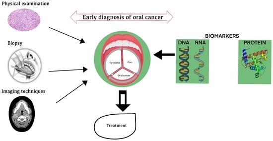
Graphical Abstract

1. Introduction

Cancer can be caused by the uncontrollable multiplication of abnormal growth of cells, which can spread and cause damage to other organs. Oral cancer (OC) is a kind of cancer that can occur in any area of the mouth (oral cavity) or throat (back) (oropharynx). It generally begins as a tiny inexplicable growth or sore in the mouthparts that later extends to the oropharynx. OC is a severe problem that can affect people all over the world. In the last several years, it has climbed in global incidence and prevalence to be the sixth most frequent tumor around the globe and the third most prevalent in South Central Asia. It accounts for approximately a quarter of all cancers around the globe. It is a severe health concern for countries in economic transition and has the highest fatality and morbidity rates among all human malignancies [1]. In contrast to other human malignancies, OC can be usually detected due to the presence of a precursor lesion or the transparency of the oral mucosa, which can be identified with certain previous training and/or alertness.
Oral squamous cell carcinoma (OSCC) is caused by highly cancerous lesions or normal epithelial linings and accounts for 84–97% of oral cancer. Inflammatory oral submucosa, fibrosis, erythroplakia, leukoplakia, candidal leukoplakia, proliferative verrucous leukoplakia (PVL), dyskeratosis congenital, and lichen planus are the symptoms of OC at the preclinical stage and are considered to be Potential Malignant Disorders [2,3]. WHO defined OPMD (i.e., oral potentially malignant condition) as precancerous lesions which lead to OC. OPMDs are a group of different oral mucosal lesions that are more likely to become cancerous. The lip, tongue, gums, palate, and other parts of the oral mucosa are the most pervasive sites of OC, as classified by the International Classification of Diseases (ICD) [4]. A lot of people who have oral cancer have a lot of different things going on in their mouths, including smoking, drinking too much alcohol, not brushing their teeth, and having long-term viral infections, such as the human papillomavirus (HPV) [5,6,7,8,9]. Tobacco use (in every form) is an important cause of cancer, particularly in underdeveloped nations. People who chew paan, which is made of piper betel leaves mixed with areca nut, lime, catechu, cinnamon, and other ingredients, are more likely to contract OC than people who smoke cigarettes. This is especially true in India, which has the highest cancer rate in the world [10]. Periodontal diseases are also a high-risk factor for OC, and they are more common in the Indian population, where it is mostly caused by the practice of chewing paan [11]. Chewing paan induces extended exposure of the oral mucosa, as well as abrasion of the epithelial linings. Smokeless tobacco consumption, both orally and nasally, has been linked to potentially malignant oral diseases and malignancies of the oral cavity. Tumorigenesis and inflammation caused by bacterial and viral infections, as well as inflammatory bowel illnesses, also play a major role in the development of cancer [11]. The exposure to harsh environmental circumstances, lack of information, and behavioral risk factors are the key indicators of this OC to be a worldwide incidence. The risk factors for OC vary by geographical area and the lifestyle habits of the people who live there. In Western nations, the primary risk factors are smoking and alcoholism. This is in contrast to South Asian countries, where betel nut eating and smoking are more prevalent [12]. This review paper provides a crisp view on conventional and recent advanced diagnostic methodologies used in the detection of OC. This paper also highlights recent molecular biomarkers used in diagnosis and detection.

2. The Pathogenesis

OSCC is a complex disease with a pathogenesis that involves interactions between a variety of carcinogenetic and genetic contributing factors to cancer’s growth and progression. As previously stated, persistent consumption of alcohol and smoking of cigarettes are the primary contributors to the development OSCC. Simultaneously, chronic inflammation, viral infections (particularly HPV type 16 and 18), and hereditary factors all contribute to the disease’s development. It is critical to understand the basic molecular processes that enable oral carcinogenesis [13], mechanisms that can lead to innovative treatment choices, in order to improve survival through improved preventive and therapy options.
OSCC helps people get a biopsy if they show signs of the disease; histological tests are then performed based on where the disease is located in their bodies [14]. OSCC is linked to changes in the oral mucosa that are caused by a variety of genetic alterations. These changes can lead to papillary hyperplasia, different stages of dysplasia (slight, minor, or major), and the growth of cancer (in situ or well-differentiated intrusive squamous cell). Most oral cancers exhibit stimulation of the extracellular environment and activity of melanoma fibroblasts. This highlights the link between tumor cell growth and basal lamina breakdown [15]. Cyclin D1 and Ki-67 have been found to be strong in OSCC that was well-differentiated [16].

3. Process Flow in the Molecular Diagnosis of OC

A molecular analysis can be performed in situ or via the separation of test sample from clinical specimens. A molecular analysis procedure is used to look at clinical specimens, such as biofluids and cell culture products. The tissue sample is preserved in formalin and afterward embedded in paraffin before being examined histopathologically in the conventional manner. In order to prepare for light microscopy, the implanted tissue is finely sliced and put on a glass slide. The tissue can then be stained with hematoxylin and eosin [17]. This method can be used to examine particular molecules in their natural topographical environment present in these tissues, often known as in situ. Antibodies (immunohistochemistry) and nucleotide probes (in situ hybridization) can be used to analyze proteins and nucleic acids, respectively.
Biofluids (e.g., saliva or blood), tissue, and extracted specific or target molecules can be examined as additional molecular components in the preparation for cell-level-based research. As a result, analyzing by using the extracted target molecules was found to be a more sensitive and particular approach that can be employed. Moreover, the sample type should be taken into main consideration when choosing the target and techniques used for the analysis. The approaches employed may have either a low or a high throughput. Low-throughput approaches can only evaluate a small number of hits or samples at once, while high-throughput techniques can examine a large number of targets simultaneously. Multiplexing can also be used, which is when a single sample is analyzed for a lot of different things at the same time or at the same time, using the same method to find out about a lot of different things. Molecular “labels”, such as fluorophores that emit light in different colors, are often used in this method. Each color is unique to a single target.

4. Need for Early Diagnosis

Overall, 15% to 50% of people who have oral cancer (OC) will live for at least five years. This is because most oral tumors are found at an advanced stage (stage III or stage IV) [3]. In contrast, if OC is detected and treated early, such as at stage I or stage II, the survival rate can surpass 80% [10,18]. In general, OC can be diagnosed by several clinical procedures, such as physical and histological examinations, biopsy, spectroscopic, radiographic techniques, and so on (Table 1). The early discovery of cancer is essential for averting additional psychological, physical, and financial losses for the patient. Numerous unique procedures have been developed as a result of advances in science and technology that offer benefits over currently used traditional diagnostic methodologies.
The molecular biology findings are useful in solving the many new issues that arise in the detection and diagnosis of OC. These methods can detect any changes at the molecular level considerably earlier than a microscope can detect them, and well before clinical changes occur. Oral lesions can also be classified based on their molecular characteristics. As a result, it is now possible to predict the toxic potential of ulcerations, meaning that the number of oral cancers will go down and that early detection and treatment will be better [19,20,21,22,23]. There has been a lot of progress in our understanding of the human genome, as well as different ways to study genetics and molecular biology. These tools can be used to quickly diagnose and treat oral lesions through molecular examination. In today’s world, molecular biology helps us comprehend pathological progressions and development at a level beyond the human sight by revealing the characteristics and functional properties of macromolecules. The application of molecular diagnostic tools to determine disease risk, their presence, and therapy efficacy is referred to as molecular diagnostics. Microarrays, deep sequencing methods, and proteomics methods are often used to study the molecular level of OC and for its early diagnosis. In the past, high-throughput methods were used to find biomarkers in saliva and plasma or predictive markers in tissue specimens that were used to find biomarkers [24].

5. Current Diagnostic Methods for Detection of OC

5.1. Visual Oral Examination

Doctors need to know as soon as possible if an OPMD has a chance of becoming cancerous so that they can keep an eye on it, treat it, and improve survival rates. Currently, the usual screening approach for detecting oral mucosal lesions is a visual oral examination (VOE). Because white or red lesions and long-term ulcers can be hard to tell apart at first glance, VOE is very dependent on the physician’s knowledge [25,26]. It is very important to distinguish lesions with a greater risk of malignant transformation because this will have a big impact on the treatment success. Though histological evaluation acts as the gold standard for clinical diagnosis and therapy, it may be insufficient to distinguish lesions that require active treatment, particularly if they are subtle or in a non-dysplasia stage.
A quick diagnosis is very important for preventing oral premalignant disorders from becoming cancerous and increasing the chances that the patients will live long lives. For the right diagnosis, many different procedures need to be performed, such as swabbing the exterior of the abscess and looking at the biological data of oral precancerous lesions. It is hard to tell, but specialists need to be capable of distinguishing between the features of ulcerations just by looking at them, without changing the cells in the area [27,28,29,30,31,32,33,34,35,36,37,38]. While incisional biopsy with histopathology is the gold standard for identifying dental pathology, it is hurtful for patients and results in a late diagnosis, due to the completion of histology. The autofluorescence approach is a new noninvasive tool for analyzing a soft-tissue injury. It can be utilized to locate oral precursor malignant lesions, as well as the proper place for biopsies within the changed mucosa. The procedure’s biggest drawback is the risk of false-positive outcomes [27,28,39]. Identifying the potential biomarkers will improve the ability to exactly analyze and forecast the likelihood and danger of OPMDs evolving into an OC lesion, which requires active treatment if found.

5.2. Physical Examination

The physical examination is the first and most important stage in diagnosing oral cancer. Typically, it is processed in two stages: a comprehensive visual evaluation, followed by palpation. External body components such as lymph nodes, salivary glands, and lips were inspected initially, followed by an inspection of the buccal cavity’s interior. In the superficial anatomy, abnormalities, irregularities, edema, and fluctuation are observed. Soft-tissue thickening, lumps, discomfort, difficulty moving the jaw, chewing and swallowing, earache, and other symptoms are typical. The parotid gland (biggest salivary gland) is palpated both within and outside the mouth, as well as the submandibular and sublingual glands [40]. Physical observations are compared to the patient’s clinical picture by the examiner. Additionally, morphological changes, as well as texture and color aberrations, are described [41].

5.3. Histopathological Examination

OC can range from benign tumors to extremely aggressive tumors with a high proclivity for invasion. Histological examinations demonstrate how oral carcinogenesis progresses from benign dysplasia to highly invasive malignancies. Histological investigation is essential to identify cell proliferation and development difficulties, cellular and cytoplasmic atypia, and abnormalities in the surface epithelium or deep tissue cytoarchitecture [42]. The crucial stage in a histological examination is identifying the correct region and proper sampling of the oral lesion, as the histopathological alterations can also develop in places where there is no indication of oral lesions upon physical examination. Thus, for the diagnosis of benign or malignant tumors, a method that recognizes both histopathological and molecular changes is desirable, as genomic changes can occur in normal tissues prior to the emergence of microscopic and clinical morphological changes [43].

5.4. Vital Staining Techniques

The vital staining technique includes labeling cells or tissues in their living state. This method of optical tissue staining for cancer detection provides an alternative to physical examination [44]. Toluidine blue staining is used to detect oral mucosal abnormalities. Toluidine blue is an acidophilic metachromatic dye that has a good attachment for acidic tissue components, coloring the nuclear material of DNA and RNA-rich tissues. This approach, when used in conjunction with Lugol’s iodine, aids in the differentiating between inflammatory lesions. Such a combination accurately predicts the stage of tumors, ranging from benign to malignant lesions, making it an advantageous visual staining technique for evaluating OC before treatment [45].

5.5. Biopsy

Biopsy is a process where part of tissue or sample of cells is surgically extracted from the suspicious area from the body and submitted to a pathology facility for microscopic analysis. This is the sole approach to determine if an oral cavity or oropharyngeal carcinoma is present [46]. For a reliable histological diagnosis after a biopsy, proper treatment of the tissue is essential. Improper sample handling might lead to a faulty biopsy, which necessitates repeating the process. Exfoliative cytology and incisional biopsy are two types of biopsies that are performed depending on the individual need. Brush biopsy involves scraping the surface mucosa from the oral lesion to extract the cells from transepithelial region. This technique is a straightforward, safe, painless, cost-effective, and highly sensitive method. Small white and red lesions in the mouth can be checked to make sure that they are not dysplastic. Compared to other biopsy procedures, brush biopsy holds 90% of sensitivity and specificity [47].
Oral exfoliative cytology (OEC) is a simple, non-aggressive method which is comfortable for the patient and can be used to detect OC early. OEC concentrates on the individual cell’s morphological and staining features, thus necessitating the involvement of expert cytopathologists [48]. Despite its benefits, OEC is neither specific nor sensitive, and as a result, it is mostly utilized for screening from a large population, regular monitoring of precancerous lesions, and identification of appropriate biopsy sites in large lesions [49]. Once the cancer is detected by OEC, incisional biopsy is performed, in which a small portion tissue sample is carefully chosen for diagnosis. Though it does not cover the whole lesion, the incisional biopsy is reasonably accurate [50]. In circumstances when it is impossible to remove the entire lesion, e.g., a broad white patch or lichen planus, an incisional biopsy is employed. In addition, this approach is favored when the clinical diagnosis is unknown [51].

5.6. Imaging Techniques

OC is diagnosed by using a variety of technological imaging methods, such as computed tomography (CT), magnetic resonance imaging (MRI), and positron emission tomography (PET) [52].

5.6.1. Magnetic Resonance Imaging

The structures in the mouth cavity, as well as surrounding regions, may be seen in detail by using MRI. Discrimination of soft tissues by MRI assists in determining the degree of the tumor’s site dissemination, invasion depth, and lymphadenopathy [53]. This is because it has a better contrast resolution and can look at things from many different angles. This makes it easy to see how far OC has spread into the soft tissues next to it. Thus, an MRI is needed to check for advanced oral and oropharyngeal cancers before they can be treated.

5.6.2. Computed Tomography

The CT scan uses X-ray radiation and a computer to make pictures of the body. It looks for the cancerous lesion and sees if it has spread to many other body parts. Because CT scans are readily available and very inexpensive, they are frequently used as a routine imaging tool for the identification of cancers. However, it has been noted that CT scans cannot detect lesions in their early stages.

5.6.3. Positron Emission Tomography

PET scans have been used to determine whether tumor cells have spread to lymph nodes or other organs. After administering a radioactive dye intravenous or oral, gamma rays emitted by positron decays are analyzed. It is a precise approach for determining the lymph node staging. Even if more lymph node metastases are identified away from the afflicted region, it does not necessitate any changes in therapy [54]. A PET scan utilizing fluorodeoxyglucose can be used to evaluate the condition of lymph nodes prior to surgery (FDG). As a result, PET scanning is crucial for detecting oral cancer in its early stages [55].

6. Biomarker Detection

Biomarkers are cell components that are highly expressed in body fluids or tumor cells at the beginning of illness. In the context of OC, the biological cells exhibited in blood or saliva act as cancer-detection indicators. Specifically, the progression of cancer consists of three stages: beginning, promotion, and advancement. All three steps are associated with substantial modifications in the cell’s processes, including metabolome, transcriptome, and proteome processes [56]. These changes occur as a result of mutations in certain genes or proteins, which result in the modification or stopping of important metabolic and structural pathways. Some of the most promising biomarkers related to the progression of OC are Ki-67 antigen, TSG p16, TSG p53, and DNA ploidy. Early identification of OSCC is aided by salivary biomarkers such as circulating tumor DNA, miRNAs, and extracellular vesicles [57]. In OSCC, p53 expression is regarded as a key indicator of carcinogenesis. The advent of the p53 mutant gene is confirmed by using immunohistochemical staining [58]. Apart from that, many advanced techniques and high-throughput molecular methodologies are used to detect the biomarkers (Figure 1 and Table 2).

6.1. Next-Generation-Sequencing-Based Biomarkers

Squamous cell carcinoma progresses because of changes at the genomic level. Next-generation sequencing (NGS) technology is a good way to find the mutations and copy number changes that cause these changes [15]. Stransky et al. performed whole-exome investigations on head and neck SCC from 74 patients and observed matched DNA pairs between tumors and peripheral whole blood [59]. This team had identified 130 coding mutations per tumor. Given that 14 percent of all cancers were HPV-positive, results from sequencing research show how these would have a genetic variation that was half that of HPV-negative tumors. This supports the idea that there are molecular differences among the two types of SCC. With the help of NGS which possess high sensitivity, the molecular characterization among the two SCCs may go even further, subdividing HPV-negative tumors not only by their precise location, but also by the frequency of specific nucleotide alterations. Aside from these findings, the presence of mutant NOTCH1 in 11% of all tumors examined was noteworthy, since it would be the first study linking genetic alterations in this gene to the genesis and progression of SCC. This research helps us better understand how this kind of cancer grows. It shows that the changes that happen at different stages of squamous differentiation have a big impact on the many genetic processes that drive SCC development [15,60]. Another interesting finding is that a mutation at TP53 had a key influence in the development of this disease for the younger patients who are preferably non-smokers [61]. Pickering and his colleagues did a very detailed genomic study of OSCC in order to find out what causes the disease to become malignant and what biomarkers can be used to classify, predict, and treat it [62]. Pickering and his team used fresh-frozen malignant cells and nonmalignant cells from 38 patients with OSCC to study genome-wide copy number changes. They found strong correlations between gene expression and comparative copy numbers for 1721 genes.
The NOTCH1 gene had many missense and truncating mutations in around one-tenth of the tumor samples, and this was confirmed by using a panel of HNSCC cell lines. With the previously described study group, they found comparable results. The absence of protein expression, along with other tests, such as in vivo experiments using mouse models, supports the hypothesis that the Notch signaling system had a major role in tumor-suppressive mechanisms in SCC [16]. This new but widely used method can help us understand not only the main molecular devices that play a role in SCC emergence and development, but also other possible participants that play a role in the tumor transformation process.
Pushalkar and his study team sought to investigate how bacteria might play a role in tumorigenesis, especially with SCC [63]. They collected saliva samples from three people who had OSCC and two people who were healthy (control). Then the bacterial gDNA was extracted, processed, and pyro-sequenced. After analyzing the data, the researchers discovered several bacterial species related to eight distinct taxonomic phyla, with the Firmicutes phylum being the best defined in samples compared to controls. At the species level, NGS analyses revealed how microorganisms are expressed variably in the dental environments of SCC individuals versus healthy participants, suggesting that these microbes might contribute to the course of the disease. The same research group then looked at matched pairs of cancerous and non-cancerous tissue from SCC patients. This shows how important it is to use unique techniques such as this to understand the many and complex aspects of OSCC transition [64].

6.2. Transcriptomics-Based Biomarkers

MicroRNA is a group of functional non-coding RNA molecules that have about 22 nucleotides each. They contribute to gene control after transcription, and they have about 22 nucleotides in each. Many diseases start and get worse when there are problems with the way microRNAs are made. This is because microRNAs control so many important biological processes, such as advancement, divergence, and cell cycles [14,15]. Tus miRNAs might be used as biomarkers for a variety of illnesses, including neurodevelopmental disorders (Ohnishi et al. 2014), cancer, and cardiovascular disease [24,65]. A microarray analysis of 20 human whole-blood specimens on 1200 miRNAs revealed OSCC-specific signature biomarkers. Then qRT-PCR was used to validate the most important miRNAs, which showed a two-fold upregulation for miR-494 and miR-3651 and a two-fold downregulation for miR-186. Another study with microarray data using Affymetrix U133A platform revealed that 51 genes were upregulated and found that most of genes were related to OSCC [66]. Their findings also showed a new set of OSCC-related genes (RHEB, SOD2, SKP2, IFI16, IFI44, STAT1, and GREM1) and three genes (MMP1, SOCS3, and ACOX1) that differentiate the normal tissue from the cancer [67]. Microarray and qPCR were used to examine a group of 23 CXC-chemokine ligands and receptors that were then connected to therapeutic response by using a logistic regression technique. CXCL10 expression was found to be substantially associated with radiotherapy response, with the group with CXCL10 overexpression having a poor outcome [68]. Similar study was also observed where miRNAs showed different levels of expression between normal tissue and OSCC tissue. In this study, miR-375 showed the greatest inhibition, whereas miR-31 recorded overexpression. Moreover, 61 miRNAs were found to establish a 93 percent accurate molecular categorization of OSCC [65]. Lajer and his group found that HPV causes changes to 21 miRNAs, the most important of which are miR-127-3p and miR-363 [69]. The influence of HPV might explain why HPV-infected malignancies have variable clinical outcomes on miRNA profile.
MiR-375 downregulation, miR-127 overexpression, and miR-137 hypermethylation were discovered in another study of OSCC. Another interesting biomarker which is epigenetically activated in tumor tissue was observed with miR-200 and miR-205. Observing changes in miR-375 and miR-200a methylation, and also changes in miR-200c methylation, is a useful non-invasive biomarker, as shown by a study that compared the saliva of OSCC patients and healthy people. It has been suggested that salivary miRNA profiles could be used as a biomarker for initial evaluation of patients in OSCC [70]. The first lncRNA heat map for usual oral cavity vs. a pre-malignant abscess was made and publicly disclosed [71]. Many recent research studies have shown that new lncRNA can be used as non-invasive saliva biological markers for prediction and diagnosis. HOTAIR identified in saliva was shown to have considerable predictive relevance in a recent research study, with the expression level associated with lymph node metastases.

6.3. Proteomic-Based Biomarkers

Proteomics is a promising technique for identifying new biomarkers with prognostic and diagnostic potential [72,73]. The identification of new biomarkers in OSCC may be aided by analyzing cellular entire protein complements or biofluids. A key feature of various research studies [74] is a saliva proteome profiling study based on patient categorization. Tests have been performed to see how much protein is found in samples of OSCC and healthy tissue, as well as in other in vitro systems for this disease [75]. A lot of proteins that affect cellular functions and structure, cellular adhesion, or cell migration, and proteins that make cancer more likely were found to be changed in studies [76,77]. When researchers looked at the plasma proteome of mice with OSCC, they found that haptoglobin and the precursor of apolipoprotein A1 have both been upregulated. The expression of haptoglobin plasma in humans showed that there was a strong connection between increasing levels of haptoglobin and the clinical stages of OSCC. This suggests that haptoglobin could be used as a plasma diagnostic marker for identification of OSCC patients [78].
A SELDI-TOF Protein Chip technology was used to screen saliva proteins from pre- and post-treatment OSCC tests to find 26 candidates with different patterns in their saliva that could be linked to the disease. The investigation found a shortened cystatin SA-I of 14 kDa in pre-treatment saliva samples, with a deletion of three amino acids at the N-terminal end [79]. Protein Chip analysis could be a good way to screen for OSCC at an early stage, and truncated cystatin SA-I could be a good tumor prognostic marker for OSCC. Transglutaminase 3 (TGM3) downregulation was linked to OSCC histological differentiation loss in another study [80]. There were 52 proteins that had statistical significance, so a comparative proteomics study found a group of eight proteins that were more or less common in different people. Annexin A8, Peroxiredoxin-2, and Tyrosine Kinase have been observed in both diabetes and OSCC, and they were thought to be possible biomarkers for OSCC diagnosis because they were found in both [81]. A mass spectrometer study of saliva from people with cancer and healthy people found 213 new proteins. The salivary biomarkers Profilin, Cofilin, S100A9, and MMP9 have all been found to be linked to head and neck cancer. Vimentin has been linked to the epithelial–mesenchymal transition and metastasis of head and neck cancer [82].
Exosomes are endocytic membrane bound vesicles with nucleic acids (DNA and RNA) inside their lumen that originate from the cell’s cytoplasm. They are secreted in large amounts and are found abundantly in bodily fluids. Exosome surface proteins can function as antigens for certain antibodies, which can then be utilized to isolate desired exosomes via affinity-based approaches. Exosomes generated from cancer are said to reflect the tumor microenvironment and can thus be employed as a biomarker for tumor identification [83,84]. The lipid bilayer of exosomes is made up of cholesterol, phosphatidylserine, and ceramide, as well as trans-membrane receptors, proteolytic enzymes, tetraspanins, and adhesion molecules. Some proteins that are linked to cancer are also very common on the exterior of exosomes, thus making them useful as markers for differentiating between different types of cancer. Proteins such as HER2, LMP1, and MUC18 can be used to make biosensors that can detect total exosomes.

6.4. Advantage of Use of Biomarker over the Traditional Technique

The diagnostic and predictive biological markers of cancer are the future of clinical cancer management. These biomarkers are critical because they will be utilized to make therapeutic decisions that will save lives. Biomarkers will help doctors make decisions about cancer treatment in the future. It is necessary to discover biomarkers that accurately predict the prognosis of a particular disease and allow clinicians and patients to make correct treatment decisions. In the absence of viable treatments, the consensus about cancer diagnostics has been that early detection, followed by surgical surgery before the tumor had progressed, was the best approach to cancer. Few novel diagnostic biomarkers have evolved, despite the success of traditional approaches, such as biopsy, vital staining, and imaging techniques, in the early identification of cancer. A noninvasive saliva or serum biomarkers that properly predicts cancer prognosis is still needed. Patients’ cancer is still being detected too late and treated without knowing if their tumor has progressed beyond its initial stage. Biomarkers will not only assist screen, detect, diagnose, aid in prognostic assessment, monitor therapy, and forecast recurrence over the next few decades, but they will also play an important role in medical decision making. Markers that indicate responsiveness to a specific therapy are, in fact, required.

7. Future Implications

Oral cancer candidate biomarkers should be identified and classified in the fields of screening, diagnostic testing, recurrence prediction, progression, therapeutics, and metastasis in future studies. These prospective markers will aid in the prediction of outcome measures and the development of dental public health strategies. The creation of biomarkers aimed at assessing the success of oral cancer medication therapy would be extremely helpful in determining therapeutic efficacy. Researchers should apply innovative research methodologies that adhere to the requirements of analytical authenticity, clinical validity, and clinical utility, in addition to biomarker reporting and evaluation systems.

8. Conclusions

In light of the vast amount of R&D in the field of OC diagnostics, the diagnostic procedures presented in this review paper are largely those that are already in clinical use or commercially accessible and show significant potential in clinical use. There are various other approaches for diagnosing OC that were not included in this paper. Each of these diagnostic approaches reveals its own technological originality. In the near future, it is anticipated that new innovations with enhanced detection sensitivity will emerge. The availability of these very sensitive methods (next-generation sequencing, mass spectrometry, and microarray technologies) will allow for reliable diagnosis of even lower amounts of sample analytes. To aid in the efficient detection of OPMDs and OC, further efforts to strengthen and enhance clinical performance during OC diagnosis are strongly urged.

Author Contributions

Conceptualization, data curation, and methodology, V.R.U.; writing—original draft preparation, writing—review and editing, and visualization, V.R.U.; resources and supervision, P.M.N.; funding acquisition, B.S. All authors have read and agreed to the published version of the manuscript.

Funding

The authors have no relevant affiliations or financial involvement with any organization or entity with a financial interest in or financial conflict with the subject matter or materials discussed in the manuscript. This includes employment, consultancies, honoraria, stock ownership or options, expert testimony, grants or patents received or pending or royalties.

Institutional Review Board Statement

The present study did not use Human participants or experimental animal models; hence, ethical approval is not applicable.

Informed Consent Statement

Not applicable.

Data Availability Statement

The data presented in this study are available in this article.

Acknowledgments

The authors would like to thank Sree Balaji Dental College and Hospital, Pallikaranai, Chennai-600 100, India.

Conflicts of Interest

The authors declare no conflict of interest.

Sample Availability

Samples of the compounds are not available from the authors.

References

  1. Gupta, B.; Bray, F.; Kumar, N.; Johnson, N.W. Associations between oral hygiene habits, diet, tobacco and alcohol and risk of oral cancer: A case–control study from India. Cancer Epidemiol. 2017, 51, 7–14. [Google Scholar] [CrossRef]
  2. Sankaranarayanan, R.; Ramadas, K.; Thomas, G.; Muwonge, R.; Thara, S.; Mathew, B.; Rajan, B. Effect of screening on oral cancer mortality in Kerala, India: A cluster randomised controlled trial. Lancet 2005, 365, 1927–1933. [Google Scholar] [CrossRef]
  3. Ajay, P.; Ashwinirani, S.; Nayak, A.; Suragimath, G.; Kamala, K.; Sande, A.; Naik, R. Oral cancer prevalence in Western population of Maharashtra, India, for a period of 5 years. J. Oral Res. Rev. 2018, 10, 11. [Google Scholar]
  4. Gupta, S.; Gupta, R.; Sinha, D.N.; Mehrotra, R. Relationship between type of smokeless tobacco & risk of cancer: A systematic review. Indian J. Med. Res. Suppl. 2018, 148, 56–76. [Google Scholar]
  5. Kadashetti, V.; Shivakumar, K.; Chaudhary, M.; Patil, S.; Gawande, M.; Hande, A. Influence of risk factors on patients suffering from potentially malignant disorders and oral cancer: A case-control study. J. Oral Maxillofac. Pathol. 2017, 21, 455–456. [Google Scholar] [CrossRef]
  6. Malik, A.; Mishra, A.; Garg, A.; Shetty, R.; Mair, M.; Chakrabarti, S.; Nair, D.; Balasubramaniam, G.; Chaturvedi, P. Trends of oral cancer with regard to age, gender, and subsite over 16 years at a tertiary cancer center in India. Indian J. Med. Paediatr. Oncol. 2018, 39, 297. [Google Scholar] [CrossRef]
  7. Murugesan, A.; Sabarinath, S. Awareness of oral cancer among medical students in Chennai. J. Med. Radiol. Pathol. Surg. 2016, 2, 18–22. [Google Scholar] [CrossRef]
  8. Singh, V.; Singh, A.K.; Dutta, K.D.; Kumar, N.; Kumari, A. Evaluation of quality of life and the nutritional status of oral cancer treated patients as compared with the control group in Varanasi district: A cross sectional study. Int. J. Community Med. Public Health 2019, 6, 4804. [Google Scholar] [CrossRef] [Green Version]
  9. Thavarool, S.B.; Muttath, G.; Nayanar, S.; Duraisamy, K.; Bhat, P.; Shringarpure, K.; Nayak, P.; Tripathy, J.P.; Thaddeus, A.; Philip, S.; et al. Improved survival among oral cancer patients: Findings from a retrospective study at a tertiary care cancer centre in rural Kerala, India. World J. Surg. Oncol. 2019, 17, 1–7. [Google Scholar] [CrossRef]
  10. Singh, M.; Prasad, C.P.; Singh, T.D.; Kumar, L. Cancer research in India: Challenges & opportunities. Indian J. Med. Res. 2018, 148, 362–365. [Google Scholar]
  11. Laprise, H.P.; Shahul, S.A.; Madathil, A.S.; Thekkepurakkal, G.; Castonguay, I.; Varghese, S.; Shiraz, P.; Allison, N.F.; Schlecht, M.C.; Rousseau, E.L.; et al. Periodontal diseases and risk of oral cancer in Southern India: Results from the HeNCe Life study. Int. J. Cancer 2016, 139, 1512–1519. [Google Scholar] [CrossRef]
  12. Asthana, S.; Patil, R.S.; Labani, S. Tobacco-related cancers in India: A review of incidence reported from population-based cancer registries. Indian J. Med. Paediatr. Oncol. 2016, 37, 152–157. [Google Scholar] [CrossRef] [Green Version]
  13. Lukesova, E.; Boucek, J.; Rotnaglova, E.; Salakova, M.; Koslabova, E.; Grega, M.; Eckschlager, T.; Rihova, B.; Prochazka, B.; Klozar, J.; et al. High level of tregs is a positive prognostic marker in patients with HPV-Positive oral and oropharyngeal squamous cell carcinomas. Biomed. Res. Int. 2014, 2014, 303929. [Google Scholar] [CrossRef]
  14. Rivera, C.; Venegas, B. Histological and molecular aspects of oral squamous cell carcinoma (Review). Oncol. Lett. 2014, 8, 7–11. [Google Scholar] [CrossRef] [Green Version]
  15. Koontongkaew, S. The tumor microenvironment contribution to development, growth, invasion and metastasis of head and necksquamous cell carcinomas. J. Cancer 2013, 4, 66–83. [Google Scholar] [CrossRef]
  16. Ohnishi, Y.; Watanabe, M.; Wato, M.; Tanaka, A.; Kakudo, K.; Nozaki, M. Cyclin D1 expression is correlated with cell differentiation and cell proliferation in oral squamous cell carcinomas. Oncol. Lett. 2014, 7, 1123–1127. [Google Scholar] [CrossRef] [Green Version]
  17. Titford, M. A short history of histopathology technique. J. Histotechnol. 2006, 29, 99–110. [Google Scholar] [CrossRef]
  18. Varshitha, A. Prevalence of oral cancer in India. J. Pharmaceut. Sci. Res. 2015, 7, 845–848. [Google Scholar]
  19. Farooqi, A.; Shu, C.-W.; Huang, H.-W.; Wang, H.-R.; Chang, Y.-T.; Fayyaz, S.; Yuan, S.-S.F.; Tang, J.-Y.; Chang, H.-W. TRAIL, Wnt, sonic hedgehog, TGFβ, and miRNA signalings are potential targets for oral cancer therapy. Int. J. Mol. Sci. 2017, 18, 1523. [Google Scholar] [CrossRef] [Green Version]
  20. Downer, M.C.; Moles, D.R.; Palmer, S.; Speight, P.M. A systematic review of test performance in screening for oral cancer and precancer. Oral Oncol. 2004, 40, 264–273. [Google Scholar] [CrossRef]
  21. Edwards, P.C. Oral cancer screening for asymptomatic adults: Do the United States preventive services task force draft guidelines miss the proverbial forest for the trees? Oral Sur. Oral Med. Oral Path. Oral Radiol. 2013, 116, 131–134. [Google Scholar] [CrossRef] [Green Version]
  22. Liu, S.Y.; Lu, C.L.; Chiou, C.T.; Yen, C.-Y.; Liaw, G.-A.; Chen, Y.-C.; Liu, Y.-C.; Chiang, W.-F. Surgical outcomes and prognostic factors of oral cancer associated with betel quid chewing and tobacco smoking in Taiwan. Oral Oncol. 2010, 46, 276–282. [Google Scholar] [CrossRef]
  23. Baylin, S.B.; Jones, P.A. A decade of exploring the cancer epigenome—Biological and translational implications. Nat. Rev. Cancer 2011, 11, 726–734. [Google Scholar] [CrossRef]
  24. Brinkman, B.M.; Wong, D.T. Disease mechanism and biomarkers of oral squamous cell carcinoma. Curr. Opin. Oncol. 2006, 18, 228–233. [Google Scholar] [CrossRef]
  25. Mehrotra, R.; Gupta, D.K. Exciting new advances in oral cancer diagnosis: Avenues to early detection. Head Neck Oncol. 2011, 3, 33. [Google Scholar] [CrossRef] [Green Version]
  26. Speight, P.M.; Epstein, J.; Kujan, O.; Lingen, M.W.; Nagao, T.; Ranganathan, K.; Vargas, P. Screening for oral cancer—A perspective from the Global Oral Cancer Forum. Oral Surg. Oral Med. Oral Pathol. Oral Radiol. 2017, 123, 680–687. [Google Scholar] [CrossRef]
  27. Mascolo, M.; Siano, M.; Ilardi, G.; Russo, D.; Merolla, F.; De Rosa, G.; Staibano, S. Epigenetic disregulation in oral cancer. Int. J. Mol. Sci. 2012, 13, 2331–2353. [Google Scholar] [CrossRef] [Green Version]
  28. Galeano, M.; Altavilla, D.; Cucinotta, D.; Russo, G.T.; Calò, M.; Bitto, A.; Marini, H.; Marini, R.; Adamo, E.B.; Seminara, P.; et al. Recombinant human erythropoietin stimulates angiogenesis and wound healing in the genetically diabetic mouse. Diabetes 2004, 53, 2509–2517. [Google Scholar] [CrossRef] [Green Version]
  29. Siriwardena, S.B.S.M.; Tsunematsu, T.; Qi, G.; Ishimaru, N.; Kudo, Y. Invasion-related factors as potential diagnostic and therapeutic targets in oral squamous cell carcinoma—A review. Int. J. Mol. Sci. 2018, 19, 1462. [Google Scholar] [CrossRef] [Green Version]
  30. Sahibzada, H.A.; Khurshid, Z.; Khan, R.S.; Naseem, M.; Siddique, K.M.; Mali, M.; Zafar, M.S. Salivary IL-8, IL-6 and TNF-α as potential diagnostic biomarkers for oral cancer. Diagnostics 2017, 7, 21. [Google Scholar] [CrossRef] [Green Version]
  31. Chuang, J.-Y.; Huang, Y.-L.; Yen, W.-L.; Chiang, I.-P.; Tsai, M.-H.; Tang, C.-H. Syk/JNK/AP-1 signaling pathway mediates interleukin-6-promoted cell migration in oral squamous cell carcinoma. Int. J. Mol. Sci. 2014, 15, 545–559. [Google Scholar] [CrossRef] [PubMed] [Green Version]
  32. Ho, M.T.; Kang, H.S.; Huh, J.S.; Kim, Y.M.; Lim, Y.; Cho, M. Effects of the novel compound DK223 ([1E,2E-1,2-bis(6-methoxy-2H-chromen-3-yl)methylene]hydrazine) on migration and proliferation of human keratinocytes and primary dermal fibroblasts. Int. J. Mol. Sci. 2014, 15, 13091–13110. [Google Scholar] [CrossRef] [PubMed] [Green Version]
  33. Hameedaldeen, A.; Liu, J.; Batres, A.; Graves, G.S.; Graves, D.T. FOXO1, TGF-β regulation and wound healing. Int. J. Mol. Sci. 2014, 15, 16257–16269. [Google Scholar] [CrossRef] [PubMed] [Green Version]
  34. Cervino, G.; Fiorillo, L.; Laino, L.; Herford, A.S.; Lauritano, F.; Giudice, G.L.; Famà, F.; Santoro, R.; Troiano, G.; Iannello, G.; et al. Oral health impact profile in celiac patients: Analysis of recent findings in a literature review. Gastroenterol. Res. Pract. 2018, 2018, 7848735. [Google Scholar] [CrossRef] [PubMed]
  35. Majtan, J.; Jesenak, M. β-Glucans: Multi-functional modulator of wound healing. Molecules 2018, 23, 806. [Google Scholar] [CrossRef] [PubMed] [Green Version]
  36. Seo, G.Y.; Hyun, C.; Koh, D.; Park, S.; Lim, Y.; Kim, Y.M.; Cho, M. A novel synthetic material, BMM, accelerates wound repair by stimulating re-epithelialization and fibroblast activation. Int. J. Mol. Sci. 2018, 19, 1164. [Google Scholar] [CrossRef] [Green Version]
  37. Ligi, L.; Croce, G.; Mosti, J.; Raffetto, D.; Mannello, F. Chronic venous insufficiency: Transforming growth factor-β isoforms and soluble endoglin concentration in different states of wound healing. Int. J. Mol. Sci. 2017, 18, 2206. [Google Scholar] [CrossRef] [Green Version]
  38. Horng, H.-C.; Chang, W.-H.; Yeh, C.-C.; Huang, B.-S.; Chang, C.-P.; Chen, Y.-J.; Tsui, K.-H.; Wang, P.-H. Estrogen effects on wound healing. Int. J. Mol. Sci. 2017, 18, 2325. [Google Scholar] [CrossRef]
  39. Cicciù, M.; Herford, A.S.; Cervino, G.; Troiano, G.; Lauritano, F.; Laino, L. Tissue fluorescence imaging (VELscope) for quick non-invasive diagnosis in oral pathology. J. Craniofacial Surg. 2017, 28, e112–e115. [Google Scholar] [CrossRef]
  40. Weinberg, M.A.; Estefan, D.J. Assessing oral malignancies. Am. Fam. Physician 2002, 65, 1–5. [Google Scholar]
  41. Mangalath, U.; Mikacha, M.K.; Abdul Khadar, A.H.; Aslam, S.; Francis, P.; Kalathingal, J. Recent trends in prevention of oral cancer. J. Int. Soc. Prev. Community Dent. 2014, 4, 131. [Google Scholar] [CrossRef] [PubMed]
  42. Dascalu, I.T. Histopathological aspects in oral squamous cell carcinoma. J. Dent. Sci. 2018, 3, 000173. [Google Scholar] [CrossRef]
  43. Hasan, S.; Elongovan, S. Conventional and advanced diagnostic aids in oral cancer screening—The journey so far. Int. J. Pharm. Pharmaceut. Sci. 2015, 7, 29–33. [Google Scholar]
  44. Sreeshyla, H.; Sudheendra, U.; Shashidara, R. Vital tissue staining in the diagnosis of oral precancer and cancer: Stains, technique, utility, and reliability. Clin. Cancer Investig. J. 2014, 3, 141. [Google Scholar] [CrossRef]
  45. Nagaraju, K.; Prasad, S.; Ashok, L. Diagnostic efficiency of toluidine blue with Lugol’s iodine in oral premalignant and malignant lesions. Indian J. Dent. Res. 2010, 21, 223. [Google Scholar] [CrossRef]
  46. Chaudhari, A.; Hegde-Shetiya, S.; Shirahatti, R.; Agrawal, D. Comparison of different screening methods in estimating the prevalence of precancer and cancer amongst male inmates of a jail in Maharashtra, India. Asian Pac. J. Cancer Prev. 2013, 14, 859–864. [Google Scholar] [CrossRef]
  47. Mehrotra, R.; Mishra, S.; Singh, M.; Singh, M. The efficacy of oral brush biopsy with computer-assisted analysis in identifying precancerous and cancerous lesions. Head Neck Oncol. 2011, 3, 39. [Google Scholar] [CrossRef] [Green Version]
  48. Jaitley, S.; Agarwal, P.; Upadhyay, R. Role of oral exfoliative cytology in predicting premalignant potential of oral submucous fibrosis: A short study. J. Cancer Res. Ther. 2015, 11, 471–474. [Google Scholar] [CrossRef]
  49. Shaila, M.; Shetty, P.; Pai, P. A new approach to exfoliative cytology: A comparative cytomorphometric study. Indian J. Cancer 2016, 53, 193. [Google Scholar] [CrossRef]
  50. Sylvie, A.L.; Hagen, K.B.E. Oral soft tissue biopsy:an overview. J. Can. Dent. Assoc. 2012, 78, c75. [Google Scholar]
  51. Zargaran, M. A review of biopsy in dentistry: Principles, techniques, and considerations. J. Dent. Mater. Tech. 2014, 3, 47–54. [Google Scholar]
  52. Pałasz, P.; Adamski, Ł.; Gorska-Chrząstek, M.; Starzynska, A.; Studniarek, M. Contemporary diagnostic imaging of oral squamous cell carcinoma – a review of literature. Pol. J. Radiol. 2017, 82, 193–202. [Google Scholar] [CrossRef] [PubMed] [Green Version]
  53. Singh, A.; Thukral, C.L.; Gupta, K.; Sood, A.S.; Singla, H.; Singh, K. Role of MRI in evaluation of malignant lesions of tongue and oral cavity. Pol. J. Radiol. 2017, 82, 92–99. [Google Scholar] [CrossRef] [Green Version]
  54. Schoder, H.; Carlson, D.L.; Kraus, D.H.; Stambuk, H.E.; Gönen, M.; Erdi, Y.E.; Yeung, H.W.D.; Huvos, A.G.; Shah, J.P.; Larson, S.M.; et al. 18F-FDG PET/CT for detecting nodal metastases in patients with oral cancer staged N0 by clinical examination and CT/MRI. J. Nucl. Med. 2006, 47, 755–762. [Google Scholar]
  55. Masthan, K.M.K.; Aravindha Babu, N.; Dash, K.C.; Elumalai, M. Advanced diagnostic aids in oral cancer. Asian Pac. J. Cancer Prev. 2012, 13, 3573–3576. [Google Scholar] [CrossRef] [PubMed] [Green Version]
  56. Padmavathi, G.; Bordoloi, D.; Banik, K.; Kunnumakkara, A.B. Cancer biomarkers: Important tools for cancer diagnosis and prognosis. In Next Generation Point-of-Care Biomedical Sensors Technologies for Cancer Diagnosis; Springer: Singapore, 2017; pp. 1–29. [Google Scholar]
  57. Cristaldi, M.; Mauceri, R.; Di Fede, O.; Giuliana, G.; Campisi, G.; Panzarella, V. Salivary biomarkers for oral squamous cell carcinoma diagnosis and follow-up: Current status and perspectives. Front. Physiol. 2019, 10, 1476. [Google Scholar] [CrossRef]
  58. Ulaganathan, G.; Mohamed Niazi, K.; Srinivasan, S.; Balaji, V.; Manikandan, D.; Shahul Hameed, K.; Banumathi, A. A clinicopathological study of various oral cancer diagnostic techniques. J. Pharm. Bioallied Sci. 2017, 9, S4–S10. [Google Scholar] [CrossRef] [PubMed]
  59. Stransky, N.; Egloff, A.M.; Tward, A.D.; Kostic, A.D.; Cibulskis, K.; Sivachenko, A.; Kryukov, G.V.; Lawrence, M.S.; Sougnez, C.; McKenna, A.; et al. The mutational landscape of head and neck squamous cell carcinoma. Science 2011, 333, 1157–1160. [Google Scholar] [CrossRef] [Green Version]
  60. Soon, W.W.; Hariharan, M.; Snyder, M.P. High-throughput sequencing for biology and medicine. Mol. Syst. Biol. 2013, 9, 640. [Google Scholar] [CrossRef]
  61. Li, R.; Faden, D.L.; Fakhry, C.; Langelier, C.; Jiao, Y.; Wang, Y.; Wilkerson, M.D.; Pedamallu, C.S.; Old, M.; Lang, J.; et al. Clinical, genomic and metagenomic characterization of oral tongue squamous cell carcinoma in patients who do not smoke. Head Neck 2014, 37, 1642–1649. [Google Scholar] [CrossRef]
  62. Pickering, C.R.; Zhang, J.; Yoo, S.Y.; Bengtsson, L.; Moorthy, S.; Neskey, D.M.; Zhao, M.; Alves, M.V.O.; Chang, K.; Drummond, J.; et al. Integrative genomic characterization of oral squamous cell carcinoma identifies frequent somatic drivers. Cancer Discov. 2013, 3, 770–781. [Google Scholar] [CrossRef] [PubMed] [Green Version]
  63. Pushalkar, S.; Mane, S.P.; Ji, X.; Li, Y.; Evans, C.; Crasta, O.R.; Morse, D.; Meagher, R.; Singh, A.; Saxena, D. Microbial diversity in saliva of oral squamous cell carcinoma. FEMS Immunol. Med. Microbiol. 2011, 61, 269–277. [Google Scholar] [CrossRef] [PubMed] [Green Version]
  64. Pushalkar, S.; Ji, X.; Li, Y.; Estilo, C.; Yegnanarayana, R.; Singh, B.; Li, X.; Saxena, D. Comparison of oral microbiota in tumor and non-tumor tissues of patients with oral squamous cell carcinoma. BMC Microbiol. 2012, 12, 144. [Google Scholar] [CrossRef] [PubMed] [Green Version]
  65. Shamaa, A.A.; Zyada, M.M.; Wagner, M.; Awad, S.S.; Osman, M.M.; Abdel Azeem, A.A. The significance of Epstein Barr virus (EBV) & DNA topoisomerase II alpha (DNA-Topo II alpha) immunoreactivity in normal oral mucosa, oral epithelial dysplasia (OED) and oral squamous cell carcinoma (OSCC). Diagn. Pathol. 2008, 3, 45. [Google Scholar]
  66. Kim, K.Y.; Cha, I.H. Possibility of the use of public microarray database for identifying significant genes associated with oral squamous cell carcinoma. Genom. Inform. 2012, 10, 23–32. [Google Scholar] [CrossRef]
  67. Neville, B.W.; Day, T.A. Oral cancer and precancerous lesions. CA Cancer J. Clin. 2002, 52, 195–215. [Google Scholar] [CrossRef]
  68. Rentoft, M.; Coates, P.J.; Loljung, L.; Wilms, T.; Laurell, G.; Nylander, K. Expression of CXCL10 is associated with response to radiotherapy and overall survival in squamous cell carcinoma of the tongue. Tumor Biol. 2014, 35, 4191–4198. [Google Scholar] [CrossRef]
  69. Lajer, C.B.; Nielsen, F.C.; Friis-Hansen, L.; Norrild, B.; Borup, R.; Garnaes, E.; Rossing, M.A.; Specht, L.; Therkildsen, M.H.; Nauntofte, B.; et al. Different miRNA signatures of oral and pharyngeal squamous cell carcinomas: A prospective translational study. Br. J. Cancer 2011, 104, 830–840. [Google Scholar] [CrossRef]
  70. Wiklund, E.D.; Gao, S.; Hulf, T.; Sibbritt, T.; Nair, S.; Costea, D.E.; Villadsen, S.B.; Bakholdt, V.; Bramsen, J.B.; Sørensen, J.A.; et al. MicroRNA alterations and associated aberrant DNA methylation patterns across multiple sample types in oral squamous cell carcinoma. PLoS ONE 2011, 6, e27840. [Google Scholar] [CrossRef]
  71. Gibb, E.A.; Enfield, K.S.; Stewart, G.L.; Lonergan, K.M.; Chari, R.; Ng, R.T.; Zhang, L.; MacAulay, C.E.; Rosin, M.P.; Lam, W.L. Long non-coding RNAs are expressed in oral mucosa and altered in oral premalignant lesions. Oral Oncol. 2011, 47, 1055–1061. [Google Scholar] [CrossRef]
  72. Herrmann, P.C.; Liotta, L.A.; Petricoin, E.F. Cancer proteomics: The state of the art. Dis. Markers 2001, 17, 49–57. [Google Scholar] [CrossRef] [PubMed] [Green Version]
  73. Turhani, D.; Krapfenbauer, K.; Thurnher, D.; Langen, H.; Fountoulakis, M. Identification of differentially expressed, tumor-associated proteins in oral squamous cell carcinoma by proteomic analysis. Electrophoresis 2006, 27, 1417–1423. [Google Scholar] [CrossRef]
  74. Wu, J.Y.; Yi, C.; Chung, H.R.; Wang, D.J.; Chang, W.C.; Lee, S.-Y.; Lin, C.-T.; Yang, Y.-C.; Yang, W.-C.V. Potential biomarkers in saliva for oral squamous cell carcinoma. Oral Oncol. 2010, 46, 226–231. [Google Scholar] [CrossRef] [PubMed]
  75. Mlynarek, A.M.; Balys, R.L.; Su, J.; Hier, M.P.; Black, M.J.; Alaoui-Jamali, M.A. A cell proteomic approach for the detection of secretable biomarkers of invasiveness in oral squamous cell carcinoma. Arch. Otolaryngol. Head Neck Surg. 2007, 133, 910–918. [Google Scholar] [CrossRef] [PubMed] [Green Version]
  76. Lo, W.Y.; Tsai, M.H.; Tsai, Y.; Hua, C.H.; Tsai, F.J.; Huang, S.Y.; Tsai, C.-H.; Lai, C.-C. Identification of over-expressed proteins in oral squamous cell carcinoma (OSCC) patients by clinical proteomic analysis. Clin. Chim. Acta 2007, 376, 101–107. [Google Scholar] [CrossRef] [PubMed]
  77. Lo, W.Y.; Lai, C.C.; Hua, C.H.; Tsai, M.H.; Huang, S.Y.; Tsai, C.H.; Tsai, F.-J. S100A8 is identified as a biomarker of HPV18-infected oral squamous cell carcinomas by suppression subtraction hybridization, clinical proteomics analysis, and immunohistochemistry staining. J. Proteome Res. 2007, 6, 2143–2151. [Google Scholar] [CrossRef]
  78. Lai, C.H.; Chang, N.W.; Lin, C.F.; Lin, C.D.; Lin, Y.J.; Wan, L.; Lin, C.W. Proteomics-based identification of haptoglobin as a novel plasma biomarker in oral squamous cell carcinoma. Clin. Chim. Acta 2010, 411, 984–991. [Google Scholar] [CrossRef] [PubMed]
  79. Shintani, S.; Hamakawa, H.; Ueyama, Y.; Hatori, M.; Toyoshima, T. Identification of a truncated cystatin SA-I as a saliva biomarker for oral squamous cell carcinoma using the SELDI ProteinChip platform. Int. J. Oral Maxillofac. Surg. 2010, 39, 68–74. [Google Scholar] [CrossRef]
  80. Negishi, A.; Masuda, M.; Ono, M.; Honda, K.; Shitashige, M.; Satow, R.; Sakuma, T.; Kuwabara, H.; Nakanishi, Y.; Kanai, Y.; et al. Quantitative proteomics using formalin-fixed paraffin-embedded tissues of oral squamous cell carcinoma. Cancer Sci. 2009, 100, 1605–1611. [Google Scholar] [CrossRef]
  81. Jancsik, V.A.; Gelencser, G.; Maasz, G.; Schmidt, J.; Molnar, G.A.; Wittmann, I.; Olasz, L.; Mark, L. Salivary proteomic analysis of diabetic patients for possible oral squamous cell carcinoma biomarkers. Pathol. Oncol. Res. 2014, 20, 591–595. [Google Scholar] [CrossRef]
  82. Sivadasan, P.; Palit, P.; Pinto, S.; Sekhar, N.; Harsha, H.C.; Pandey, A.; Suresh, A.; Hicks, W., Jr.; Gupta, V.; Sirdeshmukh, R.; et al. Salivary proteome for diagnosis and prognosis of oral cancer. Oral Oncol. 2013, 49, S27. [Google Scholar] [CrossRef]
  83. Sharma, S.; Zuniga, F.; Rice, G.E.; Perrin, L.C.; Hooper, J.D.; Salomon, C. Tumor-derived exosomes in ovarian cancer - liquid biopsies for early detection and realtime monitoring of cancer progression. Oncotarget 2017, 8, 104687–104703. [Google Scholar] [CrossRef] [PubMed] [Green Version]
  84. Cheng, N.; Du, D.; Wang, X.; Liu, D.; Xu, W.; Luo, Y.; Lin, Y. Recent advances in biosensors for detecting cancer-derived exosomes. Trends Biotechnol. 2019, 37, 1236–1254. [Google Scholar] [CrossRef] [PubMed]
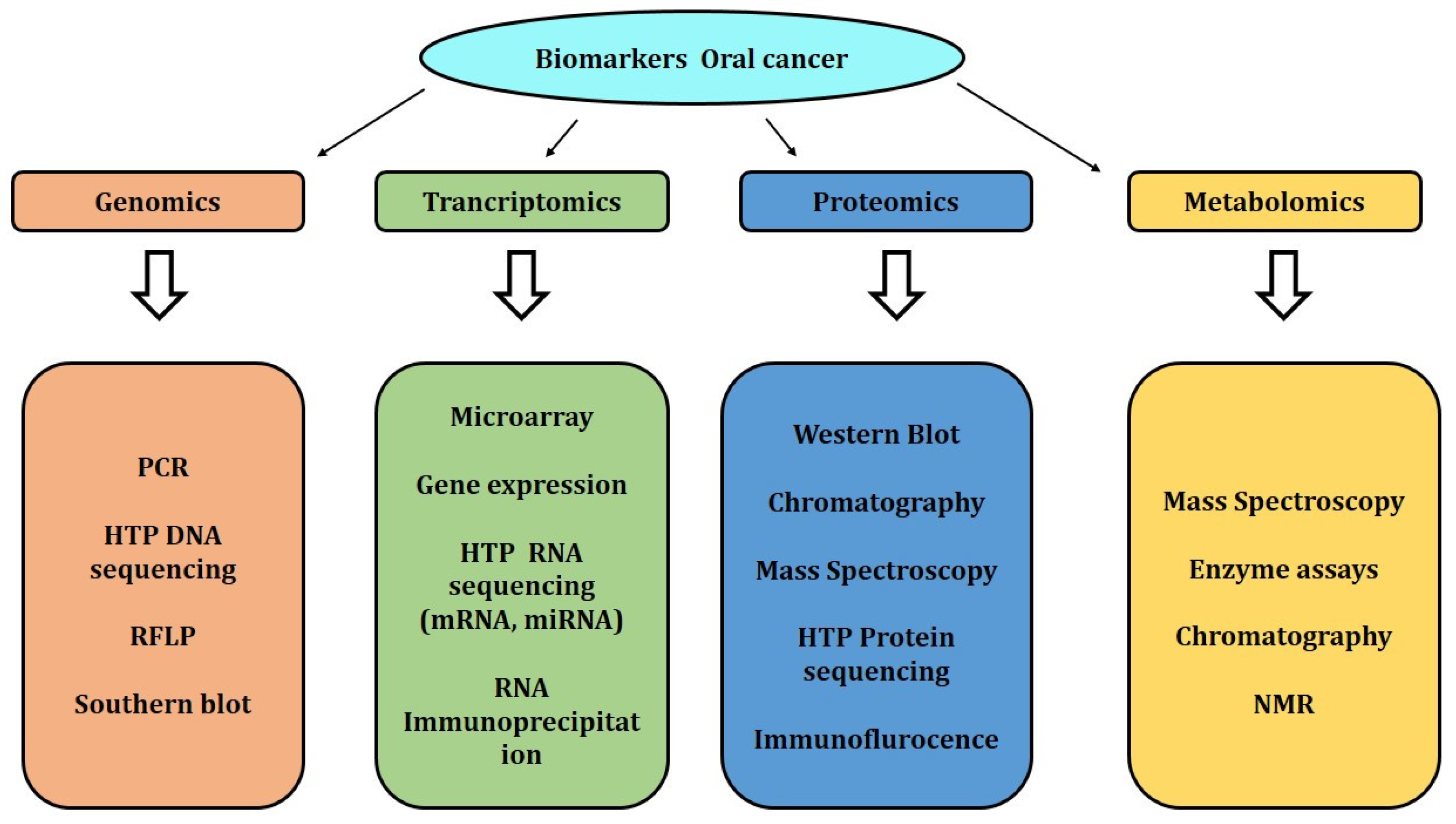
Figure 1. Various molecular methods involved in the detection of biomarkers.
Applsci 12 04926 g001
Table 1. Traditional methods for detecting oral cancer.
MethodSample TypeHighlight
Physical examination (visual)Oral cavity (in situ)Low cost and decreased death and morbidity
Vital staining (visual)Oral tissueLow cost and non-invasive
HistopathologyOral tissueLarge sample can be analyzed
Biopsy
Brush biopsyOral cellsNon-invasive, painless, and low cost
Liquid biopsyBiofluidsFast, less invasive, and complete sample profile
Incisional biopsyOral tissueLess sample; specific and accurate
Exfoliative biopsyEpithelial cellsLow cost and minimum skills
Imaging Techniques
CT scanTissue (in situ)Rapid, good visualization
MRI scanTissue (in situ)Non-invasive and high-resolution image
Optical coherence topographyOral tissueNon-invasive and high-resolution image
Molecular method
PCRBiofluids, cellsHigh sensitivity and reproducibility
Mass spectroscopyTissue (proteins/lipids)Accurate and high specificity
Table 2. Biomarkers associated with the data source and technologies used to identify oral cancer and OSCC.
Sl. No.BiomarkerSample TypeTechnique Used
Protein biomarkers
1CollagenOral tissueDiffuse reflectance spectroscopy
2Elastin
3Keratin
4FAD
5NADH
6Cytokeratin-19Oral tissueIncisional biopsy
7Secretory leukocyte protease inhibitorOral cellsBrush biopsy
8Epidermal growth factor receptor (EGFR)Oral epithelial cell liningsExfoliative biopsy
9Serum proteinsSerumHPLC
10ProteasesSalivaELISA
11ANXA2
12CA2
13CD44
14CRNN
15CST3
16CSTA
17DSG3
18FLNA
19FSCN1
20GANAB
21GSTP1
22HMGCS1
23HSPA5
24IGFBP3
25ISG15
26KNG1
27LDHA
28LGALS3BP
29MMP1
30MMP3
31MMP9
32PRDX2
33S100A9
34SPARC
35STAT1
36TIMP1
37TYMP
38YWHAB
39Interleukin-6 (IL-6)
40 Interleukin-8 (IL-8)
41Interleukin 1a (IL-1a)
42Interleukin 1b (IL-1b)
43TNF-a
44Tissue polypeptide antigen (TPA)
45Cyfra 21-1
46Cancer antigen 125 (CA 125)
47Telomerase
48Mac-2 binding protein (M2BP)SalivaELISA, shotgun proteomics
49CD44SalivaImmunoblot analysis
50CD59
51Profilin
52MRP14
53GlutathioneSalivaHPLC
54Squamous cell carcinoma antigen 2SalivaELISA, shotgun proteomics
55Involucrin
56Calcyclin
57Cathepsin-G
58Azurocidin
59Transaldolase
60Carbonic anhydrase I
61Calgizzarin
62Myeloblastin
63Vitamin D–binding protein
64Immunoglobulin heavy-chain constant region gamma (IgG)SalivaLC/MS
65S100 calcium-binding protein
66Cofilin-1
67Transferrin
68Fibrin
69Alpha-1-antitrypsin (AAT)Saliva2DE
70Secretory leukocyte peptidase inhibitor (SLPI)SalivaMS-based approaches
71Cystatin A
72Keratin 36
73Thioredoxin
74Haptoglobin (HAP)
75Salivary zinc finger
76Protein 510 peptide
77A-amylase
78Albumin
79OPN (osteopontin)Human biopsyWestern blot
80EZM2Tissue-bank sample
81DEPDC1BHuman biopsyImmunoprecipitation, Northern blot, Western blot
82P63Human biopsyMicroarray gene expression
83DeltaNp63Human biopsyCell membrane immunoreactivity, microscope
84EIC
85Podoplanin
86EGFRHuman biopsyFISH
DNA-based biomarkers
87DNA (promoter hypermethylation)SalivaPCR, qPCR, microarrays
88Histone family 3 (HA3)
89S100 calcium-binding protein P (S100P)
90Spermidine/spermine N1- acetyltransferase EST (SAT)
91Ornithine decarboxylase antizyme 1 (OAZ)
92P53 gene codon 63
93CDH1Oral tissueDNA Microarray
94MMP3
95SPARC
96POSTN
97TNC
98TGM3
99HMGA1
100PABPC1
101NT5E
102FOS
103FASN
104P53
105Cytochrome co-oxidase ISalivaPCR, qPCR, microarrays
106Cytochrome co-oxidase II
107DAPK
108DCC
109TIMP-31
110TIMP-3
111MGMT
112CCNA1
113MINT-31
114DNMT3BHuman biopsyMicroarray gene expression
RNA-based biomarkers
115miR-21Tissue-bank samplemirVana™, microarray gene expression, qPCR
116Has-miR-101Human biopsyMicroarray gene expression
117 miR-155-5p Tissue Gene expression, micro-RNA expression
118 miR-216a
119 miR-21-3p
120 miR-96-5p
121 miR-141-3p
122 miR-130b-3p
123 miR-21-5p
124 miR-483-5p SerumGene expression, micro-RNA expression
125 miR-31-5p Saliva Gene expression, micro-RNA expression
126 miR-31 Human tissue qRT-PCR
127 miR-21
128 miR-92b
129 miR-34a Human tissue Human biopsy, TaqMan miRNA assays
130 miR-139-5p Saliva Agilent miRNA microarray, qPCR
131 miR-203 Saliva qPCR
132IL-1b, IL-8SalivaELISA
133Dual-specificity phosphatase 1 (DUSP1)SalivaqPCR and microarrays
134H3 histone family 3A(H3F3A)
135Long noncoding HOTAIR
136miR-125a, miR-200a, miR-31
Metabolomics-based biomarkers
137CadaverineSalivaCapillary electrophoresis–time-of-flight mass spectrometry (CE–TOF-MS) and HPLC with quadrupole/TOF-MS.
138Alanine
139Serine
140Glutamine
141Piperidine
142Taurine piperidine
143Choline
144Pyrroline hydroxycarboxylic acid
145Beta-alanine
146Alpha-aminobutyric acid betaine
147Tyrosine
148Leucine þ isoleucine
149Histidine
150Tryptophan
151Glutamic acid
152Threonine
153Carnitine
154Pipercolic acid
155Lactic acid
156Phenylalanine
157Valine
158HypoxanthineSalivaCapillary electrophoresis–time-of-flight mass spectrometry
159Guanine
160Guanosine
161Trimethylamine N-oxide
162Spermidine
163Pipecolate
164Methionine
Publisher’s Note: MDPI stays neutral with regard to jurisdictional claims in published maps and institutional affiliations.