Case Report

Treatment of Medial Collateral Ligament Injuries of the Knee with Focused Extracorporeal Shockwave Therapy: A Case Report

Department of Neurosciences, Physical Medicine and Rehabilitation School, University of Padua, 35128 Padua, Italy
*
Author to whom correspondence should be addressed.
Appl. Sci. 2022, 12(1), 234; https://doi.org/10.3390/app12010234
Submission received: 24 November 2021 / Revised: 21 December 2021 / Accepted: 22 December 2021 / Published: 27 December 2021

Abstract

:
Medial collateral ligament (MCL) injuries are the most frequent ligamentous injuries of the knee. Focused extracorporeal shock wave therapy (f-ESWT) is progressively expanding its field of application to many musculoskeletal pathologies. Although there is evidence surrounding the efficacy of f-ESWT in tendinopathies, no studies have described the usefulness of ultrasound (US)-guided f-ESWT in the treatment of ligament injuries. Herein, we report a case of a MCL injury treated with f-ESWT. Moreover, our case shows the importance of using ultrasonography in determining the effect of treatment. A 60-year-old man with a focal area of lesion in the deep fibers underwent 4 weekly sessions of US-guided f-ESWT to the injured ligament area. His pain decreased to a visual analog scale (VAS) of 3 at the end of the treatment and was completely resolved at the 1-month follow-up visit, with these results being maintained at 4-month follow-up. The US examination showed an initial deposition of “newly formed tissue” at the site of previous injury of the proximal MCL insertion, and a reduction in MCL thickness together with an improvement in echostructure. Based on this result, we speculate that non-surgical ligament injuries could be a new indication for f-ESWT. However, further investigation on the effects of f-ESWT for ligament injuries is needed.

1. Introduction

The medial collateral ligament (MCL) is a very complex apparatus connecting the medial surface of the femoral condyle to the tibia. Its function is to resist forces applied from the outside of the knee preventing the medial or inner part of the joint from widening. Moreover, the MCL is considered a static stabilizer. Its structure is triangular and slightly expansible. Its origin is located proximally on the medial epicondyle of the femur next to the adductor tubercle, whereas its distal attachment lies below the medial condyle of the tibia on its medial surface [1].
MCL injuries are the most frequent type of ligamentous injury of the knee, with a reported annual incidence of 0.24–7.3 per 1000 and a male-to-female ratio of 2:1 [1,2]. The primary mechanism of injury to the MCL results from a valgus force with the foot being fixed to the ground [3]. The severity of the damage can be subdivided into three grades, depending on the state of laxity highlighted by the valgus stress test at 30° knee flexion [4]: 3–5 mm (grade 1), 6–10 mm (grade 2), and >10 mm (grade 3). A further classification from the American Medical Association subdivided the MCL injuries into three grades based on the severity of ligament damage: grade I with minimal torn fibers without loss of integrity, grade II with a partial MCL tear, and grade III with disruption of the ligament [5].
MCL injuries often present as isolated lesions; however, they may be associated with other ligamentous injuries, more frequently involving the anterior cruciate ligament [1]. The diagnosis of an MCL lesion is mainly based on clinical evaluation and imaging. In particular, ultrasounds (USs) are a safe, non-invasive and fast procedure with very high reliability. USs can be easily used to study the ligament and characterize its impairment, evaluating the morphological features of the damage. Moreover, USs are useful in the classification of the lesion severity and are consequently very helpful in guiding the therapeutic decision. Finally, USs may be used as an outcome measure to evaluate the efficacy of treatment.
Several nonoperative or operative treatment options have been reported. When evaluating treatment plans, nonoperative options, such as bracing, are used to manage isolated grade 1 and 2 MCL injuries, while grade 3 lesions may require surgical intervention with a repair or reconstruction, depending on the presence of joint instability [6,7]. However, there are no unequivocal treatment recommendations, probably due to the conflicting classification schemes, especially in chronic injuries [8]. Since the spontaneous healing of ligaments is often incomplete, optimal treatment should induce the healing of injured tissues and the resolution of symptoms.
Among non-invasive therapeutic modalities, extracorporeal shockwave therapy (ESWT) has been progressively expanding its field of application to many musculoskeletal pathologies. Particularly, focused-ESWT (f-ESWT) generates a pressure field that is focused to an accurate area, where maximal pressure is achieved, at a determined depth of the tissues. In daily clinical practice, the pathologies commonly treated with ESWT include tendinopathies and enthesopathies (calcified and not) and bone, muscle, and skin diseases [9,10,11].
The rationale for the use of ESWT is based on the induction of a controlled inflammation in a tissue in order to sustain its regeneration [9]. This has been well demonstrated by several studies of basic and clinical research. For these reasons, besides the known efficacy of ESWT in different conditions, the application of ESWT in ligament injures may be considered. The scientific literature scarcely explored this application and the majority of the studies considered animal models [12,13]. To the best of our knowledge, there is no study that has described the usefulness of US-guided focused ESWT (f-ESWT) in the treatment and evaluation of ligament injury in human subjects. Herein, we report a case of MCL injury treated with f-ESWT. Moreover, our case shows the importance of using ultrasonography to determine the effect of treatment.

2. Materials and Methods
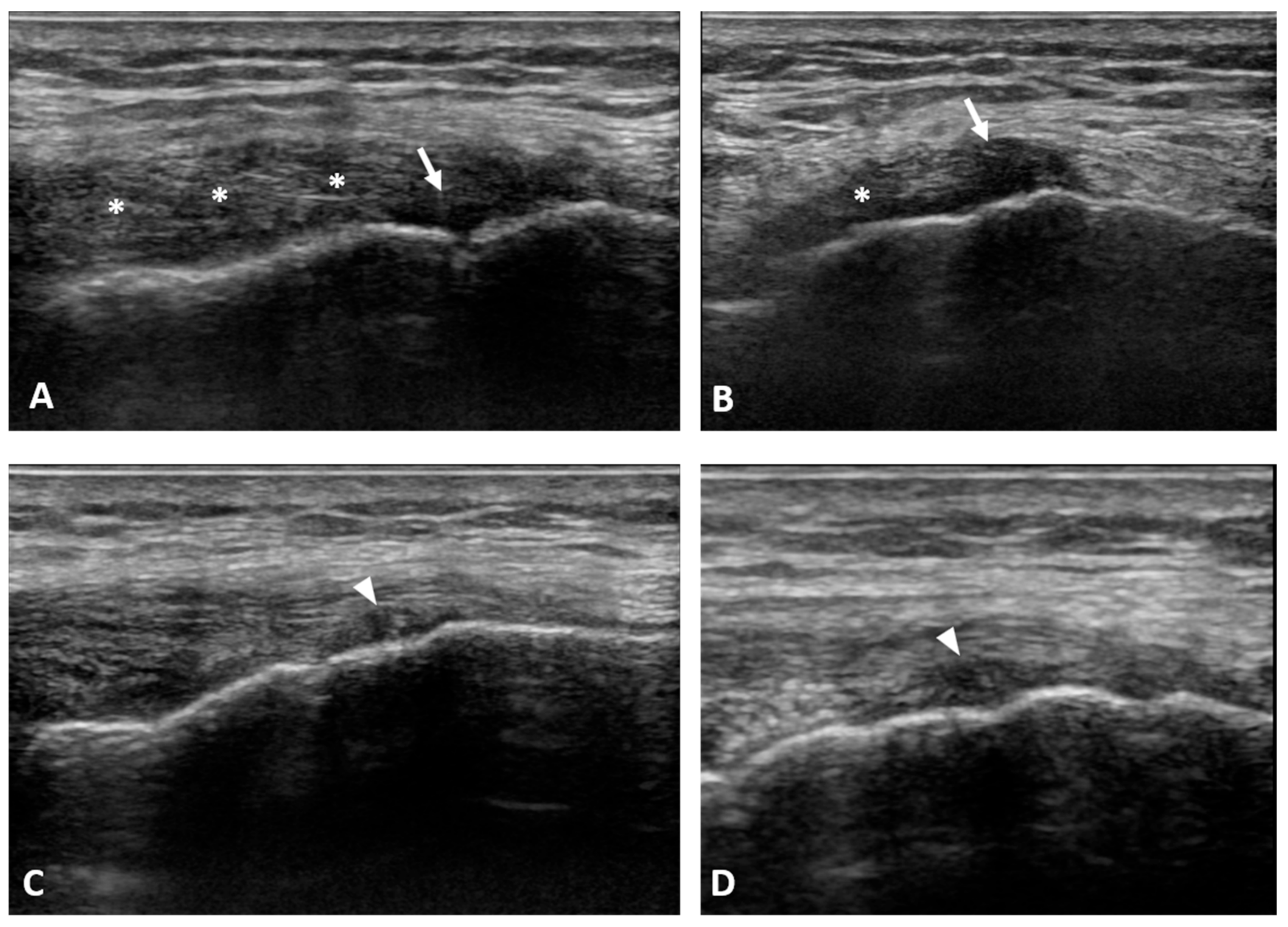
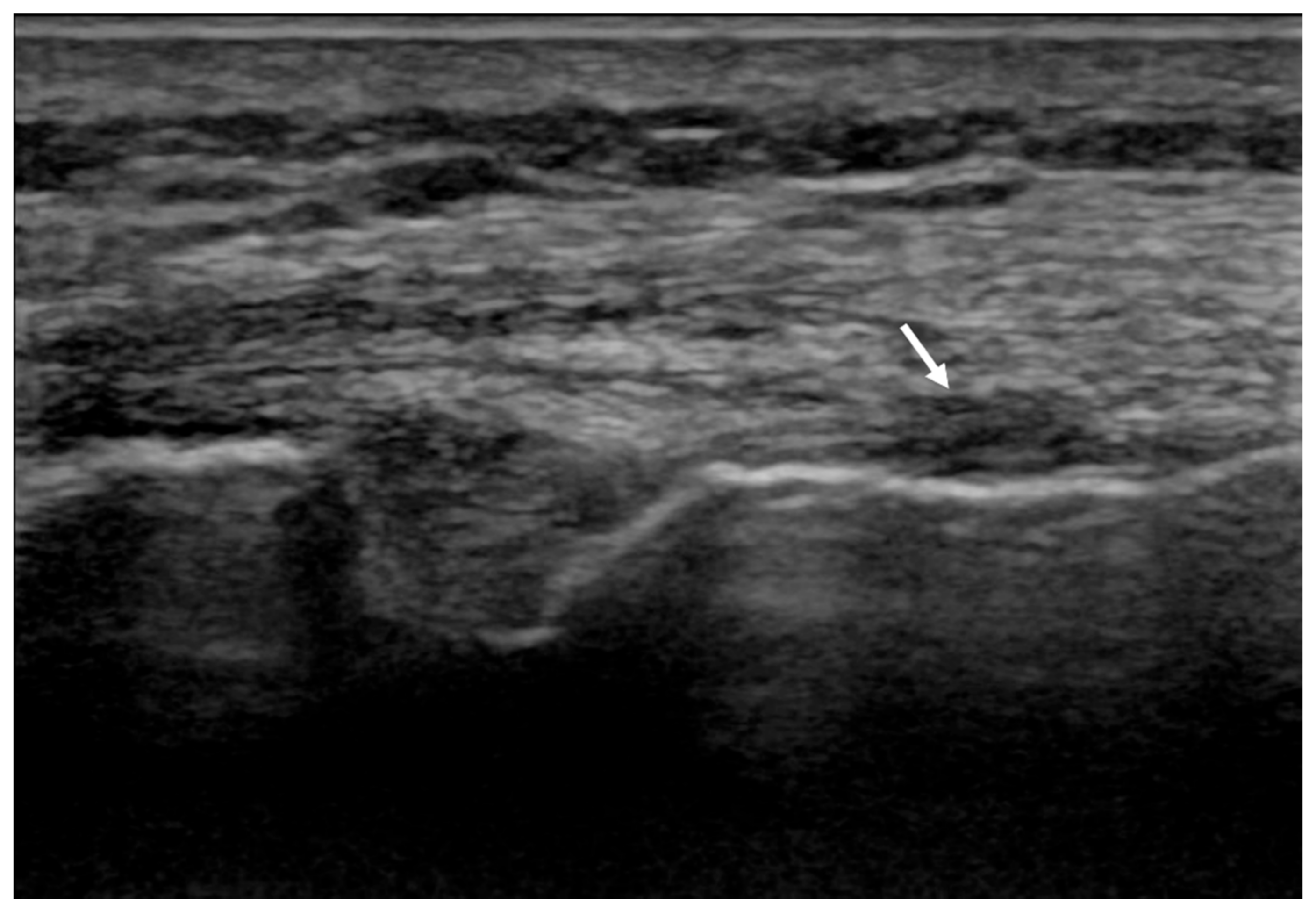
A 60-year-old male patient was referred to our physical medicine and rehabilitation clinics with a 12-month history of left knee pain without traumatic events except for a referred slight distortion trauma during walking 1 year earlier. He presented to the clinic with worsening pain in the last month that he rated as 6 on the visual analogue scale (VAS).
Patient reported medial knee pain that increased during weight bearing, walking, jumping, and running; it was absent if the knee was unloaded and at night. Patient was a non-smoker with a history of bullous pemphigus treated with topic corticosteroid application during periods of symptom exacerbation.
He previously underwent the following conservative treatments: oral analgesia, non-steroidal anti-inflammatory drugs (NSADs), and prohibition of sporting activities (hiking, running). However, the patient continued to complain of persistent pain.
Left-knee magnetic resonance imaging (MRI) from the previous year showed low-grade patella-femoral chondropathy and initial femorotibial osteoarthritis. Physical examination revealed bilateral high and externally rotated patellae. Pain on palpation along the medial collateral ligament with a positive valgus stress test on the left knee was associated with slight atrophy of the left quadricep femoris muscle. US examination at the painful area revealed a thickened and hypoechoic MCL ligament at its femoral insertion with a focal area of lesion at the deep fibers (Figure 1A,B).
We recommended 4 sessions of US-guided f-ESWT. It was also recommended to use a knee brace with medial support during standing and walking and avoidance of running, swimming, and cycling for 4 weeks. With the patient lying in a supine position and the affected limb slightly externally rotated and in a 30° knee flexion, an US examination was performed to identify the site of lesion. The target area was then marked with a pen to accurately deliver shock waves. Pressure pulses were focused on the marked area. A session of f-ESWT (device: Duolith® SD1 ultra, STORZ Medical, Tägerwilen, Switzerland) was given in the marked area under ultrasound guidance at a 4 Hz frequency for a total of 2000 shots using an Energy Flux Density (EFD) of 0.10 mJ/mm2. The treatment cycle consisted of 4 sessions of f-ESWT administered weekly. The patient received the f-ESWT in the same position as described above.
At the end of the treatment cycle, the patient was instructed to follow a home-based daily exercise protocol that included the range of motion exercises, quadricep strengthening activities, and hamstring stretching exercises until the 1-month and 4-month follow-up visits.

3. Results

The knee pain of patient slowly decreased during the 4 weeks after starting ESWT, with a VAS score of 3, at the last treatment session.
At the 1-month follow-up visit, a clinical and ultrasonographic evaluation was performed. Patient reported a complete resolution of knee pain (VAS 0). The US examination of MCL of the affected knee showed an initial deposition of “newly formed tissue” at the site of previous injury of the proximal MCL insertion. Moreover, a reduction in MCL thickness together with an improvement in echostructure was observed (Figure 1C,D). Such findings were associated with an improvement in knee function with the resumption of regular daily activity.
The clinical evaluation at 4 months post treatment revealed the continued complete absence of pain (VAS 0) with resumption of regular sports activities (walking, jogging) without any symptoms. Moreover, the US examination of the MCL showed a thinner ligament at the femoral insertion, with a clear improvement in echostructure (Figure 2).

4. Discussion

This is the first study to report a case of conservative treatment of non-surgical MCL injury with US-guided f-ESWT.
The spontaneous healing of tendons and ligaments is slow and often incomplete [14,15]. Moreover, prolonged immobilization should be avoided, since it has been demonstrated to enhance collagen degradation and bone resorption at the ligament insertion point [16]. However, due to the extra-articular location, MCL has a high healing potential compared with intra-articular ligaments (such as anterior cruciate ligament of the knee); therefore, most of the injuries can be treated conservatively and they rarely require a surgical intervention [8]. Despite this, due to the histological and volumetric characteristics of the mass of healing tissue, the chemical and biomechanical properties of the injuried MCL often fail to return to normal [17].
Among conservative therapies for the management of tendon injuries, ESWTs (and in particular f-ESWT) have gained increasing interest in recent years due to the induced biological responses, including tissue regeneration, wound healing, angiogenesis, and bone remodelling. Additionally, f-ESWT may also alleviate pain by stimulating analgesia [9]. However, to the best of our knowledge, few articles have described the effects of f-ESWT on ligament injury of in vitro and in vivo models, and no scientific works have been published regarding the usefulness of f-ESWT in the treatment of ligament injuries in humans [18,19].
Tendons and ligaments are types of fibrous connective tissue that connect muscle to bone and bone to bone, respectively. They represent crucial structures for the transmission of forces and the stabilization of the musculoskeletal system. Structurally and physiologically, tendons and ligaments are very similar. Additionally, the delineation between the two is not always clear and many of their characteristics overlap and differ at the same time (ECM composition, collagen I/collagen III and collagen/elastin ratio composition, orientation of collagen fibers) [20,21,22]. For this reason, it seems plausible to use the same type of physical energy as a form of conservative treatment even in the case of ligamentous injuries.
Previous studies have demonstrated the potential of ESWT in promoting bone healing and increasing the realignment of tendon and ligament fibers in vivo [23]. The hypothesis of f-ESWT leading to tissue regeneration is based on the mechanotransduction model: a mechanical stimulus applied to the cytoskeleton stimulates protein biosynthesis and proliferation. In vitro studies showed a protective role in tenocytes and an increased synthesis of type I collagen. Moreover, a significant increase in glycosaminoglycans (GAGs) and protein synthesis in tendons was observed [12]. These actions seems to be related to specific cellular signaling pathway activation, thus leading to increase in ATP release and activation, enhancing cell proliferation, differentiation, and regeneration [24,25].
Caminoto et al. evaluated the effects of ESWT on extra-cellular matrix components of affected ligaments in the hind limbs of horses, using ultrasonographic, ultrastructural, and immunocytochemical techniques [13]. Four weeks after treatment, ESWT-treated tissue demonstrated smaller, newly formed collagen fibrils and a greater expression of transforming growth factor beta (TGF-β), indicating that ESWT appears to facilitate the healing process. Moreover, a TGF-β1 inhibitor activity on macrophage-mediated ECM degradation during wound healing has been demonstrated [26].
Another study reported that low-energy f-ESWT can lead to M2 macrophage phenotype differentiation, which is typically characterized by anti-inflammatory properties. Indeed, M2 macrophage activity induces IL-4, IL-5, IL-9, and IL-13 biosynthesis, as well as collagen synthesis, angiogenesis, and tissue remodeling [27].
f-ESWT for treatment of ligament injury represents a previously unexplored approach in humans. This study demonstrates that mild-energy shockwave therapy could represent a feasible and safe treatment for the management of non-surgical LCM injuries. Moreover, ultrasonography represents a very good modality not only for diagnosing extra-articular ligament injuries but also for evaluating the effect of ESWT during the follow-up period.
In conclusion, although further studies are needed to confirm the result, the reported case potentially opens the doors to expanding the potential applications of f-ESWT. This physical agent may be used for the conservative treatment of ligament lesions and may aid in the management of patients with these conditions. Additionally, the result should stimulate specific studies regarding this application. In particular, properly designed randomized placebo-controlled trials with longer follow-up periods should be conducted in order to generate sufficient evidence.

Author Contributions

L.T.: conceptualization, investigation, data curation, writing—original draft preparation; D.C.: writing—review and editing; A.B.: writing—review and editing; S.M.: supervision. All authors have read and agreed to the published version of the manuscript.

Funding

The APC was funded by STORZ Medical.

Institutional Review Board Statement

Ethical review and approval were waived for this treatment since it was part of ordinary clinical activity. All procedures performed in the study were in accordance with the ethical standards of the institutional and/or national research committee(s) and with the Helsinki Declaration.

Informed Consent Statement

Informed consent was obtained from the patient.

Conflicts of Interest

The authors declare no conflict of interest.

References

  1. Memarzadeh, A.; Melton, J.T. Medial collateral ligament of the knee: Anatomy, management and surgical techniques for reconstruction. Orthop. Trauma 2019, 33, 91–99. [Google Scholar] [CrossRef]
  2. Delong, J.M.; Waterman, B.R. Surgical Techniques for the Reconstruction of Medial Collateral Ligament and Posteromedial Corner Injuries of the Knee: A Systematic Review. Arthrosc. J. Arthrosc. Relat. Surg. 2015, 31, 2258–2272.e1. [Google Scholar] [CrossRef] [PubMed]
  3. Fetto, J.F.; Marshall, J.L. Medial collateral ligament injuries of the knee: A rationale for treatment. Clin. Orthop. Relat. Res. 1978, 132, 206–218. [Google Scholar] [CrossRef]
  4. Varelas, A.N.; Erickson, B.J.; Cvetanovich, G.L.; Bach, B.R. Medial Collateral Ligament Reconstruction in Patients with Medial Knee Instability: A Systematic Review. Orthop. J. Sports Med. 2017, 5, 2325967117703920. [Google Scholar] [CrossRef]
  5. American Medical Association. Committee on the Medical Aspects of Sports. In Standard Nomenclature of Athletic Injuries; American Medical Association: Chicago, IL, USA, 1966; pp. 99–100. [Google Scholar]
  6. Reider, B.; Sathy, M.R.; Talkington, J.; Blyznak, N.; Kollias, S. Treatment of Isolated Medial Collateral Ligament Injuries in Athletes with Early Functional Rehabilitation: A Five-year Follow-up Study. Am. J. Sports Med. 1994, 22, 470–477. [Google Scholar] [CrossRef]
  7. Indelicato, P.A.; Hermansdorfer, J.; Huegel, M. Nonoperative Management of Complete Tears of the Medial Collateral Ligament of the Knee in Intercollegiate Football Players. Clin. Orthop. Relat. Res. 1990, 256, 174–177. [Google Scholar] [CrossRef]
  8. Phisitkul, P.; James, S.L.; Wolf, B.; Amendola, A. MCL injuries of the knee: Current concepts review. Iowa Orthop. J. 2006, 26, 77–90. [Google Scholar]
  9. Simplicio, C.L.; Purita, J.; Murrell, W.; Santos, G.S.; dos Santos, R.G.; Lana, J.F.S.D. Extracorporeal shock wave therapy mechanisms in musculoskeletal regenerative medicine. J. Clin. Orthop. Trauma 2020, 11, S309–S318. [Google Scholar] [CrossRef]
  10. Schroeder, A.N.; Tenforde, A.S.; Jelsing, E.J. Extracorporeal Shockwave Therapy in the Management of Sports Medicine Injuries. Curr. Sports Med. Rep. 2021, 20, 298–305. [Google Scholar] [CrossRef]
  11. Tognolo, L.; Giordani, F.; Biz, C.; Bernini, A.; Ruggieri, P.; Stecco, C.; Frigo, A.C.M.S. Myofascial points treatment with focused extracorporeal shock waves (f-ESW) for plantar fasciitis: An open label randomized clinical trial. Eur. J. Phys. Rehabil. Med. 2021. [Google Scholar] [CrossRef]
  12. Bosch, G.; de Mos, M.; van Binsbergen, R.; van Schie, H.T.M.; van de Lest, C.H.A.; van Weeren, P.R. The effect of focused extracorporeal shock wave therapy on collagen matrix and gene expression in normal tendons and ligaments. Equine Vet. J. 2009, 41, 335–341. [Google Scholar] [CrossRef]
  13. Caminoto, E.H.; Alves, A.L.G.; Amorim, R.L.; Thomassian, A.; Hussni, C.A.; Nicoletti, J.L.M. Ultrastructural and immunocytochemical evaluation of the effects of extracorporeal shock wave treatment in the hind limbs of horses with experimentally induced suspensory ligament desmitis. Am. J. Vet. Res. 2005, 66, 892–896. [Google Scholar] [CrossRef]
  14. Paavola, M.; Kannus, P.; Järvinen, T.A.; Khan, K.; Józsa, L.J.M. Achilles tendinopathy. J. Bone Jt. Surg. Am. 2002, 84, 2062–2076. [Google Scholar] [CrossRef]
  15. Maffulli, N.; Kader, D. Tendinopathy of tendo Achillis. J. Bone Jt. Surg. Br. 2002, 84, 1–8. [Google Scholar] [CrossRef]
  16. Reider, B. Medial Collateral Ligament Injuries in Athletes. Sports Med. 1996, 21, 147–156. [Google Scholar] [CrossRef]
  17. Anderson, D.R.; Weiss, J.A.; Takai, S.; Ohland, K.J.; Woo, S.L.-Y. Healing of the medial collateral ligament following a triad injury: A biomechanical and histological study of the knee in rabbits. J. Orthop. Res. 1992, 10, 485–495. [Google Scholar] [CrossRef]
  18. Lin, T.-Y.; Chen, J.-T.; Chen, Y.-Y.; Chen, T.-W.; Lee, C.-L.; Chen, C.-H.; Huang, M.-H. The efficacy of ultrasound-guided extracorporeal shockwave therapy in patients with cervical spondylosis and nuchal ligament calcification. Kaohsiung J. Med. Sci. 2015, 31, 337–343. [Google Scholar] [CrossRef] [Green Version]
  19. Wang, C.-J.; Ko, J.-Y.; Chou, W.-Y.; Hsu, S.-L.; Ko, S.-F.; Huang, C.-C.; Chang, H.-W. Shockwave therapy improves anterior cruciate ligament reconstruction. J. Surg. Res. 2014, 188, 110–118. [Google Scholar] [CrossRef]
  20. Leong, N.L.; Kator, J.L.; Clemens, T.L.; James, A.; Dds, M.E.; Jiang, J. Tendon and Ligament Healing and Current Approaches to Tendon and Ligament Regeneration. J. Orthop. Res. 2020, 38, 7–12. [Google Scholar] [CrossRef]
  21. Bobzin, L.; Roberts, R.R.; Chen, H.-J.; Crump, J.G.; Merrill, A.E. Development and maintenance of tendons and ligaments. Development 2021, 148, 186916. [Google Scholar] [CrossRef]
  22. Rumian, A.P.; Wallace, A.L.; Birch, H. Tendons and ligaments are anatomically distinct but overlap in molecular and morphological features—A comparative study in an ovine model. J. Orthop. Res. 2007, 25, 458–464. [Google Scholar] [CrossRef]
  23. Waguespack, R.W.; Burba, D.J.; Hubert, J.D.; Vidal, M.A.; Lomax, L.G.; Chirgwin, S.R.; Lopez, M.J. Effects of Extracorporeal Shock Wave Therapy on Desmitis of the Accessory Ligament of the Deep Digital Flexor Tendon in the Horse. Vet. Surg. 2011, 40, 450–456. [Google Scholar] [CrossRef]
  24. Weihs, A.M.; Fuchs, C.; Teuschl, A.; Hartinger, J.; Slezak, P.; Mittermayr, R.; Redl, H.; Junger, W.G.; Sitte, H.; Rünzler, D. Shock Wave Treatment Enhances Cell Proliferation and Improves Wound Healing by ATP Release-coupled Extracellular Signal-regulated Kinase (ERK) Activation. J. Biol. Chem. 2014, 289, 27090–27104. [Google Scholar] [CrossRef] [Green Version]
  25. Burnstock, G.; Verkhratsky, A. Long-term (trophic) purinergic signalling: Purinoceptors control cell proliferation, differentiation and death. Cell Death Dis. 2010, 1, e9. [Google Scholar] [CrossRef] [Green Version]
  26. Werner, F.; Feinberg, M.W.; Jain, M.K.; Sibinga, N.E.; Wiesel, P.; Wang, H.; Lee, M.E. Transforming growth factor-b1 inhibits cytokinemediated induction of human metalloelastase in marcophages. J. Biol. Chem. 2000, 275, 25766–25773. [Google Scholar] [CrossRef] [Green Version]
  27. Lana, J.F.; Macedo, A.; Ingrao, I.L.G.; Huber, S.C.; Santos, G.S.; Santana, M.H.A. Leukocyte-rich PRP for knee osteoarthritis: Current concepts. J. Clin. Orthop. Trauma 2019, 10, S179–S182. [Google Scholar] [CrossRef]
Figure 1. The pre-treatment ultrasound (US) examination at the painful area revealed a thickened and hypoechoic MCL ligament at its proximal (femoral) insertion (asterisks) with a focal area of the lesion (arrows) at the deep fibers (A,B). At 1-month follow-up the US examination showed an initial deposition of “newly formed tissue” at the site of previous injury of the proximal MCL insertion (arrowheads), with an initial reduction in MCL thickness (C,D).
Applsci 12 00234 g001
Figure 2. The 4-month follow-up ultrasound (US) examination of the MCL at the site of the previous injury (arrow).
Applsci 12 00234 g002
Publisher’s Note: MDPI stays neutral with regard to jurisdictional claims in published maps and institutional affiliations.

