Fluorescence Lifetime Phasor Analysis and Raman Spectroscopy of Pigmented Organic Binders and Coatings Used in Artworks
Abstract
1. Introduction
2. Materials and Methods
2.1. Samples
2.2. Fluorescence Lifetime Setup
Phasor Analysis
2.3. Raman Spectroscopy
3. Results
4. Discussion
5. Conclusions
Author Contributions
Funding
Institutional Review Board Statement
Acknowledgments
Conflicts of Interest
References
- Lakowicz, J.R. Principles of Fluorescence Spectroscopy, 3rd ed.; Springer: London, UK, 2006. [Google Scholar] [CrossRef]
- Hansell, P. Ultraviolet and Fluorescence Recording. In Photography for the Scientist; Engel, C., Ed.; Academic Press: London, UK; New York, NY, USA, 1968; pp. 363–382. [Google Scholar]
- Mairinger, F. UV-, IR- and x-ray Imaging. In Non-Destructive Microanalysis of Cultural Heritage Materials, Comprehensive Analytical Chemistry; Janssens, K., Van Grieken, R., Eds.; Elsevier: Amsterdam, The Netherlands, 2004; Volume 42, pp. 15–66. [Google Scholar]
- Comelli, D.; Valentini, G.; Cubeddu, R.; Toniolo, L. Fluorescence lifetime imaging for the analysis of works of art: Application to fresco paintings and marble sculptures. J. Neutron Res. 2006, 14, 81–90. [Google Scholar] [CrossRef]
- De la Rie, E.R. Fluorescence of Paint and Varnish Layers. Parts 1–3. Stud. Conserv. 1982, 27, 1–7, 65–69, 102–108. [Google Scholar] [CrossRef]
- Ghirardello, M.; Valentini, G.; Toniolo, L.; Alberti, R.; Gironda, M.; Comelli, D. Photoluminescence imaging of modern paintings: There is plenty of information at the microsecond timescale. Microchem. J. 2020, 154, 104618. [Google Scholar] [CrossRef]
- Anglos, D.; Georgiou, S.; Fotakis, C. Lasers in the analysis of cultural heritage materials. J. Nano Res. 2009, 8, 47–60. [Google Scholar] [CrossRef]
- Romani, A.; Clementi, C.; Miliani, C.; Favaro, G. Fluorescence Spectroscopy: A Powerful Technique for the Noninvasive Characterization of Artwork. Acc. Chem. Res. 2010, 43, 837–846. [Google Scholar] [CrossRef]
- Verri, G.; Saunders, D. Xenon Flash for Refectance and Luminescence (Multispectral) Imaging in Cultural Heritage Applications. Br. Mus. Tech. Bull. 2014, 8, 83–92. [Google Scholar]
- Thoury, M.; Elias, M.; Frigerio, J.M.; Barthou, C. Nondestructive varnish identification by ultraviolet fluorescence spectroscopy. Appl. Spectrosc. 2007, 61, 1275–1282. [Google Scholar] [CrossRef]
- Nevin, A.; Comelli, D.; Valentini, G.; Cubeddu, R. Total Synchronous Fluorescence Spectroscopy Combined with Multivariate Analysis: Method for the Classification of Selected Resins, Oils, and Protein-Based Media Used in Paintings. Anal. Chem. 2009, 81, 1784–1791. [Google Scholar] [CrossRef]
- Nevin, A.; Spoto, G.; Anglos, D. Laser spectroscopies for elemental and molecular analysis in art and archaeology. Appl. Phys. A 2012, 106, 339–361. [Google Scholar] [CrossRef]
- Striova, J.; Dal Fovo, A.; Fontana, R. Reflectance imaging spectroscopy in heritage science. Riv. Nuovo Cim. 2020, 43, 515–566. [Google Scholar] [CrossRef]
- Clementi, C.; Miliani, C.; Verri, G.; Sotiropoulou, S.; Romani, A.; Brunetti, B.G.; Sgamellotti, A. Application of the Kubelka-Munk Correction for Self-Absorption of Fluorescence Emission in Carmine Lake Paint Layers. Appl. Spectrosc. 2009, 63, 1323–1330. [Google Scholar] [CrossRef] [PubMed]
- Nevin, A.; Cesaratto, A.; Bellei, S.; D’Andrea, C.; Toniolo, L.; Valentini, G.; Comelli, D. Time-resolved photoluminescence spectroscopy and imaging: New approaches to the analysis of cultural heritage and its degradation. Sensors 2014, 14, 6338–6355. [Google Scholar] [CrossRef]
- Elias, M.; Magnain, C.; Barthou, C.; Nevin, A.; Comelli, D.; Valentini, G. UV-fluorescence spectroscopy for identification of varnishes in works of art: Influence of the underlayer on the emission spectrum. In O3A: Optics for Arts, Architecture, and Archaeology II; International Society for Optics and Photonics: Bellingham, WA, USA, 2009; Volume 7391, p. 739104. [Google Scholar]
- Verri, G.; Clementi, C.; Comelli, D.; Cather, S.; Piqueé, F. Correction of ultraviolet-induced fluorescence spectra for the examination of polychromy. Appl. Spectrosc. 2008, 62, 1295–1302. [Google Scholar] [CrossRef] [PubMed]
- Nevin, A.; Comelli, D.; Valentini, G.; Anglos, D.; Burnstock, A.; Cather, S.; Cubeddu, R. Time-resolved fluorescence spectroscopy and imaging of proteinaceous binders used in paintings. Anal. Bioanal. Chem. 2007, 388, 1897–1905. [Google Scholar] [CrossRef]
- Nevin, A.; Anglos, D. Assisted interpretation of laser-induced fluorescence spectra of egg-based binding media using total emission fluorescence spectroscopy. Laser Chem. 2006, 2006, 82823. [Google Scholar] [CrossRef][Green Version]
- Comelli, D.; Nevin, A.B.; Verri, G.; Valentini, G.; Cubeddu, R. Time-resolved fluorescence spectroscopy and fluorescence lifetime imaging for the analysis of organic materials in wall painting replicas. In Organic Materials in Wall Paintings; Project Report; Piqué, F., Verri, G., Eds.; Getty Conservation Institute: Los Angeles, CA, USA, 2015; pp. 83–96. [Google Scholar]
- Comelli, D.; Nevin, A.; Valentini, G.; Osticioli, I.; Castellucci, E.M.; Toniolo, L.; Gulotta, D.; Cubeddu, R. Insights into Masolino’s wall paintings in Castiglione Olona: Advanced reflectance and fluorescence imaging analysis. J. Cult. Herit. 2011, 12, 11–18. [Google Scholar] [CrossRef]
- Artesani, A.; Ghirardello, M.; Mosca, S.; Nevin, A.; Valentini, G.; Comelli, D. Combined photoluminescence and Raman microscopy for the identification of modern pigments: Explanatory examples on cross-sections from Russian avant-garde paintings. Herit. Sci. 2019, 7, 17. [Google Scholar] [CrossRef]
- Comelli, D.; Toja, F.; D’Andrea, C.; Toniolo, L.; Valentini, G.; Lazzari, M.; Nevin, A. Advanced non-invasive fluorescence spectroscopy and imaging for mapping photo-oxidative degradation in acrylonitrile–butadiene–styrene: A study of model samples and of an object from the 1960s. Polym. Degrad. Stab. 2014, 107, 356–365. [Google Scholar] [CrossRef]
- Osticioli, I.; Mendes, N.F.C.; Nevin, A.; Zoppi, A.; Lofrumento, C.; Becucci, M.; Castellucci, E.M. A new compact instrument for Raman, laser-induced breakdown, and laser-induced fluorescence spectroscopy of works of art and their constituent materials. Rev. Sci. Instrum. 2009, 80, 076109. [Google Scholar] [CrossRef]
- Martínez-Hernández, A.; Oujja, M.; Sanz, M.; Carrasco, E.; Detalle, V.; Castillejo, M. Analysis of heritage stones and model wall paintings by pulsed laser excitation of Raman, laser-induced fluorescence and laser-induced breakdown spectroscopy signals with a hybrid system. J. Cult. Herit. 2018, 32, 1–8. [Google Scholar] [CrossRef]
- Accorsi, G.; Verri, G.; Bolognesi, M.; Armaroli, N.; Clementi, C.; Miliani, C.; Romani, A. The exceptional near-infrared luminescence properties of cuprorivaite (Egyptian blue). Chem. Commun. 2009, 23, 3392–3394. [Google Scholar] [CrossRef] [PubMed]
- Grazia, C.; Clementi, C.; Miliani, C.; Romani, A. Photophysical properties of alizarin and purpurin Al(III) complexes in solution and in solid state. Photochem. Photobiol. Sci. 2011, 10, 1249–1254. [Google Scholar] [CrossRef]
- Lagarto, J.L.; Shcheslavskiy, V.; Pavone, F.S.; Cicchi, R. Real-time fiber-based fluorescence lifetime imaging with synchronous external illumination: A new path for clinical translation. J. Biophotonics 2020, 13, e201960119. [Google Scholar] [CrossRef]
- Lagarto, J.L.; Villa, F.; Tisa, S.; Zappa, F.; Shcheslavskiy, V.; Pavone, F.S.; Cicchi, R. Real-time multispectral fluorescence lifetime imaging using Single Photon Avalanche Diode arrays. Sci. Rep. 2020, 10, 8116. [Google Scholar] [CrossRef]
- Osete-Cortina, L.; Doménech-Carbó, M.T. Analytical characterization of diterpenoid resins present in pictorial varnishes using pyrolysis–gas chromatography–mass spectrometry with on line trimethylsilylation. J. Chromatogr. A 2005, 1065, 265–278. [Google Scholar] [CrossRef]
- Lluveras-Tenorio, A.; Spepi, A.; Pieraccioni, M.; Legnaioli, S.; Lorenzetti, G.; Palleschi, V.; Vendrell, M.; Colombini, M.P.; Tinè, M.R.; Duce, C.; et al. A multi-analytical characterization of artists’ carbon-based black pigments. J. Therm. Anal. Calorim. 2019, 138, 3287–3299. [Google Scholar] [CrossRef]
- Matteini, M. Le Patine: Genesi, Significato, Conservazione; Nardini: Firenze, Italy, 2005; pp. 1–110. [Google Scholar]
- Lagarto, J.L.; Shcheslavskiy, V.; Pavone, F.S.; Cicchi, R. Simultaneous fluorescence lifetime and Raman fiber-based mapping of tissues. Opt. Lett. 2020, 45, 2247–2250. [Google Scholar] [CrossRef]
- Liao, S.-C.; Sun, Y.; Ulas, C. FLIM Analysis Using the Phasor Plots; ISS, Inc.: Champaign, IL, USA, 2014; Volume 61822. [Google Scholar]
- Pelagotti, A.; Pezzati, L.; Bevilacqua, N.; Vascotto, V.; Reillon, V.; Daffara, C. A study of UV fluorescence emission of painting materials. In Proceedings of the 8th International Conference on Non-Destructive Investigations and Microanalysis for the Diagnostics and Conservation of the Cultural and Environmental Heritage (Art ’05), Lecce, Italy, 15–19 May 2005; p. A97. [Google Scholar]
- Vandenabeele, P.; Wehling, B.; Moens, L.; Edwards, H.; De Reu, M.; Van Hooydonk, G. Analysis with micro-Raman spectroscopy of natural organic binding media and varnishes used in art. Anal. Chim. Acta 2000, 407, 261–274. [Google Scholar] [CrossRef]
- Schoenemann, A.; Edwards, H. Raman and FTIR microspectroscopic study of the alteration of Chinese tung oil and related drying oils during ageing. Anal. Bioanal. Chem. 2011, 400, 1173–1180. [Google Scholar] [CrossRef] [PubMed]
- Osticioli, I.; Ciofini, D.; Mencaglia, A.A.; Siano, S. Automated characterization of varnishes photo-degradation using portable T-controlled Raman spectroscopy. Spectrochim. Acta A 2017, 172, 182–188. [Google Scholar] [CrossRef]
- Nevin, A.; Comelli, D.; Osticioli, I.; Toniolo, L.; Valentini, G.; Cubeddu, R. Assessment of the ageing of triterpenoid paint varnishes using fluorescence, Raman and FTIR spectroscopy. Anal. Bioanal. Chem. 2009, 395, 2139–2149. [Google Scholar] [CrossRef] [PubMed]
- Ciofini, D.; Striova, J.; Camaiti, M.; Siano, S. Photo-oxidative kinetics of solvent and oil-based terpenoid varnishes. Polym. Degrad. Stab. 2016, 123, 47–61. [Google Scholar] [CrossRef]
- Edwards, H.G.; Farwell, D.W.; Daffner, L. Fourier-transform Raman spectroscopic study of natural waxes and resins. I. Spectrochim. Acta Part A 1996, 52, 1639–1648. [Google Scholar] [CrossRef]
- Boucherit, N.; Hugot-Le Goff, A.; Joiret, S. Raman studies of corrosion films frown on Fe and Fe-6Mo in pitting conditions. Corros. Sci. 1991, 32, 497–507. [Google Scholar] [CrossRef]
- Froment, F.; Tournié, A.; Colomban, P. Raman identification of natural red to yellow pigments: Ochre and iron-containing ores. J. Raman Spectrosc. 2008, 39, 560–568. [Google Scholar] [CrossRef]
- Hanesh, M. Raman spectroscopy of iron oxides and (oxy)hydroxides at low laser power and possible applications in environmental magnetic studies. Geophys. J. Int. 2009, 177, 941–948. [Google Scholar] [CrossRef]
- Kendrix, E.; Moscardi, G.; Mazzeo, R.; Baraldi, P.; Prati, S.; Joseph, E.; Capelli, S. Far infrared and Raman spectroscopy analysis of inorganic pigments. J. Raman Spectrosc. 2008, 39, 1104–1112. [Google Scholar] [CrossRef]
- Coccato, A.; Jehlicka, J.; Moens, L.; Vandenabeele, P. Raman spectroscopy for the investigation of carbon-based black pigments. J. Raman Spectrosc. 2015, 46, 1003–1015. [Google Scholar] [CrossRef]
- Tomasini, E.P.; Halac, E.B.; Reinoso, M.; Di Liscia, E.J.; Maier, M.S. Micro-Raman Spectroscopy of Carbon-based Black Pigments. J. Raman Spectrosc. 2012, 43, 1671–1675. [Google Scholar] [CrossRef]
- Kuimova, M.K.; Yahioglu, G.; Levitt, J.A.; Suhling, K. Molecular rotor measures viscosity of live cells via fluorescence lifetime imaging. J. Am. Chem. Soc. 2008, 130, 6672–6673. [Google Scholar] [CrossRef] [PubMed]
- Haidekker, M.A.; Theodorakis, E.A. Molecular rotors—Fluorescent biosensors for viscosity and flow. Org. Biomol. Chem. 2007, 5, 1669–1678. [Google Scholar] [CrossRef] [PubMed]
- Hötzer, B.; Ivanov, R.; Altmeier, S.; Kappl, R.; Jung, G. Determination of copper(II) ion concentration by lifetime measurements of green fluorescent protein. J. Fluoresc. 2011, 21, 2143–2153. [Google Scholar] [CrossRef] [PubMed]


| Samples Description | Acronym | Mean τ-phase [ns] with (St. Dev.) | Principal Raman Signals [cm−1] |
|---|---|---|---|
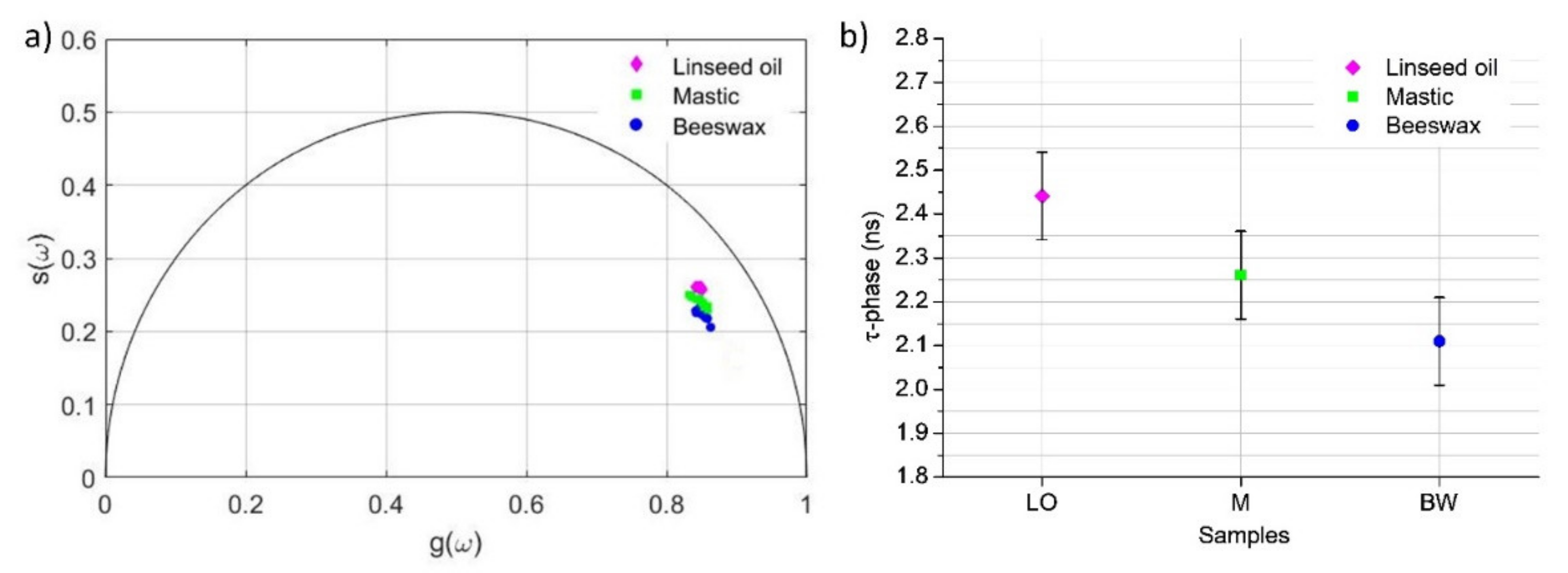
| Linseed oil | LO | 2.44 (0.10) | 727w, 866m, 972w, 1024w, 1078m, 1268ms, 1302ms, 1444s, 1658vs, 2727w, 2885m, 2932w, 3017w |
| Mastic | M | 2.26 (0.10) | 468w, 533w, 596m, 722m, 937m, 1200m, 1271w, 1315w, 1441ms, 1461sh, 1653ms, 1698sh, 2884sh, 2926m |
| Beeswax | BW | 2.11 (0.10) | 889w, 1063s, 1131s, 1172w, 1296s, 1419m, 1441s, 1464m, 2726w, 2850m, 2929sh |
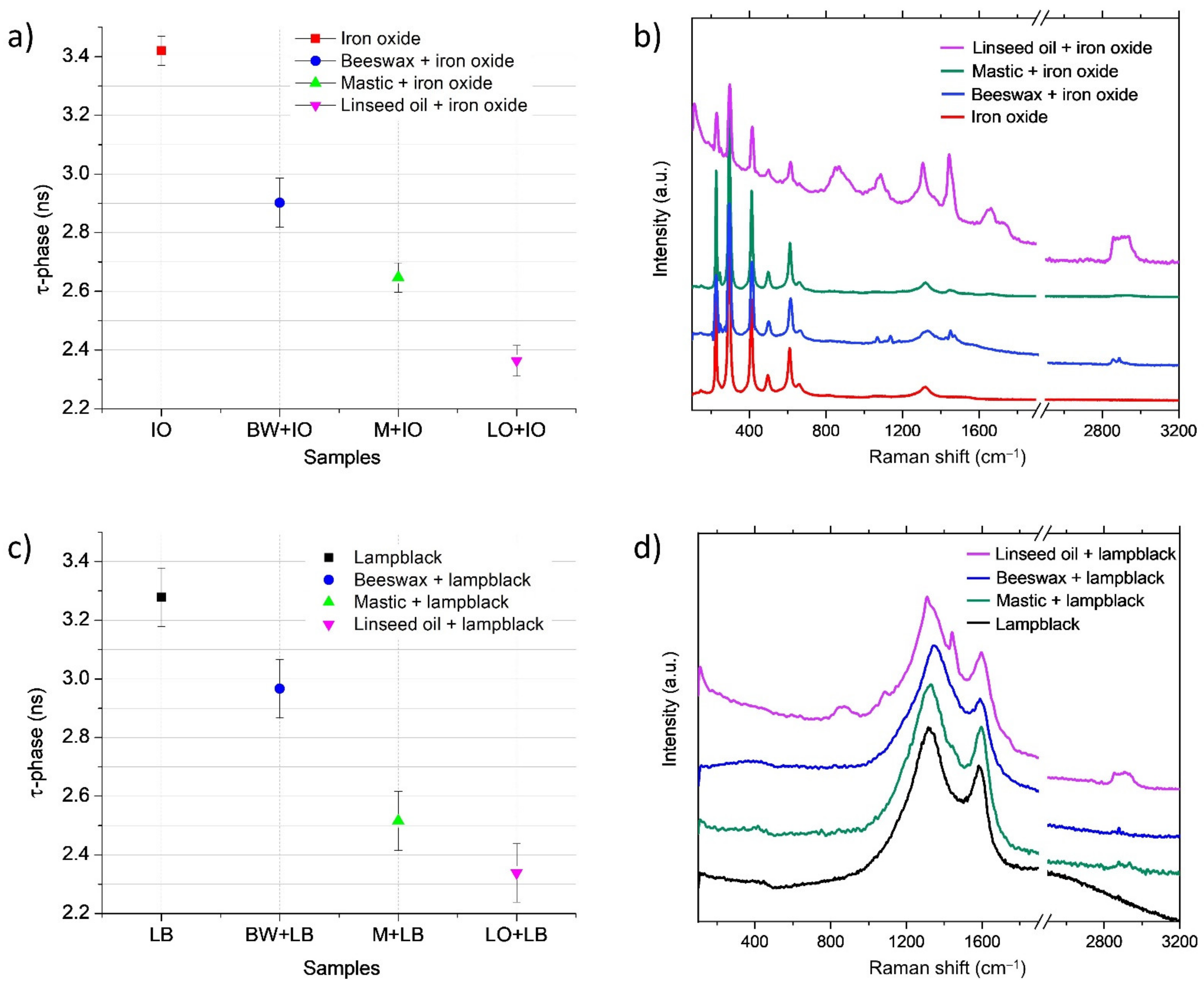
| Iron oxide | IO | 3.42 (0.10) | 146w, 225s, 293vs, 410s, 497m, 611ms, 659br, 1317br |
| Lampblack | LB | 3.28 (0.12) | 1315s,br; 1591s,br |
| Beeswax + iron oxide on glass | BW + IO | 2.90 (0.17) | 225s *, 293vs *, 411s *, 498m *, 612ms *, 671br *, 1062m, 1131m, 1321br *, 1439m, 1464br, 2848w, 2883w |
| Mastic + iron oxide on glass | M + IO | 2.65 (0.10) | 147w *, 226s *, 293vs *, 412s *, 498m *, 612ms *, 662br *, 1320br *, 1448br, 1656w,br |
| Linseed oil + iron oxide on glass | LO + IO | 2.36 (0.10) | 227s *, 294vs *, 413s *, 495m *, 611m *, 866m,br, 1085m, 1303ms, 1441ms, 1662br, 1733br |
| Beeswax + lampblack on glass | BW + LB | 2.97 (0.16) | 1315s,br *; 1597s,br * |
| Mastic + lampblack on glass | M + LB | 2.52 (0.10) | 1314s,br *; 1445br, 1588s,br *, 2880br,sh, 2935br |
| Linseed oil + lampblack on glass | LO + LB | 2.34 (0.10) | 872br, 1087w, 1313s *, 1441s, 1596s *, 1732sh, 1856m, 2910m, 2936m |
| Linseed oil on bronze | LO(b) | 2.26 (0.10) | 1064w, 1080w, 1305m, 1439ms, 1654br, 1723br |
| Oleoresin (Linseed oil + mastic on bronze) | OR(b) | 1.43 (0.10) | 1063w, 1085w,1308m, 1444m, 1647br, 2863br, 2934br |
| Linseed oil + mastic + beeswax + iron oxide on bronze | LO + M + BW + IO(b) | 1.65 (0.12) | 227s *, 293s *, 413m *, 465w, 612m,br *, 10632, 1134w, 1175w, 1296m, 1442s, 1461sh, 1653w,br, 2852m, 2883m, 2938br |
Publisher’s Note: MDPI stays neutral with regard to jurisdictional claims in published maps and institutional affiliations. |
© 2021 by the authors. Licensee MDPI, Basel, Switzerland. This article is an open access article distributed under the terms and conditions of the Creative Commons Attribution (CC BY) license (https://creativecommons.org/licenses/by/4.0/).
Share and Cite
Dal Fovo, A.; Mattana, S.; Chaban, A.; Quintero Balbas, D.; Lagarto, J.L.; Striova, J.; Cicchi, R.; Fontana, R. Fluorescence Lifetime Phasor Analysis and Raman Spectroscopy of Pigmented Organic Binders and Coatings Used in Artworks. Appl. Sci. 2022, 12, 179. https://doi.org/10.3390/app12010179
Dal Fovo A, Mattana S, Chaban A, Quintero Balbas D, Lagarto JL, Striova J, Cicchi R, Fontana R. Fluorescence Lifetime Phasor Analysis and Raman Spectroscopy of Pigmented Organic Binders and Coatings Used in Artworks. Applied Sciences. 2022; 12(1):179. https://doi.org/10.3390/app12010179
Chicago/Turabian StyleDal Fovo, Alice, Sara Mattana, Antonina Chaban, Diego Quintero Balbas, João Luis Lagarto, Jana Striova, Riccardo Cicchi, and Raffaella Fontana. 2022. "Fluorescence Lifetime Phasor Analysis and Raman Spectroscopy of Pigmented Organic Binders and Coatings Used in Artworks" Applied Sciences 12, no. 1: 179. https://doi.org/10.3390/app12010179
APA StyleDal Fovo, A., Mattana, S., Chaban, A., Quintero Balbas, D., Lagarto, J. L., Striova, J., Cicchi, R., & Fontana, R. (2022). Fluorescence Lifetime Phasor Analysis and Raman Spectroscopy of Pigmented Organic Binders and Coatings Used in Artworks. Applied Sciences, 12(1), 179. https://doi.org/10.3390/app12010179


