Article

Nano-Silver Particles Reduce Contaminations in Tissue Culture but Decrease Regeneration Rate and Slows Down Growth and Development of Aldrovanda vesiculosa Explants

by
Marzena Parzymies
Subdepartment of Ornamental Plants and Dendrology, Institute of Horticultural Production, Faculty of Horticulture and Landscape Architecture, University of Life Sciences in Lublin, Ul. Głęboka 28, 20-612 Lublin, Poland
Appl. Sci. 2021, 11(8), 3653; https://doi.org/10.3390/app11083653
Submission received: 31 March 2021 / Revised: 15 April 2021 / Accepted: 16 April 2021 / Published: 19 April 2021
(This article belongs to the Special Issue Interaction between Nanoparticles and Plants)

Abstract

:
Aldrovanda vesiculosa is a carnivorous water plant which is endangered by extinction worldwide. The number of natural stands and populations has decreased; therefore, there is a need for its active protection. The best method would be an in vitro culture. One of the main problems is disinfection of the explants. Therefore, it was decided that we should treat the explants with nano-silver particles. The explants were shoot fragments which were disinfected with sodium hypochlorite and then placed in a liquid 1/5 MS medium, supplemented with silver nanoparticles (AgNPs) at a concentration of 5 mg·dm−3. It was observed that AgNPs reduced the number of contaminations but also led to necrosis of the shoots. The shoots, which undertook regeneration in presence of AgNPs, were smaller and did not form traps; however, after being moved to fresh media twice, they started to develop normal leaves. Taking into consideration both disinfection and regeneration rates, it might be advisable to disinfect aldrovanda shoots in sodium hypochlorite only, without AgNPs. The results of the research might indicate a toxic activity of AgNPs towards water plants, which seems a big problem, as nanoparticles are commonly used in all the fields of life. However, the matter should be studied further.

1. Introduction

Aldrovanda vesiculosa L. (Drosearaceae), a waterwheel, is a carnivorous water species endangered by extinction all over the world, and it is under protection in all European countries. One of the biggest populations of the species is located in East Poland. Unfortunately, the number of natural stands and populations of Aldrovanda has decreased, both in Poland and worldwide [1]. According to Kamiński [2], in 1987, there were 14 confirmed localities of the waterwheel and, nowadays, there are only six left. The author investigated the factors affecting the survival of aldrovanda in the temperate climate of Poland, and suggested that the species should be reintroduced into new, better, and substitute localities. Aldrovanda vesiculosa does not form mature seeds in the Polish climate; therefore, it may be propagated only vegetatively. As the number of existing donor plants is low, it is important to use the methods which are the least harmful to the plants. Propagation in vitro allows for the production of the critically endangered plant species, as it allows us to obtain a lot of good quality and healthy plantlets, with no harm to the mother plants. Tissue culture has been commonly used for the propagation of many rare and endangered plant species [3,4,5].
There is little information on the micropropagation of A. vesiculosa. Kondo et al. [6] described the disinfection method and initiation of the aldrovanda cultures from seeds of the Japanese race of the species. The authors disinfected seeds with 1% benzalkonium chloride, sodium hypochlorite, and ethanol. The seeds were placed in B5 Gamborg liquid medium with 2% sucrose, the addition of 20–50 mg·dm−3 of gibberellic acid, and a few drops of antibiotic. After a day, the seeds were individually placed in the fresh medium of the same composition and kept at 22 °C under a light. After a week, 60% of the seeds germinated and formed shoots and proliferated. The authors managed to obtain regeneration from turions as well, but, at the same time, they stated that the growth of the Polish race is different.
Adamec et al. [7] studied the in vitro cultivation of aldrovanda, and they proposed the media for micropropagation of the species. The authors used the plants obtained from the Kondo cultivation. The authors studied the medium for aldrovanda micropropagation and suggested that the half strength of B5 with only 500 mg·dm−3 KNO3 was the best. They attempted to initiate the cultures from seeds but stated that, due to a high contamination rate (52.3%) and low germination (44.4%), the method was not reliable. Therefore, the authors advised using defoliated shoots and mild surface sterilization. However, the method itself was not provided.
Plant tissue cultures are commonly applied not only for the conservation of endangered plant species, but for wider use in the propagation of crop species, the production of bioactive compounds, or as models for scientific studies. However, to make all that possible, the plant material has to be disinfected before tissue culture initiation. That stage might decide the success of micropropagation, as only contamination-free cultures are used for further regeneration of plant material, which would lead to obtaining healthy and good quality plants.
To obtain in vitro cultures free of contaminations, the plant material has to be surface disinfected. It is extremely difficult to find a balance between the use of disinfection substances strong enough to get rid of any microorganisms, and mild enough to not kill the plants. The disinfection part is also highly important because remaining microorganisms usually grow fast in tissue culture. There are many sterilizing substances used for the surface disinfection of plant material. The most often used are calcium hypochlorite (CaOCl), sodium hypochlorite (NaOCl), ethanol (EtOH), hydrogen peroxide (H2O2), mercuric chloride (HgCl2), silver nitrate (AgNO3), fungicides, and antibiotics. Some disinfecting substances are harmful to plant tissues and, therefore, they negatively affect organogenesis, or even lead to necrosis due to their phytotoxicity. As such, the type, concentration and exposition time of disinfectants are important. Sometimes, it seems almost impossible to obtain pathogen-free cultures. Bacteria seem especially difficult to eliminate [8]. Therefore, researchers and plant producers look for new methods and substances.
One of the latest ideas is the use of nanomaterials, which are substances of very small size (1–100 nm) and unique properties [9]. There are different nano-substances, metals, and metal oxides, including silver (Ag), gold (Au), zinc (ZnO), copper (Cu), titanium dioside (TiO2), magnesium oxide (MgO), nickel (Ni), iron oxide (Fe3O4), and silicon (Si) [10,11]. At present, nanotechnology is present in every aspect of life—medicine, food, or the environment—and nanomaterials are available in different forms, such as powder, particles, clusters, tubes or films, and sizes. The best-known activity of nanoparticles (NPs) is its ability to eliminate different microorganisms [12]. Silver nanoparticles are the most often used nanomaterials in different sectors, especially in agriculture [13].
In the plant production sector, silver nanoparticles (AgNPs) were developed as plant growth stimulators [14] or components of new fertilizers [15], as well as plant protection products [16,17] and herbicides [18]. Recently, nanoparticles have been suggested as biostimulators that might improve plant propagation [19,20] or enhance fruit ripening [21,22]. NPs have also been used in plant tissue culture to improve plant growth, increase yield, improve seed germination [23], enhance bioactive compounds production, and enable genetic transformations [10,24,25]. AgNPs have been proven to reduce microbial contaminations in various plants and may be used for surface disinfection of explants. The antimicrobial property of silver NPs seems well studied. Its effectiveness depends on size, dimension, and type of nanoparticles [25]. According to Sarmast et al. [26], AgNPs show a high ability to eliminate microorganisms without negative effects on plant growth and development, but many researchers have studied the influence of AgNPs on explant disinfection and plant regeneration, and they indicated both positive and negative effects in the case of plants. Shokri et al. [27] suggested that substances used for sterilization are not neutral for the environment, humans or animals. Kim et al. [25] indicated that the influence of NPs on explants excised from different plant species should be studied to determine a proper method for AgNPs’ application in plant material disinfection and cultivation, especially given that plants are a basic element of the world and a source of food. Knowledge of AgNPs’ influence on plants is therefore important, especially because the common use of AgNPs increases the chance that they would be released into the environment [28,29,30]. Silver is the second most toxic metal to aquatic organisms after mercury [31], and, therefore, further studies are needed to clarify these contradictory observations concerning growth stimulation or inhibition of AgNP-treated plants [32]. Recently, there has been a different, green approach proposed. There are three possible means of nanoparticles synthesis: physical, chemical, and biological. The last one, concerning the biogenic synthesis of NPs, is described as nontoxic [33]. The “green biotechnology” methods include obtaining nanoparticles from plants’ organs, algae, or even microbes. It was possible to obtain AgNPs from the Daucus carota extract [34] or wheat straw extracted lignin [35]. The “green” methods are promising, but there is still little information on the subject.
The presented study aimed to evaluate the influence of the addition of nano-silver particles into the media on the survival rate of explants, tissue culture initiation, and regeneration ability, and then to evaluate the morphological features of regenerating shoots of Aldrovanda vesiculosa in tissue culture.
The manuscript describes two important matters. The described study is, to my knowledge, the first research concerning the influence of AgNPs on water plant growth and development in the presence of AgNPs. The toxicity of NPs towards such plants is of great importance, as the extensive use of these substances creates a risk that their residues might infiltrate natural water tanks and threaten plants, and, in turn, jeopardize biodiversity and the environment.
The other achievement described concerns the micropropagation of Aldrovanda vesiculosa, a carnivorous water plant species which is critically endangered by extinction. Because only a little amount of plant material is available, it is difficult to conduct experiments. Due to the hydration of tissues and content of different aquatic microorganisms and lake water in the traps, it is extremely difficult to obtain contamination-free in vitro cultures.

2. Materials and Methods
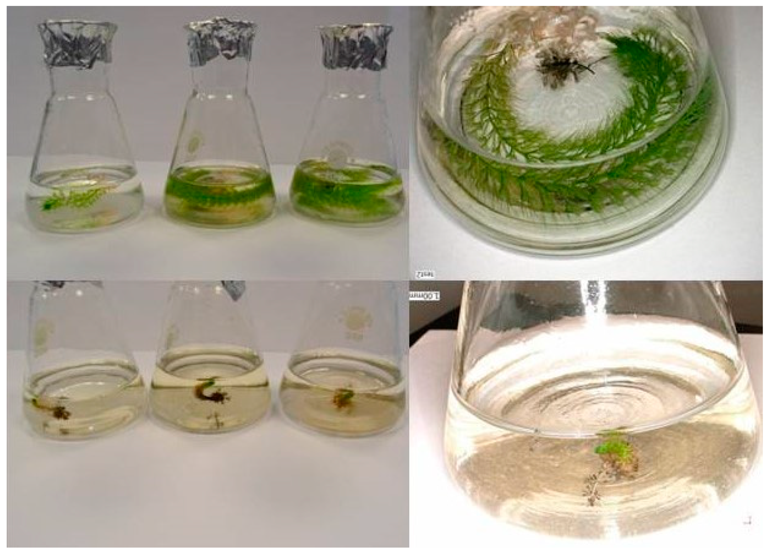
The plant material was Aldrovanda vesiculosa shoots with apex, collected from the Orchowe lake in East Poland, in June. The shoots were transported immersed in lake water in plastic containers. In the laboratory, the shoots were defoliated and the tip parts of 2–3 cm in length were used for the experiment. The explants were disinfected in the following steps: 1—rinse in water with a few drops of Ludwik (GRUPA INCO S.A., Warszawa, Poland), twice for 10 min; 2—dip in 70% ethanol (Chempur, Piekary Śląskie, Poland) for a few seconds; 3—disinfect with a 0.5% of sodium hypochlorite (NaOCl), (Chempur); 4—rinse 3 times in sterile distilled water and then place in a liquid media, described below. After 24 h, in the laminar flow chamber, the explants were taken out from the media and immersed for a few seconds in 0.5% NaOCl, 70% ethanol, or 0.5% NaOCl, followed by 70% ethanol. Then the explants were placed in the fresh liquid media of the same content, individually in the Erlenmeyer flasks of the 100 mL volume. Half of the media was supplemented with silver nanoparticles (AgNPs) solution (Sigma-Aldrich, Saint Louis, MO, USA, catalog no. 576832), at a concentration of 5 mg·dm−3.
The media used in the experiment (1/5 MS) consisted of 5-times diluted Murashige and Skoog (MS) [36] macro- and micronutrients composition, which is the most popular media and is recognized as suitable for most plant species. The media was diluted because Aldrovanda vesiculosa grows in peat bogs and lake water poor in nutrients, and, as a carnivorous plant, it is sensitive to high amounts of nitrogen.
The medium was supplemented with thiamine (vit. B1)—0.1 mg·dm−3 (Sigma-Aldrich, Saint Louis, MO, USA), pyridoxine (vit. B6)—0.5 mg·dm−3 (Sigma-Aldrich, Saint Louis, MO, USA), niacine (vit. PP)—0.5 mg·dm−3 (Sigma-Aldrich, Saint Louis, MO, USA), glycine—2.0 mg·dm−3 (Chempur, Poland), myo-inositol—100 mg·dm−3 (Sigma-Aldrich, Saint Louis, MO, USA), and sucrose—20 g·dm−3 (Chempur, Piekary Śląskie, Poland). The media pH was established at 5.5 and steam sterilized for 21 min at the temperature of 121 °C and under 1 hPa pressure. The flasks with explants were placed in a growing room at the temperature of 25 °C with 16-h photoperiod. The sources of light were fluorescent day-light lamps with 30 µmol·m−2·s−1 light intensity (Philips TLC 58W/84).
The experiment lasted for 8 weeks. The observations concerning the contamination rate and regeneration of the explants were performed once a week. At the end of the experiment, the following features were evaluated: the number of explants with the symptoms of contamination, the number of explants with the symptoms of necrosis, and the number of regenerating shoots. Morphological observations regarding the length of shoots, length of internodes, and length of petioles and traps, were also performed with the use of a Keyence VHX-950F stereoscopic microscope.
The regenerated and contamination-free shoots were placed in the fresh liquid media of the same macro- and micronutrient content, but without the addition of AgNPs, for further growth. Observations were performed after the next 8 weeks and the regenerated shoots were again placed in the fresh media. Passages were completed every 8 weeks, as it was the time when the regenerating shoots were long enough to be divided. Each time, only the green parts were used.

3. Results

It was noted that the sterilizing substance type and the presence of silver nanoparticles (AgNPs) in the media influenced disinfection efficiency (Table 1).
Based on visual observations, it was noted that the highest number of explants without the symptoms of disinfection was obtained when they had been disinfected with both ethanol and sodium hypochlorite and placed in the media supplemented with silver nanoparticles (100%). Unfortunately, that mixture caused necrosis in all the explants. Comparing the number of contamination-free explants, it might have been observed that sodium hypochlorite allowed us to obtain more disinfected stem pieces than did ethanol (138 in comparison to 36). Ethanol was the least efficient in the disinfecting of explants, as only 12% were free of fungus or bacteria. The addition of AgNPs increased the number of disinfected explants up to 24%. Looking at the data obtained, it was observed that supplementation of the media with AgNPs allowed for the obtaining of a higher number of disinfected explants in all treatments, but, at the same time, it decreased the number of regenerating shoots.
The highest number of regenerating explants was obtained when sodium hypochlorite was used for disinfection. Based on visual observations, it was noted that the addition of silver nanoparticles to the media influenced the morphological development of the shoots (Table 2, Figure 1 and Figure 2).
Shoots cultivated in the media supplemented with AgNPs regenerated much slower. They were smaller, with an average stem length of 8.24 mm and short internodes, less than 1 mm, while those cultivated in the media without AgNPs were much longer (over 10 cm), with longer internodes (2.48 mm on average). The biggest disadvantage of the use of silver ions was the lack of traps, which, in the case of a carnivorous plant species, allow to obtain nutrients. Shoots cultivated in the media without AgNPs formed traps on most of the shoots, and they were 1.52 mm long.
The shoots which regenerated after disinfection were placed in the fresh media and undertook further growth. Shoots that had been cultivated without the addition of AgNPs in the media grew normally, with a production of lateral shoots, while those that had been cultured in the presence of AgNPs were much smaller, with shorter internodes and no traps (Figure 3). However, after the second passage, the shoots started to produce longer petioles and traps (Figure 4).

4. Discussion

Disinfection of plant material is a crucial step in plants’ micropropagation, as only contamination-free explants may be used for tissue culture initiation. It is especially important in the case of plants endangered by extinction, for which only a limited amount of source explants are available. A reliable method of surface sterilization guarantees the success of tissue culture initiation, with no harm to donor specimens.
The presented paper concerns a disinfection method for Aldrovanda vesiculosa tissue culture. The species is critically endangered by extinction and present in only a few stands worldwide; therefore, it is important not to reduce existing populations. There is little information regarding surface sterilization methods for A. vesiculosa, and none of them concern the European races. Kondo et al. [6] initiated a tissue culture of the Japanese Hanyu race of aldrovanda from seeds, which were collected and surface sterilized with 1% benzalkonium chloride for 5 min, then 1–2% of sodium hypochlorite solution for 5 min, and then 70% ethanol for a few seconds. Next, the seeds were rinsed in distilled water 3–4 times and placed in a B5 Gamborg liquid medium supplemented with 2% sucrose, 20–50 mg·dm−3 of gibberellic acid, and some drops of penicillin and streptomycin, with the pH set up to 5.5 for a day. The next day, each seed was placed individually in the same medium in tubes and shaken under the light. After a week, 60% of the seeds germinated without any symptoms of contamination. The authors tried to initiate the cultures from shoot tips, but they failed, and stated that it was too difficult, as they contained many aquatic microorganisms in the traps. The authors managed to initiate the cultures from winter buds with the same method as seeds. At the same time, the authors stated that the growth of the Polish race is different.
The studies on the Polish race of A. vesiculosa were conducted by Adamec et al. [7]. However, the authors used the plant material obtained from Kondo. They tried to obtain a disinfected in vitro culture of aldrovanda from seeds, and they obtained over 44% germinated seedlings and over 52% contaminations. The authors stated that it was not sufficient, and recommended the use of defoliated shoot pieces and mild surface disinfection. However, the method was not described.
As the disinfection of aldrovanda is difficult and there is little plant material available, we decided to use silver nanoparticles to surface-disinfect the explants. Unfortunately, the use of AgNPs was disadvantageous, as it led to the obtaining of a high number of necrotic explants and inhibited regeneration of shoots. One of the latest ideas is the application of nanomaterials for the surface sterilization of plants.
There have been many experiments carried out to study the influence of AgNPs as a disinfection agent for plant material, and the harmful effects of nano-silver were proved on more than 600 microorganisms. Abdi et al. [37] applied AgNPs for the first time to control bacterial contaminations in Valeriana officinalis. They obtained single node explants from plants grown in a greenhouse and surface disinfected with 70% EtOH for 1 min, 10% Clorox for 1 min, and then applied AgNPs at a concentration of 100 mg·dm−3 for 180 min They obtained 89% contamination-free cultures and nano-silver did not affect either regeneration or rooting of plants. Similarly, Taghizadeh and Solgi [38] noted a positive effect of AgNPs application in Cynodon dactylon “Teefgreen”. They treated explants with 30% Clorox for 15 min, then rinsed them in distilled water and soaked for 30, 60, or 120 min in 100 or 200 mg·dm−3 AgNPs. The application of 200 mg·dm−3 of the solution decreased the total number of contaminations (11.1%), especially fungi ones (0%), which are the most common contaminants during tissue culture initiation. Moradpour et al. [39] obtained the highest percentage of disinfected and regenerating explants of Hevea brasiliensis when AgNPs were used at a concentration of 10 mg·dm−3 for 20 min, in comparison to mercuric chloride or sodium hypochlorite. The effect of AgNPs application was studied by Mahna et al. [40] in Arabidopsis thaliana seeds, potato leaves, and tomato cotyledons. The authors noted that the use of 100 mg·dm−3 for 1 or 5 min was ideal for disinfection, and the treatment had no negative effect on explants.
AgNPs are also applied in media supplementation. Sarmast et al. [26] observed that disinfection of Araucaria excelsa, combined with the immersion of explants in 200 mg·dm−3 AgNPs for 180 min, reduced the contamination rate from 61.5% to 11.3%, while the addition of 400 mg·dm−3 in the media decreased the contamination rate from 81.25% to 18.75%. Both methods were also used by Arab et al. [41], and the authors noted that both methods—explants immersion and media supplementation—reduced contamination rate in almond ×peach rootstock. Parzymies et al. [42] used AgNPs both as immersion and media supplementation in Pennisetum alopecuroides tissue culture initiation. They obtained the highest number of contamination-free explants when the explants were soaked in 100–250 mg·dm−3 AgNPs solution, or when the AgNPs were added to the media at a concentration of 4 mg·dm−3 for nodes and 16 mg·dm−3 for tillers. Spinoso-Castillo et al. [43] proved that the addition of 50 to 200 mg·dm−3 AgNPs to the medium completely decontaminated cultures of Vanilla planifolia. Supplementation of media with AgNPs was also used in the presented experiment, and it was observed that the treatment reduced the contamination rate, which confirms the results of other researchers.
The addition of AgNPs to the media, apart from reducing the contamination rate, may influence the growth and development of explants. Shokri et al. [44] noted that bacterial contamination was significantly reduced in the case of Rosa hybrid explants when they had been soaked in a solution of 200 mg·dm−3 of AgNPs for 20 min. However, supplementation of the medium with 100 mg·dm−3 AgNPs not only reduced the contamination rate but inhibited the phenolic compounds’ exudation as well.
The positive effect of the AgNPs media supplementation was observed in many plant species cultivated in vitro. In the case of Tecomella undulata, the number of regenerating shoots and their number increased when the MS medium contained 10 mg·dm−3 AgNPs [45], while the use of a higher concentration, 60 mg·dm−3, enhanced the number of shoots and their length [46]. The positive effect of the AgNPs use was also noted in the case of cowpea and brassica [47].
However, the use of nanomaterials gives rise to many doubts and questions, and they might be deposited in tissues [48]. The phytotoxicity of AgNPs is assessed with such features as growth potential, seed germination rate, biomass, or leaf surface area [49,50,51].
In the presented experiment, the use of AgNPs reduced the contamination rate of explants but noticeably inhibited the regeneration capacity and growth of shoots. A similar effect was proved by many researchers. Spinoso-Castillo et al. [43] studied the influence of AgNPs on Vanilla planifolia shoot regeneration and they obtained the highest shoot number of shoots, over 14, when the media contained 25 or 50 mg·dm−3 AgNPs, but the use of nano-silver at a higher concentration of 200 mg·dm−3 decreased the number of shoots to 4.55. In the case of Lupinus termis, the use of 0.1 to 0.5 mg·dm−3 of AgNPs reduced shoot and root elongation and fresh weight [51]. AgNPs reduced plant height and biomass in Capsicum annuum exposed to AgNPs 0.01 to 1 mg·dm−3 [22]. AgNPs inhibited the growth of many other plant species, such as Cucumis sativus [52], Phaseolus radiates, Sorghum bicolor [53], and Lemna minor [54].

5. Practical Applications and Future Research Perspectives

The results obtained in the presented experiment may be used for the tissue culture initiation of Aldrovanda vesiculosa worldwide, which may lead to active conservation of the species through in vitro propagation and reintroduction of the obtained plants into their natural habitat.
The conducted experiment has shown that AgNPs added to tissue culture media inhibited the growth and development of waterwheel plants. In the case of that carnivorous species, the lack of traps might prevent the plants from catching their prey. Therefore, further research should be conducted concerning the effect of AgNPs and other nanoparticles used at different concentrations on the development of both aldrovanda and other plant species in order to find a safe way to use these novel substances.

6. Conclusions

Nanomaterials have great potential to be used for plant tissue culture to disinfect the plant material, but their effect has to be tested on each plant species. Undoubtedly, AgNPs reduce microbial contaminations during tissue culture initiation, making it possible to establish a healthy culture but, on the other hand, they may have a negative effect on further regeneration of explants. In the case of Aldrovanda vesiculosa, it might be concluded that the inhibition of the regeneration capacity of explants outweighs the disinfection ability of the AgNPs used as a media supplementation. Therefore, it is advisable to surface-sterilize explants with sodium hypochlorite only.

Funding

This research was funded in part by the European Union through the Infrastructure and Environment Operational Programme, project title: Ochrona czynna aldrowandy pęcherzykowatej (Aldrovanda vesiculosa na terenie Lubelszczyzny (Active conservation of Aldrovanda vesiculosa in Lubelszczyzna region), no. POIS.02.04.00-00-0034/18.

Institutional Review Board Statement

Not applicable.

Informed Consent Statement

Not applicable.

Data Availability Statement

All the required data, which are related to the current study, are embedded in this manuscript.

Acknowledgments

The author would like to thank Alicja Świstowska, Magdalena Pogorzelec and Barbara Banach-Albińska for the technical support.

Conflicts of Interest

The author declares no conflict of interest.

References

  1. Kosiba, P.; Mróz, L.; Kamiński, R. Assessment of habitat conditions using self-organizing feature maps for reintroduction/introduction of Aldrovanda vesiculosa L. in Poland. Acta Soc. Bot. Pol. 2011, 80, 139–148. [Google Scholar] [CrossRef] [Green Version]
  2. Kamiński, R. Restitution of Aldrovanda vesiculosa L. in Poland and Designation of Factors Responsible for Its Surviving in Temperate Climate (Restytucja Aldrowandy Pęcherzykowatej i Rozpoznanie Czynników Decydujących o jej Przetrwaniu w Klimacie Umiarkowanym); Wydawnictwo Uniwersytetu Wrocławskiego: Wrocław, Poland, 2006; pp. 10–16, 30–37. [Google Scholar]
  3. Sharma, S.K.; Singh, R.; Arya, I.D. An efficient in vitro protocol for important and highly valuable medicinal plant Rauwolfia serpentina: An endangered medicinal plant of India. Vitr. Cell. Dev. Biol. Plant 2008, 44, 360–361. [Google Scholar]
  4. Holobiuc, I.; Blindu, R.; Cristea, V. Researches concerning in vitro conservation of the rare plant species Dianthus nardiformis Janka. Biotechnol. Biotechnol. Equip. 2009, 23 (Suppl. S1), 221–224. [Google Scholar] [CrossRef] [Green Version]
  5. Marszał-Jagacka, J.; Kromer, K. In vitro propagation of rare and endangered serpentine fern species. In Working with Ferns; Kumar, A., Fernández, H., Revilla, M., Eds.; Springer: New York, NY, USA, 2011; pp. 149–164. [Google Scholar] [CrossRef]
  6. Kondo, K.; Kokubugata, G.; Varghese, S.B.; Itoyama, M.; Breckpot, C.; Kromer, K.; Kamiński, R. Conservation of Aldrovanda vesiculosa by tissue culture. Carniv. Plant Newsl. 1997, 26, 86–92. [Google Scholar]
  7. Adamec, L.; Pásek, K. Medium optimization for growing Aldrovanda vesiculosa in vitro. Carniv. Plant Newsl. 2000, 29, 122–124. [Google Scholar]
  8. Leifert, C.; Cassels, A.C. Microbial hazards in plant tissue and cell cultures. Vitr. Cell. Dev. Biol. Plant 2001, 37, 133–138. [Google Scholar] [CrossRef]
  9. Rai, P.K.; Kumar, V.; Lee, S.; Raza, N.; Kim, K.H.; Ok, Y.S.; Tsang, D.C. Nanoparticle-plant interaction: Implications in energy environment, and agriculture. Environ. Int. 2018, 119, 1–19. [Google Scholar] [CrossRef] [PubMed]
  10. Ruttkay-Nedecky, B.; Krystofowa, O.; Nejdl, L.; Adam, V. Nanoparticles based on essential metals and their phytotoxicity. J. Nanobiotechnol. 2017, 15, 33. [Google Scholar] [CrossRef] [PubMed]
  11. Beyth, N.; Houri-Haddad, Y.; Domb, A.; Khan, W.; Hazan, R. Alternative antimicrobial approach: Nano-antimicrobial materials. Evid. Based Compl. Altern. Med. 2015, 2015, 246012. [Google Scholar] [CrossRef] [PubMed] [Green Version]
  12. Wang, P.; Hu, C.; Shao, L. The antimicrobial activity of nanoparticles: Present situation and prospects for the future. Int. J. Nanomed. 2017, 12, 1227–1249. [Google Scholar] [CrossRef] [Green Version]
  13. Yan, A.; Chen, Z. Impacts of silver nanoparticles on plants: A focus on the phytotoxicity and underlying mechanism. Int. J. Mol. Sci. 2019, 20, 1003. [Google Scholar] [CrossRef] [PubMed]
  14. Jasim, B.; Thomas, R.; Mathew, J.; Radhakrishnan, E.K. Plant growth and diosgenin enhancement effect of silver nanoparticles in Fenugreek (Trigonella foenum-graecum L.). Sauid Pharm. J. 2017, 25, 443–447. [Google Scholar] [CrossRef]
  15. Elmer, W.H.; White, J.C. The use of metallic oxide nanoparticles to enhance growth of tomatoes and eggplants in disease infested soil or soiless medium. Environ. Sci. Nano 2016, 3, 1072–1079. [Google Scholar] [CrossRef]
  16. Alavi, S.; Dehpour, A. Evaluation of the nanosilver colloidal solution in comparison with the registered fungicide to control greenhouse cucumber downy mildew disease in the north of Iran. Acta Hortic. 2010, 877, 1643–1646. [Google Scholar] [CrossRef]
  17. Worall, E.A.; Hamid, A.; Mody, K.T.; Mitter, N.; Pappu, H.R. Nanotechnology for plant disease management. Agronomy 2018, 8, 285. [Google Scholar] [CrossRef] [Green Version]
  18. Maruyama, C.R.; Guilger, M.; Pascoli, M.; Bileshy-José, N.; Abhilash, P.C.; Fraceto, L.F.; De Lima, R. Nanoparticles based on chitosan as carriers for the combined herbicides Imazapic and Imazapyr. Sci. Rep. 2016, 6, 19768. [Google Scholar] [CrossRef] [PubMed]
  19. Saha, N.; Gupta, S.D. Promotion of shoot regeneration of Swertia chirata by biosynthesized silver nanoparticles and their involvement in ethylene interceptions and activation of antioxidant activity. Plant Cell Tissue Org. Cult. 2018, 134, 289–300. [Google Scholar] [CrossRef]
  20. Thangavelu, R.M.; Gunasekaran, D.; Jesse, M.I.; SU, M.R.; Sundarajan, D.; Krishnan, K. Nanobiotechnology approach using plant rooting hormone synthesized silver nanoparticles as “nanobullets” for the dynamic applications in horticulture—An in vitro and ex vitro study. Arab. J. Chem. 2018, 11, 48–61. [Google Scholar] [CrossRef] [Green Version]
  21. Sah, S.; Soroosh Zadeh, A.; Rezazadeh, H.; Naghdi Badi, H. Effect of nano silver and silver nitrate on seed yield of borage. J. Med. Plants Res. 2011, 5, 706–710. [Google Scholar]
  22. Vinković, T.; Novák, O.; Strnad, M.; Goessler, W.; Jurašin, D.D.; Paradiković, N.; Vrćek, I.V. Cytokinin response in pepper plants (Capsicum annuum L.) exposed to silver nanoparticles. Environ. Res. 2017, 156, 10–18. [Google Scholar] [CrossRef]
  23. Parveen, A.; Rao, S. Effect of nanosilver on seed germination and seedling growth in Pennisetum glaucum. J. Clust. Sci. 2015, 26, 693–701. [Google Scholar] [CrossRef]
  24. Wang, P.; Lombi, E.; Zjao, F.J.; Kopittke, P.M. Nanotechnology: A new opportunity in plant sciences. Trends Plant Sci. 2016, 21, 699–712. [Google Scholar] [CrossRef] [PubMed]
  25. Kim, D.H.; Gopal, J.; Sivanesan, I. Nanomaterials in plant tissue culture: The disclosed and undisclosed. RSC Adv. 2017, 7, 36492–36505. [Google Scholar] [CrossRef] [Green Version]
  26. Sarmast, M.; Salehi, H.; Khosh-Khui, M. Nano silver treatment is effective in reducing bacterial contaminations of Araucaria excelsa R. Br. var. glauca explants. Acta Biol. Hung. 2011, 62, 477–484. [Google Scholar] [CrossRef] [PubMed]
  27. Shokri, S.; Babaei, A.; Ahmadian, M.; Hessami, S.; Arab, M.M. The effects of different concentrations of nano-silver on elimination of bacterial contaminations and phenolic exudation of rose (Rosa hybrida L.) in vitro culture. Int. J. Farm Allied Sci. 2014, 3, 50–54. [Google Scholar] [CrossRef] [Green Version]
  28. Hedberg, J.; Skoglund, S.; Karlsson, M.-E.; Wold, S.; Odnevall Wallinder, I.; Hedberg, Y. Sequential studies of silver released from silver nanoparticles in aqueous media simulating sweat, laundry detergent solutions and surface water. Environ. Sci. Technol. 2014, 48, 7314–7322. [Google Scholar] [CrossRef]
  29. Künniger, T.; Gerecke, A.C.; Ulrich, A.; Huch, A.; Vonbank, R.; Heeb, M.; Wichser, A.; Haag, R.; Kunz, P.; Faller, M. Release and environmental impact of silver nanoparticles and conventional organic biocides from coated wooden facades. Environ. Pollut. 2014, 184, 464–471. [Google Scholar] [CrossRef]
  30. Lombi, E.; Donner, E.; Scheckel, K.G.; Sekine, R.; Lorenz, C.; Goetz, N.V.; Nowack, B. Silver speciation and release in commercial antimicrobial textiles as influenced by washing. Chemosphere 2014, 111, 352–358. [Google Scholar] [CrossRef] [Green Version]
  31. Moreno-Garrido, I.; Perez, S.; Blasco, J. Toxicity of silver and gold nanoparticles on marine microalgae. Mar. Environ. Res. 2015, 111, 60–73. [Google Scholar] [CrossRef]
  32. Salachna, P.; Byczyńska, A.; Zawadzińska, A.; Piechocki, R.; Mizielińska, M. Stimulatory Effect of Silver Nanoparticles on the Growth and Flowering of Potted Oriental Lilies. Agronomy 2019, 9, 610. [Google Scholar] [CrossRef] [Green Version]
  33. Saratale, R.G.; Saratale, G.D.; Shin, H.S.; Jacob, J.J.; Pugazhendhi, A.; Bhaisare, M.; Kumar, G. New insights on the green synthesis of metallic nanoparticles using plant and waste biomaterials: Current knowledge, their agricultural and environmental applications. Environ. Sci. Pollut. Res. 2018, 25, 10164–10183. [Google Scholar]
  34. Shanmuganathan, R.; MubarakAli, D.; Prabakar, D.; Muthukumar, H.; Thajuddin, N.; Kumar, S.S.; Pugazhendhi, A. An enhancement of antimicrobial efficacy of biogenic and ceftriaxone-conjugated silver nanoparticles: Green approach. Environ. Sci. Pollut. Res. 2018, 25, 10362–10370. [Google Scholar] [CrossRef]
  35. Saratale, R.G.; Saratale, G.D.; Ghodake, G.; Cho, S.-K.; Kadam, A.; Kumar, G.; Jeon, B.-H.; Pant, D.; Bhatnagar, A.; Shin, H.S. What straw extracted lignin in silver nanoparticles synthesis: Expanding its prophecy towards antineoplastic potency and hydrogen peroxide sensing ability. Int. J. Biol. Macromol. 2019, 128, 391–400. [Google Scholar] [CrossRef] [PubMed]
  36. Murashige, T.; Skoog, F. A revised medium for rapid growth and bio assays with tobacco tissue cultures. Physiol. Plant. 1962, 15, 473–497. [Google Scholar] [CrossRef]
  37. Abdi, G.H.; Salehi, H.; Khosh-Khui, M. Nano silver: A novel nanomaterial for removal of bacterial contaminants in valerian (Valeriana officinalis L.) tissue culture. Acta Physiol. Plant. 2008, 30, 709–714. [Google Scholar] [CrossRef]
  38. Taghizadeh, M.; Solgi, M. The application of essential oils and silver nanoparticles for sterilization of bermudagrass explants in in vitro culture. Int. J. Hortic. Sci. Technol. 2014, 1, 131–140. [Google Scholar]
  39. Moradpour, M.; Aziz, M.A.; Abdullah, S.N.A. Establishment of in vitro Culture of rubber (Hevea brasiliensis) from field-derived explants: Effective role of silver nanoparticles in reducing contamination and browning. J. Nanomed. Nanotechnol. 2019, 7, 375. [Google Scholar] [CrossRef] [Green Version]
  40. Mahna, N.; Vahed, S.Z.; Khani, S. Plant in vitro culture goes nano: Nano-silver mediated decontamination of ex vitro explants. J. Nanomed. Nanotechnol. 2013, 4, 161. [Google Scholar] [CrossRef] [Green Version]
  41. Arab, M.; Yadollahi, A.; Hosseini-Mazinani, M.; Bagheri, S. Effects of antimicrobial activity of silver nanoparticles on in vitro establishment of G × N15 (hybrid almond × peach) rootstock. J. Genet. Eng. Biotechnol. 2014, 12, 103–110. [Google Scholar] [CrossRef] [Green Version]
  42. Parzymies, M.; Pudelska, K.; Poniewozik, M. The use of nano-silver for disinfection of Pennisetum alopecuroides plant material for tissue culture. Acta Sci. Pol. Hortorum Cultus 2019, 18, 128–135. [Google Scholar] [CrossRef]
  43. Spinoso-Castillo, J.L.; Chavez-Santoscoy, R.A.; Bogdanchikova, N.; Pérez-Sato, J.A.; Morales-Ramos, V.; Bello-Bello, J.J. Antimicrobial and hormetic effects of silver nanoparticles on in vitro regeneration of vanilla (Vanilla planifolia Jacks. ex Andrews) using a temporary immersion system. Plant Cell Tissue Organ Cult. (PCTOC) 2017, 129, 195–207. [Google Scholar] [CrossRef]
  44. Shokri, S.; Babaei, A.R.; Ahmadian, M.; Arab, M.M.; Hessami, S. The effects of different concentrations of nano-silver on elimination of bacterial contaminations and phenolic exudation of rose (Rosa hybrid L.) in vitro culture. Acta Hortic. 2015, 1083, 391–396. [Google Scholar] [CrossRef] [Green Version]
  45. Aghdaei, M.; Salehi, H.; Sarmast, M.K. Effects of silver nanoparticles on Tecomella undulata (Roxh.) Seem. micropropagation. Adv. Hortic. Sci. 2012, 26, 21–24. [Google Scholar]
  46. Sarmast, M.; Niazi, A.; Salehi, H.; Moghadam, A. Silver nanoparticles affect ACS expression in Tecomella undulata in vitro culture. Plant Cell Tissue Organ Cult. (PCTOC) 2014, 121, 227–236. [Google Scholar] [CrossRef]
  47. Mehta, C.M.; Srivastava, R.; Arora, S.; Sharma, A.K. Impact assessment of silver nanoparticles on plant growth and soil bacterial diversity. 3 Biotech 2016, 6, 254. [Google Scholar]
  48. Laure, C.; Castillo-Michel, H.; Sobanska, S.; Cécillon, L.; Bureau, S.; Barthés, V.; Ouerdane, L.; Carriére, M.; Sarret, G. Foliar exposure of the crop Lactuca sativa to silver nanoparticles: Evidence for internalization and changes in Ag speciation. J. Hazard. Mater. 2014, 264, 98–106. [Google Scholar] [CrossRef]
  49. Aslani, F.; Bagheri, S.; Muhd Julkapli, N.; Juraimi, A.S.; Hashemi, F.S.G.; Baghdadi, A. Effects of engineered nanomaterials on plants growth: An overview. Sci. World J. 2014, 641759. [Google Scholar] [CrossRef]
  50. Tripathi, D.K.; Tripathi, A.; Singh, S.; Singh, Y.; Vishwakarma, K.; Yadav, G.; Sharma, S.; Singh, V.K.; Mishra, R.K.; Upadhyay, R.G.; et al. Uptake accumulation and toxicity of silver nanoparticle in autotrophic plants and heterotrophic microbes. A concentric review. Front. Microbiol. 2017, 8. [Google Scholar] [CrossRef]
  51. Al-Huqail, A.A.; Hatata, M.M.; Al-Huqail, A.A.; Ibrahim, M.M. Preparation, characterization of silver phyto nanoparticles and their impact on growth potential of Lupinus termis L. seedlings. Saudi J. Biol. Sci. 2018, 25, 313–319. [Google Scholar] [CrossRef] [PubMed]
  52. Wang, P.; Lombi, E.; Sun, S.; Scheckel, K.G.; Malysheva, A.; McKenna, B.A.; Menzies, N.W.; Zhao, F.-J.; Kopittke, P.M. Characterizing the uptake, accumulation and toxicity of silver sulfide nanoparticles in plants. Environ. Sci. Nano 2017, 2, 448–460. [Google Scholar] [CrossRef]
  53. Musante, C.; White, J.C. Toxicity of silver and copper to Cucurbita pepo: Differential effects of nano and bulk-size particles. Environ. Toxicol. 2012, 27, 510–517. [Google Scholar] [CrossRef] [PubMed]
  54. Gubbins, E.J.; Batty, L.C.; Lead, J.R. Phytotoxicity of silver nanoparticles to Lemna minor L. Environ. Pollut. 2011, 159, 1551–1559. [Google Scholar] [CrossRef] [PubMed]
Figure 1. Morphological features of the regenerating explants of Aldrovanda vesiculosa, depending on the addition of silver nanoparticles (AgNPs) to the 1/5 MS culture media.
Applsci 11 03653 g001
Figure 2. The influence of the AgNPs 1/5 MS media supplementation on the Aldrovanda vesiculosa trap formation in tissue culture.
Applsci 11 03653 g002
Figure 3. Subsequent influence of the AgNPs 1/5 MS media supplementation on the Aldrovanda vesiculosa morphological features of shoots after the first 8-week passage in the AgNPs-free media.
Applsci 11 03653 g003
Figure 4. Subsequent influence of the AgNPs 1/5 MS media supplementation on the Aldrovanda vesiculosa morphological features of shoots after the third 8-week passage in the AgNPs-free media.
Applsci 11 03653 g004
Table 1. The contamination rate and regeneration of Aldrovanda vesiculosa in vitro, depending on the use of sodium hypochlorite (NaOCl) or ethanol (EtOH) for explants disinfection, and the addition of silver nanoparticles (AgNPs) to the 1/5 Murashige and Skoog (MS) culture media.
Disinfecting SubstanceAgNPsNo. of Contamination-Free Explants (%)No. of Necrotic Explants (%)No. of Regenerating Explants (%)
NaOCl613625
+775819
EtOH12012
+24240
EtOH-NaOCl625012
+1001000
Table 2. Morphological features of the regenerating explants of Aldrovanda vesiculosa, depending on the addition of AgNPs to the 1/5 MS culture media.
MediaMorphological Features of Aldrovanda Shoots
1/5 MSShoots are properly developed with visible traps
The average stem length (green part): 112 mm
Visible lateral shoots
The average length of internodes: 2.48 mm
The average length of petioles: 1.79 mm
The average length of traps: 1.52 mm
1/5 MS + AgNPsShoots are smaller, no traps formed
The average stem length (green part): 8.24 mm
The average length of internodes: 0.5 mm
Publisher’s Note: MDPI stays neutral with regard to jurisdictional claims in published maps and institutional affiliations.

