Review

Ultrasound-Guided Procedures in Common Tendinopathies at the Elbow: From Image to Needle

1
Department of Rehabilitation Medicine, First Faculty of Medicine, Charles University and General University Hospital, 128 00 Prague, Czech Republic
2
Department of Rehabilitation and Sports Medicine, Second Faculty of Medicine, Charles University and University Hospital Motol, 150 06 Prague, Czech Republic
3
Department of Orthopaedics, University J.E. Purkyně, Masaryk Hospital, 401 13 Usti nad Labem, Czech Republic
4
Institute of General Practice, First Faculty of Medicine, Charles University, 128 00 Prague, Czech Republic
5
Institute of Anatomy, First Faculty of Medicine, Charles University, 128 00 Prague, Czech Republic
*
Author to whom correspondence should be addressed.
Appl. Sci. 2021, 11(8), 3431; https://doi.org/10.3390/app11083431
Submission received: 31 March 2021 / Revised: 5 April 2021 / Accepted: 9 April 2021 / Published: 12 April 2021
(This article belongs to the Special Issue Musculoskeletal Rehabilitation)

Abstract

:
Elbow pain is a prevalent condition in musculoskeletal physicians’ settings. The majority of cases present with periarticular pathologies (varying from tendinopathy to nerve entrapment syndrome). Nevertheless, in some cases, the underlying cause can be intra-articular, e.g., loose bodies or rheumatic disease. Progress in ultrasound (US) technology has yielded high-resolution assessment of the elbow and, importantly, allows real-time, radiation-free guidance for interventions. Particularly in ambiguous cases, US imaging is necessary to arrive at the correct diagnosis. The following four clinical conditions are covered: tennis elbow, golfer’s elbow, distal biceps, and distal triceps tendinopathy. The present review illustrates cadaveric elbow anatomy, corresponding US images, and exemplary pathologies. Additionally, the authors also discuss the existing evidence on ultrasound-guided procedures in the conditions mentioned above.

1. Introduction

Pain at the elbow is a relatively common condition, particularly among athletes, manual laborers, and office workers [1]. The broad spectrum of elbow pain etiologies ranges from tendinopathy to soft tissue neoplasms. Mainly, tendinopathies are frequent in daily clinical practice. Repetitive microtrauma resulting from overload or overuse can cause collagen fibril rupture and the innate immune system’s activation [2,3]. However, histopathological studies have shown an absence of inflammatory cells in chronic tennis elbow biopsies [4,5]. Accumulating evidence identifies it as tendinosis, an asymptomatic degenerative process characterized by an abundance of fibroblasts, vascular hyperplasia, and unstructured collagen. These findings were termed “angiofibroblastic hyperplasia” by Nirschl and Alvarado [6]. In situations of repetitive stretching, multiple microtears of the tendon potentially cause an irreversible denaturing of matrix proteins and proliferation of fibrous tissue [7]. Over time, these scar tissues are vulnerable to repetitive forces, with subsequent further tears and worsening symptoms [8]. Histopathological studies have shown defects and necrosis inside the tendon fibers within tendons in patients with chronic tennis elbow, which is ascribed to a strong association with the underuse of the affected limb due to the fact of pain-related immobilization [9]. In addition, inadequate tendon angiogenesis and continuous muscle contraction can lead to tendon ischemia, further aggravating tendinosis development [10]. The elbow joint is a complex hinge joint between the humerus and the radius and ulna, providing pronation/supination of the forearm and flexion/extension of the elbow.
Due to the elbow’s complex anatomy, the underlying etiology’s determination is sometimes far from straightforward. Understanding the elbow’s anatomy, pathophysiology, and biomechanics is essential for managing pertinent pathologies [11]. The role of physical examination is essential; however, it is also limited as physical signs are often nonspecific [12,13]. Ultrasound (US) imaging has proven to be a valuable method to provide a specific diagnosis and a convenient tool to guide interventions [14,15,16,17]. Numerous interventional procedures (e.g., injections of corticosteroids and local anesthetics, dry needling, or regenerative medicines) are commonly performed to treat painful conditions at the elbow [18].
This review aims to describe the anatomy, US imaging/guidance, and the literature evidence about the most common interventional procedures targeting tendons in the elbow region, i.e., tennis elbow, golfer’s elbow, distal biceps, and distal triceps tendinopathy.

2. Materials and Methods

Representative pictures of the anatomic regions were elaborated using donated bodies with the approval of the Institute of Anatomy, First Faculty of Medicine, Charles University, Prague. Normal ultrasound images were obtained in a 32 year old asymptomatic female volunteer using the ultrasound system, UGEO HM70A, with a 3–16 Mhz linear phased array transducer (Samsung, Seoul, Korea). For elbow scanning, we recommend a comfortable semi-supine positioning of the patient [19,20]. As an alternative, the patient may also sit facing the physician with his or her elbow being supported on the examination bed/table [21]. Since most elbow structures are superficially situated, a high-frequency (8–18 MHz or higher) linear array would be preferred during all the procedures described below.

3. Common Elbow Pathologies

3.1. Tennis Elbow

Tennis elbow (i.e., lateral epicondylitis) is a painful condition of the common extensor tendon (CET) in the proximal dorsal forearm. The problem was first described by Runge [22] in 1873. The “Lawn Tennis Arm” was labeled and published in The Lancet by Morris in 1882. Patients usually complain of pain and tenderness at the lateral elbow. In most cases, the etiology is overuse due to the fact of repetitive strain from excessive grip or wrist extension, radial deviation, and/or forearm supination causing microtrauma [23]. In addition to mechanical forces, the unique origin of the extensor carpi radialis brevis muscle (ECRB) in the lateral part of the capitellum could influence repeated undersurface abrasion during repetitive flexion and extension [24]. Following the tendon’s repetitive stretching, multiple microtears can cause irreversible denaturing of matrix proteins and fibrous tissue proliferation [7]. The annual incidence of tennis elbow was reported as 2.4 per 1000 people [25], the prevalence was estimated as 1–3% [26], and the peak incidence is between 40 and 50 years of age [27]. It seems to be independent of sex or ethnic background [28]. It is also a socioeconomic problem, because workers are absent from work [29]. The diagnosis is based on clinical evaluation, US examination, and eventually an X-ray.

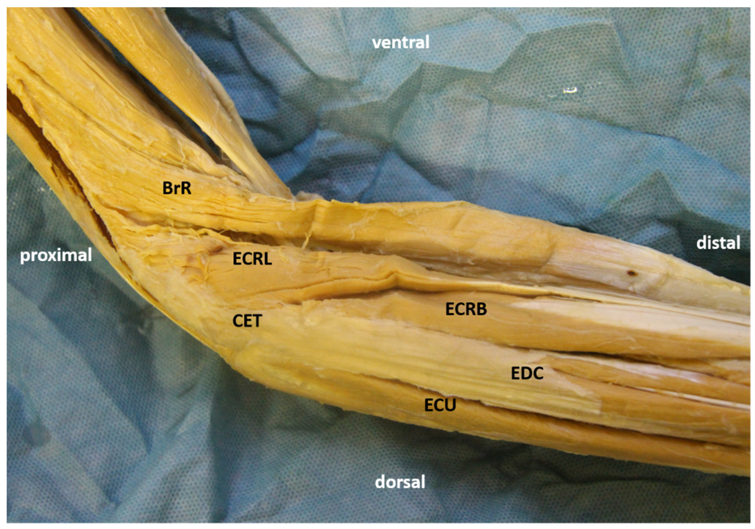
3.1.1. Essential Anatomy

A conjoint tendon, called the CET, represents the forearm’s extensor muscles’ proximal insertion, which is located in the ventral and lateral aspects of the lateral humeral epicondyle (LE) (Figure 1 and Figure 2). Its course is more distally and deeper to the brachioradialis and extensor carpi radialis longus (ECRL) muscles, which originate more proximally. The CET consists of fibers derived from the extensor carpi radialis brevis (ECRB), extensor digitorum communis (EDC), extensor digiti minimi (EDM), extensor carpi ulnaris (ECU) tendons, and receives fiber from the ECRL. The ECRB represents the anterior and deep portion, and the EDC makes up the anterior and superficial parts. The ECU forms the posterior part. The ECRB, the prime dorsiflexor of the wrist and the key tendon in tennis elbow development, originates from the LE of the humerus, from the lateral collateral ligament (LCL), and the adjacent intermuscular septum [30]. Greenbaum with coauthors [31] in a cadaveric study described no clear margins between the ECRB and EDC; thus, the authors considered those two tendons mentioned above as one common tendon. Furthermore, interconnecting tendons between the ECRB and ECRL were found in 35% of limbs [32]. Distally, the ECRB inserts onto the base of the third metacarpal. These muscles’ action extends the wrist and fingers while also supinating the forearm and abducting the wrist. The radial nerve innervates all of the muscles mentioned above. The brachioradialis, ECRL, and ECRB are supplied by direct branches from the radial nerve. Other extensors are innervated via the deep branch of the radial nerve (posterior interosseous nerve) [33]. Deep to the CET, the LCL bridges from the LE down to the radial head.

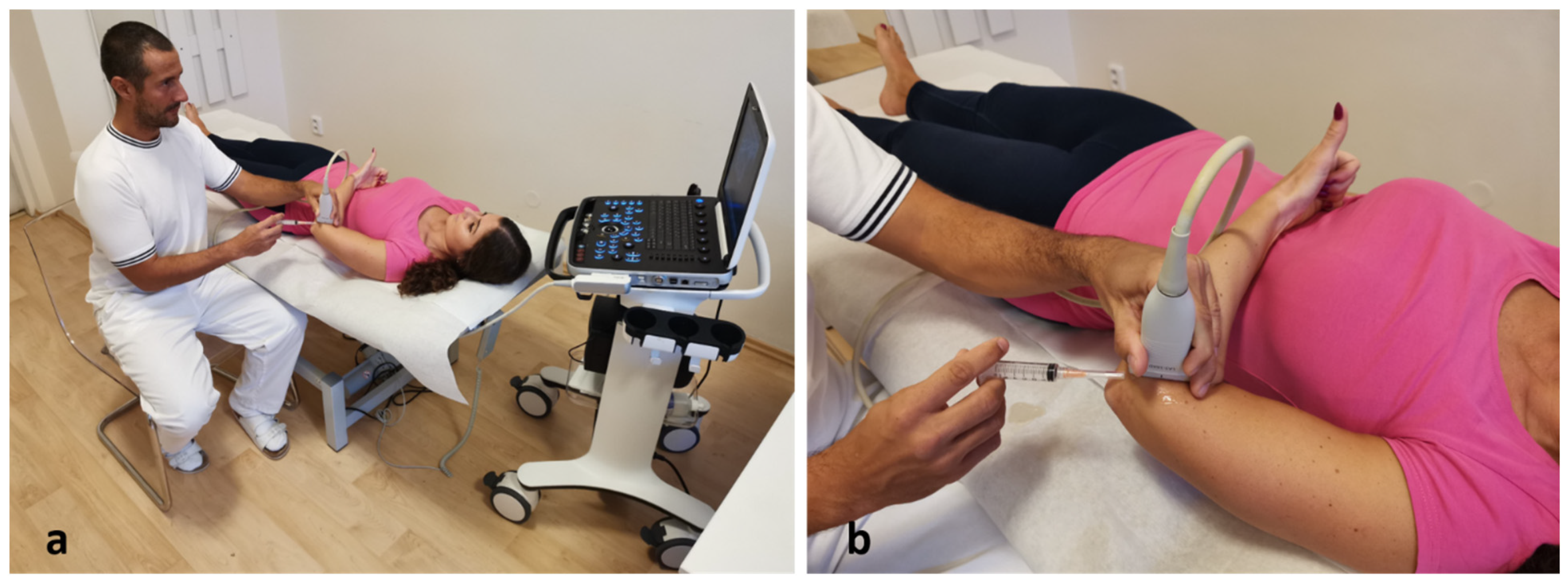
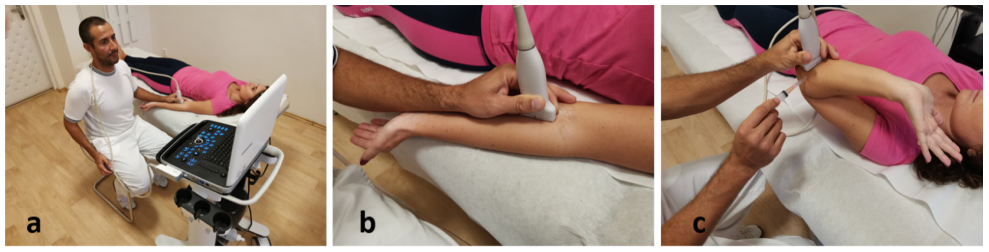
3.1.2. US scanning and Guided Injection

For US scanning of the CET, the patient may be positioned supine on the examination bed while the patient’s forearm rests on the stomach in mild supination. For the contralateral elbow, the US practitioner can easily scan the lateral compartment from his seat. Alternatively, the patient may sit opposite to the sonographer with the elbow flexed and with the shoulder internally rotated and the forearm semi-supinated. The probe is placed at the LE along the forearm’s long axis to visualize the CET in the long axis (Figure 3). The underlying bony landmarks are the LE and the radial head. The hyperechoic fibrillar layer between bone and subcutaneous tissue represents the CET (superficial) and the lateral collateral ligament (deep), which is difficult to distinguish from the CET due to the similar fibrillar echotexture. Similarly, distinguishing between individual tendons of the CET can also be challenging. To identify an individual tendon, one can use distal to proximal muscle tracking. The ECRB and EDC tendons are predominantly affected [34]. Notably, identifying the specific tendon affected by inflammation might help tailor the rehabilitation program [35] and determine a convenient target for interventions (e.g., dry-needling or regenerative medicine injections).
When intact, the CET is noncompressible, homogenous, and hyperechoic with a fibrillar tendinous pattern (Figure 4a). In pathologic cases, the examiner can observe cortical irregularities (e.g., bony spurs or erosions), tendon thickening with a loss of normal fibrillar pattern, focal or diffuse hypoechogenicity, calcifications, tears, and hypervascularity (Figure 4b–e) [36]. Furthermore, the examiner can use sonopalpation to correlate the abnormal findings with local tenderness [37].
Even though tennis elbow is, to a certain point, a self-limited disease, approximately 80% of patients get better after a year. Notably, some methods can enhance the healing process [38]. Sayegh et al. [39] reported a lack of clinical benefit comparing nonsurgical treatment to observation/placebo in intermediate to long-term clinical benefits. Nevertheless, in some patients who suffered from recalcitrant pain, despite the initial conservative therapy (physiotherapy, local/oral nonsteroidal anti-inflammatory drugs, orthotics), injection therapy can be considered. One promising option is dry-needling therapy, which, compared to ibuprofen, showed a better effect at six months follow up [40]. In clinical trials, various injectates are commonly compared to placebo, which is typically represented by saline solution (SS). A recent meta-analysis reported that SS might have some therapeutic effect itself. Acosta-Olivo et al. [41] reported a significant reduction in (visual analogue scale) VAS pain after SS injection, even after one year following administration. The authors also reported a substantial improvement in functional scores. The outcomes in the SS injection group were better than in the noninvasive group. Gao et al. [42] in their meta-analysis reported that 9 out of 10 (randomized controlled trial) RCTs showed no statistically significant difference between SS application and other solution injections such as platelet-rich plasma (PRP), autologous blood (AB), corticosteroids (CS), and botulinum toxin (BT). The most common agents to treat lateral epicondylitis are corticosteroids. Xiong et al. [43] reported a better effect from shockwave therapy with CS injection regarding VAS and grip strength after 12 weeks follow up. Another option for the treatment of the tennis elbow would be PRP injection. Platelet-rich plasma is an autologous preparation from patients’ blood that can enhance the healing process. Simental-Mendía et al. [44] found no difference in improvement in pain and joint functionality comparing PRP with placebo (SS). Several meta-analyses compared PRP and CS injection effects. Corticosteroids injection provided better outcomes regarding pain and joint function in the short term (4 to 8 weeks after application), while PRP injection improved pain and function in the long-term (6 months to one year) more effectively [45,46,47,48]. As an alternative, AB collected from the patient’s peripheral veins can also be administered into the tendon. Application of AB might enhance tissue healing. A meta-analysis by Chou et al. [49] suggested that AB is more effective at decreasing pain than CS injection, but there was no significant difference between AB and PRP. According to Arirachakaran et al.’s [50] meta-analysis, AB can improve pain disability scores but has a higher risk of complications than PRP. Kalichman et al. [51] accessed the use of botulotoxin as a possible method of treating tennis elbow in their meta-analysis. They reported a decrease in pain at 3 months follow up. The authors concluded that BT injection in the forearm might be a suitable treatment method for chronic recalcitrant tennis elbow.
One of the technical variants to perform the tennis elbow injection is the same as for the examination in the supine position. The transducer is positioned on the lateral epicondyle perpendicular to the long axis of the forearm. The needle is inserted from lateral to medial (Figure 4f). Depending on the particular procedure plan, the needle tip can reach the peritendinous space or the tendon itself to perform intralesional (e.g., PRP) injection. The approach mentioned above allows the needle course to be parallel to the probe thus providing excellent needle visibility. Alternatively, the needle can be administered from distal to proximal (Supplementary Materials Video S1) or vice versa. When injecting the tennis elbow, potentially vulnerable structures include the anterior branch of the deep brachial artery and the deep branch of the radial nerve. Using US guidance can reduce the risk of iatrogenic injury to the structures mentioned above.

3.2. Golfer’s Elbow

Golfer’s elbow (i.e., medial epicondylitis) usually presents as pain at the medial elbow. It is three- or six-fold less frequent than tendinopathy of the lateral elbow (i.e., tennis elbow). It is often associated with sports activities (particularly overhead throwing) and manual labor, e.g., carpenters and plumbers [52]. Medial epicondylitis reaches nearly 10% of all cases of epicondylitis [53]. Its prevalence is approximately 0.3–0.6% in men and 0.3–1.1% in women [28]. Repetitious wrist flexion or forearm pronation at least two hours every day, smoking, diabetes mellitus, and obesity were identified as risk factors of medial epicondylitis [54]. The pain manifests itself especially when the fingers and wrist are bent. Overhead throwers are at risk due to the elbow’s valgus torque during the acceleration phase of throwing, causing significant strain on the common flexor tendon. Some throwers may have other pathologies, including medial collateral ligament insufficiency, and some of them also show chronic signs of impingement known as “chronic valgus overload syndrome” [55]. Medial elbow tendinopathy diagnosis is usually not as easy as a lateral one [38]. In the differential diagnosis, one must also think of pain caused by ulnar or medial antebrachial cutaneous nerve entrapments [56,57]. Cervical radiculopathy of C6 and C7 could be associated with weakness and dysfunction of pronator teres (PT), flexor carpi ulnaris (FCR), palmaris longus (PL), flexor digitorum superficialis (FDS), and flexor carpi ulnaris (FCU) muscles. Their imbalance can lead to the onset of medial epicondylitis [58]. For diagnosis, US imaging and an MRI are beneficial, mainly when history and physical examination are noncontributory [59]. In adolescent pitchers, a common cause of medial elbow pain is due to the fact of repetitive valgus overload. This condition is known as “Little League elbow”. In severe cases, the medial epicondyle fracture may occur [60].

3.2.1. Essential Anatomy

The common flexor tendon (CFT), also known as the caput commune ulnare, is a conjoint tendon of the flexor–pronator musculature on the medial humeral epicondyle (ME) (Figure 5). The CFT attaches proximally to the anterior bundle of the medial collateral ligament (MCL). The part of the CFT that is the ulnar head of the pronator teres muscle (PT) is confluent with the anteromedial joint capsule [61]. The CFT is on average 3 cm long and crosses the humeroulnar joint medially [62]. Flexor–pronator musculature is a group of four muscles of the superficial layer of the anterior forearm compartment. These muscles are the PT, FCR, PL, and FCU. The PT and FCR originate from the most proximal and anterior part of the medial humeral epicondyle. They are the most susceptible to tendinopathy development from the CFT [38]. Repetitive eccentric loading of muscles, conducting wrist flexion, and forearm pronation combined with valgus overload at the elbow is considered the leading cause of golfer’s elbow [63]. The PT is inserted on the lateral border of the radius in the middle of the radial diaphysis. Other tendons of the flexor–pronator musculature undergo the flexor retinaculum of the wrist joint. The FCR tendons are inserted in the ventral part of the base of the second and third metacarpals. The PL inserts into the palmar aponeurosis. FCU is inserted onto the pisiform bone. PT, FCR, and PL are innervated by the median nerve. The FCU is innervated by the ulnar nerve. The ulnar nerve at the elbow level runs posterior to the medial epicondyle within the cubital tunnel.

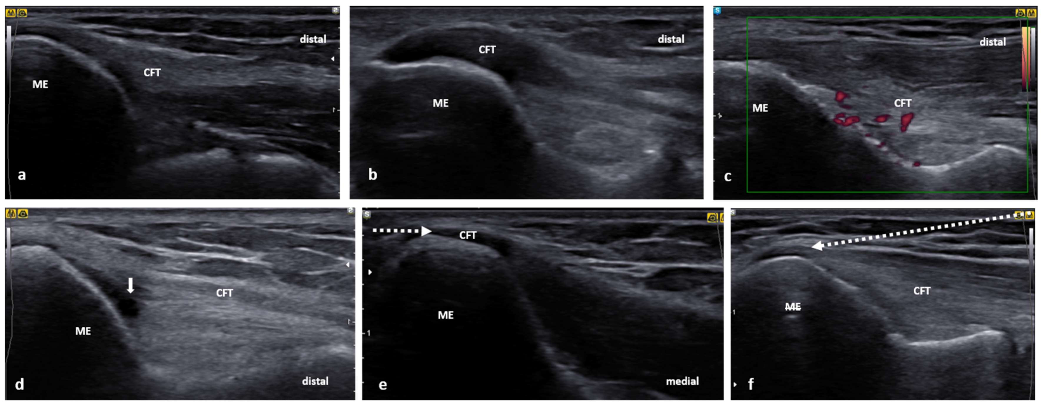
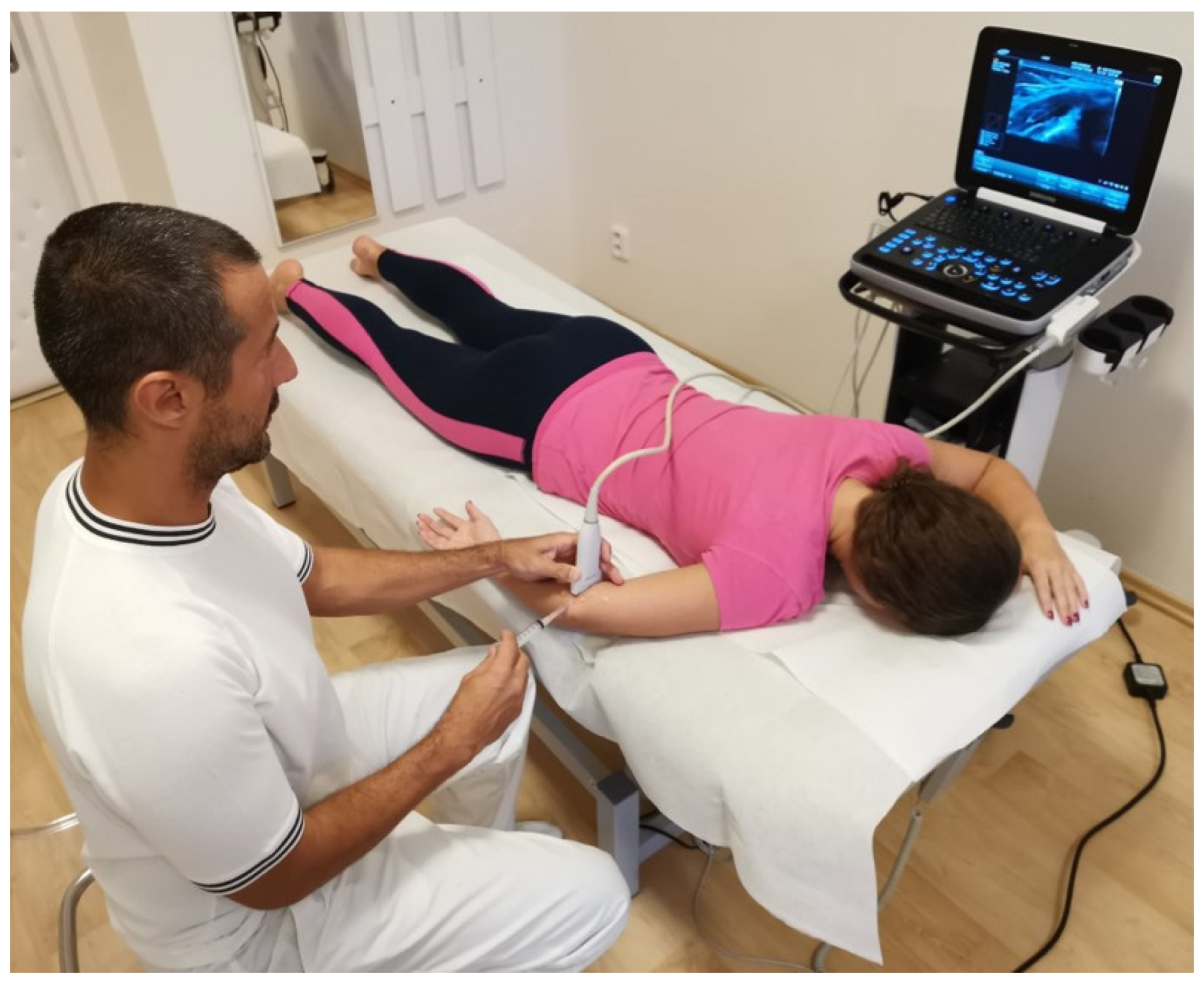
3.2.2. US scanning and Guided Injection

For US scanning of the CFT, the patient may be positioned semi-supine on the examination bed while the patient’s arm is resting on the bed with the forearm hanging over the bed’s edge. As an alternative, the patient may sit facing the examiner, leaning to the ipsilateral side with the supinated forearm resting on an examination bed. To obtain the longitudinal view of the CFT, the transducer is placed at the medial humeral epicondyle (ME) along the forearm’s long axis. The important bony landmarks would be the ME and coronoid process of the ulna. Superficial to these bony structures, the anterior bundle of the MCL and CFT can be identified (Figure 6a). Compared to CET, the CFT is broader and shorter. Normally, the CFT is proximally noncompressible and compressible distally while the softer muscle tissue prevails the stiffer tendon. A sensitivity and specificity of 95% and 92%, respectively, have been reported for the detection of clinical golfer’s elbow. Characteristic US images of medial epicondylitis show focal areas of hypoechogenicity [59]. Typically, the swelling in a golfer’s elbow is located very proximally (Figure 6b) [15]. Further signs, such as tendon thickening, cortical irregularities, intratendinous calcifications, and hypervascularity, are also common (Figure 6c,d). Notably, lesions of the anterior bundle of MCL can mimic or be concurrent with medial epicondylitis. In such a scenario, US imaging would reveal MCL structural abnormalities, e.g., thickening and discontinuity. Notably, the dynamic US valgus stress test might be beneficial to demask MCL rupture, coupled with elbow instability [19].
There is a lack of evidence for injections in medial epicondylitis. To the best of our knowledge, no meta-analysis was performed, and there are only a few RCTs. Stahl et Kaufman [64] performed RCT on 60 elbows comparing CS injection with placebo and concluded that CS injection has only short-term (6 weeks after the injection) benefit in pain reduction. The long-term effect was the same for CS and placebo. Suresh et al. [65] performed a prospective study on 20 subjects assessing pain reduction after dry-needling and autologous blood injection under US control. They found this combined method effective in reducing pain ten months after the procedure. Bohlen et al. [66] found in their cohort study (level of evidence 3) that PRP injection had a similar clinical effect as surgical treatment.
For the golfer’s elbow injection, the patient may be positioned in the prone while his or her forearm also rests pronated on the examination bed (Figure 7). The probe is positioned on the medial epicondyle perpendicular to the long axis of the forearm. The needle is inserted from lateral to medial. Again, depending on the particular procedure plan, the injection can be performed peri- or intratendinous. The parallel course of the needle to the probe provides excellent needle visibility along its whole path (Figure 6e). Alternatively, the needle can be inserted from distal to proximal (Figure 6f). When injecting a golfer’s elbow, one should take caution not to pierce the ulnar nerve, particularly in cases of ulnar nerve anterior dislocation [67] or in patients who underwent ulnar nerve anterior transposition surgery. Using US imaging, the safe needle route can be determined proceeding with the intervention.

3.3. Distal Biceps Tendinopathy

Overuse injury and tearing of the distal biceps tendon (DBT) typically presents as a sudden onset of pain at the antecubital region, usually following an acute event, e.g., lifting or catching a heavy object [68]. Distal biceps tendinopathy is rare compared to tennis or golfer’s elbow. It is believed that tendinopathy and tearing of the distal biceps tendon represent a mutual pathological process [38]. Cases of a complete tear of the distal biceps tendon are reported to account for 3% of the total cases of biceps tendon rupture, and the prevalence of this disease is estimated to be 2.55 per 100.000 [69] There is a 7.5 fold increased risk of rupture in patients who smoke [70]. Most distal biceps tendinopathy cases without the complete tear of the biceps tendon are generally associated with minor trauma or repeated activity without accompanying trauma [71].

3.3.1. Essential Anatomy

Biceps brachii muscles originate in two heads. The short head originates in the coracoid process of the scapula. The long head originates from the supraglenoid tubercle and partly from the superior part of the glenoid labrum. A DBT is a conjoint tendon of two heads. The long head inserts into a radial tuberosity proximal aspect, whereas the short head inserts into the radial tuberosity’s distal aspect (Figure 8). Attachment of the DBT to the radial tuberosity has a spreading area of approximately 3 cm2 [72]. Bifurcation of the distal biceps tendon allows functional independence and isolated pathological processes of each portion [73]. The fibers of the tendon form a spiral formation [74]. Proximal radioulnar joint was identified as a potential site of impingement of the DBT, potentially influencing the DBT vascular zone alterations [75]. The bicipitoradial bursa is located in between the DBT and the anterior part of the radial tuberosity. Enthesopathy of DBT could be an accompanying factor in the evolution of DBT tears [72]. At the elbow joint, the bicep muscle forms the biceps aponeurosis, known as the lacertus fibrosus (LF). The LF originates from the short head of the DBT and transverses medially over the CFT [38]. The biceps brachii muscle is innervated by the musculocutaneous nerve.

3.3.2. US Scanning and Guided Injection

The assessment of the DBT using US imaging can be technically challenging given its oblique course. There are different ultrasound approaches to assess the DBT. It is beneficial to examine the patient lying supine with his or her upper limb extended along the trunk. The patient is asked to actively supinate the forearm while the examination bed provides a suitable resistance. The transducer is first placed in the transverse plane. When the DBT is identified lateral to the brachial artery, the probe is turned 90° to visualize the tendon in its long axis (Figure 9a,b and Figure 10a). To reduce the unsolicited anisotropic artifact given by the tendon’s oblique course, the examiner should perform a “heel–toe” maneuver with the probe to obtain the optimal image to assess the DBT. Rotating the transducer towards the pronator teres muscle, the lacertus fibrosus insertion will appear, attaching to the superficial fascia of the muscle mentioned before. To visualize the insertion of the DBT on the radial tuberosity, the elbow is flexed, the forearm is fully pronated, and the wrist flexed to “cobra position” (Figure 9c). The transducer is placed in the transverse aspect of the forearm, at the radial head. The “cobra position” is convenient for injection at the tendon insertion. The normal DBT image demonstrates homogenous fibrillar hyperechoic echostructure. The DBT tendinopathy goes along with tendon hypoechogenicity, thickening and sometimes also calcifications and increased vascularity. When the tendon is torn and retracted, the recoiled stump may demonstrate the shadowing artifact (Figure 10b) [76]. Furthermore, bicipitoradial bursitis can also be revealed in some patients presenting with anterior elbow pain.
There is very little evidence regarding distal bicipital tendinopathy interventions and injections. Sanli et al. [77] performed a prospective cohort study on 12 patients injecting PRP with US guidance. The study showed significant improvement in pain and functional outcome at 47 months follow up. Barker et al. [78] concluded on a cohort of six patients that a US-guided PRP injection was an effective (regarding improving pain and performance scores) and a safe procedure, but further investigation involving RCT is needed. For the DBT injection, the patient can be positioned supine with the forearm in the “cobra position” to access the tendon footprint avoiding the vulnerable adjacent neurovascular structures (Figure 10c) [79]. Potentially vulnerable structures when injecting the DBT are the brachial artery and median nerve, which follow the LF. The course of the lateral cutaneous nerve of the forearm [80] and the median cubital vein should be taken into account laterally from the DBT. As such, the “cobra position” provides a potentially safer alternative to injection in the anterior side of the elbow. Ultrasound guidance aids in visualizing structures that should not be unintentionally injured.

3.4. Distal Triceps Tendinopathy

Distal triceps tendinopathy is the rarest of the tendinopathies around the elbow with little evidence in the literature. Nirschl (1988) [81] described it as a “posterior tennis elbow”. Triceps rupture represents the terminal phase of tendinopathy, again, similarly to distal biceps tendinopathy. Tendinopathies may arise from both the medial and the lateral part of the tendon [38]. An eccentric force on the contracting muscle has been proposed as a possible injury mechanism [82].
Several medical comorbidities have been described as potential predisposing risk factors for this problem, including anabolic steroid use, local steroid injections for bursitis, oral steroids, renal disease, diabetes, and familiar tendinopathy [83].

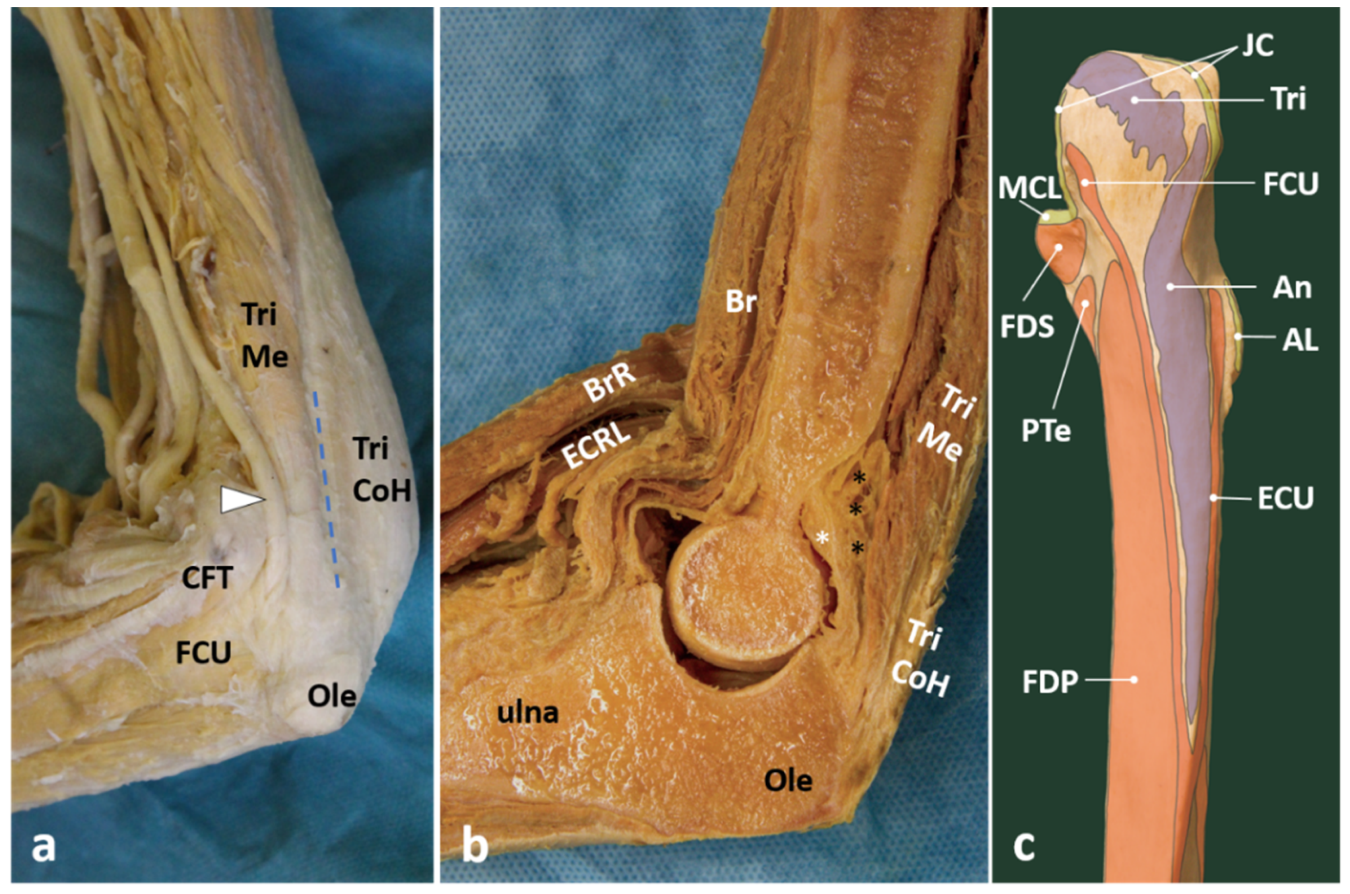
3.4.1. Essential Anatomy

The triceps brachii muscle (TBM) is formed from three heads. The long head originates in the infraglenoid tubercle of the scapula. The lateral head originates in the posterolateral aspect of the humeral shaft, proximally to the radial sulcus. The medial head originates in the posteromedial aspect of the humeral shaft, distally from the radial sulcus. The TBM tendon inserts into the olecranon (Figure 11). The attachment is approximately 12–14 mm distal to the olecranon tip with an average width of 40 mm [84]. The lateral and long heads of the TBM converge distally and form the superficial part of the triceps tendon attachment. This part attaches to the olecranon’s medial aspect, where it may converge with the anconeus muscle’s fascia [38]. The medial head forms the deep portion of the triceps inserted into the olecranon [85] and is mainly muscular at its insertion [86]. Some authors describe the space filled with fatty tissue between these attachments like “distal pretricipital space” on MRI sections and cadaver specimens [87]. The triceps muscle is innervated by the radial nerve.

3.4.2. US Scanning and Guided Injection

The triceps tendon (TT) evaluation can be performed with the patient positioned semi-supine on the examination bed. Using this positioning technique, the elbow rests on the patient’s stomach, while the elbow is free for flexion/extension dynamic assessment. The dynamic evaluation is useful to assess for the TT continuity but also to rule out the presence of fluid/loose bodies in the olecranon fossa. For the TT assessment—the longitudinal view—the examiner can see the myotendinous junction and the TT insertion footprint on the olecranon (Figure 12a). The US features of TT tendinopathy comprise local hypoechogenicity, tendon thickening, hypervascularity, calcifications, and bony irregularities/spur (Figure 12b). Triceps tendon tendinopathy is rare; however, inflammation of the overlaying olecranon bursa is more common, particularly following trauma or in patients with inflammatory diseases. The way to aspirate olecranon bursitis should be decided with respect to local anatomy (e.g., septa, adjacent structures). Notably, US guidance provides clinicians freedom in all interventional procedures [88]. There is little evidence using US-guided injections in triceps tendinopathy. Cheatham et al. [89] reported a case report of a patient with distal triceps partial tear treated with PRP injection and rehabilitation, resulting in pain-free activities of daily living and return to preinjury sports activity after four months. When injecting TT, a potentially vulnerable structure would be the ulnar nerve, which passes medially along the tendon. Using the lateral approach and the US guidance can reduce the risk of iatrogenic injury to the nerve (Figure 13).

4. Conclusions

Elbow pain resulting from overuse or trauma is a common cause of patients’ visits to physicians’ practice. This review describes four common musculoskeletal pathologies and their interventional treatment. A clinical examination often brings only incomplete information and, therefore, using US imaging daily helps us set the final diagnosis. Moreover, it can guide interventions to target a precisely treated structure and avoid vulnerable structures such as nerves or vessels. An emerging and promising tool in tendon imaging is ultrasound elastography to assess tissue properties by measuring their stiffness. Nevertheless, ultrasound elastography still requires validation to become a reliable method for tendon pathology assessment [90,91]. Future research in ultrasound imaging can be directed towards further image quality improvement. One option would be a fuzzy pre-processing operation to pretreat the images before proceeding with the clinical evaluation [92,93].

Supplementary Materials

The following are available online at https://www.mdpi.com/article/10.3390/app11083431/s1, Video S1: Ultrasound-guided tennis elbow injection: distal to proximal approach.

Author Contributions

Conceptualization, K.M. and J.J.; methodology, K.M.; data curation, K.M. and K.S.; writing—original draft preparation, K.M., J.J. and T.N.; writing—review and editing, Y.A., K.S., L.H. and O.N.; visualization, L.H., K.M. and J.J. supervision, O.N. and Y.A.; funding acquisition, Y.A. and K.M. All authors have read and agreed to the published version of the manuscript.

Funding

Open-access publication fees were supported by the Open Access Fund of the General University Hospital in Prague.

Institutional Review Board Statement

Not applicable.

Informed Consent Statement

Not applicable.

Data Availability Statement

No data sets were generated or analyzed.

Acknowledgments

We would like to express our sincere gratitude to Jan Kacvinský, an academic painter from the Institute of Anatomy, First Faculty of Medicine, Charles University in Prague, for schematic drawings of certain anatomic regions in this manuscript.

Conflicts of Interest

The authors declare no conflict of interest.

References

  1. Shiri, R.; Viikari-Juntura, E. Lateral and medial epicondylitis: Role of occupational factors. Best Pract. Res. Clin. Rheumatol. 2011, 25, 43–57. [Google Scholar] [CrossRef]
  2. Nirschl, R.P. Tennis elbow. Orthop. Clin. N. Am. 1973, 4, 787–800. [Google Scholar] [CrossRef]
  3. Bishai, S.; Plancher, K. The Basic Science of Lateral Epicondylosis: Update for the Future. Tech. Orthop. 2006, 4, 250–255. [Google Scholar] [CrossRef]
  4. Kannus, P.; Józsa, L. Histopathological changes preceding spontaneous rupture of a tendon. A controlled study of 891 patients. J. Bone Jt. Surg. Am. 1991, 73, 1507–1525. [Google Scholar] [CrossRef]
  5. Kraushaar, B.S.; Nirschl, R.P. Tendinosis of the elbow (tennis elbow). Clinical features and findings of histological, immunohistochemical, and electron microscopy studies. J. Bone Jt. Surg. Am. 1999, 81, 259–278. [Google Scholar] [CrossRef]
  6. Nirschl, R.; Alvarado, G. Tennis elbow tendinosis: Pathoanatomy, nonsurgical and surgical management. In Repetitive Motion Disorders of the Upper Extremity; American Academy of Orthopaedic Surgeons, Rosemont: Rosemont, IL, USA, 1995; pp. 467–479. [Google Scholar]
  7. Patterson-Kane, J.C.; Becker, D.L.; Rich, T. The pathogenesis of tendon microdamage in athletes: The horse as a natural model for basic cellular research. J. Comp. Pathol. 2012, 147, 227–247. [Google Scholar] [CrossRef] [PubMed] [Green Version]
  8. Kannus, P. Etiology and pathophysiology of chronic tendon disorders in sports. Scand. J. Med. Sci. Sports 1997, 7, 78–85. [Google Scholar] [CrossRef]
  9. Coombes, B.K.; Bisset, L.; Vicenzino, B. A new integrative model of lateral epicondylalgia. Br. J. Sports Med. 2009, 43, 252–258. [Google Scholar] [CrossRef] [Green Version]
  10. Boushel, R.; Langberg, H.; Green, S.; Skovgaard, D.; Bulow, J.; Kjaer, M. Blood flow and oxygenation in peritendinous tissue and calf muscle during dynamic exercise in humans. J. Physiol. 2000, 524 Pt 1, 305–313. [Google Scholar] [CrossRef]
  11. Kane, S.F.; Lynch, J.H.; Taylor, J.C. Evaluation of elbow pain in adults. Am. Fam. Phys. 2014, 89, 649–657. [Google Scholar]
  12. Kryger, A.I.; Lassen, C.F.; Andersen, J.H. The role of physical examinations in studies of musculoskeletal disorders of the elbow. Occup. Environ. Med. 2007, 64, 776–781. [Google Scholar] [CrossRef] [Green Version]
  13. Zwerus, E.L.; Somford, M.P.; Maissan, F.; Heisen, J.; Eygendaal, D.; van den Bekerom, M.P. Physical examination of the elbow, what is the evidence? A systematic literature review. Br. J. Sports Med. 2018, 52, 1253–1260. [Google Scholar] [CrossRef]
  14. Özçakar, L.; Muynck, M.D. Musculoskeletal Ultrasound in Physical Rehabilitation Medicine; Edi Ermes: Milan, Italy, 2014; ISBN 978-88-7051-420-9. [Google Scholar]
  15. Özçakar, L. Sonographic Atlas for Common Musculoskeletal Pathologies; Edi Ermes: Milan, Italy, 2017; ISBN 978-88-7051-576-3. [Google Scholar]
  16. Kara, M.; Gürçay, E.; Ekiz, T.; Sekizkardeş, M.; Yorulmaz, E.; Ata, A.M.; Chang, K.-V.; Wu, W.-T.; Akkaya, N.; Mezian, K.; et al. EURO-MUSCULUS/USPRM Global Report on Musculoskeletal Ultrasound Publications. Am. J. Phys. Med. Rehabil. 2020, 99, 847–852. [Google Scholar] [CrossRef] [PubMed]
  17. Jačisko, J.; Sobotová, K.; Mezian, K. The utility of ultrasound examination in cubital tunnel syndrome caused by heterotopic ossification. Med. Ultrason. 2020, 22, 117–118. [Google Scholar] [CrossRef]
  18. Özçakar, L. Ultrasound Imaging & Guidance for Musculoskeletal Interventions in Physical and Rehabilitation Medicine; Edi Ermes: Milan, Italy, 2019; ISBN 978-88-7051-698-2. [Google Scholar]
  19. Mezian, K.; Machač, S.; Zavareh, A.; Majerníková, L.; Vacek, J.; Navrátil, L.; Schmitz, M. Positioning Techniques to Improve the Ultrasound Evaluation of the Elbow. Ultrasound Q. 2019, 35, 136–141. [Google Scholar] [CrossRef] [PubMed]
  20. Mezian, K.; Sobotová, K.; Angerová, Y. Sonographic elbow scanning is not yoga exercise. Adv. Rheumatol. 2019, 59, 33. [Google Scholar] [CrossRef] [PubMed] [Green Version]
  21. Özçakar, L.; Kara, M.; Chang, K.V.; Hung, C.Y.; Tekın, L.; Ulaşlı, A.M.; Wu, C.H.; Tok, F.; Hsıao, M.Y.; Akkaya, N.; et al. EURO-MUSCULUS/USPRM Basic Scanning Protocols for elbow. Eur. J. Phys. Rehabil. Med. 2015, 51, 485–489. [Google Scholar] [PubMed]
  22. Cutts, S.; Gangoo, S.; Modi, N.; Pasapula, C. Tennis elbow: A clinical review article. J. Orthop. 2020, 17, 203–207. [Google Scholar] [CrossRef] [PubMed]
  23. Eygendaal, D.; Rahussen, F.T.G.; Diercks, R.L. Biomechanics of the elbow joint in tennis players and relation to pathology. Br. J. Sports Med. 2007, 41, 820–823. [Google Scholar] [CrossRef] [PubMed]
  24. Walz, D.M.; Newman, J.S.; Konin, G.P.; Ross, G. Epicondylitis: Pathogenesis, imaging, and treatment. Radiographics 2010, 30, 167–184. [Google Scholar] [CrossRef] [Green Version]
  25. Sanders, T.L.; Maradit Kremers, H.; Bryan, A.J.; Ransom, J.E.; Smith, J.; Morrey, B.F. The epidemiology and health care burden of tennis elbow: A population-based study. Am. J. Sports Med. 2015, 43, 1066–1071. [Google Scholar] [CrossRef]
  26. Allander, E. Prevalence, incidence, and remission rates of some common rheumatic diseases or syndromes. Scand. J. Rheumatol. 1974, 3, 145–153. [Google Scholar] [CrossRef]
  27. Bot, S.D.M.; van der Waal, J.M.; Terwee, C.B.; van der Windt, D.A.W.M.; Bouter, L.M.; Dekker, J. Course and prognosis of elbow complaints: A cohort study in general practice. Ann. Rheum. Dis. 2005, 64, 1331–1336. [Google Scholar] [CrossRef] [Green Version]
  28. Shiri, R.; Viikari-Juntura, E.; Varonen, H.; Heliövaara, M. Prevalence and determinants of lateral and medial epicondylitis: A population study. Am. J. Epidemiol. 2006, 164, 1065–1074. [Google Scholar] [CrossRef] [PubMed]
  29. Keijsers, R.; de Vos, R.-J.; Kuijer, P.P.F.; van den Bekerom, M.P.; van der Woude, H.-J.; Eygendaal, D. Tennis elbow. Shoulder Elb. 2019, 11, 384–392. [Google Scholar] [CrossRef] [PubMed]
  30. Nayak, S.R.; Ramanathan, L.; Krishnamurthy, A.; Prabhu, L.V.; Madhyastha, S.; Potu, B.K.; Ranade, A.V. Extensor carpi radialis brevis origin, nerve supply and its role in lateral epicondylitis. Surg. Radiol. Anat. 2010, 32, 207–211. [Google Scholar] [CrossRef] [PubMed]
  31. Greenbaum, B.; Itamura, J.; Vangsness, C.T.; Tibone, J.; Atkinson, R. Extensor carpi radialis brevis. An anatomical analysis of its origin. J. Bone Jt. Surg. Br. 1999, 81, 926–929. [Google Scholar] [CrossRef]
  32. Albright, J.A.; Linburg, R.M. Common variations of the radial writs extensors. J. Hand Surg. Am. 1978, 3, 134–138. [Google Scholar] [CrossRef]
  33. Standring, S. Gray’s Anatomy. The Anatomical Basis of Clinical Practice, 41st ed.; Churchill Livingstone/Elsevier: Edinburgh, UK, 2015. [Google Scholar]
  34. Boyer, M.I.; Hastings, H. Lateral tennis elbow: “Is there any science out there?”. J. Shoulder Elb. Surg. 1999, 8, 481–491. [Google Scholar] [CrossRef]
  35. Ricci, V.; Schroeder, A.; Özçakar, L. Ultrasound Imaging for Lateral Elbow Pain: Pinpointing the Epicondylosis. Am. J. Phys. Med. Rehabil. 2020, 99, 560–561. [Google Scholar] [CrossRef]
  36. Ricci, V.; Mezian, K.; Özçakar, L. Contemporary/Ultrasound Guidance for Musculoskeletal Interventions: Let Bygones be Bygones. Am. J. Phys. Med. Rehabil. 2021. [Google Scholar] [CrossRef] [PubMed]
  37. Pirri, C.; Stecco, C.; De Caro, R.; Foti, C.; Özçakar, L. Radiating Upper Limb Pain Due to a Large Subcutaneous Lipoma: Fascial Sono-Palpation. Pain Med. 2020, 21, 3721–3723. [Google Scholar] [CrossRef] [PubMed]
  38. Donaldson, O.; Vannet, N.; Gosens, T.; Kulkarni, R. Tendinopathies Around the Elbow Part 2: Medial Elbow, Distal Biceps and Triceps Tendinopathies. Shoulder Elb. 2014, 6, 47–56. [Google Scholar] [CrossRef] [PubMed] [Green Version]
  39. Sayegh, E.T.; Strauch, R.J. Does nonsurgical treatment improve longitudinal outcomes of lateral epicondylitis over no treatment? A meta-analysis. Clin. Orthop. Relat. Res. 2015, 473, 1093–1107. [Google Scholar] [CrossRef] [Green Version]
  40. Uygur, E.; Aktaş, B.; Özkut, A.; Erinç, S.; Yilmazoglu, E.G. Dry needling in lateral epicondylitis: A prospective controlled study. Int. Orthop. 2017, 41, 2321–2325. [Google Scholar] [CrossRef] [PubMed]
  41. Acosta-Olivo, C.A.; Millán-Alanís, J.M.; Simental-Mendía, L.E.; Álvarez-Villalobos, N.; Vilchez-Cavazos, F.; Peña-Martínez, V.M.; Simental-Mendía, M. Effect of Normal Saline Injections on Lateral Epicondylitis Symptoms: A Systematic Review and Meta-analysis of Randomized Clinical Trials. Am. J. Sports Med. 2020, 48, 3094–3102. [Google Scholar] [CrossRef]
  42. Gao, B.; Dwivedi, S.; DeFroda, S.; Bokshan, S.; Ready, L.V.; Cole, B.J.; Owens, B.D. The Therapeutic Benefits of Saline Solution Injection for Lateral Epicondylitis: A Meta-analysis of Randomized Controlled Trials Comparing Saline Injections With Nonsurgical Injection Therapies. Arthroscopy 2019, 35, 1847–1859. [Google Scholar] [CrossRef]
  43. Xiong, Y.; Xue, H.; Zhou, W.; Sun, Y.; Liu, Y.; Wu, Q.; Liu, J.; Hu, L.; Panayi, A.C.; Chen, L.; et al. Shock-wave therapy versus corticosteroid injection on lateral epicondylitis: A meta-analysis of randomized controlled trials. Phys. Sportsmed. 2019, 47, 284–289. [Google Scholar] [CrossRef]
  44. Simental-Mendía, M.; Vilchez-Cavazos, F.; Álvarez-Villalobos, N.; Blázquez-Saldaña, J.; Peña-Martínez, V.; Villarreal-Villarreal, G.; Acosta-Olivo, C. Clinical efficacy of platelet-rich plasma in the treatment of lateral epicondylitis: A systematic review and meta-analysis of randomized placebo-controlled clinical trials. Clin. Rheumatol. 2020, 39, 2255–2265. [Google Scholar] [CrossRef]
  45. Huang, K.; Giddins, G.; Wu, L.-D. Platelet-Rich Plasma Versus Corticosteroid Injections in the Management of Elbow Epicondylitis and Plantar Fasciitis: An Updated Systematic Review and Meta-analysis. Am. J. Sports Med. 2020, 48, 2572–2585. [Google Scholar] [CrossRef]
  46. Xu, Q.; Chen, J.; Cheng, L. Comparison of platelet rich plasma and corticosteroids in the management of lateral epicondylitis: A meta-analysis of randomized controlled trials. Int. J. Surg. 2019, 67, 37–46. [Google Scholar] [CrossRef]
  47. Li, A.; Wang, H.; Yu, Z.; Zhang, G.; Feng, S.; Liu, L.; Gao, Y. Platelet-rich plasma vs corticosteroids for elbow epicondylitis: A systematic review and meta-analysis. Medicine 2019, 98, e18358. [Google Scholar] [CrossRef]
  48. Mi, B.; Liu, G.; Zhou, W.; Lv, H.; Liu, Y.; Wu, Q.; Liu, J. Platelet rich plasma versus steroid on lateral epicondylitis: Meta-analysis of randomized clinical trials. Phys. Sportsmed. 2017, 45, 97–104. [Google Scholar] [CrossRef]
  49. Chou, L.-C.; Liou, T.-H.; Kuan, Y.-C.; Huang, Y.-H.; Chen, H.-C. Autologous blood injection for treatment of lateral epicondylosis: A meta-analysis of randomized controlled trials. Phys. Ther. Sport 2016, 18, 68–73. [Google Scholar] [CrossRef] [PubMed]
  50. Arirachakaran, A.; Sukthuayat, A.; Sisayanarane, T.; Laoratanavoraphong, S.; Kanchanatawan, W.; Kongtharvonskul, J. Platelet-rich plasma versus autologous blood versus steroid injection in lateral epicondylitis: Systematic review and network meta-analysis. J. Orthop. Traumatol. 2016, 17, 101–112. [Google Scholar] [CrossRef] [PubMed] [Green Version]
  51. Kalichman, L.; Bannuru, R.R.; Severin, M.; Harvey, W. Injection of botulinum toxin for treatment of chronic lateral epicondylitis: Systematic review and meta-analysis. Semin. Arthritis Rheum. 2011, 40, 532–538. [Google Scholar] [CrossRef]
  52. David, T.S. Medial elbow pain in the throwing athlete. Orthopedics 2003, 26, 94–103, quiz 104–105. [Google Scholar]
  53. Wolf, J.M.; Mountcastle, S.; Burks, R.; Sturdivant, R.X.; Owens, B.D. Epidemiology of lateral and medial epicondylitis in a military population. Mil. Med. 2010, 175, 336–339. [Google Scholar] [CrossRef] [Green Version]
  54. Taylor, S.A.; Hannafin, J.A. Evaluation and management of elbow tendinopathy. Sports Health 2012, 4, 384–393. [Google Scholar] [CrossRef] [Green Version]
  55. Lynch, J.R.; Waitayawinyu, T.; Hanel, D.P.; Trumble, T.E. Medial collateral ligament injury in the overhand-throwing athlete. J. Hand Surg. Am. 2008, 33, 430–437. [Google Scholar] [CrossRef] [PubMed]
  56. Mezian, K.; Jačisko, J.; Kaiser, R.; Machač, S.; Steyerová, P.; Sobotová, K.; Angerová, Y.; Naňka, O. Ulnar Neuropathy at the Elbow: From Ultrasound Scanning to Treatment. Front. Neurol. 2021, 12, 562. [Google Scholar]
  57. Mezian, K.; Steyeorova, P.; Vacek, J.; Navratil, L. Introduction to Neuromuscular Ultrasound. Cesk. Slov. Neurol. N. 2016, 79, 656–661. [Google Scholar] [CrossRef]
  58. Lee, A.T.; Lee-Robinson, A.L. The prevalence of medial epicondylitis among patients with c6 and c7 radiculopathy. Sports Health 2010, 2, 334–336. [Google Scholar] [CrossRef] [Green Version]
  59. Park, G.-Y.; Lee, S.-M.; Lee, M.Y. Diagnostic value of ultrasonography for clinical medial epicondylitis. Arch. Phys. Med. Rehabil. 2008, 89, 738–742. [Google Scholar] [CrossRef] [PubMed] [Green Version]
  60. Ellington, M.D.; Edmonds, E.W. Pediatric Elbow and Wrist Pathology Related to Sports Participation. Orthop. Clin. N. Am. 2016, 47, 743–748. [Google Scholar] [CrossRef]
  61. Ciccotti, M.C.; Schwartz, M.A.; Ciccotti, M.G. Diagnosis and treatment of medial epicondylitis of the elbow. Clin. Sports Med. 2004, 23, 693–705. [Google Scholar] [CrossRef] [PubMed]
  62. Amin, N.H.; Kumar, N.S.; Schickendantz, M.S. Medial epicondylitis: Evaluation and management. J. Am. Acad. Orthop. Surg. 2015, 23, 348–355. [Google Scholar] [CrossRef] [PubMed] [Green Version]
  63. Ciccotti, M.G.; Ramani, M.N. Medial epicondylitis. Tech. Hand Extrem. Surg. 2003, 7, 190–196. [Google Scholar] [CrossRef] [PubMed]
  64. Stahl, S.; Kaufman, T. The efficacy of an injection of steroids for medial epicondylitis. A prospective study of sixty elbows. J. Bone Jt. Surg. Am. 1997, 79, 1648–1652. [Google Scholar] [CrossRef] [PubMed]
  65. Suresh, S.P.; Ali, K.E.; Jones, H.; Connell, D.A. Medial epicondylitis: Is ultrasound guided autologous blood injection an effective treatment? Br. J. Sports Med. 2006, 40, 935–939; discussion 939. [Google Scholar] [CrossRef] [PubMed] [Green Version]
  66. Bohlen, H.L.; Schwartz, Z.E.; Wu, V.J.; Thon, S.G.; Finley, Z.J.; O’Brien, M.J.; Savoie, F.H. Platelet-Rich Plasma Is an Equal Alternative to Surgery in the Treatment of Type 1 Medial Epicondylitis. Orthop. J. Sports Med. 2020, 8, 2325967120908952. [Google Scholar] [CrossRef] [PubMed]
  67. Chuang, H.-J.; Hsiao, M.-Y.; Wu, C.-H.; Özçakar, L. Dynamic Ultrasound Imaging for Ulnar Nerve Subluxation and Snapping Triceps Syndrome. Am. J. Phys. Med. Rehabil. 2016, 95, e113–e114. [Google Scholar] [CrossRef]
  68. Frazier, M.S.; Boardman, M.J.; Westland, M.; Imbriglia, J.E. Surgical treatment of partial distal biceps tendon ruptures. J. Hand Surg. Am. 2010, 35, 1111–1114. [Google Scholar] [CrossRef]
  69. Kelly, M.P.; Perkinson, S.G.; Ablove, R.H.; Tueting, J.L. Distal Biceps Tendon Ruptures: An Epidemiological Analysis Using a Large Population Database. Am. J. Sports Med. 2015, 43, 2012–2017. [Google Scholar] [CrossRef] [PubMed]
  70. Safran, M.R.; Graham, S.M. Distal biceps tendon ruptures: Incidence, demographics, and the effect of smoking. Clin. Orthop. Relat. Res. 2002, 404, 275–283. [Google Scholar] [CrossRef]
  71. Lee, J.H.; Kim, K.C.; Lee, J.-H.; Ahn, K.B.; Rhyou, I.H. A Case Series of Symptomatic Distal Biceps Tendinopathy. Clin. Shoulder Elb. 2018, 21, 213–219. [Google Scholar] [CrossRef] [PubMed] [Green Version]
  72. Chew, M.L.; Giuffrè, B.M. Disorders of the distal biceps brachii tendon. Radiographics 2005, 25, 1227–1237. [Google Scholar] [CrossRef] [PubMed]
  73. Cho, C.-H.; Song, K.-S.; Choi, I.-J.; Kim, D.-K.; Lee, J.-H.; Kim, H.-T.; Moon, Y.-S. Insertional anatomy and clinical relevance of the distal biceps tendon. Knee Surg. Sports Traumatol. Arthrosc. 2011, 19, 1930–1935. [Google Scholar] [CrossRef]
  74. Cucca, Y.Y.; McLay, S.V.B.; Okamoto, T.; Ecker, J.; McMenamin, P.G. The biceps brachii muscle and its distal insertion: Observations of surgical and evolutionary relevance. Surg. Radiol. Anat. 2010, 32, 371–375. [Google Scholar] [CrossRef] [PubMed]
  75. Seiler, J.G.; Parker, L.M.; Chamberland, P.D.; Sherbourne, G.M.; Carpenter, W.A. The distal biceps tendon. Two potential mechanisms involved in its rupture: Arterial supply and mechanical impingement. J. Shoulder Elb. Surg. 1995, 4, 149–156. [Google Scholar] [CrossRef]
  76. Brigido, M.K.; De Maeseneer, M.; Morag, Y. Distal biceps brachii. Semin. Musculoskelet. Radiol. 2013, 17, 20–27. [Google Scholar] [CrossRef]
  77. Sanli, I.; Morgan, B.; van Tilborg, F.; Funk, L.; Gosens, T. Single injection of platelet-rich plasma (PRP) for the treatment of refractory distal biceps tendonitis: Long-term results of a prospective multicenter cohort study. Knee Surg. Sports Traumatol. Arthrosc. 2016, 24, 2308–2312. [Google Scholar] [CrossRef]
  78. Barker, S.L.; Bell, S.N.; Connell, D.; Coghlan, J.A. Ultrasound-guided platelet-rich plasma injection for distal biceps tendinopathy. Shoulder Elb. 2015, 7, 110–114. [Google Scholar] [CrossRef]
  79. Al-Ani, Z.; Lauder, J. Ultrasound assessment in distal biceps tendon injuries: Techniques, pearls and pitfalls. Clin. Imaging 2021, 75, 46–54. [Google Scholar] [CrossRef]
  80. Ultrasound Imaging for the Cutaneous Nerves of the Extremities and Relevant Entrapment Syndromes: From Anatomy to Clinical Implications—PubMed. Available online: https://pubmed.ncbi.nlm.nih.gov/30469370/ (accessed on 4 April 2021).
  81. Nirschl, R.P. Prevention and treatment of elbow and shoulder injuries in the tennis player. Clin. Sports Med. 1988, 7, 289–308. [Google Scholar] [CrossRef]
  82. Keener, J.D.; Chafik, D.; Kim, H.M.; Galatz, L.M.; Yamaguchi, K. Insertional anatomy of the triceps brachii tendon. J. Shoulder Elb. Surg. 2010, 19, 399–405. [Google Scholar] [CrossRef]
  83. Dunn, J.C.; Kusnezov, N.; Fares, A.; Rubin, S.; Orr, J.; Friedman, D.; Kilcoyne, K. Triceps Tendon Ruptures: A Systematic Review. Hand 2017, 12, 431–438. [Google Scholar] [CrossRef] [Green Version]
  84. Yeh, P.C.; Stephens, K.T.; Solovyova, O.; Obopilwe, E.; Smart, L.R.; Mazzocca, A.D.; Sethi, P.M. The distal triceps tendon footprint and a biomechanical analysis of 3 repair techniques. Am. J. Sports Med. 2010, 38, 1025–1033. [Google Scholar] [CrossRef] [PubMed]
  85. Madsen, M.; Marx, R.G.; Millett, P.J.; Rodeo, S.A.; Sperling, J.W.; Warren, R.F. Surgical anatomy of the triceps brachii tendon: Anatomical study and clinical correlation. Am. J. Sports Med. 2006, 34, 1839–1843. [Google Scholar] [CrossRef]
  86. Belentani, C.; Pastore, D.; Wangwinyuvirat, M.; Dirim, B.; Trudell, D.J.; Haghighi, P.; Resnick, D. Triceps brachii tendon: Anatomic-MR imaging study in cadavers with histologic correlation. Skelet. Radiol. 2009, 38, 171–175. [Google Scholar] [CrossRef]
  87. Akamatsu, F.E.; Negrão, J.R.; Rodrigues, M.B.; Itezerote, A.M.; Saleh, S.O.; Hojaij, F.; Andrade, M.; Jacomo, A.L. Is there something new regarding triceps brachii muscle insertion? Acta Cir. Bras. 2020, 35, e202001007. [Google Scholar] [CrossRef]
  88. Özçakar, L.; Onat, Ş.Ş.; Gürçay, E.; Kara, M. Are Blind Injections Ethical or Historical?: Think Twice with Ultrasound. Am. J. Phys. Med. Rehabil. 2016, 95, 158–160. [Google Scholar] [CrossRef]
  89. Cheatham, S.W.; Kolber, M.J.; Salamh, P.A.; Hanney, W.J. Rehabilitation of a partially torn distal triceps tendon after platelet rich plasma injection: A case report. Int. J. Sports Phys. Ther. 2013, 8, 290–299. [Google Scholar]
  90. Chang, K.V.; Wu, W.T.; Chen, I.J.; Lin, C.Y. Strain Ratio of Ultrasound Elastography for the Evaluation of Tendon Elasticity. Korean J. Radiol. 2020, 21, 384–385. [Google Scholar] [CrossRef]
  91. Domenichini, R.; Pialat, J.-B.; Podda, A.; Aubry, S. Ultrasound elastography in tendon pathology: State of the art. Skelet. Radiol. 2017, 46, 1643–1655. [Google Scholar] [CrossRef]
  92. Orujov, F.; Maskeliūnas, R.; Damaševičius, R.; Wei, W. Fuzzy based image edge detection algorithm for blood vessel detection in retinal images. Appl. Soft Comput. 2020, 94, 106452. [Google Scholar] [CrossRef]
  93. Versaci, M.; Morabito, F.C. Image Edge Detection: A New Approach Based on Fuzzy Entropy and Fuzzy Divergence. Int. J. Fuzzy Syst. 2021. [Google Scholar] [CrossRef]
Figure 1. Common extensor origin. The cadaveric specimen shows the location of muscular origin on the posterolateral aspect of the right elbow joint. BrR: brachioradialis muscle, CET: common extensor tendon, ECRB: extensor carpi radialis brevis muscle, ECRL: extensor carpi radialis longus muscle, ECU: extensor carpi ulnaris muscle, EDC: extensor digitorum communis muscle.
Applsci 11 03431 g001
Figure 2. Location of muscular and ligamentous attachments on distal humerus: (a) anterior, (b) medial, (c) posterior, and (d) lateral aspect of the right distal humerus. Light blue: articular surfaces. An: anconeus, Br: brachialis, BrR: brachioradialis muscle, CET: common extensor tendon, CFT: common flexor tendon, ECRB: extensor carpi radialis brevis muscle, ECRL: extensor carpi radialis longus muscle, JC: joint capsule, LCL: lateral collateral ligament, MCL: medial collateral ligament, PT: pronator teres, Tri: triceps brachii, TriMe: triceps brachii, medial head.
Applsci 11 03431 g002
Figure 3. Ultrasound-guided injection for tennis elbow: (a) position of the patient (supine, elbow flexed to 90°, internal rotation of the shoulder, thumb extension); (b) position of the probe on the lateral epicondyle perpendicular to the long axis of the forearm.
Applsci 11 03431 g003
Figure 4. Ultrasound (US) images of the CET: (a) normal image, (b) a simple thickening and hypoechogenicity of the CET, (c) US image demonstrating a prominent spur (white asterisk) on the tip of the lateral epicondyle (LE) and focal hypoechogenicity in the tendon (white arrow), (d) a severe swelling of the CET with increased vascularity on power Doppler, (e) a mild swelling of the CET with intrasubstance hyperechogenicity indicating an immature calcification (white arrowhead) without acoustic shadowing, and (f) a US-guided peritendinous CET injection showing the needle (white, dotted arrow) being inserted from medial to lateral in a short axis of the CET.
Applsci 11 03431 g004
Figure 5. Common origin of flexors from the medial epicondyle. (a) Common flexor tendon originating from the medial epicondyle. Muscles are covered by antebrachial fascia. (b) The same specimen without fascial covering; the lacertus fibrosus was removed. Black arrow: medial epicondyle, yellow arrowhead: median nerve, red arrowhead: brachial artery, black arrowhead: distal biceps tendon, CFT: common flexor tendon, FCR: flexor carpi radialis muscle, FDS: flexor digitorum superficialis, FCU: flexor carpi ulnaris muscle, LF: lacertus fibrosus (aponeurosis of the biceps brachii), PL: palmaris longus muscle, and PT: pronator teres muscle.
Applsci 11 03431 g005
Figure 6. Ultrasound images of the CFT: (a) normal image of the CFT, (b) a severe thickening and hypoechogenicity of the CFT, (c) US image demonstrating degenerative changes and hypervascularity in the CFT, (d) small ganglion (white arrow) in the CFT, (e) US-guided peritendinous CET injection showing the needle (white, dotted arrow) being inserted from medial to lateral in a short axis of the CFT, (f) the needle (white, dotted arrow) can also be inserted from distal to proximal along the long axis of the CFT.
Applsci 11 03431 g006
Figure 7. Prone position for the ultrasound-guided golfer’s elbow injection with the elbow flexed and forearm pronated while supported on the examination bed.
Applsci 11 03431 g007
Figure 8. Insertion of biceps brachii. The cadaveric specimen shows an anteromedial aspect of the right elbow joint (all specimens were in the same position). (a,b) Lacertus fibrosus (bicipital aponeurosis) fans out in the forearm’s fascia and covers the brachial artery. (c,d) The proper tendon of the biceps continues to the radial tuberosity. Its position is changing during pronation (c) and supination (d). Notably, during the supination, the bicipital tendon is moving anteriorly and superficially. (e) Area of insertion of the biceps tendon on radial tuberosity—proximally is situated tendon springs off from the long head (LH) and more distally inserting tendon from the short head (SH). Black arrow: medial epicondyle, black arrowhead: bicipital tendon, white asterisk: lateral cutaneous nerve of forearm, black asterisk: median cubital vein, white arrowhead: brachial artery, Bi: biceps brachii muscle, Br: brachialis muscle, BrR: brachioradialis muscle, CFT: common flexor tendon, LF: lacertus fibrosus (aponeurosis of the biceps brachii), PT: pronator teres muscle, RT: radial tuberosity.
Applsci 11 03431 g008
Figure 9. Ultrasound imaging and injection of distal bicep tendinopathy. (a) The physician sits adjacent to the examination bed while the patient is lying supine. The patient’s actively supinated forearm rests along the body. (b) The same position as described before in detail. (c) Ultrasound-guided injection of the distal bicep tendinopathy from the “cobra position”. The elbow and wrist are flexed, while the forearm is fully pronated. The probe is placed in the transverse aspect of the forearm at the radial head. The needle is inserted from lateral to medial.
Applsci 11 03431 g009
Figure 10. Ultrasound images of the DBT: (a) normal longitudinal image of the DBT; (b) DBT is torn and retracted, and the recoiled stump demonstrates the shadowing artifact (white arrow); (c) US-guided peritendinous injection of the DBT at its footprint (white asterisk) showing the needle (white, dotted arrow) being inserted from the “cobra position” of the patient’s forearm.
Applsci 11 03431 g010
Figure 11. The tricipital tendon (right elbow). (a) The tricipital tendon is attached to the olecranon. The tendon has a bipartite arrangement. The medial head has separate insertion from the common tendon of lateral and long heads. The dashed line indicates the border between them. (b) Attachment of the common and medial heads on the sagittal section. The medial head’s insertion is more muscular and more profound than the common head insertion. (c) Location of muscular and ligamentous attachments on proximal ulna from the posterior aspect. Black asterisks: distal pretricipital space filled with fat and connective tissue, white asterisk: the posterior fat pad inside the joint, white arrowhead: ulnar nerve, AL: annular ligament, An: anconeus muscle, Br: brachialis muscle, BrR: brachioradialis muscle, CFT: common flexor tendon, ECRL: extensor carpi radialis longus muscle, ECRB: extensor carpi radialis brevis muscle, EDig: extensor digitorum muscle, ECU: extensor carpi ulnaris muscle, FCU: flexor carpi ulnaris muscle, FDS: flexor digitorum superficialis muscle, FDP: flexor digitorum profundus muscle, JC: joint capsule, ME: medial epicondyle, Ole: olecranon, Pte: pronator teres TriMe: triceps brachii—medial head, Tri–CoH: triceps brachii–common head (lateral and long head).
Applsci 11 03431 g011
Figure 12. Ultrasound images of the distal triceps tendon (TT): (a) normal longitudinal image of the TT; (b) US image that demonstrates a prominent spur (white arrow) at the olecranon’s TT insertion site.
Applsci 11 03431 g012
Figure 13. Ultrasound imaging and injection of the distal triceps tendinopathy: (a) the patient in the decubitus position with the shoulder internally rotated; (b) the same procedure as described before in detail—the needle is inserted from lateral to medial.
Applsci 11 03431 g013
Publisher’s Note: MDPI stays neutral with regard to jurisdictional claims in published maps and institutional affiliations.

