Review

Applications of Graphene-Based Nanomaterials in Environmental Analysis

by
Orfeas-Evangelos Plastiras
1,
Eleni Deliyanni
2 and
Victoria Samanidou
1,*
1
Laboratory of Analytical Chemistry, Department of Chemistry, Aristotle University of Thessaloniki, 541 24 Thessaloniki, Greece
2
Laboratory of Chemical and Environmental Technology, Department of Chemistry, Aristotle University of Thessaloniki, 541 24 Thessaloniki, Greece
*
Author to whom correspondence should be addressed.
Appl. Sci. 2021, 11(7), 3028; https://doi.org/10.3390/app11073028
Submission received: 24 February 2021 / Revised: 13 March 2021 / Accepted: 26 March 2021 / Published: 29 March 2021
(This article belongs to the Special Issue Graphene Growth and Its Nanostructuring)

Abstract

:
Sample preparation is an essential and preliminary procedure of most chemical analyses. Due to the sample diversity, the selection of appropriate adsorbents for the effective preparation and separation of different samples turned out to be important for the methods. By exploiting the rapid development of material science, some novel adsorption materials, especially graphene-based nanomaterials, have shown supremacy in sample pretreatment. In this review, a discussion between these nanomaterials will be made, as well as some basic information about their synthesis. The focus will be on the different environmental applications that use these materials.

1. Introduction

Oftentimes, the concentration of the target compounds may be lower than the limits of detection (LOD) even for the most advanced instruments, especially for trace or ultra-trace analytes in complex matrices. With sample preparation, we eliminate the above problem by preconcentrating the low concentrations of the molecules, thus enhancing the sensitivity and rendering it possible to detect them [1]. While this step is time-consuming and sometimes costly, there are green approaches which lower the cost and the time needed to pretreat a sample. Some extra benefits that sample preparation provides are the removal of contaminants or substances that interfere with the compounds of interest (cleanup), the dissolution of a solid sample into an aqueous media or the chemical modification with the proper reagents so as to obtain better analytical parameters (lower LODs, higher recoveries, higher selectivity and sensitivity, improved accuracy and repeatability and etc.) [2].
Lately, there has been a huge interest in graphene (G) and graphene-based nanomaterials, with scientists all over the world studying and publishing a variety of topics regarding its properties and its use in the sample pretreatment of a plethora of samples, matrices and target analytes [3,4,5,6,7,8,9,10,11,12,13,14,15,16,17,18,19,20,21,22,23,24,25,26,27,28,29,30,31,32,33,34,35,36]. Some of these applications include wastewater treatment [37], such as carbon dioxide adsorption [38], photocatalysis [39] graphene oxide membranes [40] and so forth. Graphene is a single layer of two dimensions of sp2 hybridized carbon atoms arranged into a honeycomb grid. Due to this formation, G shows extraordinary properties, such as high mechanical strength, excellent optical, great electronic and thermal properties with a vast surface area of 2630 m2/g [35,36]. The combination of its high surface area and its hexagonal array with delocalized π-π electron interactions renders it ideal for the adsorption of aromatic compounds and their derivatives, which also intrigues the analytical chemists to use it as a sorbent [41]. The most common way of producing G is via the chemical oxidation and sonication of graphite to graphene oxide (GO) with the Hummers method and its subsequent reduction of GO to reduced G (rGO) (Figure 1) [42,43]. Organic analytes can be adsorbed onto G with the development of potent π-π interactions between the carbon rings of the analytes and the hexagonal carbons of G [44]. GO provides a variety of functional groups, making electrostatic interactions possible and enabling hydrogen bonding possible for the organic molecules that contain N- and/or O- functional groups. Other possible interactions between the analytes and the nanoparticles are dispersion forces, dative bonds and hydrophobicity [45]. G and GO are oftentimes modified so as to be more selective towards the compounds of interest or to enhance their adsorption properties and recovery rates, as seen in the applications that will be discussed.
The purpose of this review is to highlight some environmental applications in environmental analysis of graphene-based nanomaterials or nanocomposites that utilize the properties of GO or rGO and the ones of their guest components (e.g., Fe3O4), to show how the most used nanocomposites are synthesized and present their advantages versus common sorbents used in Solid Phase Extraction (SPE) or Magnetic Solid Phase Extraction (MSPE) (Figure 2).

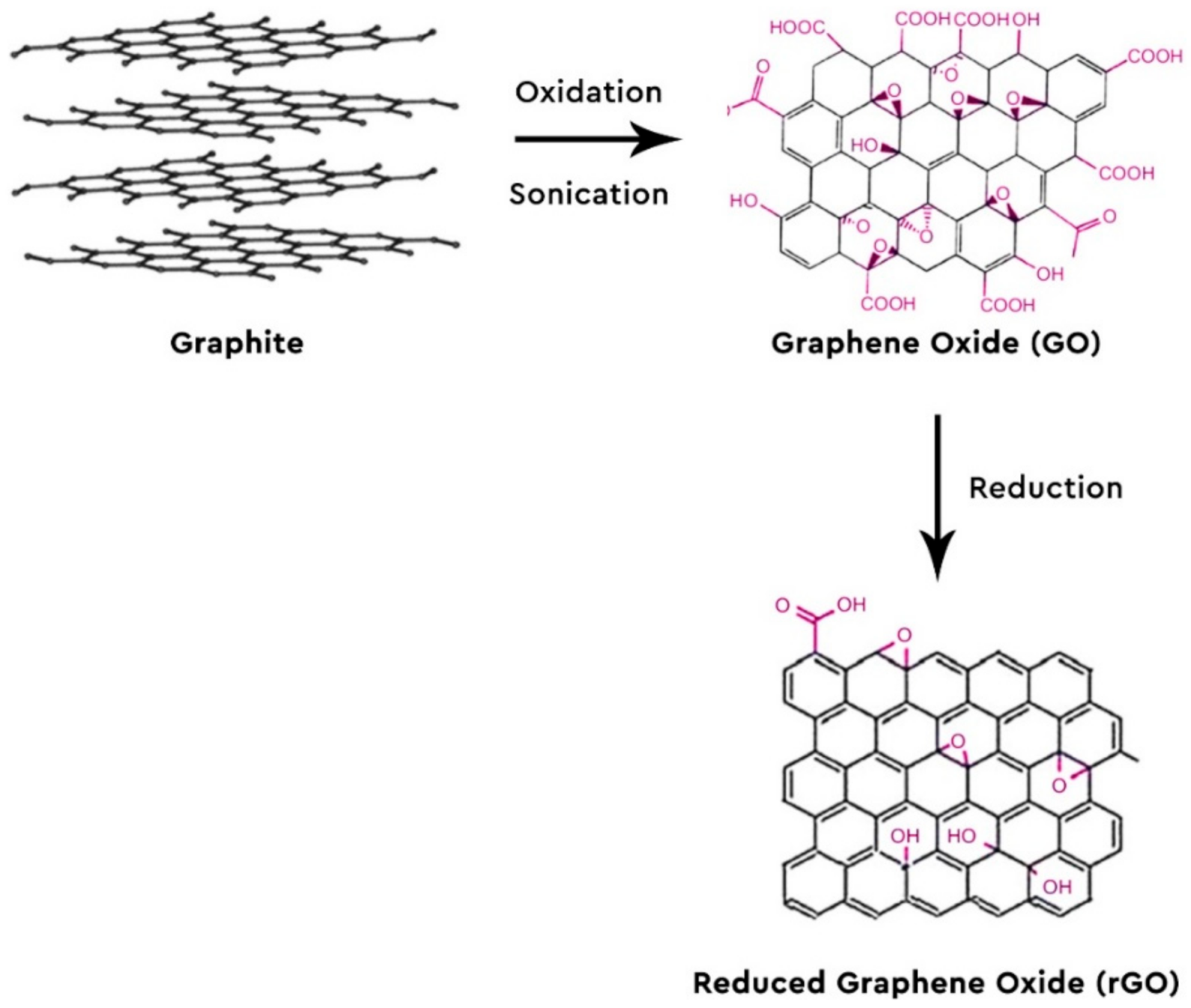
2. Graphene-Based Nanomaterials and Nanocomposites

One of the assets provided by graphene-based materials is selective adsorption. The rGO presents a large π-electron system, making it ideal for the adsorption of organic compounds that consist of aromatic rings. The GO has several functional groups that favor the adsorption of organic molecules containing oxygen- or nitrogen-functional groups.
Even though graphene-based materials (G, GO and rGO) have an enhanced ability of absorbing many and different compounds, they show a significant drawback; the difficulty of the material with the adsorbed analytes being separated from the mother solution. To combat this problem, magnetic nanoparticles (MNPs) are impregnated into rGO or GO. Thus, the combination of high adsorption capacity from G and the convenient separation of the adsorbent from the sample, given from the MNPs, makes these nanocomposites ideal for analytical purposes [43]. Moreover, Fe3O4 nanoparticles are non-toxic, have a small particle size, offer high reactivity and possess a vast specific surface area, adding to the benefits of their usage and rendering them more appealing [46,47].

2.1. Synthesis of Magnetic Nanoparticles

The common methods for synthesizing MNPs are co-precipitation, thermal decomposition, hydrothermal synthesis, colloidal and high-energy ball mill. Depending on which method was used, a specific size and dispersion of size distribution of MNPs are obtained [48].
The modified co-precipitation method introduced by Massart includes the addition of FeCl3·6H2O and FeCl2·4H2O in deionized water, with the temperature rising to 60 °C in order to acquire a yellow and clear solution and stirred vigorously. Afterwards, aqueous ammonia is inserted drop by drop until the pH reaches 10. Then, it is left for the reaction to take place for about 30 min, under continuous and vigorous stirring. Throughout the whole experiment, N2 is used as a protective gas. When the reaction finishes, the black precipitate is collected with the help of a strong magnet, it is washed many times with deionized water and ethanol. Lastly, the magnetite nanoparticles are lyophilized [49,50].
The thermal decomposition method carried out by Kluchova et al. produced maghemite nanoparticles by the solid-state isothermal decomposition in air at 400 °C of Fe(C2H3O2)2·2H2O for 1 h. Before the synthesis, the solid was homogenized in an agate mortar, following in the precursor particles having a size distribution of 1–5 μm [51].
Zheng et al. [52] produced Fe3O4 nanoparticles by hydrothermal synthesis, where 1 mmol of Fe(NO3)3·9H2O and 0.5 mmol of sodium bis(2-ethylhexyl)sulfosuccinate were added in 10 mL deionized water, following the addition of 2 mL of 50% hydrazine hydrate while stirring. This solution was put into a 20 mL Teflon-lined stainless-steel autoclave of and heated to 160 °C at a pace of 5 °C/min. For 10 h, the temperature was kept at 160 °C and then the mixture brought to room temperature naturally. The nanoparticles were filtered, washed several times with ethanol and deionized water and air-dried. The morphology and the structure of the MNPs were characterized by TEM, HRTEM and XRD.
Another procedure for synthesizing Fe3O4 magnetic nanoparticles is by the microwave-solvothermal method suggested by Li et al. [53], where 5 mmol of FeCl3·6H2O, 400 mg of Na3C6H5O7 and 50 mmol of NH4CH3CO2 were added in a solution of 70 mL of ethylene glycol. The black solution was stirred and heated and then transferred into a microwave transparent PTFE-TFM-lined alumina ceramic vessel, with the capacity of 100 mL. Then, it was heated for 15 min to 260 °C for 2 h in a microwave reaction system. Finally, the black powder of Fe3O4 was gathered with the help of a strong magnet and washed several times with ethanol.

2.2. Synthesis of Fe3O4-GO Nanocomposites

Kyzas et al. [50] presented two ways of preparing magnetic GO (mGO): by co-precipitation (mGOp) or by impregnation (mGOi). In the former method, graphite oxide is scattered in deionized water with the help of a sonicator for 30 min. Then, specific amounts of FeCl3·6H2O and FeCl2·4H2O are dissolved in deionized water at room temperature and the mixture is added drop by drop to the GO solution, under vigorous stirring and under a N2 atmosphere. After the completion of ion exchange, 28% v/v ammonia solution is added dropwise so the pH reaches the value of 10 for the synthesis of MNPs. Next, the solution is heated to 80 °C and after agitating for 45 min, the black solid is collected via centrifugation, is washed many times with CH3OH and is lyophilized. In the latter method, graphite oxide is again added in deionized water and put into a sonicator for 30 min to obtain GO and then an amount of Fe3O4 nanoparticles are added to the mixture. After another 30 min, a homogenous suspension is obtained and the nanocomposites are collected by centrifugation and freeze-dried.
The aforementioned material was studied for dye adsorption experiments, with Reactive Black 5 (C26H21N5Na4O19S6) being the target compound. From the X-ray diffraction measurements (XRD) presented in the paper, the average crystallite size of the MNPs were found to be around 18.4 nm. Scanning electron microscopy images were taken to identify the morphology of the mGOi nanoparticles and the iron distribution map of the material. Also, the magnetization of the two materials were studied and the mGOi adsorbent had 65 emu/g, which was a bit higher than the same value of mGOp. Lastly, from Fourier transform infrared (FT-IR)spectra, the identification of functional groups, such as carboxylic groups, epoxy groups and ammonium groups, was carried out.

2.3. Synthesis of GO Membranes Composites

Deng et al. [54] firstly prepared hydroxylated graphene (GOH), by using ferrous chloride and hydrogen peroxide at certain volume ratios, while adjusting the pH with diluted HCl, and GO with a modified Hummers method. Then, 1 mg of GO and 1 mg of GOH were mixed into a beaker, under ultrasonication for 30 min to form a uniform dispersion and the solution was put onto the polyethersulfone membranes of pore sizes 0.22 μm and 1 μm under vacuum.
The morphology and validation of the deposition of the material on the membrane was done via scanning electron microscopy (SEM) and X-ray diffraction (XRD). The performance of the adsorption properties was evaluated with dye compounds, such as rhodamine B and methyl orange, and with the protein albumin from bovine serum.

2.4. Synthesis of Graphene Aerogels

Zhi et al. [55] showcased in a review seven different ways of preparing graphene aerogels, either with no templates or with a template. The methods that require no template are the hydrothermal method, where a GO colloidal dispersion is prepared in ethanol or water, heated at a certain temperature and freeze-dried, dried in atmospheric and in supercritical conditions, the chemical reduction method, by using a reducing agent, the chemical cross-linking method, to enhance the interaction between the graphene oxide sheets with the usage of glutaraldehyde or poly(vinyl alcohol), and a 3D printing method, with the preparation of a high viscosity ink of SiO2 and GO suspension. The latter methods thar require a template are the chemical vapor deposition method, where 3D graphene is prepared by the chemical vapor deposition of carbon onto a metal foam, like copper or nickel, the ice template method, by freezing a GO suspension and its gradual melting to form a porous aerogel with GO, and the bubble template method, with the utilization of a surfactant under vigorous stirring so that the GO is trapped in the bubbles and begins to form.

2.5. Synthesis of GO Alginate Beads

Arshad et al. [56] prepared alginate beads of GO by adding 0.2 g sodium alginate in a 5 wt.% GO suspension in deionized water and then deposited dropwise into a 5 wt.% calcium chloride solution. The alginate beads that were formed were kept in the solution for 3 h. These beads were used for the removal of metals and organic compounds, with maximum adsorption capacities reaching 588.2 mg/g bead.

3. Analytical Applications in Environmental Samples

The environmental applications of graphene-based nanomaterials and nanocomposites are classified into two categories: in the extraction and determination of (i) organic pollutants and (ii) metal ions and rare earth elements. Some of the organic compounds that are examined with these materials are Polycyclic Aromatic Hydrocarbons (PAHs), estrogens, sulfonamides, chlorophenols and so forth, whilst the second category consists of Hg, Cr(III), Cd, Cr(VI), Au, Co, Cu, Zn, Pb(II), Ce(III), Tl(III) and other [57,58].

3.1. Applications of G and GO

Wang et al. [59] used G as a sorbent in SPE for the preconcentration and determination of trace-level amounts of Co and Ni in tap water, river water and sea water. A 3 mL SPE column was packed with 60 mg of G and frits were placed above and below the solid to prevent any loss of the adsorbent. Then, 10 mL of methanol and 10 mL of deionized water were added to the column for equilibrium, cleaning and neutralization before usage. The pH of the sorbent was modified to the value of 5. Afterwards, 100 mL of an aqueous sample loaded with 2 mL of ammonium pyrrolidine dithiocarbamate was passed through the column. The column was then rinsed with 5 mL deionized water and the adsorbed analytes were extracted with 2 mL of 1 mol/L HNO3 in acetone. Finally, the eluate was collected and then measured with the help of a flame atomic absorption spectrometry (FAAS) instrument. Also, experiments of possible reusage of the sorbent were conducted, concluding that a single SPE cartridge packed with G could be reused for over 50 times. The recoveries of Co and Ni in tap water were 96.5–97.5% and 96.2–97.2% respectively, in sea water 96.8–98.0% and 96.5–97.0% and in river waters 96.5–100.5% and 96.6–99.8%.
Su et al. [60] worked with a graphene oxide-silica composite coating in hollow-fiber solid phase microextraction (HF-SPME), which was online coupled with inductively coupled plasma mass spectrometry (ICP-MS) to determinate trace amounts of Mn2+, Co2+, Ni2+, Cu2+, Cd2+ and Pb2+ in environmental water samples. In particular, three water samples of well water, pond water and lake water were examined, which were collected and filtered with a 0.45 μm membrane filter of and then their pH was adjusted to 1 with the addition of concentrated nitric acid for storage. Afterwards, for the extraction the pH was adjusted to the value of 5 with 0.1 mol/L nitric acid and NH3·H2O. The LODs of the six metal ions Mn2+, Co2+, Ni2+, Cu2+, Cd2+ and Pb2+ were 7.5, 0.39, 20, 23, 6.7 and 28 ng/L respectively, while their recoveries were in the range of 85–107%, 88–93%, 90–92%, 91–112%, 100–107% and 87–119% respectively.
Li et al. [61] utilized graphene as their sorbent in the extraction and determination of five sulfonylurea herbicides in environmental waters by Ultra-high Performance Liquid Chromatography-Mass Spectrometry (UHPLC-MS). Once again, 3 mL SPE cartridges were packed with G and they were rinsed with 10 mL of acetonitrile (ACN) and 10 mL of distilled water. 100 mL of the sample’s solution were permeated through the cartridge and then, the cartridge was washed with 10 mL of ultra-pure water and the analytes were extracted with 6 mL of ACN, which contained 1% v/v CH3COOH. The solution eluted was evaporated under the stream of N2 until dried and reconstituted in 1 mL ACN:Water (1:1 v/v). A 0.22 μm nylon membrane filter was used for filtration prior to UHPLC injection. The method followed showed outstanding analytical performance, with the LODs ranging between 0.28–0.53 ng/L for the five herbicides, while the recoveries rates were 84.16–109.31%.
Pourjavid et al. [62] synthesized GO and used it for the column SPE of Fe(III) and Mn(II) ions in water samples, used 3-(1-methyl-1H-pyrrol-2-yl)-1H-pyrazole-5-carboxylic acid (MPPC) as a chelating reagent and the determination was done by FAAS. At optimum conditions, Mn and Fe showed 145 and 162 ng/L respectively, while their recoveries were both 102% in tap water samples, 101% and 103% in mineral water samples respectively and 102–104% and 103% in river water samples.
Sitko et al. [63] covalently bonded GO nanosheets to spherical silica via the coupling of the -NH2 groups of the spherical amino-silica with the -COOH groups of GO (GO@SiO2), which was validated by electron microscopy and X-ray photoelectron spectroscopy, for its usage in SPE to determine Pb(II) and Cu(II) by FAAS from water samples. The water samples were filtered through a 0.45 mm cellulose acetate membrane and put in storage at 4 °C. Before each extraction, the cartridge was rinsed with an acetate buffer solution to pH 5.5, as well as the analyzed solution with the addition of 0.1 mol/L HNO3 and NH3. The two metal ions were eluted from the cartridge with the addition of 2 mL of 1 mol/L HNO3. The recoveries of Cu(II) and Pb(II) were 98.7 ± 0.9% and 95.2 ± 1.0% respectively, while the LODs were 0.084 and 0.27 ng/mL.
Zawisza et al. [64] used an ethylenediamine-modified GO (GO-EDA) for the preconcentration of Cu(II), Zn(II), Fe(III), Ni(II), Co(II) and Pb(II) in water samples. The target metal ions were adsorbed onto GO-EDA by d-μSPE, then the solution was filtered and the filter paper was exposed directly to energy dispersive X-ray fluorescence spectrometry (ED-XRF). The limits of detection of the six metals were calculated as 0.08, 0.06, 0.07, 0.07, 0.10 and 0.10 ng/mL, respectively and their recoveries from the analyses from real samples were in the range of 92–110%. In a different study, Zawisza et al. [65] proposed a new and green method of sample preparation by using GO in dispersive micro-solid phase extraction (d-μSPE) for the preconcentration of trace amounts of Co(II), Ni(II), Zn(II), Cu(II) and Pb(II) of water samples determined by energy dispersive X-ray fluorescence spectrometry (ED-XRF). The GO was dispersed in aqueous samples containing the above-mentioned elements and during stirring, metal ions were sorbed onto GO. Later, the solution was filtered under vacuum and the sorbent with the adsorbed metal ions was collected onto a membrane filter. Finally, the obtained samples were analyzed directly by ED-XRF. This procedure demonstrates low LODs, 0.5, 0.7, 1.8, 1.5 and 1.4 ng/mL, respectively for each metal ion and the recoveries ranged between 94–106%.
Pourjavid et al. [66] studied the preconcentration and determination of trace amounts of Ni(II) and Co(II) ions by FAAS, after their extraction with GO on column SPE in water samples. The adsorption on GO was achieved at pH 6.0 and then the target compounds were eluted with 3 mol/L nitric acid. Experimental conditions for the optimal separation of the analyte ions such as concentration of eluent, sample volume, pH, flow rate and interference ions were investigated. An enrichment factor of 250 was achieved, whilst the LODs of Ni(II) and Co(II) were found to be 0.18 and 0.25 ng/mL, respectively.
Pytlakowska [67] utilized GO nanoparticles for the d-μSPE of heavy metals as their complexes with 2-(5-bromo-2-pyridylazo)-5-diethylaminophenol (5-Br-PADAP) in water samples. The heavy metals examined in this study were Cr(III), Co(II), Ni(II), Cu(II), Zn(II) and Pb(II) ions and their determination was achieved by ED-XRF. Under optimum conditions, the LODs ranged from 0.07 to 0.25 ng/mL and the recoveries of the ions in water and spiked water samples were 94.4–103.5%.
In an additional study, Pytlakowska et al. used glycine modified GO (GO-Gly) as a new sorbent for the preconcentration of chromium, zinc and copper ions from water samples, prior to ED-XRF determination. 500 mg/mL GO-Gly mixture were added to an aqueous sample and the pH of the sample was modified to 6 by adding 0.1 mol/L NH3·H2O and the solution was agitated for 10 min. The analyzed sample was put through a membrane filter with the use of vacuum. This filter containing GO-Gly and the adsorbed ions was air-dried and then immediately measured using the ED-XRF [68]. Furthermore, in a third study, Pytlakowska et al. [69] experimented with chemically modified GO with mercapto-groups (GO-S) for the determination of copper, chromium, lead and zinc by ED-XRF in lake, river, mineral, spring and sea waters. The extraction technique used was d-μSPE.
Ghazaghi et al. [70] presented a new, rapid, and easy technique, where they exploited the GO nanosheets for the novel separation and preconcentration of coagulating homogenous dispersive micro solid phase extraction (CHd-μSPE) of chromium, cadmium and lead in water samples. The LODs were 12, 5 and 35 ng/L respectively, while their recovery rates were 98.0% for both Pb and Cd and 97.2% for Cr.
Zhu et al. [71] utilized diethylenetriamine-functionalized multi-walled carbon nanotubes (MWCNTs) distributed in graphene oxide colloids for the selective SPE of trace amounts of Cr(III), Mn(II), Pb(II) and Fe(III) ions in wastewater. Inductively coupled plasma with optical emission spectrometry (ICP-OES) was used as a determination method of the above ions. The LODs were found to be 0.16, 0.38, 0.24 and 0.50 ng/mL respectively, while the pre-concentration factor was 75.
Chatzimitakos et al. [72] utilized G-functionalized melamine sponges (GMeS) for the microextraction of sulfonamides from environmental samples. The GMeS was prepared by a facile procedure containing only one step, complying with the principles of green chemistry. Lake water samples were adjusted to 3.0 pH and 6% w/v NaCl was added. Before the extraction, the GMeS was preconditioned by dipping it, subsequently, once in CH3OH and three times in deionized water. Then, the GMeS was deposited in 10 mL of the sample and agitated for 30 min at 600 rounds per minute. After each extraction, GMeS was put in a syringe cartridge, where it was washed multiple times with water and squeezed to wash away the excess water. The elution was done with 2 × 1 mL of acetonitrile containing 5% v/v NH3. The eluent was evaporated until dried, under a N2 stream and the precipitate was reconstituted in 100 μL H2O: Acetonitrile mixture of 70:30 v/v, put into an ultrasonicator for 1 min and was finally injected into the HPLC valve. The recoveries of the proposed method were found to be 93%–105% for lake water samples and their enrichment factor 96–99.
Mosavi et al. [73] used modified GO nanosheets with trithiocyanuric acid for the enrichment and extraction of Cu(II) and Pb(II) ions in sea water samples prior to FAAS. Before starting the SPE extraction, the pH of the solution containing both ions was modified to 6.0 with 0.1 mol/L nitric acid or NaOH. Afterwards, it was permeated through the mini-cartridge packed with a standard amount of GO-TTC. The mini-cartridge was rinsed with 2 mL distilled water. Then, the effluate containing the adsorbed metal ions from the mini-cartridge was performed with 3 mL of 3 mol/L HNO3. The amount of each ion in the effluate was determined by FAAS.
Sun et al. [74] developed lightweight GO-based sponges with high durability and compressibility for dye adsorption, specifically for methylene blue in wastewater samples. These sponges adsorbed and retained methylene blue when put in a solution that contains it and then the remaining concentration of the compound was determined by UV-vis spectrometry at 664 nm. The maximum adsorption capacity was observed at 476 mg/g sponge and its recyclability was found to be great, by adsorbing the target compound almost at 86% after the tenth cycle. Tao et al. [75] also chose methylene blue from wastewater samples as their target compound for the selective adsorption on GO-montmorillonite/sodium alginate aerogel beads (GO-MMT/SA). Once again, the determination was carried out by UV-vis spectrometry and the residual concentration was calculated. The maximum adsorption capacity of the beads was considerably lower than the sponges, which was found to be 150.66 mg/g beads.
To summarize, there are plenty of applications that utilize the extraordinary adsorption properties of graphene-based nanomaterials, with most of them being GO or modified GO, hence the low LODs and the high enrichment factors achieved. Most methods are focused towards the extraction and determination of metal ions, such as Mn(II), Cr(III), Zn(II), Pb(II) and so forth due to their toxicity in the environmental waters. A synopsis of the aforementioned papers can be found in Table 1.

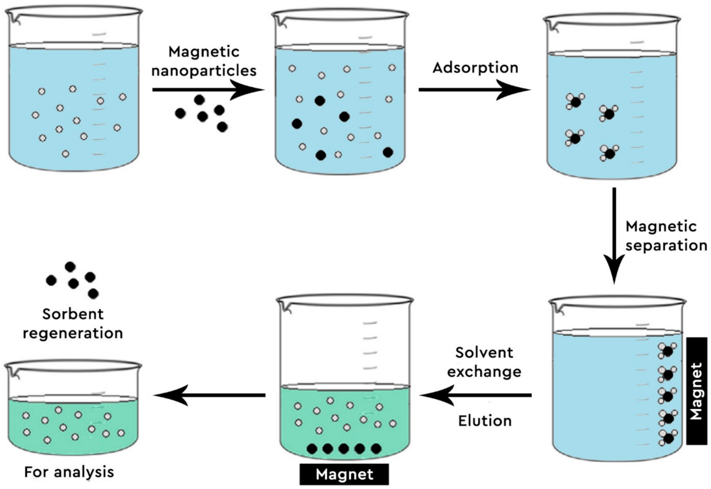
3.2. Applications of GO-Fe3O4

Han et al. [76] synthesized mGO nanocomposites for the adsorption of PAHs with the help of MSPE from tap water, sea water samples and river water. For the extraction of the analytes, 40 mg of the sorbent were conditioned with 5 mL methanol and 5 mL deionized water and then dispersed into a 50 mL aqueous sample solution into an ultrasonic to form a homogenous suspension. Then, the solution was shaken for 10 min for the adsorption to take place and with the help of an external magnetic field at the bottom of the vial, the sorbent was isolated from the mixture. After 3 min, the liquid was decanted, and the adsorbent was held at the bottom by the magnet. Afterwards, the remaining precipitate was washed with 5 mL of 10% v/v ACN and the target organic pollutants were eluted with 2 mL of ACN:CH2Cl2 (4:1) after ultrasonication for 3 min. Lastly, the 20 μL of the effluate was injected to the HPLC-UV system for analysis. The LODs from this work were 0.09–0.19 ng/mL and the recoveries were between 86.3–103.2% for the tap water samples, 76.8–101.2% for the river water samples and 80.9–99.6% for the sea water samples.
Zeng et al. [77] also used Fe3O4@GO as the sorbent for the MSPE of trace levels of 2,4,4′-trichlorobiphenyl (PCB 28) from water samples and then determined by Gas Chromatography- Mass Spectrometry (GC-MS). The recommended method was found suitable for the preconcentration and immobilization of PCB 28 from large amounts of aqueous solutions, as well as the cleaning of polychlorinated biphenyl pollution.
Shi et al. [78] proposed a facile approach of preparing mGO composites and then using them in the MSPE of trace amounts of sulfonamides (sulfadiazine, sulfadimidine and sulfathiazole) in different water samples, determined by High Performance Liquid Chromatography- Diode Array Detection (HPLC-DAD). The results showed that the LODs of the three sulfonamides were between 0.05–0.10 μg/mL and the recoveries where in the range of 67.4–119.9%. In another study, Shi et al. [79] investigated the adsorption mechanism and the pre-concentration of sulfonamides by using a porphyrin-functionalized mGO nanocomposite from environmental water samples, determined once again by HPLC-DAD. The mechanism was investigated and predicted by altering the value of the pH, so as to activate the hydrogen of the porphyrin ring, to negatively charge the material when it was synthesized and then exploit the electrostatic interactions between the adsorbent and the positively charged sulfonamides at pH 4. This speculation was validated with the Langmuir isotherm. This proposed method exhibited 0.2 μg/mL LOD and 83.7–116.7% recoveries.
Ziaei et al. [80] prepared mercapto-grafted GO-magnetic chitosan (GO-MC) as a new biosorbent for the extraction and preconcentration of mercury ion from different water samples. For the synthesis of GO-MC composite, a chitosan solution of 2% w/v was formulated by dissolving 2 g of chitosan powder into 100 mL of 2% v/v CH3COOH under ultrasonication and stirring for 2 h at room temperature. Next, 0.6 g of the MNPs were added into the chitosan solution and the mixture was agitated for another 2 h. Then, 15 mL of glutaraldehyde and 1.5 g of GO were put into the flask. The pH was adjusted to 9–10 and kept in a water bath for 90 min at 80 °C. The black precipitate was rinsed with CH3OH and deionized water, until the pH attained the value of 7 and then dried in an oven at 50 °C. Afterwards, for the surface modification of GO-MC with 3-mercaptopropyl trimethoxysilane (MTPS), 100 mg of the GO-MC was added in 100 mL distilled water with simultaneous sonication for 5 min. The MTPS was added drop by drop under continuous stirring for 6 h under nitrogen, at 60 °C. The new material was magnetically separated and washed two times with 40 mL of double distilled water and 40 mL of ethanol. Lastly, the GO-MC-MTPS was dried in an oven at 40 °C. The prepared nanocomposite material was characterized by SEM, FT-IR and energy dispersive X-ray spectroscopy (ED-X), to confirm the presence of -NHCO-, -CHO, -COOH, epoxy and -SH groups, to carry out morphology studies of the nanocomposite and verify the attachment of -SH groups onto GO-MC. The LODs achieved were 0.06 ng/mL and the recoveries varied from 95%–100% from the water samples.
Aliyari et al. [81] utilized a diethylenetriamine-functionalized mGO (mGO-DETA) for the extraction and determination of lead and cadmium (Pb(II) and Cd(II)) from water samples prior to FAAS analysis. The LOD of lead was 0.38 μg/L and of cadmium was 0.40 μg/L. The maximum adsorption capacity of the two metal ions was calculated as 172.41 and 59.88 mg/g sorbent, respectively and the recoveries were 99.2 and 103.9% for sea water samples, 98.0% and 94.4% for river water samples and 95.4 and 100.8% for well water samples.
Kazemi et al. [82,83] used d-μSPE as the extraction technique of gold and chromium (Cr(III) and Cr(VI)) ions for two different methods. For both methods, mGO was used as the sorbent and they were determined by FAAS. In the first method, under the optimum conditions, the LOD of gold ions from water and wastewater samples reached the level of 4 ng/L, with a preconcentration factor of 500. The recoveries ranged between 98.3%–102.1% for the four different water samples. In the second method, the LOD was found to be 0.1 μg/L, with an preconcentration factor of 200 and the recoveries were 98.5%–102.0% for Cr(III) ions and 97.0%–102.5% for Cr(VI) ions in drinking water, river water, spring water and sea water samples.
Ahmad et al. [84] exhibited the quantitative analysis of gold from environmental water samples by using mGO in dSPE, determined via microwave plasma-atomic emission spectrometry (MP-AES). Recoveries of analytes ranged from 97%–101% and LOD was calculated to be 5 ng/L.
Chen et al. [85] prepared a novel triethylenetetramine-functionalized mGO (TETA-mGO) composite used as a MSPE adsorbent for the swift detection of 10 trace amounts of phenolic environmental estrogens in water samples (tap water, rivel water, well water). Its synthesis was done in three steps: first the magnetic Fe3O4 nanoparticles were prepared by a solvothermal method, then they were dispersed in acetonitrile (ACN), with the usage of an ultrasonicator. In the dispersion solutions of 10 mmol of glycidylmethacrylate and 20 mmol of divinylbenzene were added, along with a certain quantity of 2,2-azobis(2-methylpropionitrile) as the initiator. The new solution was stirred at 500 rpm under nitrogen atmosphere and heated to 70 °C for 3 h. Lastly, 1% of mGO in methanol solution was prepared and TETA was dissolved and stirred for 3 h at 80 °C. The obtained material was washed three times with methanol and vacuum dried in an oven at 25 °C overnight. The determination of the above analytes was done by ultra-fast liquid chromatography coupled to MS tandem MS (UFLC-MS/MS). The proposed method exhibited LODs in the range of 0.15–1.5 ng/L, which was lower than the ones reported in other studies regarding phenolic environmental estrogens, and the recoveries ranged between 88.5% to 105.6%.
Khan et al. [86] synthesized and characterized an allylamine modified mGO-poly(vinyl acetate-co-divinylbenzene) (mGO-DVB-VA) and used in the MSPE of Pb(II), Cu(II), Ni(II), Cd(II), and Co(II) for their determination by FAAS. The preconcentration is 40 for all the metal ions and the LODs are 0.37–2.39 μg/L. This method was applied to wastewater samples.
Islam et al. [87] presented a quick and sensitive method for sequential speciation and separation of Cr(VI) and Cr(III) using d-MSPE for their determination by FAAS, with a triethylenetetramine-modified mGO (mf-GO) as a sorbent from river water samples, tannery wastewater and electroplating wastewater. The use of this sorbent results in the fast removal of Cr(VI) and of Cr(III), which is 10 and 30 min, respectively, while its adsorption capacity is 16.4 and 9.6 mg/g. The speciation is achieved by adjusting the value of the pH. The LODs of the two ions are 1.6 and 5.2 μg/L, respectively and the recoveries of spiked analyte samples ranged between 96–102%.
Arvand et al. [88] extracted imatinib and doxorubicin as cytostatic drugs from environmental water samples by using mGO nanocomposites as a sorbent for MSPE, prior to HPLC-UV analysis. After optimizing the conditions (sample pH, amount of MNPs, the effect of salt concentration and etc.), the LODs of the two drugs were 1.9 and 1.8 ng/mL, respectively, while their recoveries were 88.4% and 96.7%.
Costa dos Reis et al. [89] also used mGO as a sorbent for the MSPE of 2,4,6-trinitrotoluene in different water samples, determined by HPLC-UV. The optimal MSPE conditions reported were 0.04 g of the nanocomposite, 10 min of extraction by vortexing, 1 mL of ACN as eluent, and 6 min of desorption by ultrasonication. The method exhibited 0.3 μg/L as the LOD, while the recovery values from the reservoir, drinking water and effluent wastewater samples were 87–120%.
Dahaghin et al. [90,91] proposed two different methods regarding the usage of mGO in MSPE. The first one dealt with the determination of trace amounts of Cd(II), Pb(II) and Cu(II) ions by FAAS by utilizing a modified mGO with 2-mercaptobenzothiazole. Under optimum conditions, the LODs of the three metal ions were calculated to be 0.19, 0.24 and 0.35 ng/mL, respectively. The second method consisted of the synthesis of mGO modified with 8-hydroxyquinoline and its use for the extraction and preconcentration of traces of Cd(II) and Pb(II) ions. The LODs were 0.09 and 0.27 ng/mL and the enrichment factor was obtained at 130.43.
Molaei et al. [92] demonstrated the preparation SiO2-coated mGO modified with polypyrrole-polythiophene (mGO/SiO2@coPPy-Th) for its use as an ideal sorbent for the determination of trace levels of Cu(II), Zn(II), Cr(III), Pb(II) and Cd(II) from well, river and bottled mineral water samples prior to FAAS analysis. The recoveries of the metal ions varied from 90% to 106%, the LODs ranged from 0.15–0.65 ng/mL and the preconcentration factor was defined as 36–44 for the five metals.
Neyestani et al. [93] modified an mGO with 2-mercaptobenzothiazole for the selective MSPE of Au(III), Pd(II) and Ag(I). The metal ions’ desorption was done with a solution of thiourea and then the ions were determined by flow injection coupled with ICP-OES (FI-ICP-OES). Effects of sample volume and pH, volume and type of eluent, amount of sorbent and time of adsorption and desorption were considered. Under the optimal conditions, the method exhibited 45–76 ng/L LODs, with enrichment factor of 160, 160 and 140 for the three metal ions respectively.
Sarikhani et al. [94] synthesized a novel imidazolium-functionalized mGO nanocomposite in the MSPE for the determination of very low amounts of Cr(VI) and Cr(III) species by electrothermal atomic absorption spectrometry (ETAAS) in wastewater samples. The preparation of the material consisted of the following steps: firstly, 0.1% of GO aqueous dispersion was prepared, under ultrasonication for 3 h. A certain amount of (NH4)2Fe(SO4)2·6H2O and FeCl3 was added to the dispersion under a stream of nitrogen. To dissolve the salts, 8 mol/L NH4OH was added dropwise under sonication for 10 min and the reaction was carried out at 50 °C for 30 min. The mGO was separated by using a strong magnet and it was rinsed with deionized water and ethanol. The obtained material was suspended in an aqueous solution that also contained ethanol and NH4OH. Then, tetraethyl orthosilicate was put into the suspension under continuous agitation for 12 h at 40 degrees Celsius. After separating it with a magnet and rinsing it, the new material (GO@Fe3O4@SiO2) was dispersed in a certain volume of dried C6H5CH3 and (3-chloropropyl)-triethoxysilane (CPTS) was put into the mixture under reflux for 24 h. Meanwhile, a solution of thiocarbamate imidazole was synthesized by combining 1-(3-aminopropyl)imidazole and carbon disulfide and agitating it for 3 h at room temperature. A fixed mass of GO@Fe3O4@SiO2@CPTS was dispersed in a solution of methanol:triethylamine (volume ratio 1:1) and a certain amount of the other compound was added under reflux for another 24 h. Separated by a magnet, rinsed with methanol and ethanol and dried at 50 °C, the material was lastly mixed with n-bromobutane under stirring and a nitrogen atmosphere for 24 h at 80 °C. Finally, the adsorbent was once again separated with a strong magnet and rinsed with tetrahydrofuran and vacuum dried. The LODs of the two Cr ions altogether were 1.9 ng/L, the sorption capacity of the sorbent was 304 mg/g and the preconcentration factor was 357.
Ma et al. [95] presented a method for the determination of di-2-ethylhexyl phthalate (DEHP) by HPLC-DAD in different environmental water samples, using MSPE as an extraction technique and mGO as the sorbent. The process of the extraction is as follows: 0.05 g of mGO were added to 200 mL of the water sample that contained the analyte and the solution pH was modified to the value of 7. After shaking for 25 min, the mGO was separated from the mixture with the use of a strong magnet. The adsorbed analyte was eluted with 2 mL of CH3OH. The obtained recovery rates were 91.6%–106.5% and the LOD of DEHP was 0.35 μg/L.
Most applications on magnetic graphene-based nanomaterials are on tap, river, sea and wastewater samples, examining either the trace amounts of certain metal ions or the concentration of organic pollutants. These materials, also, demonstrate pretty high enrichment factors and very low LODs, in the range of a few ng/L–μg/L. A summary of the above works and methods can be found in Table 2.

4. Conclusions

The usage of the different graphene-based nanomaterials and nanocomposites in the removal of organic pollutants or metal ions from a variety of environmental water samples is on the rise due to the many advantages they provide versus the commonly used sorbents, such as high sorption capacity, many cycles of reusability, extraordinary active surface area, high chemical and mechanical endurance, and facile handling.
The main downside these materials demonstrate is the potential aggregation and the piling up of the nanosheets, which might cause blockage to its active adsorption groups. So as to overwhelm this setback, a wide range of functional groups that help to prevent the problem have been recommended and assessed. By improving the dispersibility of the sorbent, its extraction efficiency and the selectivity towards the target compounds, functionalization is enhanced as well. Moreover, in order to be used at an industrial level, more facile and inexpensive preparation pathways have to be introduced, that lead to recyclable and reusable materials.
As seen, most applications of these materials are fixated on SPE, MSPE and dSPE. Forthcoming developments involve the utilization of graphene-based materials and nanocomposites in other emerging extraction techniques, such as SPME (especially in HF-SPME), PT-SPE and SBSE. More and more new development paths are studied, that are greener or more specific in order to adsorb certain groups of compounds and be selective or to maximize their adsorption capacity. In the foreseeable future, these sorbents will be expected to play a vital part in the extraction of pollutants from environmental samples, whilst new and more specialized materials are synthesized and studied.

Author Contributions

Conceptualization, V.S. and E.D.; methodology, V.S. and E.D.; investigation, O.-E.P., V.S. and E.D.; writing—original draft preparation, O.-E.P., V.S. and E.D.; writing—review and editing, O.-E.P., V.S. and E.D.; supervision E.D. and V.S. All authors have read and agreed to the published version of the manuscript.

Funding

This research received no external funding.

Conflicts of Interest

The authors declare no conflict of interest.

Abbreviations

ACNAcetonitrile
d-SPEDispersive Solid Phase Extraction
d-μSPEDispersive Micro Solid Phase Extraction
d-MSPEDispersive Magnetic Solid Phase Extraction
ED-XRFEnergy Dispersive X-ray Fluorescence Spectrometry
ETAASElectrothermal Atomic Absorption Spectrometry
FAASFlame Atomic Absorption Spectrometry
FT-IRFourier-Transform Infrared Spectroscopy
GGraphene
GC-MSGas Chromatography—Mass Spectrometry
GOGraphene Oxide
HF-SPMEHollow-fiber Solid Phase Micro Extraction
HPLCHigh Pressure Liquid Chromatography
HPLC-DADHigh Pressure Liquid Chromatography—Diode-Array Detector
ICP-OESInductively Coupled Plasma Optical Emission Spectrometry
LODLimit of Detection
mGOMagnetic Graphene Oxide (Fe3O4/Graphene Oxide)
MNPMagnetic Nanoparticles
MSPEMagnetic Solid Phase Extraction
PAHPolycyclic Aromatic Hydrocarbons
PT-SPEPipette-tip Solid Phase Extraction
rGOReduced Graphene Oxide
SEMScanning Electron Microscope
SPESolid Phase Extraction
SPMESolid Phase Microextraction
UHPLC-MSUltra-High Pressure Liquid Chromatography–Mass Spectrometry

References

  1. Chen, Y.; Xia, L.; Liang, R.; Lu, Z.; Li, L.; Huo, B.; Li, G.; Hu, Y. Advanced materials for sample preparation in recent decade. TrAC Trends Anal. Chem. 2019, 120, 115652. [Google Scholar] [CrossRef]
  2. Plastiras, O.-E.; Andreasidou, E.; Samanidou, V. Microextraction Techniques with Deep Eutectic Solvents. Molecules 2020, 25, 6026. [Google Scholar] [CrossRef] [PubMed]
  3. Lu, X.; Sun, J.; Sun, X. Recent advances in biosensors for the detection of estrogens in the environment and food. TrAC Trends Anal. Chem. 2020, 127, 115882. [Google Scholar] [CrossRef]
  4. Xia, S.; Dong, J.; Chen, Y.; Wang, Y.; Chen, X. Three dimensional phytic acid-induced graphene as a solid-phase microextraction fiber coating and its analytical applications for nerolidol in tea. Chin. Chem. Lett. 2018, 29, 107–110. [Google Scholar] [CrossRef]
  5. Jiang, H.-L.; Li, N.; Cui, L.; Wang, X.; Zhao, R.-S. Recent application of magnetic solid phase extraction for food safety analysis. TrAC Trends Anal. Chem. 2019, 120. [Google Scholar] [CrossRef]
  6. Amiri, A.; Baghayeri, M.; Sedighi, M. Magnetic solid-phase extraction of polycyclic aromatic hydrocarbons using a graphene oxide/Fe3O4@polystyrene nanocomposite. Microchim. Acta 2018, 185, 1–9. [Google Scholar] [CrossRef]
  7. Yap, P.L.; Auyoong, Y.L.; Hassan, K.; Farivar, F.; Tran, D.N.; Ma, J.; Losic, D. Multithiol functionalized graphene bio-sponge via photoinitiated thiol-ene click chemistry for efficient heavy metal ions adsorption. Chem. Eng. J. 2020, 395, 124965. [Google Scholar] [CrossRef]
  8. Maciel, E.V.S.; de Toffoli, A.L.; Neto, E.S.; Nazario, C.E.D.; Lanças, F.M. New materials in sample preparation: Recent advances and future trends. TrAC Trends Anal. Chem. 2019, 119, 115633. [Google Scholar] [CrossRef]
  9. Shivakumar, R.; Bolker, A.; Tsang, S.H.; Atar, N.; Verker, R.; Gouzman, I.; Hala, M.; Moshe, N.; Jones, A.; Grossman, E.; et al. POSS enhanced 3D graphene—Polyimide film for atomic oxygen endurance in Low Earth Orbit space environment. Polymer 2020, 191, 122270. [Google Scholar] [CrossRef]
  10. Kyzas, G.Z.; Travlou, N.A.; Deliyanni, E.A. The role of chitosan as nanofiller of graphite oxide for the removal of toxic mercury ions. Colloids Surf. B Biointerfaces 2014, 113, 467–476. [Google Scholar] [CrossRef]
  11. Kyzas, G.Z.; Deliyanni, E.A.; Matis, K.A. Graphene oxide and its application as an adsorbent for wastewater treatment. J. Chem. Technol. Biotechnol. 2014, 89, 196–205. [Google Scholar] [CrossRef]
  12. Sengupta, J.; Hussain, C.M. Graphene and its derivatives for Analytical Lab on Chip platforms. TrAC Trends Anal. Chem. 2019, 114, 326–337. [Google Scholar] [CrossRef] [Green Version]
  13. Nayl, A.; Abd-Elhamid, A.; El-Shanshory, A.A.; Soliman, H.M.; Kenawy, E.-R.; Aly, H. Development of sponge/graphene oxide composite as eco-friendly filter to remove methylene blue from aqueous media. Appl. Surf. Sci. 2019, 496, 143676. [Google Scholar] [CrossRef]
  14. Gazzari, S.; Cortés-Arriagada, D. Interaction of trivalent arsenic on different topologies of Fe-doped graphene nanosheets at water environments: A computational study. J. Mol. Liq. 2019, 289, 1–7. [Google Scholar] [CrossRef]
  15. Zhang, L.; Zhang, Y.; Tang, Y.; Li, X.; Zhang, X.; Li, C.; Xu, S. Magnetic solid-phase extraction based on Fe3O4/graphene oxide nanoparticles for the determination of malachite green and crystal violet in environmental water samples by HPLC. Int. J. Environ. Anal. Chem. 2018, 98, 215–228. [Google Scholar] [CrossRef]
  16. Martín, A.; Escarpa, A. Graphene: The cutting–edge interaction between chemistry and electrochemistry. TrAC Trends Anal. Chem. 2014, 56, 13–26. [Google Scholar] [CrossRef]
  17. Mateos, R.; Vera, S.; Díez-Pascual, A.M.; Andrés, M.P.S. Graphene solid phase extraction (SPE) of synthetic antioxidants in complex food matrices. J. Food Compos. Anal. 2017, 62, 223–230. [Google Scholar] [CrossRef]
  18. Magesa, F.; Wu, Y.; Tian, Y.; Vianney, J.-M.; Buza, J.; He, Q.; Tan, Y. Graphene and graphene like 2D graphitic carbon nitride: Electrochemical detection of food colorants and toxic substances in environment. Trends Environ. Anal. Chem. 2019, 23, e00064. [Google Scholar] [CrossRef]
  19. Yang, M.; Tian, H.; Zhu, J.; He, J. Graphene Nanomaterials in Energy and Environment Applications. Handb. Graphene 2019, 5, 1–25. [Google Scholar] [CrossRef]
  20. Qi, C.; Zhao, L.; Lin, Y.; Wu, D. Graphene oxide/chitosan sponge as a novel filtering material for the removal of dye from water. J. Colloid Interface Sci. 2018, 517, 18–27. [Google Scholar] [CrossRef]
  21. Zhou, S.; Yao, W.; Wang, Z.; Ma, L.; Lu, Z.; Hou, C. The first-principles calculations to explore the mechanism of oxygen diffusion on vacancy defective graphene in marine environment. Appl. Surf. Sci. 2020, 525, 146585. [Google Scholar] [CrossRef]
  22. Zaaba, N.; Foo, K.; Hashim, U.; Tan, S.; Liu, W.-W.; Voon, C. Synthesis of Graphene Oxide using Modified Hummers Method: Solvent Influence. Proc. Eng. 2017, 184, 469–477. [Google Scholar] [CrossRef]
  23. Maggira, M.; Deliyanni, E.A.; Samanidou, V.F. Synthesis of Graphene Oxide Based Sponges and Their Study as Sorbents for Sample Preparation of Cow Milk Prior to HPLC Determination of Sulfonamides. Molecules 2019, 24, 2086. [Google Scholar] [CrossRef] [PubMed] [Green Version]
  24. García-Mesa, J.; Leal, P.M.; Guerrero, M.L.; Alonso, E.V. Simultaneous determination of noble metals, Sb and Hg by magnetic solid phase extraction on line ICP OES based on a new functionalized magnetic graphene oxide. Microchem. J. 2019, 150, 104141. [Google Scholar] [CrossRef]
  25. Shahabi, M.; Raissi, H. Comprehensive theoretical prediction of the dynamics and stability properties of Tegafur pharmaceutical agent on the Graphene based nanostructures in aqueous environment. Appl. Surf. Sci. 2018, 455, 32–36. [Google Scholar] [CrossRef]
  26. Li, W.-K.; Shi, Y.-P. Recent advances and applications of carbon nanotubes based composites in magnetic solid-phase extraction. TrAC Trends Anal. Chem. 2019, 118, 652–665. [Google Scholar] [CrossRef]
  27. Jon, C.-S.; Meng, L.-Y.; Li, D. Recent review on carbon nanomaterials functionalized with ionic liquids in sample pretreatment application. TrAC Trends Anal. Chem. 2019, 120, 115641. [Google Scholar] [CrossRef]
  28. Sun, C.; Wang, Z.; Chen, L.; Li, F. Fabrication of robust and compressive chitin and graphene oxide sponges for removal of microplastics with different functional groups. Chem. Eng. J. 2020, 393, 124796. [Google Scholar] [CrossRef]
  29. Yao, Y.; Ping, J. Recent advances in graphene-based freestanding paper-like materials for sensing applications. TrAC Trends Anal. Chem. 2018, 105, 75–88. [Google Scholar] [CrossRef]
  30. Wang, Z.; Han, Q.; Xia, J.; Xia, L.; Ding, M.; Tang, J. Graphene-based solid-phase extraction disk for fast separation and preconcentration of trace polycyclic aromatic hydrocarbons from environmental water samples. J. Sep. Sci. 2013, 36, 1834–1842. [Google Scholar] [CrossRef]
  31. Núñez, C.; Triviño, J.J.; Segura, R.; Arancibia, V. Development of a fast and sensitive method for the determination of As(III) at trace levels in urine by differential pulse anodic voltammetry using a simple graphene screen–printed electrode. Microchem. J. 2020, 159, 105393. [Google Scholar] [CrossRef]
  32. Wang, Z.; Yao, M.; Wang, X.; Li, S.; Liu, Y.; Yang, G. Influence of reaction media on synthesis of dialdehyde cellulose/GO composites and their adsorption performances on heavy metals. Carbohydr. Polym. 2019, 232, 115781. [Google Scholar] [CrossRef]
  33. Zhang, Y.; Guan, J.; Wu, J.; Ding, S.; Yang, J.; Zhang, J.; Dong, A.; Deng, L. N-alkylated chitosan/graphene oxide porous sponge for rapid and effective hemostasis in emergency situations. Carbohydr. Polym. 2019, 219, 405–413. [Google Scholar] [CrossRef]
  34. Wu, J.; Chen, L.; Mao, P.; Lu, Y.; Wang, H. Determination of chloramphenicol in aquatic products by graphene-based SPE coupled with HPLC-MS/MS. J. Sep. Sci. 2012, 35, 3586–3592. [Google Scholar] [CrossRef]
  35. Service, R.F. Carbon Sheets an Atom Thick Give Rise to Graphene Dreams. Science 2009, 324, 875–877. [Google Scholar] [CrossRef]
  36. Hou, X.; Tang, S.; Wang, J. Recent advances and applications of graphene-based extraction materials in food safety. TrAC Trends Anal. Chem. 2019, 119, 115603. [Google Scholar] [CrossRef]
  37. Sadegh, H. Development of graphene oxide from graphite: A review on synthesis, characterization and its application in wastewater treatment. Rev. Adv. Mater. Sci. 2017, 49, 38–43. [Google Scholar]
  38. Alhwaige, A.A.; Agag, T.; Ishida, H.; Qutubuddin, S. Biobased chitosan hybrid aerogels with superior adsorption: Role of graphene oxide in CO2 capture. RSC Adv. 2013, 3, 16011–16020. [Google Scholar] [CrossRef]
  39. Nidheesh, P.V. Graphene-based materials supported advanced oxidation processes for water and wastewater treatment: A review. Environ. Sci. Pollut. Res. 2017, 24, 27047–27069. [Google Scholar] [CrossRef]
  40. Wei, Y.; Zhang, Y.; Gao, X.; Ma, Z.; Wang, X.; Gao, C. Multilayered graphene oxide membranes for water treatment: A review. Carbon 2018, 139, 964–981. [Google Scholar] [CrossRef]
  41. Sitko, R.; Zawisza, B.; Malicka, E. Graphene as a new sorbent in analytical chemistry. TrAC Trends Anal. Chem. 2013, 51, 33–43. [Google Scholar] [CrossRef]
  42. Hummers, W.S.; Offeman, R.E. Preparation of Graphitic Oxide. J. Am. Chem. Soc. 1958, 80, 1339. [Google Scholar] [CrossRef]
  43. Wang, X.; Liu, B.; Lu, Q.; Qu, Q. Graphene-based materials: Fabrication and application for adsorption in analytical chemistry. J. Chromatogr. A 2014, 1362, 1–15. [Google Scholar] [CrossRef] [PubMed]
  44. Gao, Y.; Li, Y.; Zhang, L.; Huang, H.; Hu, J.; Shah, S.M.; Su, X. Adsorption and removal of tetracycline antibiotics from aqueous solution by graphene oxide. J. Colloid Interface Sci. 2012, 368, 540–546. [Google Scholar] [CrossRef] [PubMed]
  45. Valcárcel, M.; Cárdenas, S.; Simonet, B.; Moliner-Martínez, Y.; Lucena, R. Carbon nanostructures as sorbent materials in analytical processes. TrAC Trends Anal. Chem. 2008, 27, 34–43. [Google Scholar] [CrossRef]
  46. Zhang, J.; Lin, S.; Han, M.; Su, Q.; Xia, L.; Hui, Z. Adsorption Properties of Magnetic Magnetite Nanoparticle for Coexistent Cr(VI) and Cu(II) in Mixed Solution. Water 2020, 12, 446. [Google Scholar] [CrossRef] [Green Version]
  47. Jędrzak, A.; Grześkowiak, B.F.; Coy, E.; Wojnarowicz, J.; Szutkowski, K.; Jurga, S.; Jesionowski, T.; Mrówczyński, R. Dendrimer based theranostic nanostructures for combined chemo- and photothermal therapy of liver cancer cells in vitro. Colloids Surf. B Biointerfaces 2019, 173, 698–708. [Google Scholar] [CrossRef]
  48. Wierucka, M.; Biziuk, M. Application of magnetic nanoparticles for magnetic solid-phase extraction in preparing biological, environmental and food samples. TrAC Trends Anal. Chem. 2014, 59, 50–58. [Google Scholar] [CrossRef]
  49. Massart, R. Preparation of aqueous magnetic liquids in alkaline and acidic media. IEEE Trans. Magn. 1981, 17, 1247–1248. [Google Scholar] [CrossRef]
  50. Kyzas, G.Z.; Travlou, N.A.; Kalogirou, O.; Deliyanni, E.A. Magnetic graphene oxide: Effect of preparation route on reactive black 5 adsorption. Materials 2013, 6, 1360–1376. [Google Scholar] [CrossRef] [Green Version]
  51. Kluchova, K.; Zboril, R.; Tucek, J.; Pecova, M.; Zajoncova, L.; Safarik, I.; Mashlan, M.; Markova, I.; Jancik, D.; Sebela, M.; et al. Superparamagnetic maghemite nanoparticles from solid-state synthesis—Their functionalization towards peroral MRI contrast agent and magnetic carrier for trypsin immobilization. Biomaterials 2009, 30, 2855–2863. [Google Scholar] [CrossRef]
  52. Zheng, Y.H.; Cheng, Y.; Bao, F.; Wang, Y.S. Synthesis and magnetic properties of Fe3O4 nanoparticles. Mater. Res. Bull. 2006, 41, 525–529. [Google Scholar] [CrossRef]
  53. Li, C.; Wei, Y.; Liivat, A.; Zhu, Y.; Zhu, J. Microwave-solvothermal synthesis of Fe3O4 magnetic nanoparticles. Mater. Lett. 2013, 107, 23–26. [Google Scholar] [CrossRef]
  54. Deng, H.; Huang, J.; Qin, C.; Xu, T.; Ni, H.; Ye, P. Preparation of high-performance nanocomposite membranes with hydroxylated graphene and graphene oxide. J. Water Process. Eng. 2021, 40, 101945. [Google Scholar] [CrossRef]
  55. Zhi, D.; Li, T.; Li, J.; Ren, H.; Meng, F. A review of three-dimensional graphene-based aerogels: Synthesis, structure and application for microwave absorption. Compos. Part B Eng. 2021, 211. [Google Scholar] [CrossRef]
  56. Arshad, F.; Selvaraj, M.; Banat, F.; Abu Haija, M. Removal of metal ions and organics from real refinery wastewater using double- functionalized graphene oxide in alginate beads. J. Water Process. Eng. 2020, 38, 101635. [Google Scholar] [CrossRef]
  57. Manousi, N.; Rosenberg, E.; Deliyanni, E.A.; Zachariadis, G.A. Sample Preparation Using Graphene-Oxide-Derived Nanomaterials for the Extraction of Metals. Molecules 2020, 25, 2411. [Google Scholar] [CrossRef]
  58. Manousi, N.; Rosenberg, E.; Deliyanni, E.; Zachariadis, G.A.; Samanidou, V. Magnetic Solid-Phase Extraction of Organic Compounds Based on Graphene Oxide Nanocomposites. Molecules 2020, 25, 1148. [Google Scholar] [CrossRef] [Green Version]
  59. Wang, Y.-K.; Gao, S.-T.; Ma, J.-J.; Li, J.-C. Application of Graphene as a Sorbent for Simultaneous Preconcentration and Determination of Trace Amounts of Cobalt and Nickel in Environmental Water and Vegetable Samples. J. Chin. Chem. Soc. 2012, 59, 1468–1477. [Google Scholar] [CrossRef]
  60. Su, S.; Chen, B.; He, M.; Hu, B. Graphene oxide–silica composite coating hollow fiber solid phase microextraction online coupled with inductively coupled plasma mass spectrometry for the determination of trace heavy metals in environmental water samples. Talanta 2014, 123, 1–9. [Google Scholar] [CrossRef]
  61. Li, C.; Lu, A.; Wang, J.; Li, J.; Ping, H.; Luan, Y.; Chen, J.; Ha, X. Determination of five sulfonylurea herbicides in environmental waters and soil by ultra- high performance liquid chromatography with tandem mass spectrometry after extraction using graphene. J. Sep. Sci. 2014, 37, 3714–3721. [Google Scholar] [CrossRef] [PubMed]
  62. Pourjavid, M.R.; Sehat, A.A.; Arabieh, M.; Yousefi, S.R.; Hosseini, M.H.; Rezaee, M. Column solid phase extraction and flame atomic absorption spectrometric determination of manganese(II) and iron(III) ions in water, food and biological samples using 3-(1-methyl-1H-pyrrol-2-yl)-1H-pyrazole-5-carboxylic acid on synthesized graphene oxide. Mater. Sci. Eng. C 2014, 35, 370–378. [Google Scholar] [CrossRef] [PubMed]
  63. Sitko, R.; Zawisza, B.; Talik, E.; Janik, P.; Osoba, G.; Feist, B.; Malicka, E. Spherical silica particles decorated with graphene oxide nanosheets as a new sorbent in inorganic trace analysis. Anal. Chim. Acta 2014, 834, 22–29. [Google Scholar] [CrossRef] [PubMed]
  64. Zawisza, B.; Baranik, A.; Malicka, E.; Talik, E.; Sitko, R. Preconcentration of Fe(III), Co(II), Ni(II), Cu(II), Zn(II) and Pb(II) with ethylenediamine-modified graphene oxide. Microchim. Acta 2016, 183, 231–240. [Google Scholar] [CrossRef] [Green Version]
  65. Zawisza, B.; Sitko, R.; Malicka, E.; Talik, E. Graphene oxide as a solid sorbent for the preconcentration of cobalt, nickel, copper, zinc and lead prior to determination by energy- dispersive X-ray fluorescence spectrometry. Anal. Methods 2013. [Google Scholar] [CrossRef]
  66. Pourjavid, M.R.; Arabieh, M.; Yousefi, S.R.; Jamali, M.R.; Rezaee, M.; Hosseini, M.H.; Sehat, A.A. Study on column SPE with synthesized graphene oxide and FAAS for determination of trace amount of Co(II) and Ni(II) ions in real samples. Mater. Sci. Eng. C 2015, 47, 114–122. [Google Scholar] [CrossRef]
  67. Pytlakowska, K. Dispersive micro solid-phase extraction of heavy metals as their complexes with 2-(5-bromo-2-pyridylazo)-5-diethylaminophenol using graphene oxide nanoparticles. Microchim. Acta 2016, 183, 91–99. [Google Scholar] [CrossRef]
  68. Pytlakowska, K.; Kozik, V.; Matussek, M.; Pilch, M.; Hachuła, B.; Kocot, K. Glycine modified graphene oxide as a novel sorbent for preconcentration of chromium, copper, and zinc ions from water samples prior to energy dispersive X-ray fluorescence spectrometric determination. RSC Adv. 2016, 6, 42836–42844. [Google Scholar] [CrossRef]
  69. Pytlakowska, K.; Pilch, M.; Hachuła, B.; Nycz, J.E.; Kornaus, K.; Pisarski, W.A. Energy dispersive X-ray fluorescence spectrometric determination of copper, zinc, lead and chromium species after preconcentration on graphene oxide chemically modified with mercapto-groups. J. Anal. At. Spectrom. 2019, 34, 1416–1425. [Google Scholar] [CrossRef]
  70. Ghazaghi, M.; Mousavi, H.Z.; Rashidi, A.M.; Shirkhanloo, H.; Rahighi, R. Innovative separation and preconcentration technique of coagulating homogenous dispersive micro solid phase extraction exploiting graphene oxide nanosheets. Anal. Chim. Acta 2016, 902, 33–42. [Google Scholar] [CrossRef]
  71. Zhu, X.; Cui, Y.; Chang, X.; Wang, H. Selective solid-phase extraction and analysis of trace-level Cr(III), Fe(III), Pb(II), and Mn(II) Ions in wastewater using diethylenetriamine-functionalized carbon nanotubes dispersed in graphene oxide colloids. Talanta 2016, 146, 358–363. [Google Scholar] [CrossRef]
  72. Chatzimitakos, T.; Samanidou, V.; Stalikas, C.D. Graphene-functionalized melamine sponges for microextraction of sulfonamides from food and environmental samples. J. Chromatogr. A 2017, 1522, 1–8. [Google Scholar] [CrossRef]
  73. Mosavi, S.S.; Ghanemi, K.; Nickpour, Y. Graphene oxide nanosheets modified with trithiocyanuric acid for extraction and enrichment of Pb (II) and Cu (II) ions in seawater. Water Environ. J. 2018, 32, 377–383. [Google Scholar] [CrossRef]
  74. Sun, Y.; Chen, L.; Yu, J.; Yoon, B.; Lee, S.K.; Nam, J.-D.; Ci, L.; Suhr, J. Lightweight graphene oxide-based sponges with high compressibility and durability for dye adsorption. Carbon 2020, 160, 54–63. [Google Scholar] [CrossRef]
  75. Tao, E.; Ma, D.; Yang, S.; Hao, X. Graphene oxide-montmorillonite/sodium alginate aerogel beads for selective adsorption of methylene blue in wastewater. J. Alloys Compd. 2020, 832, 154833. [Google Scholar] [CrossRef]
  76. Han, Q.; Wang, Z.; Xia, J.; Chen, S.; Zhang, X.; Ding, M. Facile and tunable fabrication of Fe3O4/graphene oxide nanocomposites and their application in the magnetic solid-phase extraction of polycyclic aromatic hydrocarbons from environmental water samples. Talanta 2012, 101, 388–395. [Google Scholar] [CrossRef]
  77. Zeng, S.; Gan, N.; Weideman-Mera, R.; Cao, Y.; Li, T.; Sang, W. Enrichment of polychlorinated biphenyl 28 from aqueous solutions using Fe3O4 grafted graphene oxide. Chem. Eng. J. 2013, 218, 108–115. [Google Scholar] [CrossRef]
  78. Shi, P.; Ye, N. Magnetite–graphene oxide composites as a magnetic solid-phase extraction adsorbent for the determination of trace sulfonamides in water samples. Anal. Methods 2014, 6, 9725–9730. [Google Scholar] [CrossRef]
  79. Shi, P.; Ye, N. Investigation of the adsorption mechanism and preconcentration of sulfonamides using a porphyrin-functionalized Fe3O4-graphene oxide nanocomposite. Talanta 2015, 143, 219–225. [Google Scholar] [CrossRef]
  80. Ziaei, E.; Mehdinia, A.; Jabbari, A. A novel hierarchical nanobiocomposite of graphene oxide–magnetic chitosan grafted with mercapto as a solid phase extraction sorbent for the determination of mercury ions in environmental water samples. Anal. Chim. Acta 2014, 850, 49–56. [Google Scholar] [CrossRef]
  81. Aliyari, E.; Alvand, M.; Shemirani, F. Simultaneous separation and preconcentration of lead and cadmium from water and vegetable samples using a diethylenetriamine-modified magnetic graphene oxide nanocomposite. Anal. Methods 2015, 7, 7582–7589. [Google Scholar] [CrossRef]
  82. Kazemi, E.; Dadfarnia, S.; Shabani, A.M.H. Dispersive solid phase microextraction with magnetic graphene oxide as the sorbent for separation and preconcentration of ultra-trace amounts of gold ions. Talanta 2015, 141, 273–278. [Google Scholar] [CrossRef] [PubMed]
  83. Kazemi, E.; Shabani, A.M.H.; Dadfarnia, S.; Izadi, F. Speciation and determination of chromium ions by dispersive micro solid phase extraction using magnetic graphene oxide followed by flame atomic absorption spectrometry. Int. J. Environ. Anal. Chem. 2017, 97, 1080–1093. [Google Scholar] [CrossRef]
  84. Ahmad, H.; Jalil, A.A.; Triwahyono, S. Dispersive solid phase extraction of gold with magnetite-graphene oxide prior to its determination via microwave plasma-atomic emission spectrometry. RSC Adv. 2016, 6, 88110–88116. [Google Scholar] [CrossRef]
  85. Chen, X.-H.; Pan, S.-D.; Ye, M.-J.; Li, X.-P.; Zhao, Y.-G.; Jin, M.-C. Magnetic solid-phase extraction based on a triethylenetetramine-functionalized magnetic graphene oxide composite for the detection of ten trace phenolic environmental estrogens in environmental water. J. Sep. Sci. 2016, 39, 762–768. [Google Scholar] [CrossRef]
  86. Khan, M.; Yilmaz, E.; Sevinc, B.; Sahmetlioglu, E.; Shah, J.; Jan, M.R.; Soylak, M. Preparation and characterization of magnetic allylamine modified graphene oxide-poly(vinyl acetate-co-divinylbenzene) nanocomposite for vortex assisted magnetic solid phase extraction of some metal ions. Talanta 2016, 146, 130–137. [Google Scholar] [CrossRef]
  87. Islam, A.; Ahmad, H.; Zaidi, N.; Kumar, S. A graphene oxide decorated with triethylenetetramine-modified magnetite for separation of chromium species prior to their sequential speciation and determination via FAAS. Microchim. Acta 2016, 183, 289–296. [Google Scholar] [CrossRef]
  88. Arvand, M.; Masouleh, A.N. Magnetic solid-phase extraction of imatinib and doxorubicin as cytostatic drugs by Fe3O4/graphene oxide nanocomposite. J. Iran. Chem. Soc. 2017, 14, 1673–1682. [Google Scholar] [CrossRef]
  89. Dos Reis, L.C.; Vidal, L.; Canals, A. Graphene oxide/Fe3O4 as sorbent for magnetic solid-phase extraction coupled with liquid chromatography to determine 2,4,6-trinitrotoluene in water samples. Anal. Bioanal. Chem. 2017, 409, 2665–2674. [Google Scholar] [CrossRef] [Green Version]
  90. Dahaghin, Z.; Mousavi, H.Z.; Sajjadi, S.M. Trace amounts of Cd(II), Cu(II) and Pb(II) ions monitoring using Fe3O4 @graphene oxide nanocomposite modified via 2-mercaptobenzothiazole as a novel and efficient nanosorbent. J. Mol. Liq. 2017, 231, 386–395. [Google Scholar] [CrossRef]
  91. Dahaghin, Z.; Mousavi, H.Z.; Sajjadi, S.M. Synthesis and Application of Magnetic Graphene Oxide Modified with 8-Hydroxyquinoline for Extraction and Preconcentration of Trace Heavy Metal Ions. ChemistrySelect 2017, 2, 1282–1289. [Google Scholar] [CrossRef]
  92. Molaei, K.; Bagheri, H.; Asgharinezhad, A.A.; Ebrahimzadeh, H.; Shamsipur, M. SiO2-coated magnetic graphene oxide modified with polypyrrole–polythiophene: A novel and efficient nanocomposite for solid phase extraction of trace amounts of heavy metals. Talanta 2017, 167, 607–616. [Google Scholar] [CrossRef]
  93. Neyestani, M.R.; Shemirani, F.; Mozaffari, S.; Alvand, M. A magnetized graphene oxide modified with 2-mercaptobenzothiazole as a selective nanosorbent for magnetic solid phase extraction of gold(III), palladium(II) and silver(I). Microchim. Acta 2017, 184, 2871–2879. [Google Scholar] [CrossRef]
  94. Sarikhani, Z.; Manoochehri, M. Determination of Ultra Trace Cr(III) and Cr(VI) Species by Electrothermal Atomic Absorption Spectrometry after Simultaneous Magnetic Solid Phase Extraction with the Aid of a Novel Imidazolium-Functionalized Magnetite Graphene Oxide Nanocomposite. Bull. Chem. Soc. Jpn. 2017, 90, 746–753. [Google Scholar] [CrossRef]
  95. Ma, L.; Huang, J.; Zhou, M. Magnetic graphene oxide for efficient solid phase extraction of DEHP. IOP Conf. Ser. Mater. Sci. Eng. 2019, 544. [Google Scholar] [CrossRef]
Figure 1. Synthesis of graphene oxide (GO) and reduced G (rGO) from graphite powder.
Applsci 11 03028 g001
Figure 2. Steps of Magnetic Solid Phase Extraction (MSPE).
Applsci 11 03028 g002
Table 1. Recent applications of graphene-based nanomaterials in environmental analysis.
AdsorbentAnalyte(s)ApplicationsSample
Preparation
Analytical
Technique
LODsEF 1Reference
GCo(II), Ni(II)Tap, river and sea waterSPEFAAS0.36, 0.51 μg/L200[59]
GO-silicaMn(II), Co(II), Ni(II), Cu(II), Cd(II), Pb(II)Well, pond and lake waterHF-SPMEICP-MS7.5, 0.39, 20, 23, 6.7, 28 ng/L10[60]
GSulfonylurea herbicidesEnvironmental water samplesSPEUHPLC-MS0.28–0.53 ng/LN/R 2[61]
GOMn(II), Fe(III)Tap, mineral, river waterSPEFAAS0.145, 0.162 μg/L325[62]
GO-silicaCu(II), Pb(II)Mineral, waste and sea waterSPEFAAS0.084, 0.27 μg/L200–250[63]
GO-EDAFe(III), Co(II), Ni(II), Cu(II), Zn(II), Pb(II)Waste water from industryd-μSPEED-XRF0.07, 0.10, 0.07, 0.08, 0.06, 0.10 μg/LN/R[64]
GOCo(II), Ni(II), Cu(II), Zn(II), Pb(II)Waste water from industryd-μSPEED-XRF0.5, 0.7, 1.8, 1.5, 1.4 μg/LN/R[65]
GOCo(II), Ni(II)Mineral and spring waterSPEFAAS0.25, 0.18 μg/L250[66]
GOCr(III), Co(II), Ni(II), Cu(II), Zn(II), Pb(II)Environmental water samplesd-μSPEED-XRF0.07–0.25 μg/LN/R[67]
GO-GlyCr(III), Cu(II), Zn(II)Tap, river, estuarine and lake waterd-μSPEED-XRF0.15, 0.07, 0.08 μg/L1575, 890, 810[68]
GO-SCu(II), Zn(II), Pb(II), Cr(III)Lake, river, mineral, spring and sea waterd-μSPEED-XRF0.06–0.10 μg/L520–3120[69]
GOPb(II), Cd(II), Cr(III)River waterCHd-μSPEETAAS0.035, 0.005, 0.012 μg/L14.7, 16.1, 15.4[70]
GO-MWCNTCr(III), Fe(III), Pb(II), Mn(II)WastewaterSPEICP-OES0.16, 0.50, 0.24, 0.38 μg/L75[71]
GMeSSulfonamidesLake waterSPEHPLC0.10–0.29 μg/kg 396–99[72]
GO-TTCPb(II), Cu(II)Sea waterSPEFAAS0.32, 0.13 μg/L83.3[73]
GO-spongesMethylene blueWastewaterN/RUV-visN/RN/R[74]
GO-MMT/SAMethylene blueWastewaterN/RUV-visN/RN/R[75]
1 EF = Enrichment Factor., 2 N/R = Not Reported., 3 Only the Limit of Quantification (LOQ) was reported.
Table 2. Main applications of magnetic graphene-based nanomaterials in environmental samples analysis.
AdsorbentAnalyte(s)ApplicationsSample
Preparation
Analytical
Technique
LODsEFReference
mGOPAHsTap, river, sea waterMSPEHPLC-UV0.09–0.19 μg/L25[76]
mGOPCB 28School sewage, river waterMSPEGC-MS0.027–0.059 μg/L200[77]
mGOSulfonamidesTap, river waterMSPEHPLC-DAD0.05–0.10 mg/LN/R[78]
mGO-porphyrinSulfonamidesTap, river waterMSPEHPLC-DAD0.2 mg/LN/R[79]
GO-MC-MTPSHg(II)Tap, sea waterMSPECV-AAS 10.06 μg/L80[80]
mGO-DETACd(II), Pb(II)Sea, river, well waterMSPEFAAS0.40, 0.38 μg/L150, 167[81]
mGOCr(III), Cr(VI), Au(III)Drinking, river, spring, sea, waste waterd-μSPEFAAS0.1, 0.1, 0.004 μg/L200, 200, 500[82,83]
mGOAu(III)Tap, lake, sea waterdSPEMP-AES5 ng/L60[84]
TETA-mGOPhenolic estrogensTap, river, well waterMSPEUFLC-MS/MS0.15–1.5 ng/L10,000[85]
mGO-DVB-VAPb(II), Cd(II), Cu(II), Ni(II), Co(II)Waste waterMSPEFAAS0.37–2.39 μg/L40[86]
mf-GOCr(III), Cr(VI)River, tannery and electroplating waste waterd-MSPEFAAS5.2, 1.6 μg/L10[87]
mGOImatinib, DoxorubicinWell, waste waterMSPEHPLC-UV1.9, 1.8 μg/LN/R[88]
mGO2,4,6-trinitrotolueneReservoir, drinking, waste waterMSPEHPLC-UV0.3 μg/L153[89]
mGO-MBT 2Cd(II), Cu(II), Pb(II)Tap, lake, sea waterMSPEFAAS0.19, 0.35, 0.24 μg/L400[90]
mGO-HQ 3Cd(II), Pb(II)Water samplesMSPEFAAS0.09, 0.27 μg/L130.43[91]
mGO/SiO2@coPPy-ThCu(II), Cr(III), Zn(II), Cd(II), Pb(II)Well, river, bottled mineral waterMSPEFAAS0.15–0.65 μg/L36–44[92]
mGO-MBTAu(III), Pd(II), Ag(I)Waste waterMSPEFI-ICP-OES45–76 ng/L160, 160, 140[93]
mGO-imidazoliumCr(III), Cr(VI)Waste waterMSPEETAAS1.9 ng/L357[94]
mGOdi-2-ethylhexyl phthalateWater samplesMSPEHPLC-DAD0.35 μg/L100[95]
1 Cold Vapor Atomic Absorption Spectrometry, 2 Magnetic Graphene Oxide modified with 2-mercaptobenzothiazole, 3 Magnetic Graphene Oxide modified with 8-hyrdoxyquinolone.
Publisher’s Note: MDPI stays neutral with regard to jurisdictional claims in published maps and institutional affiliations.

