Article

In Vitro Model for Lumbar Disc Herniation to Investigate Regenerative Tissue Repair Approaches

1
Institute of Orthopaedic Research and Biomechanics, Ulm University, 89081 Ulm, Germany
2
Institute for Laser Technologies in Medicine and Metrology, Ulm University, 89081 Ulm, Germany
*
Author to whom correspondence should be addressed.
Appl. Sci. 2021, 11(6), 2847; https://doi.org/10.3390/app11062847
Submission received: 26 February 2021 / Revised: 15 March 2021 / Accepted: 21 March 2021 / Published: 22 March 2021
(This article belongs to the Special Issue Intervertebral Disc Regeneration)

Abstract

:

Featured Application

Development of a lumbar disc herniation model with which regenerative tissue repair approaches can be investigated under physiological loading conditions and worst-case scenarios out of patients’ daily-life.

Abstract

Lumbar disc herniation (LDH) is the most common reason for low back pain in the working society. New regenerative approaches and novel implants are directed towards the restoration of the disc or its biomechanical properties. Aiming to investigate these new therapies under physiological conditions, in this study, a model for LDH was established by developing a new physiological in vitro test method. In 14 human lumbar motion segments, different daily-life and worst-case activities were simulated successfully by applying a physiological range of motion and axial loading in order to create physiological intradiscal pressure. An LDH could be provoked in 11 of the 14 specimens through vertical and round annular defects of different sizes. Interestingly, the defect and the LDH hardly influenced the biomechanical properties of the disc. For the investigation of regenerative approaches in further experiments, the recommendation based on the results of this study is to create an LDH in non-degenerated motion segments by the application of the new physiological in vitro test method after setting the round annular defects to a size of 4 mm in diameter.

1. Introduction

Lumbar disc herniation is the most common reason for low back pain in the working society [1]. The incidence ranges from 1 to 5 of 1000 persons per year but shows a peak in patients with an age ranging from 30 to 50 years [2]. The lower lumbar region is mostly affected with a proportion of over 95% of all lumbar disc herniations, whereas 50% occur in L4-5 and 46% in L5-S1 [3,4]. An intervertebral disc prolapse with extrusion of nucleus pulposus can lead to a compression of nerve roots which can cause pain, numbness and even paralysis.
For a long time, therapies targeted the reduction of these symptoms by surgical nerve decompression, often in combination with implants. New regenerative approaches and novel implants are directed towards the restoration of the disc itself with the goal to improve the biomechanical properties [5,6,7] and the biological functions of an intact disc [8,9,10,11]. This motivates the development of new materials as well as the use of innovative technologies such as 3D bioprinting which offers the opportunity to create highly complex and multi-dimensional biomaterials [12,13,14]. The investigation of such new treatment options also requires biomechanical experiments where a lumbar disc herniation can be provoked under realistic conditions in order to challenge the new treatments under physiological worst-case scenarios.
Therefore, it is of considerable interest to understand the mechanisms of when, where, and how lumbar disc herniations develop. It is known that most lumbar disc herniations result from endplate junction failures (65%) rather than an annulus fibrosus rupture (35%) [15,16]. However, which activities lead to the development of such lumbar disc herniations, are yet unknown. Activities that were associated with a higher risk for a gradual development of lumbar disc herniations include flexional movements or lifting weights [17,18,19,20]. Further, it has been assumed that complex and extreme loading conditions might facilitate disc herniations.
Various in vitro studies investigated the influence of these different loading conditions or motions on structural failures of the lumbar motion segments [2,15,21,22,23,24,25,26,27,28,29,30]. The results of those studies confirmed that the combination of bending forward in combination with lateral bending and axial rotation while lifting heavy objects is considered to be harmful [31,32,33]. Nevertheless, in most of these studies, an exaggerated range of motion (ROM) or loading values had to be applied in order to simulate the long-term effects expected from physiological movements [15,30]. So far, no herniation model could be created that allows the investigation of new regenerative therapies under physiological loading conditions and worst-case scenarios. Furthermore, the precise biomechanical mechanism of the development of lumbar disc herniations is still not completely understood. It is still uncertain during which physiologic activity a disc herniation most likely occurs.
Therefore, the aim of this study was to establish a lumbar disc herniation model by using a new biomechanical in vitro test method which simulates patients’ daily life activities. With this new test method, lumbar disc herniations should be produced through standardized annular defects and dynamic physiological loading. This lumbar disc herniation model should allow the investigation of novel implants and regenerative therapies under worst-case loading scenarios that really happen during patients’ daily life activities.

2. Materials and Methods

2.1. Specimens

In this study, we used 14 single lumbo-sacral motion segments (L2-3, L3-4, L4-5, L5-S1) from eight different adult human donors with a median age of 35.5 years, ranging from 19 to 53 years (Table 1). Ethical approval was provided by the ethical committee board. Sagittal T1 and T2-weighted and transverse T2-weighted magnetic resonance imaging (MRI) was performed to evaluate disc quality and to exclude specimens with disc damage such as disc herniations. Only discs with a low degree of disc degeneration (according to Pfirrmann 1 or 2) were chosen and included in this study. CT scans were performed to exclude specimens with bony fractures, tumors or other problems. The segments were then dissected removing all soft tissue and keeping the disc and all functional ligaments intact. The cranial and caudal vertebrae were embedded into polymethylmethacrylate (PMMA, Technovit 3040, Haereus Kulzer, Wehrheim, Germany) and flanges were mounted to enable a proper fixation in the testing machines.
In all specimens, a right lateral laminotomy was performed to allow access to and observation of the posterior annulus fibrosus with an endoscopic camera. Prior to the calibration of the physiological testing protocol, the specimens were hydrated in physiological saline solution for 1 h to ensure physiologically high hydrostatic pressure in the nucleus pulposus. Before dynamically loading the specimens with the new physiological test protocol, the specimens were rehydrated for 30 min [34] and kept moist throughout the experiment [35].

2.2. New Physiological Loading Protocol

A new physiological loading protocol was developed to simulate typical daily in vivo activities associated with lumbar disc herniations. Therefore, activities were chosen that either consist of extreme flexion like tying shoes or with additional loading like lifting boxes. Furthermore, the influence of activities with complex motion patterns on the risk of lumbar disc herniations was investigated by simulating sweeping the floor or lifting boxes while turning. For each activity, physiological in vivo segmental motion [37] and intradiscal pressures (IDP, [38]) were replicated in a dynamic disc loading simulator [39]. This dynamic disc loading simulator is able to deform the specimens in six degrees of freedom and thereby allows the replication of complex motion in all anatomical planes, as well as additional axial loading or compression to simulate resultant muscle forces or body weight. In order to monitor the IDP that results from this complex deformation and loading of the specimen, an IDP sensor (Mammendorfer Institut für Physik und Medizin GmbH, Hattenhofen, Germany) was implanted into the nucleus pulposus from the right lateral side of the disc.
From a neutral position, the starting activity of the patient in a standing position was approached. Then, the dynamic activities tying shoes, sweeping the floor, lifting boxes (20 kg) and the corresponding complex activity lifting boxes while turning were performed subsequently (Supplementary Materials: Video S1). Each activity consisted either of pure axial loading (standing) or in combination with bending. For the simulation of tying shoes and lifting boxes, the specimens were bent in flexion; for the simulation of sweeping the floor or lifting boxes while turning in flexion, lateral bending and axial rotation were applied simultaneously (Table 2). The motion values that were applied for each activity resulted from an in vivo study performed by Pearcy in 1985 [37]. The amount of the necessary axial load for every individual specimen was determined from in vivo IDP measurements from the study by Wilke et al. [38]. During a quasistatic calibration cycle prior to testing, the equivalent motion of each activity was applied and axial compression was subsequently adjusted (Table 2) until the equivalent in vivo IDP value had been created within the disc. However, in vivo IDP was known for the activities of standing, tying shoes, and lifting boxes. For the simulation of the complex activities (sweeping the floor and lifting boxes while turning), rotations in all motion directions were applied. The axial load was maintained with respect to the corresponding simple activity.
The dynamic loading protocol applied was motion controlled for all ranges of motion and force controlled for the axial compression in order to create the IDP. Each activity was simulated for six cycles at 0.1 Hz. IDP was monitored constantly during dynamic loading. The specimens were loaded first in the intact state of the intervertebral disc and then after setting annular defects.

2.3. Defects

In the posterior part of the annulus fibrosus, vertical (scalpel cut 0.4 mm × 4.0 mm, 1.0 mm × 5.5 mm, 1.2 mm × 6.5 mm) or round defects (Ø 4 mm, Ø 6 mm, Ø 8 mm) were created in the specimens and enlarged consecutively. The posterior annulus fibrosus was observed during the simulation of the activities using an endoscopic camera in order to evaluate whether and at which cycle of which activity a herniation with clear nucleus extrusion occurred. The system consists of a high-performance light unit (D-Light-N, 20t33420 Karl Storz SE & Co. KG, Tuttlingen, Germany), a fiber optic light transmission incorporated telescope (PA-NOVIEW, Richard Wolf GmbH, Knittlingen, Germany) and an ultra-compact USB 3.0 camera (xiQ, XIMEA GmbH, Münster, Germany). After occurrence of a herniation, the defect was not further enlarged. In every case a herniation occurred through a vertical defect; scans in an ultra-high field MRI (11.7 T, BioSpec 117/16, Bruker Corp., Billerica, MA, USA) were taken using isotropic voxels with a resolution of 100 µm.

2.4. Flexibility Tests

For comparison of the biomechanical properties of the specimens with intact, injured, or herniated disc, standardized torsion tests were carried out. Hence, quasistatic flexibility tests were performed in all testing conditions: in the intact disc, after setting the defects and after a herniation was provoked during dynamical loading by use of the new physiological loading protocol. Therefore, a universal spine loading simulator [40] was used to apply pure moments of ±7.5 Nm in flexion-extension, lateral bending, and axial rotation [41]. Maximum ROM and neutral zone (NZ) were measured by a motion tracking system (Vicon Nexus 1.4.116, Vicon Motion Systems Ltd., Oxford, UK) with six cameras (Type MX13, Vicon Motion Systems Ltd., Oxford, UK). Maximum IDP that was reached in the extrema of the flexibility test, was recorded by the same IDP sensor that was already implanted into the nucleus pulposus and that was also used for the calibration of the dynamic loading protocol.

2.5. Statistical Analysis
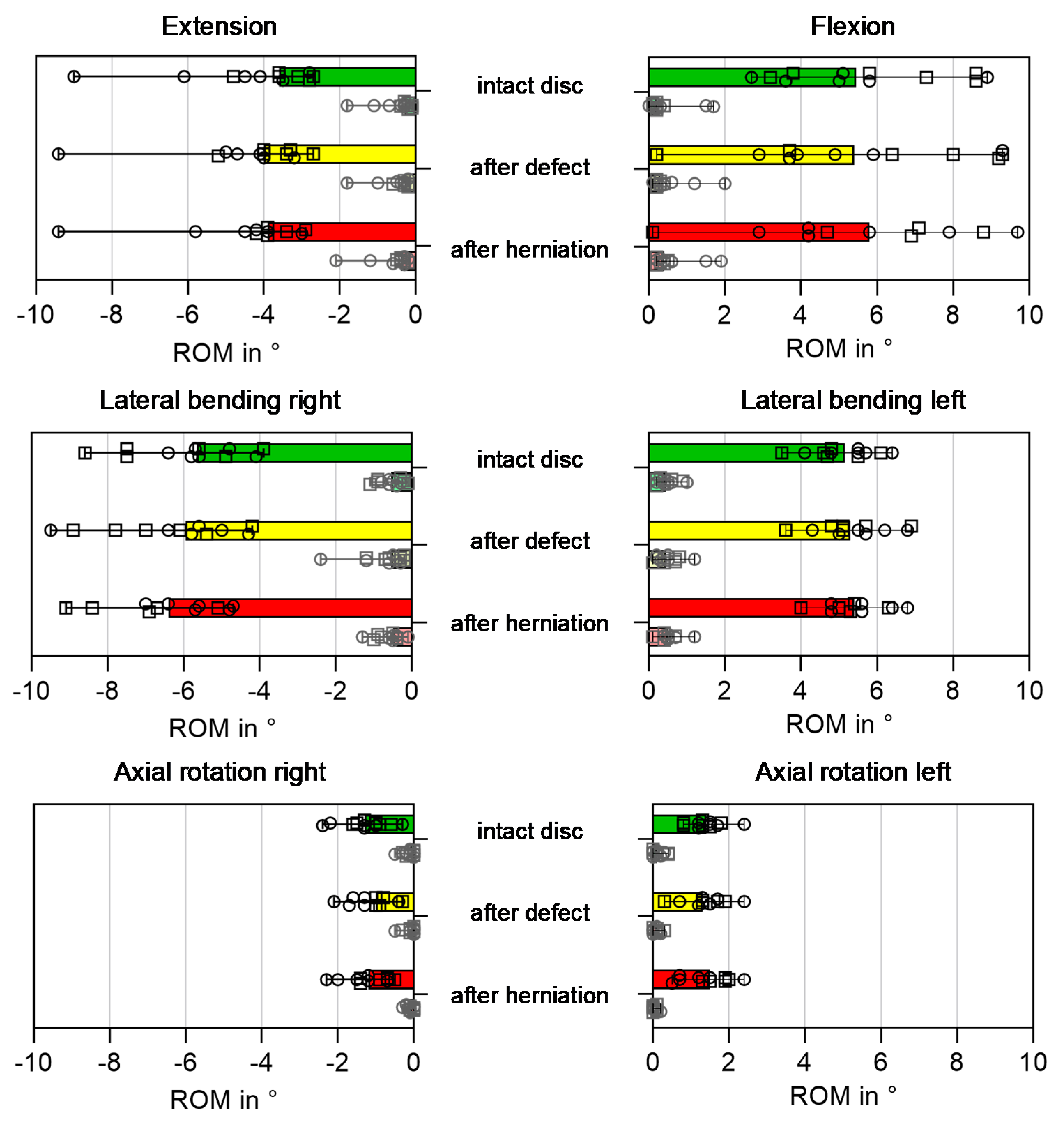
Statistical analysis was performed using a Friedman-Test with Bonferroni Post-Hoc correction in SPSS Software (IBM SPSS Statistics Vol. 27; IBM Corp., Armonk, NY, USA). The significance level was set to α ≤ 0.05.

3. Results

In this study, a new test method could be developed which simulates physiological daily-life activities in a dynamic way. In vivo motion values could be successfully replicated. By combining it with an axial compression, in vivo IDP could be created during the calibration cycle. During the dynamic validation cycle, the in vivo IDP values for standing were reached, but exceeded for tying shoes and lifting boxes (Table 2).

3.1. Provocation of Disc Herniation

With this method, a prolapse with clear nucleus extrusion (Figure 1a) could be provoked in 11 of the 14 specimens (Supplementary Materials: Video S2). A previously set defect was necessary in all specimens (Table 3).

3.1.1. Influence of Shape and Size of the Annular Defect

After setting the first vertical defect, a herniation could only be provoked in one specimen. Enlarging the defect to a size of 1.0 mm × 5.5 mm led to a herniation in one other specimen. In three other specimens, a herniation could be provoked after the annular defect had a size of 1.2 mm × 6.5 mm. One of those specimens showed an intermittent herniation with nucleus pulposus material protruding through the annulus fibrosus in the phase of load application and wandering back into the interior of the disc when unloading. In one specimen, no herniation could be provoked at all. In the specimens with round annular defects, a herniation could be provoked in six specimens after setting a defect with a diameter of Ø 4 mm. In two specimens, no herniation could be provoked at all, neither after widening the defect diameter to Ø 6 mm nor to Ø 8 mm.

3.1.2. Influence of Daily-Life Activities

In seven of the 11 herniated discs (Table 3), the activity lifting boxes led to a herniation, whereby four herniations occurred under pure flexion and three herniations occurred under complex motion. One herniation could be provoked while simulating sweeping floor. Three discs with a round defect (Ø 4 mm) already herniated during the flexibility test before dynamic loading.

3.2. Ultra-High Field MR Imaging

The herniations and defects of the specimens with vertical defects could be observed and further investigated through highly resolved images of the ultra-high field MRI (Figure 1b,c). It could be observed that the herniated nucleus pulposus material always extruded through the defect that was previously set. The sequester extruded to the posterocentral or posterolateral side of the disc.

3.3. Biomechanical Parameters

The herniation itself and the defect only led to a slight increase in ROM and NZ by overall about 1° (Figure 2), and a very slight decrease of IDP (Figure 3). After setting the vertical defects, IDP slightly decreased by 0.2 MPa and 0.1 MPa after setting the round defects. It could be observed that stronger disc injury resulted in a wider range especially for NZ, but also for ROM and IDP values. However, no significant differences could be observed either between specimens with vertical and round shaped defect or between the different conditions of the specimens.
Interestingly, the specimens that did not herniate at all showed slightly lower IDP values or higher ROM either already in the intact state or after creation of the defect.

4. Discussion

In this study, a new physiological test method was developed that allows the replication of different physiological activities in vitro. With this dynamical test method, it was possible to provoke lumbar disc herniations under experimental conditions. A lumbar disc herniation model was developed that can be used for the biomechanical investigation of new regenerative therapies in order to prevent or treat intervertebral disc herniations.
The simulation of different physiological daily-life activities of patients resulted in intravertebral disc herniations, but only after an annular defect was previously set. The annular defects mimicked a structural failure of the annulus fibrosus which has also been observed in patients with herniated intervertebral discs through clinical MRI. Moreover, annular tears have also been identified in asymptomatic individuals. It could be concluded, that an intervertebral disc herniation with nucleus extrusion through an annular defect might be the consequence of substantial annular fissures or tears [28].
However, a herniation could not be provoked in every specimen, even with a large annular defect. The observation of this specimen with ultra-high field MRI might indicate that structural changes have already occurred inside the disc. Compared to the other intervertebral discs, the nucleus pulposus did not look as homogeneous as in the herniated discs and showed disturbances in the signal response of the MRI (Figure 4). It can be assumed that the ability of the disc to create hydrostatic pressure was already slightly impaired. This might underlie the assumption of Wilke et al. that the risk of getting a herniation is higher in younger patients with non-degenerated discs [42], but only if annular defects coexist. This goes in accordance with the findings of Adams et al. that the prevalence of disc herniations increases with age because the annulus fibrosus or endplate junction becomes increasingly injured over time [2]. Additionally, the findings of this study confirm the hypothesis that the risk of an intervertebral disc herniation might be generally higher in the morning after the intervertebral disc has been rehydrated by recovering during night rest [43].
In all specimens, the extrusion of nucleus pulposus occurred either to the central region or to the opposite site to where the combined motion was exerted. This behavior was already observed by Adams and Hutton [2].
Three discs with a round annular defect were already herniated during the flexibility test before dynamic loading. During the flexibility test, the IDP values created by pure motion, were comparable to the in vivo IDP in a standing position. A reason for the early occurrence of a nucleus extrusion in the discs with round annular defects could be that the initial damage led to a larger expansion of the defect due to high fiber strains generated by the dynamical loading [44], compared to the thinner initial vertical defect. Hence for further experiments, we would suggest using a model with a round defect in order to guarantee the successful provocation of a nucleus extrusion.
The defect and the extrusion of nucleus pulposus material through the aforesaid led to a migration of nucleus material. Interestingly, this migration only caused a small decrease of IDP and increase of ROM. From former in vitro studies [42,45] it was known, that treating a disc herniation by partial nucleotomy significantly decreased the IDP and increased ROM. From these findings, it can be concluded that only changes in nucleus volume lead to a significant change of the biomechanical properties, whereas volume migrations do not.
In this study, only herniations with clear nucleus pulposus extrusion through an annular defect could be provoked and hence, investigated. So far, no intervertebral disc herniations caused by endplate junction failures [16] could be investigated by using this test method and it cannot be answered yet as to which activities might lead to endplate junction failures. However, using this new test method, other activities could be simulated in vitro and crucial motion of loading scenarios could be identified that might lead to endplate junction failures.
Furthermore, the defects were set artificially. In previous in vitro studies, it could be shown that annular defects are usually initiated from the inside of the disc and migrate to the outer layers of the annulus fibrosus [26]. Such annular defects were provoked during long-term excessive dynamical loading or under exaggerated loading conditions [2,15,26]. Long-term and excessive dynamical test protocols result in a completely dehydrated disc and especially nucleus pulposus which is unable to rehydrate completely under experimental conditions [34]. Hence, after previously excessive loading of the specimens, it would not have been possible to generate hydrostatic pressure conditions inside the discs [22], which was the basis for this new test method.
Another limitation of this study is that no in vivo IDP values were available for the activities with complex, combined motion. It was assumed that the IDP does change, but the axial load does not change between corresponding activities with single flexion or combined flexion, lateral bending, and axial rotation. The results of the validation might confirm this approach as hydrostatic IDP values highly depend on the individual disc quality. Nevertheless, it would be beneficial to investigate in greater detail how external loads and muscle forces are distributed in the intervertebral disc.

5. Conclusions

In this study, a new lumbar disc herniation model was established by which regenerative tissue repair approaches as well as novel implants, such as nucleus implants or annulus sealing methods for the treatment of lumbar disc herniations could be challenged under normal and physiological worst-case scenarios. Therefore, a new biomechanical in vitro test method was developed that dynamically simulates daily-life activities. By means of this test method, disc herniations could be successfully produced and the influence of shape and size of annular defects investigated. Based on these results, it is recommended to produce a disc herniation in non- or only low degenerated human lumbar discs through a round annular defect.

Supplementary Materials

The following are available online at https://www.mdpi.com/2076-3417/11/6/2847/s1, Table S1: Ultra-high field MRI-IVDs, Video S1: Dynamic disc loading simulator with simulation of physiological activities, Video S2: Lumbar Disc Herniation.

Author Contributions

Conceptualization, H.-J.W. and L.Z. (Laura Zengerle); methodology, L.Z. (Laura Zengerle) and E.D.; software, L.Z. (Laura Zengerle) and E.D.; validation, E.D. and L.Z. (Laura Zengerle); formal analysis, E.D., L.Z. (Laura Zengerle), B.K. and L.Z. (Lena Zöllner); investigation, L.Z. (Laura Zengerle), E.D. and L.Z. (Lena Zöllner); resources, H.-J.W.; data curation, E.D., L.Z. (Lena Zöllner); writing—original draft preparation, L.Z. (Laura Zengerle); writing—review and editing, H.-J.W., E.D., B.K., L.Z. (Lena Zöllner); visualization, B.K., E.D., L.Z. (Laura Zengerle), L.Z. (Lena Zöllner); supervision, H.-J.W., L.Z. (Laura Zengerle); project administration, H.-J.W.; funding acquisition, H.-J.W. All authors have read and agreed to the published version of the manuscript.

Funding

This research was funded by the German Research Foundation (DFG), grant number Wi1352/14-3.

Institutional Review Board Statement

The study was conducted according to the guidelines of the Declaration of Helsinki, and approved by the Institutional Review Board (or Ethics Committee) of Ulm University (protocol code 35/16 and date of approval 3 October 2016).

Informed Consent Statement

Not applicable.

Data Availability Statement

The data presented in this study are available on request from the corresponding author. The data are not publicly available due to confidentiality reasons.

Acknowledgments

The authors thank the Core Facility Small Animal Imaging and the Ulm University Center for Translational Imaging MoMAN for their support. The authors further acknowledge Youping Tao, Jan Ulrich Jansen, Theresa Schilpp and Saskia Brendle for their help in the experiments.

Conflicts of Interest

The authors declare no conflict of interest.

References

  1. Evans, W.; Jobe, W.; Seibert, C. A cross-sectional prevalence study of lumbar disc degeneration in a working population. Spine 1989, 14, 60–64. [Google Scholar] [CrossRef]
  2. Adams, M.A.; Hutton, W.C. The mechanics of prolapsed intervertebral disc. Int. Orthop. 1982, 6, 249–253. [Google Scholar] [CrossRef]
  3. Spangfort, E.V. The lumbar disc herniation. A computer-aided analysis of 2504 operations. Acta Orthop. Scand. 1972, 142, 1–95. [Google Scholar] [CrossRef] [Green Version]
  4. White, A.A.; Panjabi, M.M.; Company, J.B.L. Clinical Biomechanics of the Spine; J. B. Lippincott Company: Philadelphia, PA, USA, 1978. [Google Scholar]
  5. Iatridis, J.C.; Nicoll, S.B.; Michalek, A.J.; Walter, B.A.; Gupta, M.S. Role of biomechanics in intervertebral disc degeneration and regenerative therapies: What needs repairing in the disc and what are promising biomaterials for its repair? Spine J. 2013, 13, 243–262. [Google Scholar] [CrossRef] [PubMed] [Green Version]
  6. Sloan, S.R., Jr.; Wipplinger, C.; Kirnaz, S.; Navarro-Ramirez, R.; Schmidt, F.; McCloskey, D.; Pannellini, T.; Schiavinato, A.; Härtl, R.; Bonassar, L.J. Combined nucleus pulposus augmentation and annulus fibrosus repair prevents acute intervertebral disc degeneration after discectomy. Sci. Transl. Med. 2020, 12. [Google Scholar] [CrossRef] [PubMed]
  7. Zengerle, L.; Köhler, A.; Debout, E.; Hackenbroch, C.; Wilke, H.-J. Nucleus replacement could get a new chance with annulus closure. Eur. Spine J. 2020, 29, 1733–1741. [Google Scholar] [CrossRef] [PubMed]
  8. Bowles, R.D.; Setton, L.A. Biomaterials for intervertebral disc regeneration and repair. Biomaterials 2017, 129, 54–67. [Google Scholar] [CrossRef]
  9. Huang, Y.C.; Hu, Y.; Li, Z.; Luk, K.D.K. Biomaterials for intervertebral disc regeneration: Current status and looming challenges. J. Tissue Eng. Regen. Med. 2018, 12, 2188–2202. [Google Scholar] [CrossRef]
  10. Huang, Y.C.; Urban, J.P.; Luk, K.D. Intervertebral disc regeneration: Do nutrients lead the way? Nat. Rev. Rheumatol. 2014, 10, 561–566. [Google Scholar] [CrossRef] [PubMed]
  11. Sakai, D.; Andersson, G.B. Stem cell therapy for intervertebral disc regeneration: Obstacles and solutions. Nat. Rev. Rheumatol. 2015, 11, 243–256. [Google Scholar] [CrossRef]
  12. Campisi, M.; Shin, Y.; Osaki, T.; Hajal, C.; Chiono, V.; Kamm, R.D. 3D self-organized microvascular model of the human blood-brain barrier with endothelial cells, pericytes and astrocytes. Biomaterials 2018, 180, 117–129. [Google Scholar] [CrossRef]
  13. Foresti, R.; Rossi, S.; Pinelli, S.; Alinovi, R.; Barozzi, M.; Sciancalepore, C.; Galetti, M.; Caffarra, C.; Lagonegro, P.; Scavia, G. Highly-defined bioprinting of long-term vascularized scaffolds with Bio-Trap: Complex geometry functionalization and process parameters with computer aided tissue engineering. Materialia 2020, 9, 100560. [Google Scholar] [CrossRef]
  14. Foresti, R.; Rossi, S.; Pinelli, S.; Alinovi, R.; Sciancalepore, C.; Delmonte, N.; Selleri, S.; Caffarra, C.; Raposio, E.; Macaluso, G.; et al. In-vivo vascular application via ultra-fast bioprinting for future 5D personalised nanomedicine. Sci. Rep. 2020, 10, 3205. [Google Scholar] [CrossRef] [PubMed] [Green Version]
  15. Berger-Roscher, N.; Casaroli, G.; Rasche, V.; Villa, T.; Galbusera, F.; Wilke, H.J. Influence of Complex Loading Conditions on Intervertebral Disc Failure. Spine 2017, 42, E78–E85. [Google Scholar] [CrossRef]
  16. Rajasekaran, S.; Bajaj, N.; Tubaki, V.; Kanna, R.M.; Shetty, A.P. ISSLS Prize winner: The anatomy of failure in lumbar disc herniation: An in vivo, multimodal, prospective study of 181 subjects. Spine 2013, 38, 1491–1500. [Google Scholar] [CrossRef] [PubMed]
  17. Ahsan, M.K.; Matin, T.; Ali, M.I.; Ali, M.Y.; Awwal, M.A.; Sakeb, N. Relationship between physical work load and lumbar disc herniation. Mymensingh Med. J. 2013, 22, 533–540. [Google Scholar] [PubMed]
  18. Kelsey, J.L.; Githens, P.B.; Walter, S.D.; Southwick, W.O.; Weil, U.; Holford, T.R.; Ostfeld, A.M.; Calogero, J.A.; O’Connor, T.; White, A.A. An epidemiological study of acute prolapsed cervical intervertebral disc. J. Bone Joint Surg. Am. 1984, 66, 907–914. [Google Scholar] [CrossRef] [PubMed]
  19. Mundt, D.J.; Kelsey, J.L.; Golden, A.L.; Panjabi, M.M.; Pastides, H.; Berg, A.T.; Sklar, J.; Hosea, T. An epidemiologic study of sports and weight lifting as possible risk factors for herniated lumbar and cervical discs. The Northeast Collaborative Group on Low Back Pain. Am. J. Sports Med. 1993, 21, 854–860. [Google Scholar] [CrossRef]
  20. Pietila, T.A.; Stendel, R.; Kombos, T.; Ramsbacher, J.; Schulte, T.; Brock, M. Lumbar disc herniation in patients up to 25 years of age. Neurol. Med. Chir. 2001, 41, 340–344. [Google Scholar] [CrossRef] [Green Version]
  21. Hutton, W.C.; Adams, M.A. Can the lumbar spine be crushed in heavy lifting? Spine 1982, 7, 586–590. [Google Scholar]
  22. Adams, M.A.; Freeman, B.J.; Morrison, H.P.; Nelson, I.W.; Dolan, P. Mechanical initiation of intervertebral disc degeneration. Spine 2000, 25, 1625–1636. [Google Scholar] [CrossRef]
  23. Gordon, S.J.; Yang, K.H.; Mayer, P.J.; Mace, A.H., Jr.; Kish, V.L.; Radin, E.L. Mechanism of disc rupture. A preliminary report. Spine 1991, 16, 450–456. [Google Scholar] [CrossRef]
  24. Lu, Y.M.; Hutton, W.C.; Gharpuray, V.M. Do bending, twisting, and diurnal fluid changes in the disc affect the propensity to prolapse? A viscoelastic finite element model. Spine 1996, 21, 2570–2579. [Google Scholar] [CrossRef] [PubMed]
  25. McNally, D.S.; Adams, M.A.; Goodship, A.E. Can intervertebral disc prolapse be predicted by disc mechanics? Spine 1993, 18, 1525–1530. [Google Scholar] [CrossRef] [PubMed]
  26. Wade, K.R.; Robertson, P.A.; Thambyah, A.; Broom, N.D. How healthy discs herniate: A biomechanical and microstructural study investigating the combined effects of compression rate and flexion. Spine 2014, 39, 1018–1028. [Google Scholar] [CrossRef]
  27. Wade, K.R.; Robertson, P.A.; Thambyah, A.; Broom, N.D. “Surprise” Loading in Flexion Increases the Risk of Disc Herniation Due to Annulus-Endplate Junction Failure: A Mechanical and Microstructural Investigation. Spine 2015, 40, 891–901. [Google Scholar] [CrossRef]
  28. Wade, K.R.; Schollum, M.L.; Robertson, P.A.; Thambyah, A.; Broom, N.D. ISSLS Prize Winner: Vibration Really Does Disrupt the Disc: A Microanatomical Investigation. Spine 2016, 41, 1185–1198. [Google Scholar] [CrossRef] [PubMed]
  29. Edwards, W.T.; Ordway, N.R.; Zheng, Y.; McCullen, G.; Han, Z.; Yuan, H.A. Peak stresses observed in the posterior lateral anulus. Spine 2001, 26, 1753–1759. [Google Scholar] [CrossRef]
  30. Wade, K.; Berger-Roscher, N.; Rasche, V.; Wilke, H. Disc wall structural abnormalities can act as initiation sites for herniation. Eur. Cell Mater. 2020, 40, 227–238. [Google Scholar] [CrossRef]
  31. Behjati, M.; Arjmand, N. Biomechanical Assessment of the NIOSH Lifting Equation in Asymmetric Load-Handling Activities Using a Detailed Musculoskeletal Model. Hum. Factors 2018, 61, 191–202. [Google Scholar] [CrossRef] [PubMed]
  32. Gunzburg, R.; Parkinson, R.; Moore, R.; Cantraine, F.; Hutton, W.; Vernon-Roberts, B.; Fraser, R. A cadaveric study comparing discography, magnetic resonance imaging, histology, and mechanical behavior of the human lumbar disc. Spine 1992, 17, 417–426. [Google Scholar] [CrossRef]
  33. Andersson, G.B. Epidemiologic aspects on low-back pain in industry. Spine 1981, 6, 53–60. [Google Scholar] [CrossRef]
  34. Pflaster, D.S.; Krag, M.H.; Johnson, C.C.; Haugh, L.D.; Pope, M.H. Effect of test environment on intervertebral disc hydration. Spine 1997, 22, 133–139. [Google Scholar] [CrossRef]
  35. Wilke, H.J.; Jungkunz, B.; Wenger, K.; Claes, L.E. Spinal segment range of motion as a function of in vitro test conditions: Effects of exposure period, accumulated cycles, angular-deformation rate, and moisture condition. Anat. Rec. 1998, 251, 15–19. [Google Scholar] [CrossRef]
  36. Pfirrmann, C.W.; Metzdorf, A.; Zanetti, M.; Hodler, J.; Boos, N. Magnetic resonance classification of lumbar intervertebral disc degeneration. Spine 2001, 26, 1873–1878. [Google Scholar] [CrossRef] [PubMed]
  37. Pearcy, M.J. Stereo radiography of lumbar spine motion. Acta Orthop. Scand. 1985, 212, 1–45. [Google Scholar] [CrossRef]
  38. Wilke, H.J.; Neef, P.; Caimi, M.; Hoogland, T.; Claes, L.E. New in vivo measurements of pressures in the intervertebral disc in daily life. Spine 1999, 24, 755–762. [Google Scholar] [CrossRef]
  39. Wilke, H.J.; Kienle, A.; Maile, S.; Rasche, V.; Berger-Roscher, N. A new dynamic six degrees of freedom disc-loading simulator allows to provoke disc damage and herniation. Eur. Spine J. 2016, 25, 1363–1372. [Google Scholar] [CrossRef] [PubMed] [Green Version]
  40. Wilke, H.J.; Claes, L.; Schmitt, H.; Wolf, S. A universal spine tester for in vitro experiments with muscle force simulation. Eur. Spine J. 1994, 3, 91–97. [Google Scholar] [CrossRef]
  41. Wilke, H.J.; Wenger, K.; Claes, L. Testing criteria for spinal implants: Recommendations for the standardization of in vitro stability testing of spinal implants. Eur. Spine J. 1998, 7, 148–154. [Google Scholar] [CrossRef] [Green Version]
  42. Wilke, H.J.; Ressel, L.; Heuer, F.; Graf, N.; Rath, S. Can prevention of a reherniation be investigated? Establishment of a herniation model and experiments with an anular closure device. Spine 2013, 38, E587–E593. [Google Scholar] [CrossRef] [PubMed] [Green Version]
  43. Belavy, D.L.; Adams, M.; Brisby, H.; Cagnie, B.; Danneels, L.; Fairbank, J.; Hargens, A.R.; Judex, S.; Scheuring, R.A.; Sovelius, R.; et al. Disc herniations in astronauts: What causes them, and what does it tell us about herniation on earth? Eur. Spine J. 2016, 25, 144–154. [Google Scholar] [CrossRef] [PubMed] [Green Version]
  44. Schmidt, H.; Kettler, A.; Heuer, F.; Simon, U.; Claes, L.; Wilke, H.J. Intradiscal pressure, shear strain, and fiber strain in the intervertebral disc under combined loading. Spine 2007, 32, 748–755. [Google Scholar] [CrossRef] [PubMed] [Green Version]
  45. Heuer, F.; Ulrich, S.; Claes, L.; Wilke, H.J. Biomechanical evaluation of conventional anulus fibrosus closure methods required for nucleus replacement. Laboratory investigation. J. Neurosurg. Spine 2008, 9, 307–313. [Google Scholar] [CrossRef] [PubMed]
Figure 1. Provoked herniation with clear nucleus extrusion with (a) endoscopic view of the posterior annulus fibrosus and imaging from ultra-high field MRI (11.7 T) (b) in the transverse and (c) sagittal plane.
Applsci 11 02847 g001
Figure 2. Range of motion (ROM) in ° in flexion-extension, left/right lateral bending and left/right axial rotation for the intact disc, after setting the vertical (□) or round (ο) defects, respectively, assessed with the universal spine tester under pure moments of ±7.5 Nm.
Applsci 11 02847 g002
Figure 3. Intradiscal pressure (IDP) in MPa in flexion-extension, left/right lateral bending and left/right axial rotation for the intact disc, after setting the vertical (□) or round (ο) defects, respectively, assessed with the universal spine tester under pure moments of ±7.5 Nm.
Applsci 11 02847 g003
Figure 4. Specimen in which no herniation could be provoked (a) endoscopic view of the posterior annulus fibrosus and imaging from ultra-high field MRI (11.7 T) (b) in the transverse and (c) sagittal plane cutting through the defect that can be clearly seen, indicated with a green arrow in (b). In both ultra-high field MRI sections (b,c), disturbances of the nucleus pulposus structure and maybe air inclusions could be detected (orange circles). Those disturbances could not be detected in the (d) MRI scans performed prior to testing.
Applsci 11 02847 g004
Table 1. Motion segments from human donors that were used for this study for the different defect groups. Only segments with hardly no or low degeneration (degree of degeneration according to Pfirrmann [36] 1–2) and no relevant diagnoses such as present disc herniation that led to exclusion of the specimen were chosen.
Defect GroupDonorSexAge in YearsSegmentDegeneration
(acc. to Pfirrmann [36])
Relevant Diagnoses
vertical cut2130male26L2-L32
L4-L53–4disc herniation 1
2133female24L2-L31
L4-L51
2134male26L3-L41
L5-S14disc herniation 1
2138male33L2-L31
L4-L52
median (range) 26 (24, 33) 1 (1, 2) 2
round hole2036male40L2-L31
L4-L51
2072male53L3-L41–2
L5-S11–2
2129n.a.19L2-L31
L4-L51
2132male31L2-L31
L4-L51
median (range) 35.5 (19, 53) 1 (1, 1–2)
overallmedian (range) 35.5 (19, 53) 1 (1, 2)
1 The diagnosis disc herniation led to exclusion of those specimens. 2 Median (range) based on specimens that were included in the study.
Table 2. Daily-life activities (standing, tying shoes, sweeping floor, lifting boxes, lifting boxes while turning) simulated by application of the equivalent physiological range of motion (ROM), exemplarily for L4-L5 [25] in the main bending directions, and specimen-specific axial loading in kN in order to achieve the intradiscal pressure (IDP) in MPa that was measured in vivo [26]. The resultant IDP peaks that could be measured during dynamic loading show deviations from the target IDP that was used for quasistatic calibration.
ActivitiesStandingTying ShoesSweeping FloorLifting BoxesLifting Boxes while Turning
Applsci 11 02847 i001 Applsci 11 02847 i002 Applsci 11 02847 i003 Applsci 11 02847 i004 Applsci 11 02847 i005
bending directions (with ROM for L4-L5 [25])neutral (0°)flexion 13°flexion (13°)
left lateral bending (2°)
left axial rotation (2°)
flexion 13°flexion (13°)
left lateral bending (2°)
left axial rotation (2°)
IDP in MPa [26]0.51.1n.a.2.3n.a.
Axial load in kN0.45
(0.24–0.54)
0.97
(0.24–2.66)
1.45
(0.23–2.63)
2.13
(1.14–3.57)
1.9
(1.13–3.58)
IDP peaks in MPa during dyn. loading0.5
(0.4–0.8)
1.7
(1.4–3.5)
1.9
(1.4–3.3)
3.0
(2.7–3.6)
2.8
(1.9–3.2)
Table 3. Lumbar disc herniations (LDH) that could be provoked in the specimens with different defects during the simulation of physiological activities indicating when the herniation occurred.
Disc ConditionDefect SizeNumber of HerniationsPhysiological Activity (Cycle) When LDH Occurred
intact disc-0data
vertical defect0.4 mm × 4.0 mm1 Applsci 11 02847 i006(n = 1)
vertical defect1.0 mm × 5.5 mm1 Applsci 11 02847 i007(n = 1)
vertical defect1.2 mm × 6.5 mm3 Applsci 11 02847 i008(n = 1)
Applsci 11 02847 i009(n = 2)
One specimen with vertical defect did NOT herniate
round defectØ 4 mm6before dynamic loading
Applsci 11 02847 i010(n = 3) 1
(n = 3)
round defectØ 6 mmno further LDH
round defectØ 8 mmno further LDH
1 In three specimens, the LDH already occurred during the flexibility test (under pure moments with no preload) before dynamic loading. Two specimens with round defect did NOT herniate at all, even with 8 mm.
Publisher’s Note: MDPI stays neutral with regard to jurisdictional claims in published maps and institutional affiliations.

