
Modified Ankle Joint Neuromechanics during One-Legged Heel Raise Test after an Achilles Rupture and Its Associations with Jumping

 , , ,
, , ,
Abstract
Featured Application
Abstract
1. Introduction
2. Materials and Methods
2.1. Participants
2.2. Isometric EMG Measurement
2.3. EMG, Kinematic and Kinetic Data during Heel-Raise Exercise
2.4. One-Legged Jumping Test
2.5. Self-Reported Questionnaire
2.6. Statistical Analyses
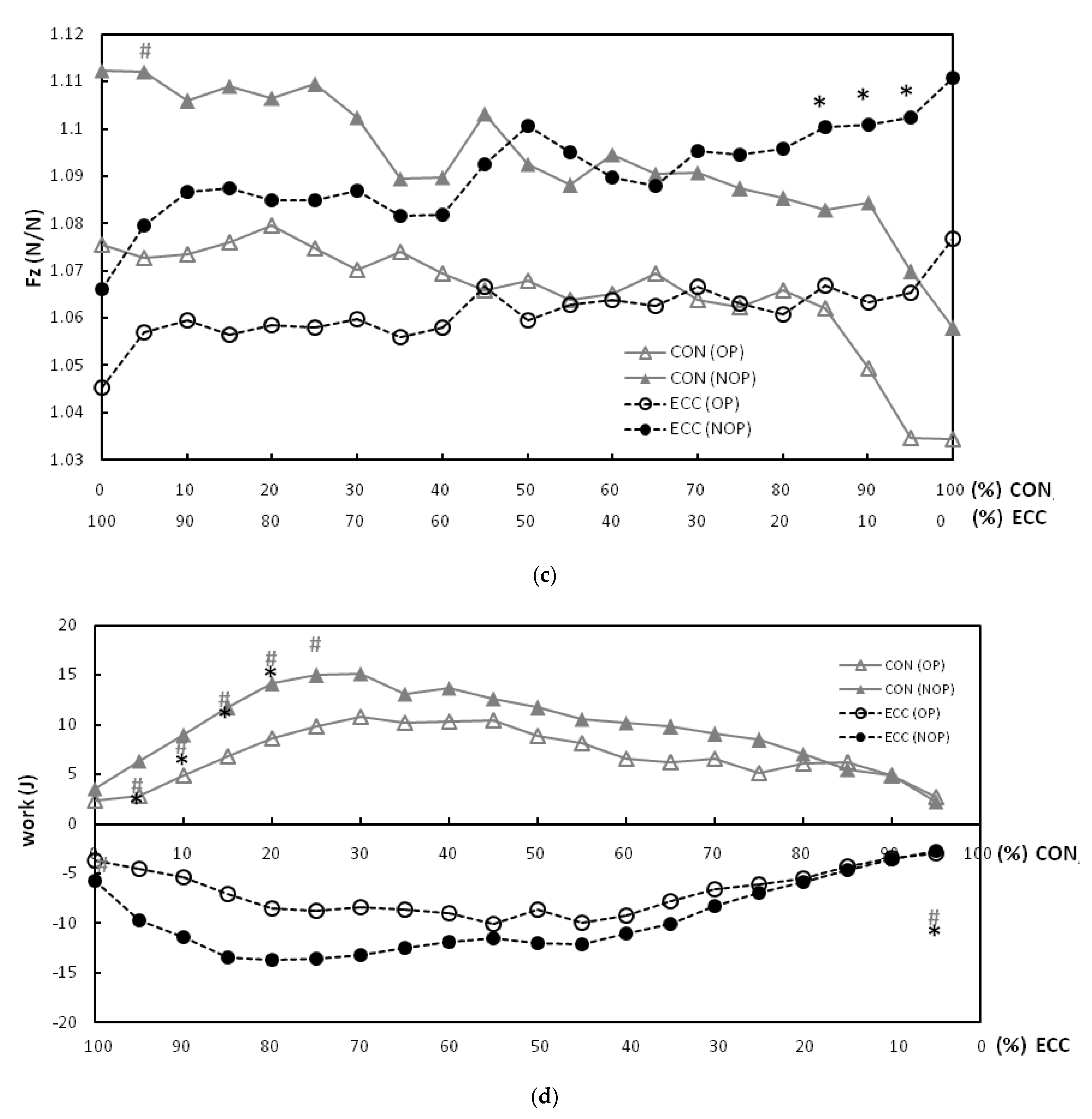
3. Results
4. Discussion
5. Conclusions
Author Contributions
Funding
Institutional Review Board Statement
Informed Consent Statement
Data Availability Statement
Conflicts of Interest
References
- Brorsson, A.; GrävareSilbernagel, K.; Olsson, N.; Nilsson Helander, K. Calf muscle performance deficits remain 7 years after an Achilles’ tendon rupture. Am. J. Sports Med. 2018, 46, 470–477. [Google Scholar] [CrossRef] [PubMed]
- Brorsson, A.; Willy, R.W.; Tranberg, R.; GrävareSilbernagel, K. Heel-rise height deficit 1 year after Achilles’ tendon rupture relates to changes in ankle biomechanics 6 years after injury. Am. J. Sports Med. 2017, 45, 3060–3068. [Google Scholar] [CrossRef]
- Mullaney, M.J.; McHugh, M.P.; Tyler, T.F.; Nicholas, S.J.; Lee, S.J. Weakness in end-range plantar flexion after Achilles’ tendon repair. Am. J. Sports Med. 2006, 34, 1120–1125. [Google Scholar] [CrossRef] [PubMed]
- Olsson, N.; Nilsson-Helander, K.; Karlsson, J.; Eriksson, B.I.; Thomée, R.; Faxén, E.; Silbernagel, K.G. Major functional deficits persist 2 years after acute Achilles’ tendon rupture. Knee Surg. Sports Traumatol. Arthrosc. 2011, 19, 1385–1393. [Google Scholar] [CrossRef] [PubMed]
- Bostick, G.P.; Jomha, N.M.; Suchak, A.A.; Beaupré, L.A. Factors associated with calf muscle endurance recovery 1 year after Achilles’ tendon rupture repair. J. Orthop. Sports Phys. Ther. 2010, 40, 345–351. [Google Scholar] [CrossRef] [PubMed]
- Peng, W.C.; Chao, Y.H.; Fu, A.S.N.; Fong, S.S.M.; Rolf, C.; Chiang, H.; Chen, S.; Wang, H.K. Muscular morphomechanical characteristics after an Achilles’ repair. Foot Ankle Int. 2019, 40, 568–577. [Google Scholar] [CrossRef] [PubMed]
- Silbernagel, K.G.; Steele, R.; Manal, K. Deficits in heel-rise height and Achilles’ tendon elongation occur in patients recovering from an Achilles’ tendon rupture. Am. J. Sports Med. 2012, 40, 1564–1571. [Google Scholar] [CrossRef] [PubMed]
- Orishimo, K.F.; Schwartz-Balle, S.; Tyler, T.F.; McHugh, M.P.; Bedford, B.B.; Lee, S.J.; Nicholas, S.J. Can weakness in end-range plantar flexion after Achilles’ tendon repair be prevented? Orthop. J. Sports Med. 2018, 6. [Google Scholar] [CrossRef]
- Lidstone, D.E.; van Werkhoven, H.; Needle, A.R.; Rice, P.E.; McBride, J.M. Gastrocnemius fascicle and achilles tendon length at the end of the eccentric phase in a single and multiple countermovement hop. J. Electromyogr. Kinesiol. 2018, 38, 175–181. [Google Scholar] [CrossRef] [PubMed]
- Fukutani, A.; Herzog, W. Influence of stretch magnitude on the stretch-shortening cycle in skinned muscle fibres. J. Exp. Biol. 2019, 222, jeb206557. [Google Scholar] [CrossRef]
- Fukutani, A.; Kurihara, T.; Isaka, T. Influence of joint angular velocity on electrically evoked concentric force potentiation induced by stretch-shortening cycle in young adults. Springerplus 2015, 4, 82. [Google Scholar] [CrossRef] [PubMed]
- Häggmark, T.; Eriksson, E. Hypotrophy of the soleus muscle in man afterAchilles’tendonrupture. Discussion of findings obtained by computed tomography and morphologic studies. Am. J. Sports Med. 1979, 7, 121–126. [Google Scholar] [CrossRef] [PubMed]
- Heikkinen, J.; Lantto, I.; Flinkkila, T.; Ohtonen, P.; Niinimaki, J.; Siira, P.; Laine, V.; Leppilahti, J. Soleus atrophy is common after the nonsurgical treatment of acute Achilles’ tendon ruptures: A randomized clinical trial comparing surgical and nonsurgical functional treatments. Am. J. Sports Med. 2017, 45, 1395–1404. [Google Scholar] [CrossRef] [PubMed]
- Zellers, J.A.; Marmon, A.R.; Ebrahimi, A.; GrävareSilbernagel, K. Lower extremity work along with triceps surae structure and activation is altered with jumping after Achilles’ tendon repair. J. Orthop. Res. 2019, 37, 933–941. [Google Scholar] [CrossRef]
- Strom, A.C.; Casillas, M.M. Achilles’ tendon rehabilitation. Foot Ankle Clin. 2009, 14, 773–782. [Google Scholar] [CrossRef] [PubMed]
- Maffulli, N.; Kenward, M.G.; Testa, V.; Capasso, G.; Regine, R.; King, J. Clinical diagnosis of Achilles’ tendinopathy with tendinosis. Clin. J. Sport Med. 2003, 13, 11–15. [Google Scholar] [CrossRef]
- O’Reilly, M.A.; Massouh, H. Pictorial review, the sonographic diagnosis of pathology in the Achilles’ tendon. Clin. Radiol. 1993, 48, 202–206. [Google Scholar] [CrossRef]
- Peng, W.C.; Chang, Y.P.; Chao, Y.H.; Fu, S.N.; Rolf, C.; Shih, T.T.; Su, S.C.; Wang, H.K. Morphomechanical alterations in the medial gastrocnemius muscle in patients with a repaired Achilles’ tendon, associations with outcome measures. Clin. Biomech. 2017, 43, 50–57. [Google Scholar] [CrossRef] [PubMed]
- Wang, H.K.; Chiang, H.; Chen, W.S.; Shih, T.T.; Huang, Y.C.; Jiang, C.C. Early neuromechanical outcomes of the triceps surae muscle-tendon after an Achilles’ tendon repair. Arch. Phys. Med. Rehabil. 2013, 94, 1590–1598. [Google Scholar] [CrossRef]
- Chaudhry, S.; Morrissey, D.; Woledge, R.C.; Bader, D.L.; Screen, H.R. Eccentric and concentric loading of the triceps surae, an in vivo study of dynamic muscle and tendon biomechanical parameters. J. Appl. Biomech. 2015, 31, 69–78. [Google Scholar] [CrossRef]
- Hou, W.H.; Yeh, T.S.; Liang, H.W. Reliability and validity of the Taiwan Chinese version of the Lower Extremity Functional Scale. J. Formos. Med. Assoc. 2014, 113, 313–320. [Google Scholar] [CrossRef] [PubMed]
- Bressel, E.; Larsen, B.T.; McNair, P.J.; Cronin, J. Anklejointproprioception and passive mechanical properties of the calf muscles after an Achilles tendon rupture, a comparison with matched controls. Clin. Biomech. 2004, 19, 284–291. [Google Scholar] [CrossRef] [PubMed]
- Maganaris, C.N.; Baltzopoulos, V.; Sargeant, A.J. Differences in human antagonistic ankle dorsiflexor coactivation between legs, can they explain the moment deficit in the weaker plantarflexor leg? Exp. Physiol. 1998, 83, 843–855. [Google Scholar] [CrossRef] [PubMed]
- Werkhausen, A.; Albracht, K.; Cronin, N.J.; Meier, R.; Bojsen-Møller, J.; Seynnes, O.R. Modulation of muscle-tendon interaction in the human triceps surae during an energy dissipation task. J. Exp. Biol. 2017, 220, 4141–4149. [Google Scholar] [CrossRef]
- Un, C.P.; Lin, K.H.; Shiang, T.Y.; Chang, E.C.; Su, S.C.; Wang, H.K. Comparative and reliability studies of neuromechanical leg muscle performances of volleyball athletes in different divisions. Eur. J. Appl. Physiol. 2013, 113, 457–466. [Google Scholar] [CrossRef]
- Oda, H.; Sano, K.; Kunimasa, Y.; Komi, P.V.; Ishikawa, M. Neuromechanical modulation of the Achilles tendon during bilateral hopping in patients with unilateral Achilles tendon rupture, over 1 year after surgical repair. Sports Med. 2017, 47, 1221–1230. [Google Scholar] [CrossRef]
- Geertsen, S.S.; Lundbye-Jensen, J.; Nielsen, J.B. Increased central facilitation of antagonist reciprocal inhibition at the onset of dorsiflexion following explosive strength training. J. Appl. Physiol. 2008, 105, 915–922. [Google Scholar] [CrossRef]
- Pensini, M.; Martin, A.; Maffiuletti, N.A. Central versus peripheral adaptations following eccentric resistance training. Int. J. Sports Med. 2002, 23, 567–574. [Google Scholar] [CrossRef] [PubMed]
- Franchi, M.V.; Atherton, P.J.; Reeves, N.D.; Flück, M.; Williams, J.; Mitchell, W.K.; Selby, A.; Beltran Valls, R.M.; Narici, M.V. Architectural, functional and molecular responses to concentric and eccentric loading in human skeletal muscle. Acta Phys. 2014, 210, 642–654. [Google Scholar] [CrossRef]
- Suydam, S.M.; Buchanan, T.S.; Manal, K.; Silbernagel, K.G. Compensatory muscle activation caused by tendon lengtheningpostAchilles’tendon rupture. Knee Surg. Sports Traumatol. Arthrosc. 2015, 23, 868–874. [Google Scholar] [CrossRef]
- Kawakami, Y.; Ichinose, Y.; Fukunaga, T. Architectural and functional features of human triceps surae muscles during contraction. J. Appl. Physiol. 1998, 85, 398–404. [Google Scholar] [CrossRef] [PubMed]
- Chino, K.; Oda, T.; Kurihara, T.; Nagayoshi, T.; Yoshikawa, K.; Kanehisa, H.; Fukunaga, T.; Fukashiro, S.; Kawakami, Y. In vivo fascicle behavior of synergistic muscles in concentric and eccentric plantar flexions in humans. J. Electromyogr. Kinesiol. 2008, 18, 79–88. [Google Scholar] [CrossRef] [PubMed]
- Ishikawa, M.; Komi, P.V.; Grey, M.J.; Lepola, V.; Bruggemann, G.P. Muscle-tendon interaction and elastic energy usage in human walking. J. Appl. Physiol. 2005, 99, 603–608. [Google Scholar] [CrossRef] [PubMed]
- Godinho, I.; Pinheiro, B.N.; Júnior, L.D.S.; Lucas, G.C.; Cavalcante, J.F.; Monteiro, G.M.; Uchoa, P.A.G. Effect of reduced ankle mobility on jumping performance in young athletes. Motricidade 2014, 15, 46–51. [Google Scholar]
- Panoutsakopoulos, V.; Kotzamanidou, M.C.; Papaiakovou, G.; Kollias, I.A. The ankle joint range of motion and its effect on squat jump performance with and without arm swing in adolescent female volleyball players. J. Funct. Morphol. Kinesiol. 2021, 6, 14. [Google Scholar] [CrossRef]
- Papaiakovou, G. Kinematic and kinetic differences in the execution of vertical jumps between people with good and poor ankle joint dorsiflexion. J. Sports Sci. 2013, 31, 1789–1796. [Google Scholar] [CrossRef]
- Yun, S.J.; Kim, M.H.; Weon, J.H.; Kim, Y.; Jung, S.H.; Kwon, O.Y. Correlation between toe flexor strength and ankle dorsiflexion ROM during the countermovement jump. J. Phys. Ther. Sci. 2016, 28, 2241–2244. [Google Scholar] [CrossRef]
- Kubo, K.; Kanehisa, H.; Takeshita, D.; Kawakami, Y.; Fukashiro, S.; Fukunaga, T. In vivo dynamics of human medial gastrocnemius muscle-tendon complex during stretch-shortening cycle exercise. Acta Physiol. Scand. 2000, 170, 127–135. [Google Scholar] [CrossRef] [PubMed]


| Characteristics | Achilles Repair |
|---|---|
| Age (y) | 40.5 (19.5) |
| Gender (n): Male/Female | 21/5 |
| Repaired leg (n): Right/Left | 15/11 |
| Body height (cm) | 173.6 (7.0) |
| Post-surgery period (months) | 4.5 (5.15) |
| Body weight (kg) | 74.8 (12.5) |
| Injury mechanism (n): Sports/Non-sports injury | 24/2 |
| LEFS score | 75 (6.5) |
| RMS EMG | Repaired Leg | Non-Injured Leg | p Value | |
|---|---|---|---|---|
| TA | CON | 0.20 (0.10–0.55) | 0.16 (0.05–0.36) | 0.027 * |
| ECC | 0.13 (0.07–0.48) | 0.12 (0.04–0.32) | 0.049 * | |
| SOL | CON | 0.99 (0.55–1.53) | 0.86 (0.48–2.45) | 0.023 * |
| ECC | 0.75 (0.31–1.28) | 0.49 (0.34–1.13) | 0.002 * | |
| MG | CON | 0.95 (0.59–1.37) | 0.89 (0.47–1.47) | 0.351 |
| ECC | 0.64 (0.45–0.95) | 0.54 (0.28–1.06) | 0.204 | |
| LG | CON | 0.87 (0.43–1.25) | 0.91 (0.48–1.51) | 0.647 |
| ECC | 0.57 (0.29–0.86) | 0.54 (0.35–0.93) | 0.744 | |
| nOLH (%) | 0.75 (0.40–1.01) | 0.95 (0.68–1.10) | <0.001 * |
| Kinematic | Kinetic | |||
|---|---|---|---|---|
| CON phase | Ankle joint (°) | Joint angular velocity (rad/s) | Normalized ground reaction force Fz | Mechanical work (J) |
| Sol RMS EMG | −0.011 | −0.072 | −0.404 | −0.327 |
| (0.952) | (0.684) | (0.021) | (0.056) | |
| MG RMS EMG | 0.490 * | −0.065 | −0.041 | 0.040 |
| (0.004) | (0.726) | (0.812) | (0.948) | |
| ECC phase | ||||
| Sol RMS EMG | 0.013s | −0.281 | −0.153 | −0.560 * |
| (0.959) | (0.113) | (0.417) | (0.001) | |
| MG RMS EMG | 0.247 | −0.199 | 0.058 | −0.127 |
| (0.180) | (0.276) | (0.751) | (0.490) | |
| Kinematic and Kinetic Data | Spearman Rank Correlation Coefficients |
|---|---|
| Ankle joint (°) | −0.192 (0.293) |
| Joint angular velocity (rad/s) | 0.575 (0.001) * |
| Ground reaction force Fz (n) | −0.077 (0.675) |
| Mechanical work (J) | −0.471 (0.007) * |
Publisher’s Note: MDPI stays neutral with regard to jurisdictional claims in published maps and institutional affiliations. |
© 2021 by the authors. Licensee MDPI, Basel, Switzerland. This article is an open access article distributed under the terms and conditions of the Creative Commons Attribution (CC BY) license (http://creativecommons.org/licenses/by/4.0/).
Share and Cite
Shih, K.-S.; Chen, P.-Y.; Yeh, W.-L.; Ma, H.-L.; Farn, C.-J.; Hou, C.-H.; Peng, W.-C.; Wang, H.-K. Modified Ankle Joint Neuromechanics during One-Legged Heel Raise Test after an Achilles Rupture and Its Associations with Jumping. Appl. Sci. 2021, 11, 2227. https://doi.org/10.3390/app11052227
Shih K-S, Chen P-Y, Yeh W-L, Ma H-L, Farn C-J, Hou C-H, Peng W-C, Wang H-K. Modified Ankle Joint Neuromechanics during One-Legged Heel Raise Test after an Achilles Rupture and Its Associations with Jumping. Applied Sciences. 2021; 11(5):2227. https://doi.org/10.3390/app11052227
Chicago/Turabian StyleShih, Kao-Shang, Pei-Yu Chen, Wen-Ling Yeh, Hsiao-Li Ma, Chui-Jia Farn, Chun-Han Hou, Wei-Chen Peng, and Hsing-Kuo Wang. 2021. "Modified Ankle Joint Neuromechanics during One-Legged Heel Raise Test after an Achilles Rupture and Its Associations with Jumping" Applied Sciences 11, no. 5: 2227. https://doi.org/10.3390/app11052227
APA StyleShih, K.-S., Chen, P.-Y., Yeh, W.-L., Ma, H.-L., Farn, C.-J., Hou, C.-H., Peng, W.-C., & Wang, H.-K. (2021). Modified Ankle Joint Neuromechanics during One-Legged Heel Raise Test after an Achilles Rupture and Its Associations with Jumping. Applied Sciences, 11(5), 2227. https://doi.org/10.3390/app11052227