Article

Immunohistochemical Differences in Squamous Precancerous and Cancerous Lesions of the Oral Cavity and the Larynx: Preliminary Data

by
Piero Luigi Aló
1,
Miriam Cicciarelli
2,
Francesca De Felice
3,*,
Claudia Quintiliani
1,4,
Alessandro Corsi
2 and
Antonella Polimeni
5
1
Department DAF, Anatomic Pathology, ASL Frosinone, Via Armando Fabi, 03100 Frosinone, Italy
2
Department of Molecular Medicine, “Sapienza” University of Rome, Policlinico Umberto I, Viale Regina Elena 324, 00161 Rome, Italy
3
Department of Radiological, Oncological and Pathological Sciences, Section of Radiotherapy, “Sapienza” University of Rome, Policlinico Umberto I, Viale Regina Elena 324, 00161 Rome, Italy
4
Anatomic Pathology, Università degli Studi di Bari “Aldo Moro”-DETO-Policlinico Bari, Piazzale Giulio Cesare, 70125 Bari, Italy
5
Department of Oral and Maxillo Facial Sciences, “Sapienza” University of Rome, Policlinico Umberto I, Via Caserta 6, 00161 Rome, Italy
*
Author to whom correspondence should be addressed.
Appl. Sci. 2021, 11(5), 2048; https://doi.org/10.3390/app11052048
Submission received: 5 December 2020 / Revised: 18 February 2021 / Accepted: 19 February 2021 / Published: 25 February 2021
(This article belongs to the Special Issue Current Concept and Emerging Treatments in Oral Diseases)

Abstract

:
The aim of this study is to assess immune cell populations in squamous precancerous (preinvasive) and cancerous lesions of the oral cavity and larynx. Qualitative and quantitative immunohistochemical analyses were performed to determine the expressions of CD4, CD8, CD15, CD57 and CD68. The expressions of programmed death-ligand 1 (PD-L1), p16 and Ki67 were also assessed. Squamous cell lesions from forty-one patients were included in the study. Sixteen samples were categorized as precancerous (preinvasive) lesions and twenty-five as invasive squamous cell carcinoma. Invasive lesions showed a negative correlation with CD57+ cells (ρ = −0.69) and a positive correlation with Ki67 (ρ = 0.61). The amount of CD4+ lymphocytes was higher in invasive lesions. There were no differences in PD-L1 and p16 immunoreactivity. Our analysis showed differences in the immunohistochemical profile between preinvasive and invasive squamous cell lesions. In the near future, this study should be useful in driving treatment strategy in both preinvasive and invasive squamous cell lesions of the oral cavity and larynx. However, studies on larger series of patients focusing on site-specific lesions are required.

1. Introduction

Oral cavity squamous cell carcinoma (SCC) and laryngeal SCC are the 18th and 23rd most common cancers worldwide, respectively, with a 5-year survival rate of approximately 60% in both cases and up to 20% of these carcinomas arise from precancerous (preinvasive, dysplastic) lesions [1,2,3]. In addition to tobacco and alcohol abuse, Human Papilloma Virus (HPV) infection is nowadays documented as an independent risk factor for those lesions that originate from the epithelium of the upper aerodigestive tract [4,5]. Despite timely evaluation, early diagnosis and remarkably improved treatment strategies, including traditional excision, cryosurgery and carbon dioxide laser ablation—in the case of preinvasive lesions—and conservative surgery with or without radiotherapy and systemic therapy—in case of invasive SCC—remain crucial because of the highly possible evolution of a dysplastic lesion in invasive carcinoma and the high recurrence rate of invasive SCC [3,4,6].
Therefore, there is an urgent need to identify robust biomarkers to potentially stratify patients to individualized treatment approaches. In recent years, the importance of immune cell populations in the microenvironment of both precancerous and cancerous scenarios has been affirmed [7,8,9]. A plethora of evidence now supports the central role of numerous cell types, such as CD4+, CD8+ T cells, dendritic cells, natural killer cells, and programmed death 1 (PD1)/programmed death-ligand 1 (PD-L1) pathways, in the cancer immunoediting process [7,8,9]. This consists of three sequential phases, in which innate and adaptive immunity promote transformed cell death (elimination phase), controlling net tumor cell outgrowth (equilibrium phase) until these tumor cells acquire resistance to the immune system (escape phase) [10]. However, the role of this immune response in the natural history of preinvasive squamous cell lesions and invasive SCC of the oral cavity and larynx needs to be better defined [7,8,9].
In this context, the main purpose of this preliminary study is to comparatively assess the immune cell populations in preinvasive squamous cell lesions and in invasive SCC of the oral cavity and larynx through qualitative and quantitative immunohistochemistry. Although we are aware of the limits of the study that include the limited number of patients/samples and the different sites of the aerodigestive tract considered, we feel that it could provide a rationale to identify potential targets to be exploited to develop novel therapeutic approaches.

2. Materials and Methods

2.1. Cases Selection

Formalin-fixed paraffin-embedded (FFPE) tissue samples collected between January 2018 and December 2019 from forty-one patients with a pathologic diagnosis of either preinvasive squamous cell lesion lesions or SCC in the oral cavity or larynx were included in this study. All FFPE specimens were retrospectively identified from the archives of the Section of Pathology of the “F. Spaziani” Hospital (Frosinone, Italy).

2.2. Tissue Samples

Microscopic characteristics of the lesions were assessed analyzing 2 µm-thick sections of each sample stained with hematoxylin–eosin. According to World Health Organization criteria [11], preinvasive lesions were classified as mild, moderate and severe epithelial dysplasia, whereas SCC was graded as well, moderately and poorly differentiated.

2.3. Immunohistochemistry

Section of each sample (2 µm-thick) and positively charged glass slides (Tomo, Matsunami) were used for immunohistochemical analysis. Assays were performed within Ventana ULTRA and XT platform, Roche Diagnostics, according to recommendations. Slides were incubated with the following antihuman primary antibodies: CD4 (clone SP31, Ventana Medical Systems, Roche Diagnostics), CD8 (clone SP57, Ventana Medical Systems, Roche Group), CD15 (clone MMA, Ventana Medical Systems, Oro Valley, AZ, USA), CD57 (clone NK-1, Cell Marque), CD68 (clone KP1, Ventana Medical Systems), PD-L1 (clone SP263; Ventana Medical Systems), p16INK4a (kit CINTec p16 Histology, Ventana Medical Systems) and Ki67 (clone 30-9, Ventana Medical Systems). The primary antibodies for CD4, CD8, CD15, CD57 and CD68 were used to identify T-helper lymphocytes, T-suppressor/cytotoxic lymphocytes, neutrophil granulocytes, natural killer cells and cells of the monocyte/macrophage lineage, respectively.
The ULTRAView Detection Kit (Ventana Medical Systems, Roche Group) was employed for CD4, CD8, CD15, CD57, CD68, p16INK4a and Ki67 and the OptiView DAB IHC Detection Kit (Ventana Medical Systems, Roche Group) was employed for PD-L1. For PD-L1 immunostain, positive (on-slide placental tissue) and negative (buffer, no primary antibody) controls were used.

2.4. Quantitative Immunohistochemical Analysis

The count of immunoreactive cells for each antibody was made in five randomly chosen fields with 400x magnification. For CD4, CD8, CD15, CD57 and CD68, the number of positive cells was scored as 0 (less than 5% immunoreactive cells), 1 (from 5% to <15%), 2 (from 15% to <30%) and 3 (≥30%). PD-L1 immunoreactivity was defined as cytoplasmic and/or membranous and dichotomized as low (0, <1) and high (1, ≥1) Combined Positive Scores (CPSs) [12]. The immunoreactivity for p16 was dichotomized in negative (value 0) versus positive (value 1) and positivity was defined by strong nuclear and cytoplasmic immunoreactivity in at least 70% of the lesional cells, as described previously [13]. The Ki67 labeling index (LI) was estimated by counting 300 cells from areas representative of the lesions, as described previously [14]. Preinvasive lesions were scored as 1 (positive cells localized in the basal layer of the epithelium), 2 (positive cells localized in both the basal and parabasal layers of the epithelium) and 3 (positive cells localized in basal, parabasal and spinous layers of the epithelium). SCCs were scored as 1 (positive cell in the periphery of the tumor nests), 2 (positive cells in both periphery and central part of the tumor nests) and 3 (diffuse positive cells in the tumor nests).

2.5. Statistical Analysis

Statistical analysis was performed using R-Studio 0.98.1091 software. Standard descriptive statistics were used to evaluate the distribution of each variable. Continuous variables were reported as means ± standard deviation and categorical variables as frequencies or percentages. The distribution of variables between precancerous lesions and cancerous lesions was compared with an χ2 test or Fisher’s exact test and Mann–Whitney U test, as appropriate. Correlations between factors were assessed by correlation matrices and the use of either Pearson (r; parametric) or Spearman (ρ; nonparametric) correlation methods where appropriate. A p-value < 0.05 was considered significant.

3. Results

3.1. Patients, Sites and Histologic Classification of the Lesions

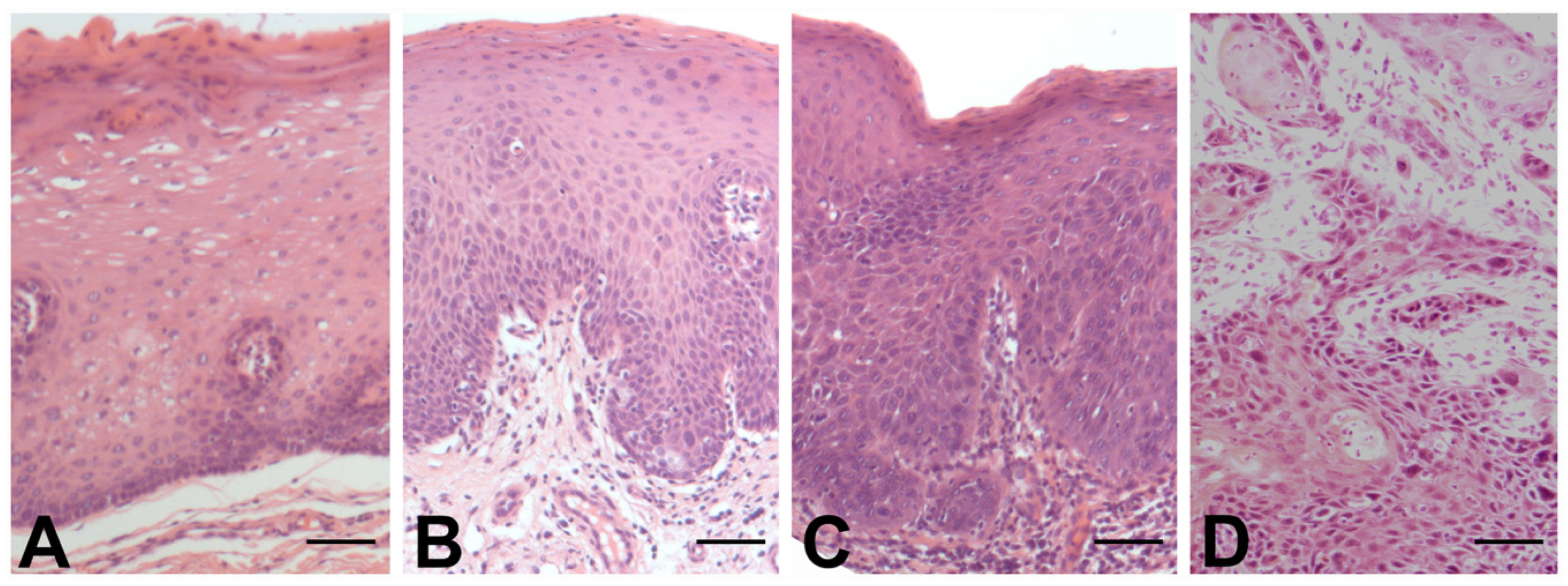
Of the forty-one samples included in this study, thirty-five were males and six females. All patients were adults. The sites of the lesions and their histologic classification as either preinvasive or as SCC are illustrated in Figure 1. Among the dysplastic lesions, one (larynx) was classified as mild, seven (one in the oral cavity and six in the larynx) as moderate and eight (one in the oral cavity and seven in the larynx) as severe dysplasia. Among the SCCs, seven (three in the oral cavity and four in the larynx) were classified as well differentiated, nine (five in the oral cavity and four in the larynx) as moderately differentiated and nine (four in the oral cavity and five in the larynx) as poorly differentiated. Representative histological images of preinvasive lesions and SCC are illustrated in Figure 2.

3.2. Qualitative and Quantitative Immunohistochemistry

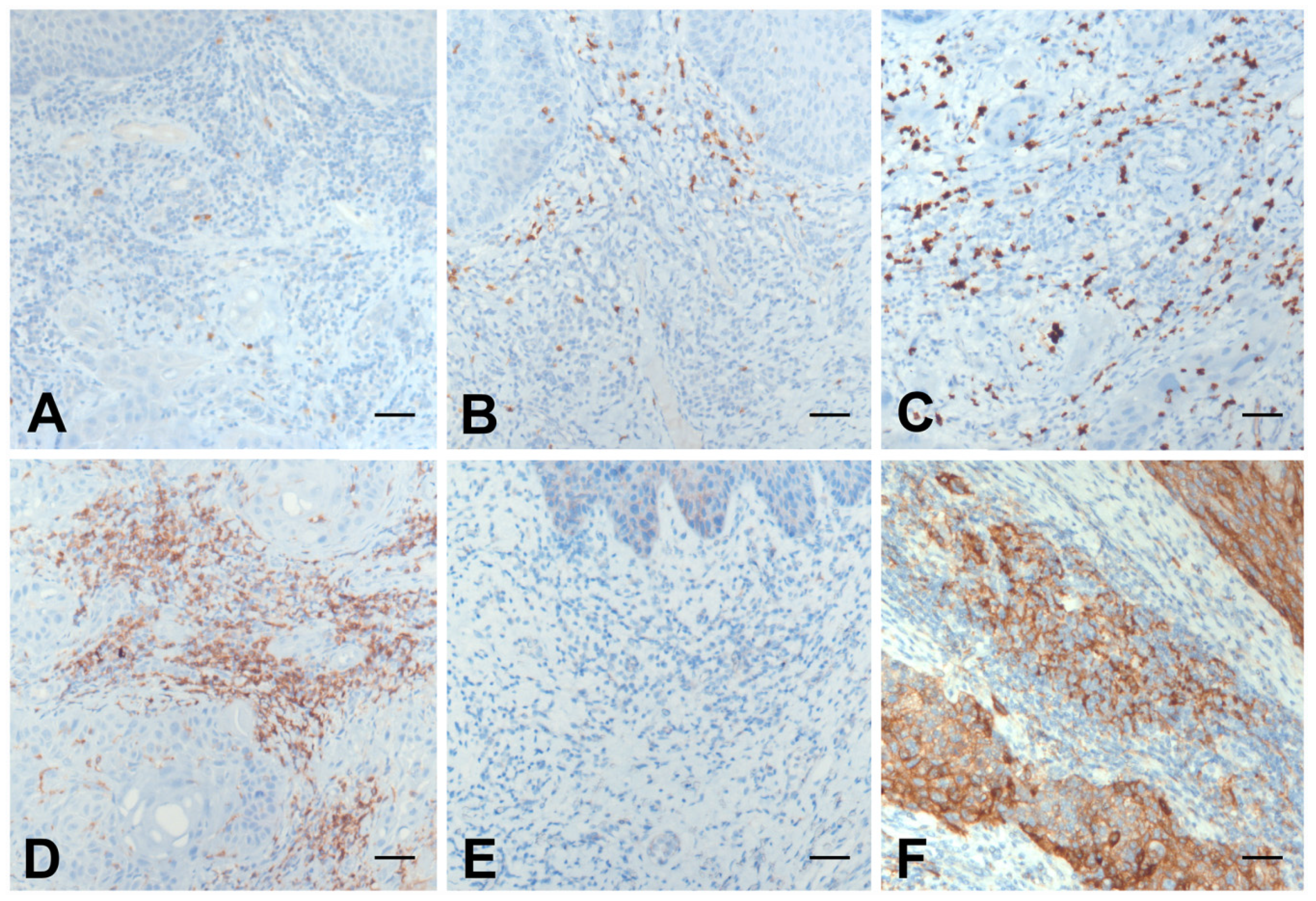
Representative images of immunohistochemical stains scored as 0, 1, 2 and 3 and of PD-L1 are illustrated in Figure 3. T-helper (CD4+) lymphocytes, T-suppressor/cytotoxic (CD8+) lymphocytes, neutrophil granulocytes (CD15+) and cells of the monocyte/macrophage (CD68+) were observed in all samples. In contrast, natural killer cells (CD57+) were observed in all preinvasive lesions and only in seven SCCs (two well differentiated, one moderately differentiated and four poorly differentiated). High PD-L1 CPS was detected in nine preinvasive lesions (three moderate dysplasia and six severe dysplasia) and in sixteen SCCs (four well differentiated, seven moderately differentiated and five poorly differentiated). Statistical comparison (Figure 4) failed to reveal significant differences except for CD4 and CD57, which were statistically significantly higher (p = 0.044) and lower (p < 0.001), respectively, in SCC compared to preinvasive lesions.
Positive immunostaining for p16 was detected in seven preinvasive lesions (two moderate and five severe epithelial dysplasia) and in seven SCCs (one well differentiated, four moderately differentiated and two poorly differentiated). Statistical comparison failed to reveal significant differences in p16 immunoreactivity between preinvasive and SCC (p = 0.306).
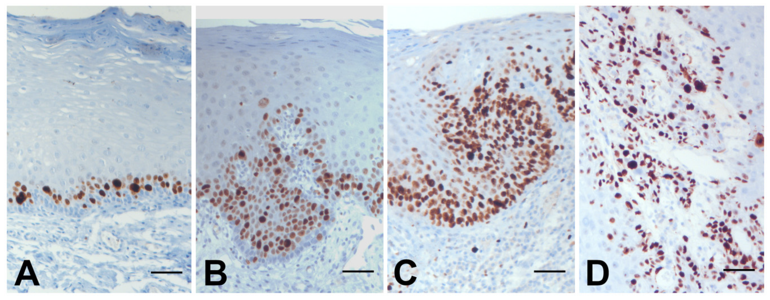
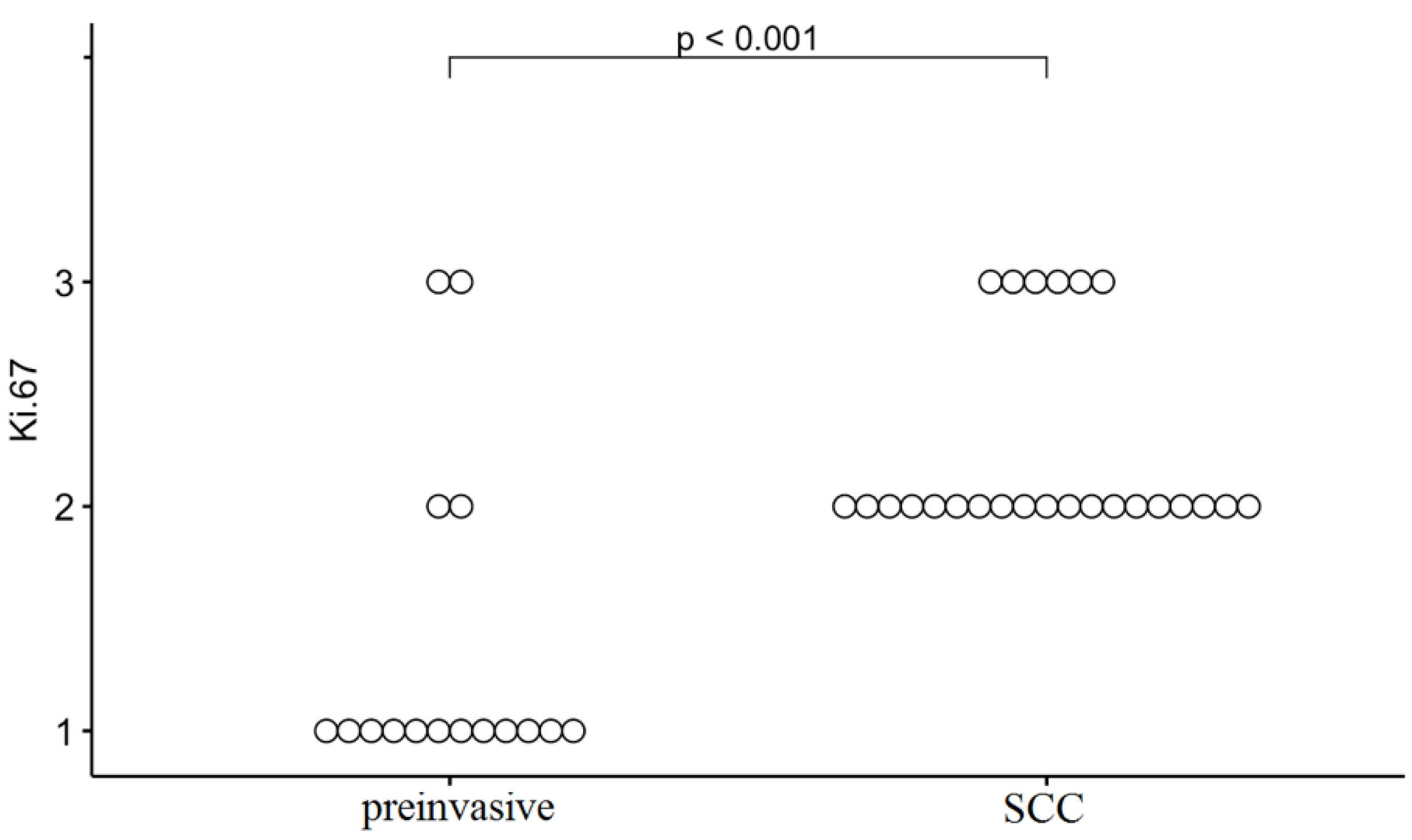
Ki67 expression (Figure 5 and Figure 6) was significantly higher in SCCs compared to preinvasive lesions (p < 0.001).
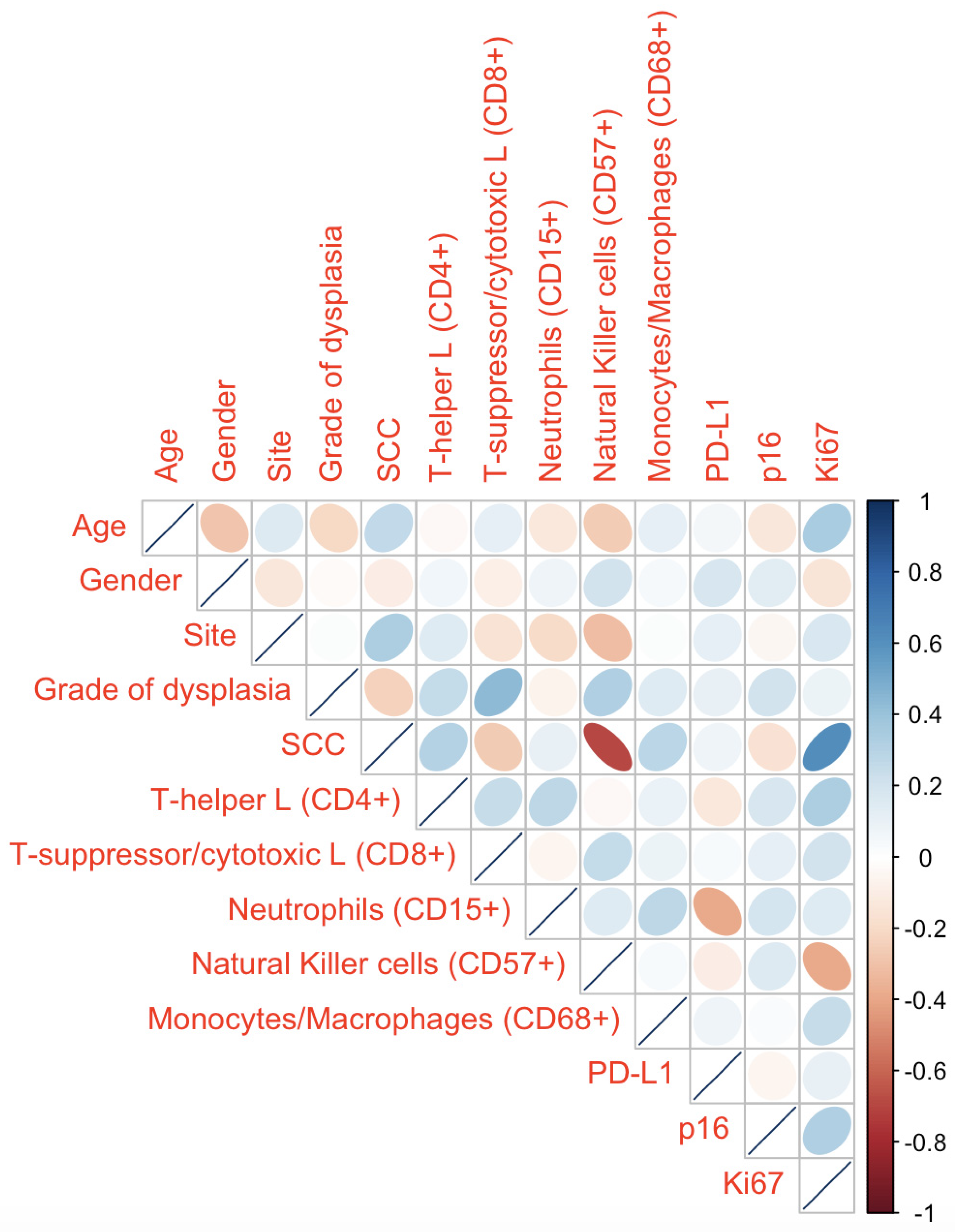
A correlation matrix was used to summarize data. The coefficients between the different clinic-pathologic variables are reported in Figure 7. SCC showed a negative correlation with CD57 (ρ = −0.69) and a positive correlation with Ki67 (ρ = 0.61).

4. Discussion

This analysis revealed that CD57+ natural killer cells predominated in preinvasive squamous cell lesions (p < 0.001) and CD4+ cells were dominant in oral cavity and laryngeal SCCs (p = 0.044). In addition, proliferation activity (Ki67+) positively correlated with SCC but not with preinvasive lesions (ρ = 0.61). No significant differences were observed in the amount of the other immune cells (CD8+ T-suppressor/cytotoxic lymphocytes, CD15+ neutrophil granulocytes and CD68+ cells of the monocyte/macrophage lineages) as also for p16 and PD-L1 between preinvasive squamous cell lesions and SCC.
Overall, these data are in agreement with the concept of the two-stage model of carcinogenesis in the oral and laryngeal mucosa and the cancer immunoediting process [15]. From preinvasive lesions to invasive SCC, the amount and the quality of immune cells changed and it is hard to define a clear “black and white” classification for which different cell types are procancer or anticancer.
It is certain that T lymphocytes, B lymphocytes, natural killer cells, dendritic cells, monocytes, macrophages and neutrophil granulocytes are key instruments in the immune response and tumor progression [16]. In our analysis, the negative correlation of CD57 (ρ = −0.69) with SCC is likely related to the fact that natural killer cells play a major role in the elimination phase [17], whereas the high CD4+ expression in SCC supports the evidence that CD4+ T cells “are a critical cornerstone of optimal anti-tumor immunity” [18]. Indeed, it is accepted that CD4+ T cells have a synergistic role with CD8+ T cells in the immune response. However, once the escape phase is activated, CD4+ cells seem to play a role in favoring tumor cell survival [18,19].
The expression of PD-L1 has been considered a point of balance between the host immune response and cancer escape ability [20]. A vast body of literature indicates that PD-L1 is mainly expressed on the surface of tumor cells and antigen-presenting cells in head and neck SCC. Based on the randomized phase III CheckMate 141 trial [21] and nonrandomized phase Ib KEYNOTE-012 trial [22], immune checkpoint PD1/PD-L1 blockade therapy (nivolumab and pembrolizumab, respectively) become the standard of care in the recurrent/metastatic setting [4,23]. In contrast, little is known on the role of PD1/PD-L1 pathway in preinvasive lesions. A recent meta-analysis study focused on the prevalence of PD-L1 expression in preinvasive lesions compared to invasive SCC in the head and neck region [20]. Aware of the high heterogeneity and the moderate quality of the included studies, PD-L1 expression was reported to be less frequent in preinvasive lesions than in invasive SCC. We were not able to confirm this result likely due to the low number and site heterogeneity of the examined cases. However, there is no doubt that much remains to be learned on the interactions between the multiple PD-L1-expressing cell types within the tumor microenvironment [24,25].
We also assessed the p16 status without recording significant differences between preinvasive lesions and SCC. This absence of difference should not surprise. Despite a causal association between HPV infection and SCC development being well established over the years, nowadays, p16 detection is mandatory to correctly stage primary tumors at diagnosis, in particular in the oropharynx, and its expression is a surrogate marker for HPV-related lesions, both preinvasive and invasive [26,27]. Therefore, p16 expression should not be considered as a marker for the differential diagnosis of preinvasive versus invasive lesion.
Regarding the positive correlation between Ki67 and SCC in our series, it is well known that Ki67 expression is an important biological mechanism in oncogenesis. The significance of Ki67 in the head and neck preinvasive and invasive squamous cell lesions has been discussed over the last years, but at present it is highly correlated with the increased aggressiveness of the lesion [28,29]. Thus, as expected, Ki67 was in our series significantly higher in SCC compared to preinvasive lesions.
Our study has intrinsic limitations, mainly due to its retrospective nature. For example, cases were excluded if there was insufficient archival material for full immunohistochemical analysis. As a result, the patient cohort was relatively small. In addition, the site distribution of the lesions was heterogeneous and risk factors (i.e., smoking and alcohol habits) and survival data were not included in the analysis. Further works, including analysis of a larger series of patients/samples focusing on specific sites of the aerodigestive tract, are currently under development. As involved in head and neck cancer, we believe that improving our knowledge on the tissue immunologic profile of preinvasive and invasive squamous cell lesions is one of the major goals in this field, in particular for the risk stratification of these patients.

5. Conclusions

In this study, we comparatively evaluated, through immunohistochemistry, preinvasive and invasive squamous cell lesions of the oral cavity and larynx. CD57+ cells were prominent in precancerous lesions whereas CD4+ cells and proliferation activity predominated in invasive lesions. Differences were not observed for the other markers used in this study (CD8, CD15, CD68, p16 and PD-L1). Further investigations are needed to validate these results expanding the number of cases/lesions and focusing on specific sites to define which immunohistochemical markers may identify patients with either preinvasive or invasive lesions at higher risk of transformation in SCC or of recurrence, respectively, in order to potentially implement appropriate therapy.

Author Contributions

Conceptualization, P.L.A. and A.P.; Formal analysis, F.D.F.; Data curation, P.L.A., M.C., F.D.F. and C.Q.; Writing—Original Draft Preparation, F.D.F.; Writing—Review and Editing, P.L.A., F.D.F., A.C. and A.P. All authors have read and agreed to the published version of the manuscript.

Funding

This research received no external funding.

Institutional Review Board Statement

The study was conducted according to the guidelines of the Declaration of Helsinki, and approved by the Institutional Review Board (Department of Radiological, Oncological and Anatomo-pathological Sciences, Sapienza University of Rome, Italy, 29/01/2021).

Informed Consent Statement

Written informed consent was not deemed necessary owing to the retrospective nature of the study.

Data Availability Statement

Data are available from the authors upon reasonable request.

Conflicts of Interest

The authors declare no conflict of interest.

Ethical Statements

Because of the retrospective nature of the study, written informed consent was not deemed necessary. The study was conducted in accordance with the Declaration of Helsinki and approved by the Institutional Review Board (Department of Radiological, Oncological and Anatomo-pathological Sciences, Sapienza University of Rome, Italy, 29/01/2021).

References

  1. Bray, F.; Ferlay, J.; Soerjomataram, I.; Siegel, R.L.; Torre, L.A.; Jemal, A. Global cancer statistics 2018: GLOBOCAN estimates of incidence and mortality worldwide for 36 cancers in 185 countries. CA Cancer J. Clin. 2018, 68, 394–424. [Google Scholar] [CrossRef] [PubMed] [Green Version]
  2. Reibel, J. Prognosis of oral pre-malignant lesions: Significance of clinical, histopathological, and molecular biological characteristics. Crit. Rev. Oral Biol. Med. 2003, 14, 47–62. [Google Scholar] [CrossRef]
  3. Awadallah, M.; Idle, M.; Patel, K.; Kademani, D. Management update of potentially premalignant oral epithelial lesions. Oral Surg. Oral Med. Oral Pathol. Oral Radiol. 2018, 125, 628–636. [Google Scholar] [CrossRef] [Green Version]
  4. National Comprehensive Cancer Network Clinical Practice Guidelines in Oncology (NCCN Guidelines) Head and Neck Cancers, Version 1.2021. Available online: http://www.nccn.org (accessed on 21 November 2020).
  5. Tenore, G.; Nuvoli, A.; Mohsen, A.; Cassoni, A.; Battisti, A.; Terenzi, V.; Della Monaca, M.; Raponi, I.; Brauner, E.; De Felice, F.; et al. Tobacco, Alcohol and Family History of Cancer as Risk Factors of Oral Squamous Cell Carcinoma: Case-Control Retrospective Study. Appl. Sci. 2020, 10, 3896. [Google Scholar] [CrossRef]
  6. Rocchetti, F.; Tenore, G.; Montori, A.; Cassoni, A.; Cantisani, V.; Di Segni, M.; Di Gioia, C.R.T.; Carletti, R.; Valentini, V.; Polimeni, A.; et al. Preoperative evaluation of tumor depth of invasion in oral squamous cell carcinoma with intraoral ultrasonography: A retrospective study. Oral Surg. Oral Med. Oral Pathol Oral Radiol. 2020, 131, 130–138. [Google Scholar] [CrossRef]
  7. Jones, T.M. Tumour-infiltrating lymphocytes in the risk stratification of squamous cell carcinoma of the head and neck. Br. J. Cancer. 2014, 110, 269–270. [Google Scholar] [CrossRef] [Green Version]
  8. Stasikowska-Kanicka, O.; Wągrowska-Danilewicz, M.; Danilewicz, M. Immunohistochemical Analysis of Foxp3+, CD4+, CD8+ Cell Infiltrates and PD-L1 in Oral Squamous Cell Carcinoma. Pathol. Oncol. Res. 2018, 24, 497–505. [Google Scholar] [CrossRef] [PubMed] [Green Version]
  9. Chaves, A.L.F.; Silva, A.G.; Maia, F.M.; Lopes, G.F.; Muniz, L.V.; Soares, J.M.A.; Santos, H.B.; A Barbosa, L.; Andrade, A.; Paula, L.F.B.; et al. CD4, CD8 and PD1 immunoexpression in oral premalignant lesions (PML) and oral squamous cell carcinoma (OSCC) and its association with malignant transformation. J. Clin. Oncol. 2018, 36, 113. [Google Scholar] [CrossRef]
  10. Vesely, M.D.; Schreiber, R.D. Cancer immunoediting: Antigens, mechanisms, and implications to cancer immunotherapy. Ann. N. Y. Acad. Sci. 2013, 1284, 1–5. [Google Scholar] [CrossRef] [PubMed] [Green Version]
  11. El-Naggar, A.K.; Chan, J.K.C.; Takata, T.; Grandis, J.R.; Slootweg, P.J. The fourth edition of the head and neck World Health Organization blue book: Editors’ perspectives. Hum. Pathol. 2017, 66, 10–12. [Google Scholar] [CrossRef]
  12. Kulangara, K.; Hanks, D.A.; Waldroup, S.; Peltz, L.; Shah, S.; Roach, C.; Juco, J.W.; Emancipator, K.; Stanforth, D. Development of the combined positive score (CPS) for the evaluation of PD-L1 in solid tumors with the immunohistochemistry assay PD-L1 IHC 22C3 pharmDx. J. Clin. Oncol. 2017, 35, e14589. [Google Scholar] [CrossRef]
  13. Golusiński, P.; Pazdrowski, J.; Szewczyk, M.; Misiołek, M.; Pietruszewska, W.; Klatka, J.; Okła, S.; Kaźmierczak, H.; Marszałek, A.; Filas, V.; et al. Is immunohistochemical evaluation of p16 in oropharyngeal cancer enough to predict the HPV positivity? Rep. Pract. Oncol. Radiother. 2017, 22, 237–242. [Google Scholar] [CrossRef] [PubMed] [Green Version]
  14. Takkem, A.; Barakat, C.; Zakaraia, S.; Zaid, K.; Najmeh, J.; Ayoub, M.; Seirawan, M.Y. Ki-67 Prognostic Value in Different Histological Grades of Oral Epithelial Dysplasia and Oral Squamous Cell Carcinoma. Asian Pac. J. Cancer Prev. 2018, 19, 3279–3286. [Google Scholar] [CrossRef] [Green Version]
  15. The Two-Stage Model of Carcinogenesis National Research Council (US) Committee on Risk Assessment Methodology; National Academies Press (US): Washington, DC, USA, 1993.
  16. Galli, F.; Aguilera, J.V.; Palermo, B.; Markovic, S.N.; Nisticò, P.; Signore, A. Relevance of immune cell and tumor microenvironment imaging in the new era of immunotherapy. J. Exp. Clin. Cancer Res. 2020, 39, 89. [Google Scholar] [CrossRef]
  17. Kurosawa, S.; Harada, M.; Matsuzaki, G.; Shinomiya, Y.; Terao, H.; Kobayashi, N.; Nomoto, K. Early-appearing tumour-infiltrating natural killer cells play a crucial role in the generation of anti-tumour T lymphocytes. Immunology 1995, 85, 338–346. [Google Scholar]
  18. Tay, R.E.; Richardson, E.K.; Toh, H.C. Revisiting the role of CD4+ T cells in cancer immunotherapy-new insights into old paradigms. Cancer Gene Ther. 2021, 28, 5–17. [Google Scholar] [CrossRef]
  19. Ostroumov, D.; Fekete-Drimusz, N.; Saborowski, M.; Kühnel, F.; Woller, N. CD4 and CD8 T lymphocyte interplay in controlling tumor growth. Cell Mol. Life Sci. 2018, 75, 689–713. [Google Scholar] [CrossRef] [Green Version]
  20. Girolami, I.; Pantanowitz, L.; Munari, E.; Martini, M.; Nocini, R.; Bisi, N.; Molteni, G.; Marchioni, D.; Ghimenton, C.; Brunelli, M.; et al. Prevalence of PD-L1 expression in head and neck squamous precancerous lesions: A systematic review and meta-analysis. Head Neck. 2020, 42, 3018–3030. [Google Scholar] [CrossRef] [PubMed]
  21. Ferris, R.L.; Blumenschein, G., Jr.; Fayette, J.; Guigay, J.; Colevas, A.D.; Licitra, L.; Harrington, K.; Kasper, S.; Vokes, E.E.; Even, C. Nivolumab for recurrent squamous-cell carcinoma of the head and neck. N. Engl. J. Med. 2016, 375, 1856–1867. [Google Scholar] [CrossRef]
  22. Chow, L.Q.; Haddad, R.; Gupta, S.; Mahipal, A.; Mehra, R.; Tahara, M.; Berger, R.; Eder, J.P.; Burtness, B.; Lee, S.H. Antitumor activity of pembrolizumab in biomarker-unselected patients with recurrent and/or metastatic head and neck squamous cell carcinoma: Results from the Phase Ib KEYNOTE-012 expansion cohort. J. Clin. Oncol. 2016, 34, 3838–3845. [Google Scholar] [CrossRef]
  23. Wang, X.; Teng, F.; Kong, L.; Yu, J. PD-L1 expression in human cancers and its association with clinical outcomes. Onco Targets Ther. 2016, 9, 5023–5039. [Google Scholar] [PubMed] [Green Version]
  24. Dave, K.; Ali, A.; Magalhaes, M. Increased expression of PD-1 and PD-L1 in oral lesions progressing to oral squamous cell carcinoma: A pilot study. Sci. Rep. 2020, 10, 9705. [Google Scholar] [CrossRef] [PubMed]
  25. Yagyuu, T.; Hatakeyama, K.; Imada, M.; Kurihara, M.; Matsusue, Y.; Yamamoto, K.; Obayashi, C.; Kirita, T. Programmed death ligand 1 (PD-L1) expression and tumor microenvironment: Implications for patients with oral precancerous lesions. Oral Oncol. 2017, 68, 36–43. [Google Scholar] [CrossRef]
  26. Buajeeb, W.; Poomsawat, S.; Punyasingh, J.; Sanguansin, S. Expression of p16 in oral cancer and premalignant lesions. J. Oral Pathol. Med. 2009, 38, 104–108. [Google Scholar] [CrossRef]
  27. Lydiatt, W.M.; Patel, S.G.; O’Sullivan, B.; Brandwein, M.S.; Ridge, J.A.; Migliacci, J.C.; Loomis, A.M.; Shah, J.P. Head and Neck cancers-major changes in the American Joint Committee on cancer eighth edition cancer staging manual. CA Cancer J. Clin. 2017, 67, 122–137. [Google Scholar] [CrossRef]
  28. Zuzul, I.; Pavic, I.; Granic, M.; Kuna, T.; Kotarac Knezevic, A.; Andabak Rogulj, A. The Significance of Ki-67 in Head and Neck Cancers: Review Article. RJPBCS 2018, 9, 34–38. [Google Scholar]
  29. Olimid, D.A.; Simionescu, C.E.; Mărgăritescu, C.; Florescu, A. Immunoexpression of Ki67 and cyclin D1 in oral squamous carcinomas. Rom. J. Morphol. Embryol. 2012, 53 (Suppl. 3), 795–798. [Google Scholar] [PubMed]
Figure 1. Sites of the lesions and their histological classification.
Applsci 11 02048 g001
Figure 2. Representative histological images of mild ((A), larynx), moderate ((B), hard palate) and severe ((C), tongue) dysplasia and of squamous cell carcinoma (SCC) ((D), tongue). Hematoxylin–eosin. Bars: 100 μm.
Applsci 11 02048 g002
Figure 3. Representative images of immunohistochemical stains scored as 0 ((A); CD57, larynx SCC), 1 ((B); CD8, tongue preinvasive lesion), 2 ((C); CD15, tongue SCC) and 3 ((D); CD4, larynx SCC) and of PD-L1 (E,F), graded as low (0, <1, (E), larynx preinvasive lesion) and high (1, ≥1, (F), tongue SCC). Bars: 100 μm.
Applsci 11 02048 g003
Figure 4. Comparative statistical analysis of CD4, CD8, CD15, CD57, CD68 and PD-L1 Combined Positive Scores (CPSs), the last being graded as low (0, <1) and high (1, ≥1) between preinvasive squamous cell lesions and SCC. Each circle represents a patient.
Applsci 11 02048 g004
Figure 5. Representative images of the immunohistochemical stain for Ki67 in mild ((A), larynx), moderate ((B), hard palate) and severe ((C), tongue) dysplasia and in SCC ((D), tongue). Bars: 100 μm.
Applsci 11 02048 g005
Figure 6. Comparative statistical analysis of Ki67 between preinvasive squamous cell lesions and SCC. Each circle represents a patient.
Applsci 11 02048 g006
Figure 7. Correlation matrix between different clinical and pathologic variables. Each ellipse in the table represents a correlation between the two variables. The diagonal indicates that each variable perfectly correlates with itself. L is for lymphocytes.
Applsci 11 02048 g007
Publisher’s Note: MDPI stays neutral with regard to jurisdictional claims in published maps and institutional affiliations.