Article

Amoxicillin in Water: Insights into Relative Reactivity, Byproduct Formation, and Toxicological Interactions during Chlorination

1
Department of Biology, University of Naples Federico II, 80126 Naples, Italy
2
Department of Chemical Sciences, University of Naples Federico II, 80126 Naples, Italy
3
Associazione Italiana per la Promozione delle Ricerche su Ambiente e Salute umana, 82030 Dugenta, Italy
*
Author to whom correspondence should be addressed.
Appl. Sci. 2021, 11(3), 1076; https://doi.org/10.3390/app11031076
Submission received: 23 December 2020 / Revised: 15 January 2021 / Accepted: 21 January 2021 / Published: 25 January 2021

Abstract

:
In recent years, many studies have highlighted the consistent finding of amoxicillin in waters destined for wastewater treatment plants, in addition to superficial waters of rivers and lakes in both Europe and North America. In this paper, the amoxicillin degradation pathway was investigated by simulating the chlorination process normally used in a wastewater treatment plant to reduce similar emerging pollutants at three different pH values. The structures of 16 isolated degradation byproducts (DPs), one of which was isolated for the first time, were separated on a C-18 column via a gradient HPLC method. Combining mass spectrometry and nuclear magnetic resonance, we then compared commercial standards and justified a proposed formation mechanism beginning from the parent drug. Microbial growth inhibition bioassays with Escherichia coli, Klebsiella pneumoniae, and Staphylococcus aureus were performed to determine the potential loss of antibacterial activity in isolated degradation byproducts. An increase of antibacterial activity in the DPs was observed compared to the parent compound.

1. Introduction

The presence of pharmaceutical compounds in natural water bodies, even at low concentrations, raises health concerns. Pharmaceutical substances, used to prevent and fight diseases, are produced to guarantee their maximum effectiveness and, at the same time, ensure their resistance to inactivation until they perform their intended functions. Thus, these compounds can be excreted through feces and urine in the form of metabolite mixtures and their unchanged product, which flow into wastewater treatment plants (WWTPs). The recent widespread detection of these compounds in such environments [1,2,3,4,5,6] have led to their designation as emerging contaminants, as they are still unregulated. The environmental persistence and high biological activity that characterizes them make these substances harmful, even at low concentrations. These contaminants can, in fact, cause alterations to the endocrine system [7] and an increase in microbial resistance to drugs [8,9,10,11]. They can also be adsorbed by plants [12] and bioaccumulated [13] in the food chain. Additional risks are associated with biodiversity loss [7], infertility, and cancer [14,15]. One of the categories of drugs on which the attention of the scientific community is most focused is that of antibiotics present in the aquatic environment and in foods, which have the possibility of inducing the formation of antibiotic-resistant bacteria and the health risks that may derive from them [16,17].
Amoxicillin (AMO) is among the most prescribed antibiotics for human use in Italy [18] and other European countries [19], which is a drug synthesized in large amounts and used in aquaculture farms to cope with the most common fish diseases. In Italy, it is estimated that more than 210 t are used annually, of which 86 ± 8% [20] are excreted in parental form with a theoretical environmental load estimated at around 190 t/year. Risk assessment studies for aquatic species and humans are under development, but despite the small amount of ecotoxicological data, some studies found it possible to establish compounds, such as AMO, in surface waters at non-negligible risk levels for aquatic species organisms [21,22].
AMO has been detected at µg/L concentrations in the influent and effluent of WWTPs and surface water [23,24], while its levels in pharmaceutical industry effluents may reach mg/L concentrations [25].
Although the treatment processes used in the plants shows high AMO removals [26,27,28], even if the percentage of removal depends on the duration of the treatment [29], at the same time, they have the disadvantage of increasing effluent toxicity and producing its transformation compounds, which may be more toxic than the product from which they derive [30]. As a consequence, WWTP effluents and the practice of reusing sewage sludge in agriculture to recover nitrogen compounds useful for soil fertilization can contribute to its introduction into water bodies and its diffusion in the terrestrial environment of the degradation byproducts (DPs) of the drug [31,32,33,34]. Humans can be exposed to DPs through the consumption of aquatic organisms, agricultural products, or drinking water.
In this paper, DPs of AMO were investigated under the same conditions as the chlorination process normally used in WWTPs to reduce similar emerging pollutants [35,36] at three different pH values. In particular, a chromatographic profile of the possible DPs was obtained with an experiment at low concentrations of AMO (about 10−5 M), which were then isolated and structurally determined by repeating the chlorination experiment at concentrations levels at least 100 times higher. The structures of 16 isolated DPs, one of which was isolated for the first time, were determined by combining mass spectrometry, using as source a matrix-assisted laser desorption/ionization and as mass analyzer a time-of-flight analyzer (MALDI-MS/TOF) and nuclear magnetic resonance (NMR) data. They were then justified by a proposed formation mechanism. Microbial growth inhibition bioassays with Escherichia coli (ATCC 25922), Klebsiella pneumoniae (ATCC 20081), and Staphylococcus aureus (ATCC 6538) were performed to determine the changes in AMO antibacterial activity. E. coli, K. pneumoniae, and S. aureus were used as indicator microorganisms in the antimicrobial assays, since these bacteria are important human pathogens with high stability against antibiotics.

2. Materials and Methods

2.1. Drug and Reagents

Amoxicillin (99.5%) was purchased from Sigma-Aldrich (Milan, Italy). All other chemicals and solvents were purchased from Fluka (Saint-Quentin Fallavier, France) and were of HPLC grade and used as received. For the antimicrobial assessment, tryptic soy broth (TSB, Difco, Becton-Dickenson Labs) was used. All the chemicals were of analytical grade and supplied by Sigma-Aldrich. Double distilled water (Microtech) was used to prepare the dilution water and treatments. The microbial growth was measured with automatic plate reader (Synergy HTX, BioTek Instruments, Winooski, VT, USA).

2.2. Chlorination Reaction

2.2.1. Apparatus and Equipment

Column chromatography (CC) was carried out with Kieselgel 60 (230–400 mesh, Merck, Darmstadt, Germany). HPLC was performed on a Shimadzu LC-8A system using a Shimadzu SPD-10A VP UV-VIS detector (Shimadzu, Milan, Italy). Semipreparative HPLC was performed using an RP Gemini C18-110A preparative column (10 µm particle size, 250 mm × 21.2 mm i.d., Phenomenex, Bologna, Italy) with a flow rate of 7 mL min−1. The 1H- and 13C NMR spectra were recorded with a NMR spectrometer, operated at 400 MHz and at 25 °C (Bruker DRX, Bruker Avance) and referenced in ppm to the residual solvent signals (CDCl3, at δH 7.27 and δC 77.0). The proton-detected heteronuclear correlations were measured using a gradient heteronuclear single-quantum coherence (HSQC) experiment, optimized for 1JHC = 155 Hz, and a gradient heteronuclear multiple bond coherence (HMBC) experiment, optimized for nJHC = 8 Hz. The MALDI-TOF mass spectrometric analyses were performed on a Voyager-De Pro MALDI mass spectrometer (PerSeptive Biosystems, Framingham, MA, USA). The UV/Vis spectra were recorded with a Perkin Elmer Lambda 7 spectrophotometer. The IR spectra were recorded with a Jasco FT/IR-430 instrument equipped with a single reflection ATR accessory.

2.2.2. Chlorination Experiments

A 10−5 M AMO solution was treated for 10 min with 10% hypochlorite (molar ratio AMO/NaOCl 1:1 concentration, spectroscopically determined λmax 292 nm, ε 350 dm3/mol cm) at room temperature [37], simulating the conditions used in a typical WWTP. The experiment was repeated at pH = 3 in a common H3PO4/KH2PO4 (20 mM) buffer (Figure 1a), at pH = 7 in KH2PO4/K2HPO4 (20 mM) buffer (Figure 1b) and at pH = 9 (Figure 2a). The presence of AMO was quantified using a Lambda 12 UV-Vis spectrophotometer (Perkin Elmer, 940 Winter Street, Waltham, MA 02451, USA). Absorbance peaks were determined at 230 nm (Figure 2b). The absorbance values were converted into concentration using a calibration curve prepared from standard solutions with known AMO concentrations. In this latter case, the pH of the solution, measured and recorded continuously by a pH-meter, increased immediately from the initial pH of 8.0 to 10.5, and the pH remained at this value during the reaction. An aliquot of the solution was taken every 5 min, quenched by sodium thiosulphate excess, filtered, dried by lyophilization, and dissolved in a saturated sodium bicarbonate solution before being extracted with ethyl acetate. The course of the reaction was monitored by HPLC. The main degradation byproducts (DP4 and DP6—DP10 for the ethyl acetate fraction and DP1—DP3, DP5 and DP11—DP16 for the aqueous fraction; Scheme 1 and Figure 3) were identified by comparing their retention times with those of commercially available standard compounds or isolated by performing preparative experiments with an AMO solution at a concentration higher than 10−3 M treated with 5% hypochlorite at room temperature for 5 min. The degradation byproducts obtained were isolated via column chromatography and HPLC and completely characterized using NMR and mass spectrometry (MS) analysis. DP1—DP16 were isolated in relative percentages of 1.01, 0.89, 2.25, 2.02, 1.56, 1.36, 2.21, 2.05, 3.01, 2.24, 1.25, 1.11, 1.23, 1.45, 2.25, and 0.23%, respectively. The proposed mechanism of their formation from AMO is shown in Figure 4. DP16, isolated for the first time, was determined by combining mass spectrometry (MS) and nuclear magnetic resonance (NMR) data.

2.2.3. Chlorination Procedure and Product Isolation

Amoxicillin (1 g, 2.74 mmol), dissolved in milliQ water (2 L), was treated for 5 min with 5% hypochlorite (molar ratio AMO/NaOCl 1:2; concentration spectroscopically determined at a λmax of 292 nm, ε = 350 dm3/mol cm) at room temperature [38]. The pH of the solution increased immediately from the initial pH of 8.0 to 10.5, and the pH remained at this value during the reaction. After 5 min, the solution was quenched using an excess of thiosulphate, with respect to NaOCl, and dried by lyophilization, and the residue was dissolved in a saturated Na2CO3 solution and extracted with ethyl acetate (EA). The EA fraction (351 mg) was separated with the silica gel column chromatography (CC) using a gradient of methylene chloride/methanol (100:0 to 10:90, v/v) to yield 15 fractions. The EA5 fraction (25 mg), eluted with methylene chloride/methanol (90:10, v/v), was analyzed via HPLC using a Supelcosil LC-18 column, 25 cm × 4.6 mm ID, and 5 µm particles. The solvent system was a gradient of acetonitrile/tetrahydrofuran/water (A, 30:10:60, v/v/v) and acetonitrile/water (B, 60:40, v/v), starting with 0% B for 1 min and installing a gradient to obtain 100% B over 20 min, at a solvent flow rate of 1.5 mL/min. The column effluent was monitored at 360 nm. Identification of DP6 and DP4 was achieved by comparison with standard compounds. The fraction EA7 (33 mg), eluted with methylene chloride/methanol (75:25, v/v), was analyzed via HPLC using a Supelcosil LC-8 column, 15 cm × 4.6 mm I.D., and 5 µm particles. The solvent system used was a gradient of acetic acid/methanol (A, 1:99, v/v) and acetic acid/water (B, 1:99, v/v), starting with 65% B for 1 min and installing a gradient to obtain 100% A over 25 min and returning to 65% B for 5 min at a solvent flow rate of 1.5 mL/min. The column effluent was monitored at 280 nm. Identification of DP7 and DP8 was achieved by comparison with standard compounds. The fraction EA8 (29 mg), eluted with methylene chloride/methanol (70:30, v/v), was dried, dissolved in an appropriate volume of methylene chloride (100 μL), and analyzed using a gas chromatograph with a flame ionization detector (Shimadzu 2010 series, Milano, Italy). The gas chromatograph was equipped with an EquityTM-5 capillary column (30 m × 0.25 mm I.D. × 0.25 μm film thickness). The following parameters were set during the experiments: detector temperature, 340 °C, carrier gas, helium (25 cm/s), injected samples, and 1.0 μL, introduced into the injector using an AOC-20i auto sampler (Shimadzu, Milano, Italy) and heated to 225 °C with a split ratio of 100:1. The initial temperature was 40 °C with a 2 min hold, followed by a 8 °C/min ramp to 300 °C, with a 2 min hold. Identification of DP9 and DP10 was achieved by comparison with standard compounds.
The aqueous fraction (W, 959 mg) was dried by lyophilization, re-dissolved in methanol, and separated with the silica gel CC using a gradient of ethyl acetate/methanol (100:0 to 0:100, v/v) to yield 27 fractions. The fraction W8 (39 mg), eluted with ethyl acetate/methanol (70:30, v/v), was analyzed via HPLC using a Discovery RP-Amide C16 column, 15 cm × 4.6 mm I.D., and 5.0 µm particles. The solvent system used was a mixture of 0.1% trifluoroacetic acid in acetonitrile/water (25:75), at a solvent flow rate of 1.0 mL/min. The column effluent was monitored at 254 nm. The identification of DP1—DP3 and DP5 was achieved by comparison with a standard compound. The W13 fraction (78 mg), eluted with ethyl acetate/methanol (60:40, v/v), was dried, dissolved in an appropriate volume of water/ethanol (50:50, v/v), and analyzed using a Shimadzu 2010 series GC FID (Shimadzu, Milano, Italy). The gas chromatograph was equipped with an 80/120 CarbopackTM B AW/6.6% PEG 20M (2 m × 2 mm I.D., glass). The following parameters were set during the experiments: carrier gas, nitrogen, injected samples, and 1.0 μL, introduced into the injector using an AOC-20i auto sampler (Shimadzu, Milano, Italy). The initial temperature was 80 °C with a 2 min hold, followed by a 4 °C/min ramp to 200 °C with a 2 min hold. The identification of DP11, DP14, and DP15 was achieved by comparison with a standard compound. The fraction W15 (131 mg), eluted with ethyl acetate/methanol (50:50, v/v), was analyzed via HPLC using an octadecyl-silica ODS (2) column (15 cm × 4.6 mm I.D.). The solvent system was a mixture of acetic acid, tetrahydrofuran, methanol, and water (1/2/10/87, v/v/v/v) at a solvent flow rate of 1.0 mL/min. The column effluent was monitored at 264 nm. The identification of compound DP13 was achieved by comparison with a standard compound. The fraction W22 (23 mg), eluted with methanol, was analyzed via HPLC with an electron capture detector (ECD), using a RP-18 column (25 cm × 4.6 mm ID). The solvent system used was a mixture of 25% hexadecyltrimethylammonium chloride, KH2PO4, water, and methanol (1:7.5:500:500, v/w/v/v) at a solvent flow rate of 1.5 mL/min. The identification of DP12 was achieved by comparison with a standard compound [39]. The structures of all the degradation byproducts are shown in Figure 3.

2.3. Spectral Data

DP1: (R)-2-Amino-2-(4-hydroxyphenyl)acetic acid. White powder. NMR spectra were in accordance with those reported in the literature [40].
DP2: 2-(4-Hydroxyphenyl)-2-iminoacetic acid. White powder. NMR spectra are in accordance with those reported in the literature [26].
DP3: 2-(4-Hydroxyphenyl)-2-oxoacetic acid. White powder. NMR spectra conform to those recorded for the commercially available standard.
DP4: 4-Hydroxybenzamide. White powder. NMR spectra are in accordance with those reported in the literature [41].
DP5: 4-Hydroxybenzoic acid. White powder. NMR spectra conform to those recorded for the commercially available standard.
DP6: 4-Hydroxybenzaldehyde. White oil. NMR spectra conform to those recorded for the commercially available standard.
DP7: Hydroquinone. White powder. NMR spectra conform to those recorded for the commercially available standard.
DP8: Phenol. White powder. NMR spectra conform to those recorded for the commercially available standard.
DP9: 2-Chlorophenol. White powder. NMR spectra are in accordance with those reported in the literature [42].
DP10: 4-Chlorophenol. White powder. NMR spectra are in accordance with those reported in the literature [43].
DP11: 3-Methylbutanoic acid. White powder. NMR spectra are in accordance with those reported in the literature [44].
DP12: Oxalic acid. White powder. NMR spectra conform to those recorded for the commercially available standard.
DP13: (2E,4E)-Hexa-2,4-dienedioic acid. White powder. NMR spectra conform to those recorded for the commercially available standard.
DP14: 2-Aminoacetic acid. White powder. NMR spectra conform to those recorded for the commercially available standard.
DP15: Acetic acid. White liquid. NMR spectra conform to those recorded for the commercially available standard.
DP16: 3-Chloro-5-hydroxy-4-[(4-hydroxybenzoyl)oxy]benzoic acid. White powder. 1H-NMR (400 MHz, CDCl3): δ 8.13 (d, J = 8.0 Hz, 2H, H-2, H-6), 7.46 (d, J = 7.6 Hz, 1H, H-6′), 7.16 (d, J = 7.6 Hz, 1H, H-5′), 6.89 (d, J = 8.0 Hz, 2H, H-3, H-5). 13C-NMR (100 MHz, CDCl3): δ 128.64 (C-1), 131.61 (C-1 and C-6), 115.74 (C-3 and C-5), 154.83 (C-4), 160.62 (C-7), 157.66 (C-1′), 141.4 (C-2′), 128.64 (C-3′), 154.83 (C-4′), 125.69 (C-5′), 125.08 (C-6′). ESI-MS (positive ions): m/z calculated for C13H8Cl2O4 m/z 297.98 [M]+; found 300.08 [M + H]+, 261.52 [M − HCl]+.

2.4. Measurement of Antibiotic Activity

Microbial growth inhibition tests were performed on AMO and samples were isolated from NaOCl experiments at initial concentrations of 5 mg/L and used E. coli (ATCC 25922), K. pneumoniae (ATCC 20081), and S. aureus (ATCC 6538) as reference strains. A preculture of bacteria was grown in tris-buffered solution (TBS) overnight at 37 °C and then diluted with the same medium for a concentration of 103 cell/mL. Bacteria were inoculated into 96 wells with samples and incubated at 37 °C for 24 h. The growth of bacteria was evaluated by the degree of turbidity of the culture measuring the absorbance at 600 nm.
Negative and positive controls were included in each test. Negative tests were carried out on TBS, containing 0.001% of DMSO (used with the aim of dissolving AMO) per liter of solution.

3. Results and Discussion

3.1. Chlorination Experiments

The AMO chlorination experiments were performed by mimicking the conditions of a typical WWTP, in which a 10−5 M solution of the drug was treated for 10 min with 10% hypochlorite (AMO/hypochlorite molar ratio of 1:1; concn.) at room temperature [45,46,47] for different pH values (Figure 1 and Figure 2).
The measurements of the AMO concentration as a function of time at the two different buffered pH values show how degradation was greater at pH = 7, with a percentage of about 80% after just 20–25 min of treatment. When pH = 3, it was just under with the same time. Regardless of the pH value, the AMO concentration remained practically constant in the absence of NaOCl, with a degradation percentage of no more than 5–7% at the higher pH.
It is interesting to note that the presence of AMO practically disappeared after 15 min when it was in contact with hypochlorite. Thus, it remained almost constant in its absence. Hypochlorite, on the other hand, decomposed faster than AMO degraded, reducing by more than 95% after just 10 min, which indicates how all the active species presented in the solution contributed to drug degradation.
The measurements of the quantity of non-degraded AMO clearly shows how the percentage of degradation rapidly increased to 60% with a NaOCl/AMO ratio of 0.5, which was almost total with a NaOCl/AMO ratio = 0.75. The data reported in the literature for other emerging micropollutants generally observed longer reaction times, even in the order of hours. Oxidant concentrations even doubled that of the pollutant to ensure the complete mineralization of the latter, which was not infrequently after a double or triple treatment [44,45,46,47].

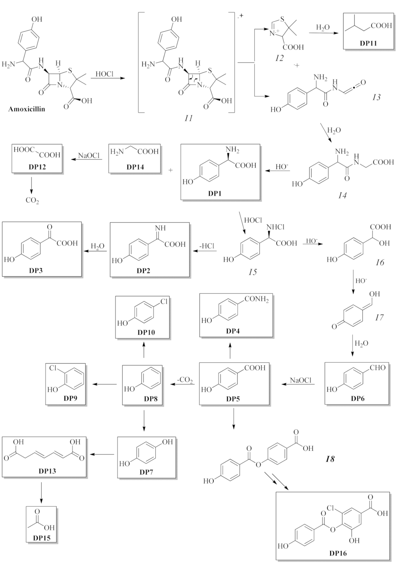
3.2. Structure Elucidation of Degradation Byproducts DP1—DP16

AMO chlorination-produced degradation byproducts DP1—DP16 (Figure 3) were isolated by chromatographic processes and identified on the basis of their physical features (Scheme 1).
In AMO treatment with an unbuffered pH value, the changes of the drug were monitored with HPLC. Its main degradation byproducts (DP1—DP16, Figure 3) were identified by comparing their retention times with those of the standard compounds and by employing NMR and MS analyses. The concentrations of DP1—DP16 were at a maximum after 5 min and were in the range of 3.01–0.89%.
The first three DPs (DP1—DP3) were C6C2 skeletal compounds obtained from the hydrolysis of the amide bond of the phenylethanoic acid residue and the subsequent oxidation of the alkyl chain. The DP4—DP6 had a C6C1 skeleton and, thus, it was easy to hypothesize that they were products derived from the decarboxylation of the previous three. Moreover, the DP7—DP10 had a C6C1 skeleton with an oxidized or chlorinated aromatic ring. DP11—DP15 products were di- or mono-carboxylic acids, which were final oxidation products. A separate discussion should be had for DP16, which is a phenylbenzoic ester chlorinated on the alcoholic part and clearly obtained from the esterification of two oxidation byproducts. The plausible mechanism of the DP formation from AMO is shown in Figure 4.
The reaction could start by a single-electron transfer from the lone electron pair of the amino group to HClO, which formed the corresponding radical cation and chloride. This aminyl radical cation (I1) could undergo a β-lactam cycle when two fragments, I2 and I3, formed. The first fragment formed via hydrolysis and gave DP11. The I3 fragment first hydrolyzed the ketene function to a carboxylic function, giving the intermediate I4 that led to DP1 and DP14. The second fragment oxidized to DP12 and CO2. From the product DP1, it was possible to obtain DP2—DP3 with a C6C2 skeleton, DP4—DP6 with a C6C1 skeleton, and DP7—DP10 with a C6C0 skeleton through a series of oxidations, decarboxylations, and chlorations. Finally, DP13 and DP15 was obtained by opening the aromatic ring. A slightly different argument to justify DP16, the synthesis of which could come from the chlorination and subsequent oxidation of intermediate I8, in turn was obtainable from the esterification of two DP5 molecules.

3.3. Antibiotic Activity Data

Figure 5 shows the antimicrobial activity of AMO and its DPs against S. aureus. Partial activity was developed at 5 mg/L AMO when the inhibition did not exceed 28% for the parent compound.
It was evident that 56% of DPs showed residual activity to S. aureus and this was more pronounced for DP6, where activity was exclusively due to oxidation byproducts with 74% of antibiotic activity. DP1, DP8, and DP9 showed decreased antibiotic activity. Only DP13, DP14, and DP16 were revealed to have no antibiotic effects.
Similar tests with E. coli and K. pneumoniae revealed that both bacteria were resistant at 5 mg/L AMO, with no significant antimicrobial activity (data not shown). The related DPs appeared to have no antibiotic and/or toxic effect against E. coli and K. pneumoniae (data not shown).
According to Dimitrakopoulou [48], E. coli and K. pneumoniae revealed a resistance up to 25 mg/L AMO, even if the suggested ranges for Minimum Inhibitory Concentrations (MICs) were 0.25–128 mg/L for Enterobacteriaceae (i.e., E. coli and K. pneumoniae) [49].

4. Conclusions

This paper investigated the fate of AMO by following the degradation treatment via chlorination. The reaction was carried out by simulating the conditions of a typical WWTP, using excess sodium hypochlorite at 3 different pH values. After the chlorination treatment, chromatographic techniques were used to isolate 16 degradation byproducts, including byproduct isolated for the first time, which were fully characterized using MS and NMR analyses and compared with parental samples. AMO underwent almost complete mineralization: 95–96% at pH 9, almost 80% at pH 7, and just under 70% at pH 3 after only a few minutes of treatment. We hypothesized a possible mechanism for the degradation of AMO and the formation of its byproducts. The antibiotic activity of AMO depended on the test bacteria in question. With regard to E. coli and K. pneumoniae, no antimicrobial activity occurred, regardless of how low AMO concentrations were or how low transformation byproducts were. Conversely, S. aureus was less resistant to AMO, and this effect remained partially or totally in its reaction byproducts. If it was known from the literature that amoxicillin was degraded by at least 75% in WWTPs, it was not clear what formed or the possible toxicity of the byproducts.

Author Contributions

G.L. (Giovanni Luongo) performed the chlorination experiments; M.G., A.S., G.L. (Giovanni Libralato), and L.S. performed the acute and chronic toxicity tests; A.Z., L.P., and G.D.F. designed the research study and wrote the paper. All authors have read and agreed to the published version of the manuscript.

Funding

We acknowledge AIPRAS-Onlus (Associazione Italiana per la Promozione delle Ricerche sull’Ambiente e la Salute umana) for the grants in support of this investigation.

Institutional Review Board Statement

Not applicable.

Informed Consent Statement

Not applicable.

Conflicts of Interest

The authors have no conflict of interest to declare.

References

  1. Pal, A.; Gin, K.Y.-H.; Lin, A.Y.-C.; Reinhard, M. Impacts of emerging organic contaminants on freshwater resources: Review of recent occurrences, sources, fate and effects. Sci. Total Environ. 2010, 408, 6062–6069. [Google Scholar] [CrossRef] [PubMed]
  2. Kasprzyk-Hordern, B.; Dinsdale, R.M.; Guwy, A.J. The occurrence of pharmaceuticals, personal care products, endocrine disruptors and illicit drugs in surface water in South Wales. Water Res. 2008, 42, 3498–3518. [Google Scholar] [CrossRef] [PubMed]
  3. Gabarrón, S.; Gernjak, W.; Valero, F.; Barceló, A.; Petrovic, M.; Rodríguez-Roda, I. Evaluation of emerging contaminants in a drinking water treatment plant using electrodialysis reversal technology. J. Hazard. Mater. 2016, 309, 192–201. [Google Scholar] [CrossRef] [PubMed]
  4. Huerta-Fontela, M.; Galceran, M.T.; Ventura, F. Occurrence and removal of pharmaceuticals and hormones through drinking water treatment. Water Res. 2011, 45, 1432–1442. [Google Scholar] [CrossRef]
  5. Bayer, A.; Asner, R.; Schüssler, W.; Kopf, W.; Weiß, K.; Sengl, M.; Letzel, M. Behavior of sartans (antihypertensive drugs) in wastewater treatment plants, their occurrence and risk for the aquatic environment. Environ. Sci. Pollut. Res. 2014, 21, 10830–10839. [Google Scholar] [CrossRef]
  6. Boix, C.; Ibáñez, M.; Sancho, J.V.; Parsons, J.R.; de Voogt, P.; Hernández, F. Biotransformation of pharmaceuticals in surface water and during waste water treatment: Identification and occurrence of transformation products. J. Hazard. Mater. 2016, 302, 175–187. [Google Scholar] [CrossRef]
  7. Mills, L.J.; Chichester, C. Review of evidence: Are endocrine-disrupting chemicals in the aquatic environment impacting fish populations? Sci. Total Environ. 2005, 343, 1–34. [Google Scholar] [CrossRef] [Green Version]
  8. Christensen, F.M. Pharmaceuticals in the environment: A human risk? Regul. Toxicol. Pharmacol. 1998, 28, 212–221. [Google Scholar] [CrossRef]
  9. Stuer-Lauridsen, F.; Birkved, M.; Hansen, L.P.; Lutzhoft, H.C.H.; Halling-Sorensen, B. Environmental risk assessment of human pharmaceuticals in Denmark after normal therapeutic use. Chemosphere 2000, 40, 783–793. [Google Scholar] [CrossRef]
  10. Merlin, C.; Bonot, S.; Courtois, S.; Block, J.-C. Persistence and dissemination of the multiple-antibiotic-resistance plasmid pB10 in the microbial communities of wastewater sludge microcosms. Water Res. 2011, 45, 2897–2905. [Google Scholar] [CrossRef]
  11. Andersson, D.I.; Hughes, D. Evolution of antibiotic resistance at non-lethal drug concentrations. Drug Resist. Updates 2012, 15, 162–172. [Google Scholar] [CrossRef] [PubMed]
  12. Boumendjel, A.; Tawe, G.S.; Bum, E.N.; Chabrol, T.; Beney, C.; Sinniger, V.; Haudecoeur, R.; Marcourt, L.; Challal, S.; Ferreira–Queiroz, E.; et al. Occurrence of the synthetic analgesic tramadol in an African medicinal plant. Angew. Chem. Int. Ed. Engl. 2013, 52, 11780–11784. [Google Scholar] [CrossRef] [PubMed]
  13. Jean, J.; Perrodin, Y.; Pivot, C.; Trepo, D.; Perraud, M.; Droguet, J.; Tissot-Guerraz, F.; Locher, F. Identification and prioritization of bioaccumulable pharmaceutical substances discharged in hospital effluents. J. Environ. Manag. 2012, 103, 113–121. [Google Scholar] [CrossRef] [PubMed]
  14. Fowler, P.A.; Bellingham, M.; Sinclair, K.D.; Evans, N.P.; Pocar, P.; Fischer, B.; Schaedlich, K.; Schmidt, J.-S.; Amezaga, M.R.; Bhattacharya, S.; et al. Impact of endocrine-disrupting compounds (EDCs) on female reproductive health. Mol. Cell. Endocrinol. 2012, 355, 231–239. [Google Scholar] [CrossRef] [PubMed]
  15. Hess-Wilson, J.K.; Knudsen, K.E. Endocrine disrupting compounds and prostate cancer. Cancer Lett. 2006, 241, 1–12. [Google Scholar] [CrossRef] [PubMed]
  16. Austin, B. Antibiotic pollution from fish farms: Effects on aquatic microflora. Microbiol. Sci. 1985, 2, 113–117. [Google Scholar]
  17. Miranda, C.D.; Castillo, G. Resistance to antibiotic and heavy metals of motile aeromonads from Chilean freshwater. Sci. Total Environ. 1998, 224, 167–176. [Google Scholar] [CrossRef]
  18. Lalumera, G.M.; Calamari, D.; Galli, P.; Castiglioni, S.; Crosa, S.; Fanelli, R. Preliminary investigation on the environmental occurrence and effects of antibiotics used in aquaculture in Italy. Chemosphere 2004, 54, 661–668. [Google Scholar] [CrossRef]
  19. Jones, O.A.; Voulvoulis, N.; Lester, J.N. Aquatic environmental assessment of the top 25 English prescription pharmaceuticals. Water Res. 2002, 36, 5013–5022. [Google Scholar] [CrossRef]
  20. Garcia-Reiriz, A.; Damiani, P.C.; Olivieri, A.C. Different strategies for the direct determination of amoxicillin in human urine by second-order multivariate analysis of kinetic-spectrophotometric data. Talanta 2007, 71, 806–815. [Google Scholar] [CrossRef]
  21. Calderon-Preciado, D.; Matamoros, V.; Bayona, J.M. Occurrence and potential crop uptake of emerging contaminants and related compounds in an agricultural irrigation network. Sci. Total Environ. 2011, 412–413, 14–19. [Google Scholar] [CrossRef] [PubMed]
  22. CSTEE (Scientific Committee on Toxicity, Ecotoxicity and the Environment). Opinion on draft CPMP discussion paper on environmental risk assessment of medicinal products of human use [Non-genetically modified organism (non-GMO) containing]. In Proceedings of the 24th CSTEE Plenary Meeting, Brussels, Belgium, 12 June 2001.
  23. Schreiber, F.; Szewzyk, U. Environmentally relevant concentrations of pharmaceuticals influence the initial adhesion of bacteria. Aquatic Toxicol. 2008, 87, 227–233. [Google Scholar] [CrossRef] [PubMed]
  24. Andreozzi, R.; Caprio, V.; Ciniglia, C.; De Champdoré, M.; Lo Giudice, R.; Marotta, R.; Zuccato, E. Antibiotics in the environment: Occurrence in Italian STPs, fate, and preliminary assessment on algal toxicity of amoxicillin. Environ. Sci. Technol. 2004, 38, 6832–6838. [Google Scholar] [CrossRef]
  25. Arslan-Alaton, I.; Dogruel, S.; Baykal, E.; Gerone, G. Combined chemical and biological oxidation of penicillin formulation effluent. J. Environ. Manag. 2004, 73, 155–163. [Google Scholar] [CrossRef] [PubMed]
  26. Navalon, S.; Alvaro, M.; Garcia, H. Reaction of chlorine dioxide with emergent water pollutants: Product study of the reaction of three β-lactam antibiotics with ClO2. Water Res. 2008, 42, 1935–1942. [Google Scholar] [CrossRef]
  27. Kurt, A.; Mert, B.K.; Özengin, N.; Sivrioğlu, Ö.; Yonar, T. Treatment of antibiotics in wastewater using advanced oxidation processes (AOPs). In Physico-Chemical Wastewater Treatment And Resource Recovery; IntechOpen: London, UK, 2017; Volume 175. [Google Scholar] [CrossRef] [Green Version]
  28. Moles, S.; Mosteo, R.; Gómez, J.; Szpunar, J.; Gozzo, S.; Castillo, J.R.; Ormad, M.P. Towards the removal of antibiotics detected in wastewaters in the POCTEFA territory: Occurrence and TiO2 photocatalytic pilot-scale plant performance. Water 2020, 12, 1453. [Google Scholar] [CrossRef]
  29. Abbassi, B.E.; Saleem, M.A.; Zytner, R.G.; Gharabaghi, B.; Rudra, R. Antibiotics in wastewater: Their degradation and effect on wastewater treatment efficiency. J. Food Agric. Environ. 2016, 14, 95–99. [Google Scholar]
  30. Calisto, V.; Domingues, M.R.M.; Erny, G.L.; Esteves, V.I. Direct photodegradation of carbamazepine followed by micellar electrokinetic chromatography and mass spectrometry. Water Res. 2011, 45, 1095–1104. [Google Scholar] [CrossRef]
  31. Persoone, G.; Marsalek, B.; Blinova, I.; Törökne, A.; Zarina, D.; Manusadzianas, L.; Nalecz-Jawecki, G.; Tofan, L.; Stepanova, N.; Kolar, B. A practical and user-friendly toxicity classification system with microbiotests for natural waters and wastewaters. Environ. Toxicol. 2003, 18, 395–402. [Google Scholar] [CrossRef]
  32. Romanucci, V.; Siciliano, A.; Guida, M.; Libralato, G.; Saviano, L.; Luongo, G.; Previtera, L.; Di Fabio, G.; Zarrelli, A. Disinfection by-products and ecotoxic risk associated with hypochlorite treatment of irbesartan. Sci. Total Environ. 2020, 712, 135625. [Google Scholar] [CrossRef]
  33. Luongo, G.; Previtera, L.; Ladhari, A.; Di Fabio, G.; Zarrelli, A. Peracetic acid vs. sodium hypochlorite: Degradation and transformation of drugs in wastewater. Molecules 2020, 25, 2294. [Google Scholar] [CrossRef] [PubMed]
  34. Luongo, G.; Guida, M.; Siciliano, A.; Libralato, G.; Saviano, L.; Amoresano, A.; Previtera, L.; Di Fabio, G.; Zarrelli, A. Oxidation of diclofenac in water by sodium hypochlorite: Identification of new degradation by-products and their ecotoxicological evaluation. J. Pharm. Biomed. Anal. 2021, 194, 113762. [Google Scholar] [CrossRef] [PubMed]
  35. Chusaksri, S.; Sutthivaiyakit, S.; Sedlak, D.L.; Sutthivaiyakit, P. Reactions of phenylurea compounds with aqueous chlorine: Implications for herbicide transformation during drinking water degradation. J. Hazard. Mater. 2012, 209, 484–491. [Google Scholar] [CrossRef]
  36. Sandín-España, P.; Magrans, J.O.; García-Baudín, J.M. Study of clethodim degradation and by-product formation in chlorinated water by HPLC. Chromatographia 2005, 62, 133–137. [Google Scholar] [CrossRef]
  37. ISO 6341:2012. Water Quality-Determination of the Inhibition of the Mobility of Daphnia magna Straus (Cladocera, Crustacea)—Acute Toxicity Test. International Organisation for Standardisation, Geneva, Switzerland. 2012. Available online: https://www.iso.org/standard/54614.html (accessed on 23 December 2020).
  38. Bedner, M.; MacCrehan, W.A. Transformation of acetaminophen bychlorination produces the toxicants 1,4-benzoquinone and N-acetyl-p-benzoquinone imine. Environ. Sci. Technol. 2006, 40, 516–522. [Google Scholar] [CrossRef] [PubMed]
  39. Hurst, J.W.; Mckim, J.M.; Martin, R.A., Jr. HPLC Determination of oxalic acid in cocoa. J. Liq. Chromatogr. 1986, 9, 2781–2789. [Google Scholar] [CrossRef]
  40. Liu, C.; Molinski, T.F. Preparation of α-amino acids by oxidative oxazoline–oxazinone rearrangement–hydrogenation (OOOH). Scope and limitations. Chem. Asian J. 2011, 6, 2022–2027. [Google Scholar] [CrossRef] [PubMed]
  41. Gowda, R.R.; Chakraborty, D. Fe-III-catalyzed synthesis of primary amides from aldehydes. Eur. J. Org. Chem. 2011, 201, 2226–2229. [Google Scholar] [CrossRef]
  42. Fujita, M.; Nagai, M.; Inoue, T. Carbon-13 nuclear magnetic resonance spectral study. Effect of O-methylation of ortho-substituted phenols on the aryl carbon shielding and its application to interpretation of the spectra of some flavonoids. Chem. Pharm. Bull. 1982, 30, 1151–1156. [Google Scholar] [CrossRef] [Green Version]
  43. Bovonsombat, P.; Ali, R.; Khan, C.; Leykajarakul, J.; Pla-on, K.; Aphimanchindakul, S.; Natchapon, P.; Nisit, T.; Anchalee, A.; Punpongjareorn, N. Facile p-toluenesulfonic acid-promoted para-selective monobromination and chlorination of phenol and analogues. Tetrahedron 2010, 66, 6928–6935. [Google Scholar] [CrossRef]
  44. Trincado, M.; Grützmacher, H.; Vizza, F.; Bianchini, C. Domino rhodium/palladium-catalyzed dehydrogenation reactions of alcohols to acids by hydrogen transfer to inactivated alkenes. Chem. Eur. J. 2010, 16, 2751–2757. [Google Scholar] [CrossRef] [PubMed]
  45. Romanucci, V.; Siciliano, A.; Galdiero, E.; Guida, M.; Luongo, G.; Liguori, R.; Di Fabio, G.; Previtera, L.; Zarrelli, A. Degradation by-products and ecotoxic risk associated with hypochlorite treatment of tramadol. Molecules 2019, 24, 693. [Google Scholar] [CrossRef] [PubMed] [Green Version]
  46. Zarrelli, A.; Della Greca, M.; Parolisi, A.; Iesce, M.R.; Cermola, F.; Isidori, M.; Lavorgna, M.; Passananti, M.; Previtera, L. Chemical fate and genotoxic risk associated with hypochlorite treatment of nicotine. Sci. Total Environ. 2012, 426, 132–138. [Google Scholar] [CrossRef] [PubMed] [Green Version]
  47. Zarrelli, A.; Della Greca, M.; Iesce, M.R.; Lavorgna, M.; Temussi, F.; Schiavone, L.; Criscuolo, E.; Parrella, A.; Previtera, L.; Isidori, M. Ecotoxicological evaluation of caffeine and its derivatives from a simulated chlorination step. Sci. Total Environ. 2014, 470, 453–458. [Google Scholar] [CrossRef] [Green Version]
  48. Dimitrakopoulou, D.; Rethemiotaki, I.; Frontistis, Z.; Xekoukoulotakis, N.P.; Venieri, D.; Mantzavinos, D. Degradation, mineralization and antibiotic inactivation of amoxicillin by UV-A/TiO2 photocatalysis. J. Environ. Manag. 2012, 98, 168–174. [Google Scholar] [CrossRef]
  49. Andrews, J.M. Determination of minimum inhibitory concentrations. J. Antimicrob. Chemother. 2001, 48, 5–16. [Google Scholar] [CrossRef] [Green Version]
Figure 1. Time-conversion plot for the reaction of amoxicillin (AMO) with one equivalent NaOCl at buffered pH = 3.0 (a) and pH = 7 (b). ●: AMO consumption by reaction with NaOCl (green); ○: Disappearance of AMO in the absence of NaOCl (red).
Applsci 11 01076 g001
Figure 2. (a) Time-conversion plot for the reaction of AMO with one equivalent NaOCl at pH = 9.0. ■: NaOCl consumption in the presence of AMO (green); ●: AMO consumption by reaction with NaOCl (blue); □: Disappearance of NaOCl in the absence of AMO (red); ○: Disappearance of AMO in the absence of NaOCl (black); (b) AMO disappearance by NaOCl at pH basic no-buffered after a 5 min reaction.
Applsci 11 01076 g002
Figure 3. Chemical structures of amoxicillin and its degradation byproducts.
Applsci 11 01076 g003
Scheme 1. Isolation of 16 identified degradation byproducts.
Applsci 11 01076 sch001
Figure 4. Plausible mechanism for the formation of DP1—DP16.
Applsci 11 01076 g004
Figure 5. Antibacterial activity of AMO and its DPs against S. aureus. Groups with the same letter were not significantly different (Tukey post hoc, p < 0.05).
Applsci 11 01076 g005
Publisher’s Note: MDPI stays neutral with regard to jurisdictional claims in published maps and institutional affiliations.

