Article

TQ-6, a Novel Ruthenium Derivative Compound, Possesses Potent Free Radical Scavenging Activity in Macrophages and Rats

1
Department of Neurology, Yuan’s General Hospital, Kaohsiung 802, Taiwan
2
Department of Cardiovascular Center, Cathay General Hospital, Taipei 106, Taiwan
3
Graduate Institute of Medical Sciences, College of Medicine, Taipei Medical University, Taipei 110, Taiwan
4
Division of Cardiology, Department of Internal Medicine, School of Medicine, College of Medicine, Fu Jen Catholic University, New Taipei City 242, Taiwan
5
Department of Chemistry, North Eastern Hill University, Shillong 793022, India
6
International Ph.D. Program for Cell Therapy and Regeneration Medicine, College of Medicine, Taipei Medical University, Taipei 110, Taiwan
7
Department of Pharmacology, School of Medicine, College of Medicine, Taipei Medical University, Taipei 110, Taiwan
8
Translational Medicine Center, Shin Kong Wu Ho-Su Memorial Hospital, Taipei 111, Taiwan
*
Authors to whom correspondence should be addressed.
Appl. Sci. 2021, 11(3), 1008; https://doi.org/10.3390/app11031008
Submission received: 17 December 2020 / Revised: 11 January 2021 / Accepted: 20 January 2021 / Published: 23 January 2021

Abstract

:
Reactive oxygen species (ROS) play major role in inducing inflammation and related diseases. Our previous studies have revealed that the ruthenium (II)-compound, [Ru(η6-cymene)2-(1H-benzoimidazol-2-yl)-quinoline Cl]BF4 (TQ-6), inhibits hydroxyl radical (OH) formation in human platelets. TQ-6 also have protective effect against induced inflammation in macrophages and hepatic injury in mice through NF-κB signaling. However, the free radical formation inhibitory mechanism of TQ-6 in macrophages is unclear. Therefore, this study detected the antioxidative ability of TQ-6 in both a cell-free system and in LPS-induced macrophages through electron spin resonance (ESR) spectrometry. TQ-6 reduced 1,1-diphenyl-2-picrylhydrazyl (DPPH), galvinoxyl, and superoxide radicals in a cell-free system and OH formation in macrophages. Additionally, TQ-6 activated the nuclear factor erythroid 2-related factor 2 (Nrf2) signaling pathway and upregulated the antioxidant protein heme oxygenase-1 (HO-1) to elevate anti-inflammatory activity in LPS-induced macrophage cells and inhibited carrageenan-induced paw edema in a rat model. Therefore, TQ-6 may prevent oxidative stress and also act as an effective therapeutic agent for the treatment of oxidant-related diseases.

1. Introduction

Oxidative stress induce inflammation via reactive oxygen species (ROS) formation, which are reflected the strong inflammatory mediators. Several ROS are produced inside lipopolysaccharide (LPS)-stimulated macrophages, such as the superoxide radical anion, which is produced primarily, but not exclusively, by NADPH oxidase (NOX) [1]. NOX is present in various cells, especially specialized phagocytic and endothelial cells [1]. Polymorphonuclear neutrophils lead to increased ROS production at the site of inflammation, resulting in endothelial dysfunction and tissue damage [2]. Because antioxidants reduce inflammation, therefore, an antioxidant drug may be an anti-inflammatory drug and vice versa.
Nuclear factor erythroid 2-related factor 2 (Nrf2) is a transcription factor that can obstruct inflammatory events. Antioxidant gene expression mediated by Nrf2 is proposed to diminish the macrophage M1 phenotype and ROS production. Since Nrf2 stimulators are reported to be an efficient therapeutic strategy for several diseases [3], such as inflammatory disorders and cardiovascular diseases [3], the Nrf2 signaling pathway is considered to be a critical cascade in inflammation. Nrf2 mitigates inflammatory reactions by inducing anti-inflammatory enzyme heme oxygenase-1 (HO-1), and negatively regulating the expression of proinflammatory cytokines and chemokines [4]. Therefore, developing new pharmacological interventions that induce Nrf2 and HO-1 is crucial for the therapeutic control of inflammatory diseases and oxidative stress.
Increasing attention has been paid to antioxidant-derived metal complexes as novel methods of protecting living organisms and cells against damage caused by oxidative stress and free radicals [5]. Transition metal complexes have been reported to have various biological activities through free radical scavenging properties [6]. Previous research has also found that ruthenium complexes exhibited anticancer activity [7]. Ruthenium complexes have been reported to exhibit strong scavenging activity on 2, 2′-azinobis (3-ethylbenzothiazoline-6-sulfonic acid) (ABTS) and DPPH radicals and antiproliferative effect against some cancer cell lines [8]. Our previous study showed that [Ru(η6-cymene)2-(1H-benzoimidazol-2-yl)-quinoline Cl]BF4 (TQ-6), a synthetic ruthenium (II)-derived compound, exhibited potent anti-inflammatory activity in vitro and effectively inhibited liver injury in vivo. This research further suggested that nuclear factor (NF)-κB is a potential target for the TQ-6’s preventive effects in LPS-induced inflammation and liver injury [9]. In addition, we showed that TQ-6 inhibited OH formation and platelet activation in collagen-stimulated platelets, suggesting that free radical scavenging action can be involved in TQ-6-mediated inhibition of in vivo thrombogenesis [10]. The present study examined the antioxidant property of TQ-6 by determining its radical scavenging and Nrf2/HO-1 induction potential in vitro. Moreover, the anti-inflammatory action of TQ-6 was compared with the nonsteroidal anti-inflammatory drug indomethacin in the rat paw edema model.

2. Materials and Methods

2.1. Materials

Fetal bovine serum (FBS), Dulbecco’s modified Eagle medium (DMEM), L-glutamine penicillin/streptomycin, and anti-α-tubulin monoclonal antibodies were purchased from Invitrogen (Thermo Fisher Scientific, Waltham, MA, USA). Lipopolysaccharide (LPS) (Escherichia coli 0127:B8), 5,5-dimethyl-1-pyrroline N-oxide (DMPO), and dimethyl sulfoxide (DMSO), and were purchased from Sigma-Aldrich (St. Louis, MO, USA). 1,1-diphenyl-2-picrylhydrazyl (DPPH) and galvinoxyl were purchased from Nacalai Tesque (Kyoto, Japan). Anti-HO-1 and Nrf2 polyclonal antibodies (pAb) were purchased from Enzo, Farmingdale, New York and Genetex, Irvine, CA, USA, respectively. Amersham, Buckinghamshire, UK was supplied horseradish peroxidase-conjugated donkey anti-rabbit and anti-mouse immunoglobulin G (IgG). GE Healthcare Life Sciences (Waukesha, WI, USA) supplied enhanced chemiluminescence (ECL) western blotting detection reagents and Hybond™-P polyvinylidene difluoride (PVDF) blotting membranes.

2.2. Synthesis of TQ-6

TQ-6 and its ligand were synthesized according to the method described by Hsiao et al. [10] and it dissolved in DMSO for analysis. The final concentration of stock solution of TQ-6 in DMSO was 100 mM, which was diluted in >99% DMSO.

2.3. DPPH and Galvinoxyl Radical Scavenging Assay

DPPH or galvinoxyl (500 μM) and TQ-6 (10 and 20 μM) were added to a reaction mixture containing distilled water and stirred well by using pipette for few sec. After 30 min incubation at 37 °C, electron spin resonance (ESR) spectra were measured with the conditions of 20 mW, 9.78 GHz, 100 G scan and 5 × 10⁴ receiver gain. The variation amplitude 1 G, and the time constant 164 ms was set. Individual sample was scanned for 42 s, with sum of three scans. A quartz flat cell was used to document the ESR spectra signals for the detection of DPPH and Galvinoxyl Radicals.

2.4. Superoxide Scavenging Assay

A xanthine/xanthine oxidase (X/XO) system was adopted to produce superoxide to a quantity necessary to assay the superoxide-scavenging activity of TQ-6 as defined previously [11]. Concisely, PBS (pH 7.4) mixture solution contained 0.1 mM DTPA, 10 mM 5-Diethoxyphosphoryl-5-methyl-1-pyrroline N-oxide (DEPMPO), 1 mM hypoxanthine, and 0.2 U/mL XO with or without TQ-6. Following 1 min incubation at 37 °C, the incubation mixture was moved to a flat cell to measure ESR spectra.

2.5. Fenton Reaction Analysis by ESR Spectrometry

ESR spectrometry (Bruker EMX ESR, Billerica, MA, USA) analysis was done as defined earlier [12]. The suspension of the Fenton reaction (50 μM FeSO4 + 2 mM H2O2) was preincubated with 0.1% DMSO or TQ-6 (10 and 20 μM) for 3 min, and 100 μM DMPO was added to the suspension before ESR spectrometry.

2.6. Cell Cultivation

The American Type Culture Collection (ATCC) was supplied RAW 264.7 cells (TIB-71), they were cultivated in DMEM contained with 10% FBS, 100 U/mL penicillin G, and 100 mg/mL streptomycin at 37 °C in a moistened atmosphere of 5% CO2/95% air [9].

2.7. Measurement of OH Formation

The ESR method was used to measure OH as labelled before [10]. RAW 264.7 cells (5 × 10⁵ cells) were pretreated with TQ-6 (10 and 20 μM) or DMSO for 20 min, and LPS (1 μg/mL) was subsequently added. ESR spectra were recorded as mentioned in the Section 2.3. The ESR spectrum analysis was performed by using WIN-EPR, version 921,201 supplied by BRUKER-FRANZEN Analytik GmbH (Bremen, Germany).

2.8. Quantitative Real-Time PCR (RT-qPCR)

To analyze the expression of target genes, a StepOne Real-Time PCR system (Applied Biosystems; Thermo Fisher Scientific, Inc.) was applied to run RT-qPCR and it was performed by using Fast SYBR®-Green Master mix (Thermo Fisher Scientific, Inc.) per the manufacturer’s instructions. Glyceraldehyde 3-phosphate dehydrogenase (GAPDH) was used as positive control. The conditions of RT-qPCR cycling were Hot-start activation at 95 °C for 20 s; 40 cycles of denaturation at 95 °C for 3 s and annealing/extension at 60 °C for 30 s. The specific primers were showed in Table 1. A comparative CT method (2-ΔΔCq) was applied to measure the intensity of PCR bands [13].

2.9. Immunoblotting

According to the method defined by Sheu et al. [14], the immunoblotting assay was done to detect the targeting protein expression. For this, cells at a density of 8 × 10⁵/dish were cultured and treated with TQ-6 or DMSO for 20 min and then stimulated with LPS (1 μg/mL). The extracted proteins (50 μg) using the lysis buffer (50 mM HEPES, 5 mM EDTA, 50 mM NaCl and 1% Triton X-100) were separated in a 12% SDS-PAGE, and then transferred to PVDF membranes (0.45 μm). Using 5% skimmed milk, the membranes were blocked and titrated with primary antibodies against Nrf2 and HO-1 followed by HRP-linked anti-mouse IgG or anti-rabbit IgG secondary antibodies. The immune-reactive bands were visualized using ECL system and their densities were measured by the Biolight Windows Application, V2000.01 (Bio-Profil, Vilber Lourmat, France).

2.10. Carrageenan-Induced Rat Paw Edema Measurement

A total of 30 male Wistar rats (200–250 g) were procured from BioLASCO, Taipei, Taiwan. The Institutional Animal Care and Use Committee, Taipei Medical University (LAC-2015-0267) approved animal experiments and care procedures. The animals were maintained at a condition with 22 ± 4 °C temperature, 50 ± 20% humidity and 12 h light/dark cycle in animal center. They had access to standard rodent pellet food and water ad libitum. The rats were anesthetized by 5% isoflurane. Animals were grouped as: (i) vehicle control (20 μL, 0.1% DMSO); (ii) carrageenan control; (iii) standard anti-inflammatory drug indomethacin (5 mg/kg b.w.); and (iv) and (v) 1 and 2 mg/kg of TQ-6, respectively. A carrageenan-induced paw edema was measured as described previously [15], with some minor modifications. Paw thickness was measured before (0 h) or after 1, 2, 4, 6, and 24 h carrageenan injection.

2.11. Statistical Analysis

Results are presented as the mean ± standard error (SEM). One-way analysis of variance (one-way ANOVA) was applied to measure the data. The significant differences among the groups were compared by using the Newman-Keuls method. A p value <0.05 was considered as statistically significant.

3. Results

3.1. TQ-6 Reduces DPPH and Galvinoxyl Free Radical Formation

The DPPH radical scavenging activity is commonly used for evaluating the antioxidant activity of drugs or compounds [16]. These radicals are counteracted by antioxidants via donating either electron or hydrogen atoms [17]. As shown in Figure 1A, TQ-6 (20 μM) increased DPPH scavenging capacity compared with DMSO.
Galvinoxyl radical is also a relatively stable radical commonly used in antioxidant assays [17,18]. TQ-6 (20 μM) scavenges galvinoxyl radicals, showing considerable radical-scavenging activity (Figure 1B). Moreover, TQ-6 is more potent on scavenging galvinoxyl radical than DPPH. These results indicated that TQ-6 showed stronger scavenging activity against DPPH and galvinoxyl radicals.

3.2. Effect of TQ-6 on Superoxide Anion Formation-Derived from X/XO and OH Formation

Figure 2A shows the superoxide-scavenging activity of TQ-6 via decreased the spectrum intensity of DEPMPO/superoxide adduct (DEPMPO-OOH). ESR radical spectrum signal heights were concentration dependently inhibited by 10 and 20 μM TQ-6. In addition, OH radical formation was observed in Fenton reaction solution (Figure 2B), and treatment with 10 or 20 μM TQ-6 diminished Fenton reaction-induced OH.

3.3. TQ-6 Inhibits OH and ROS Formation

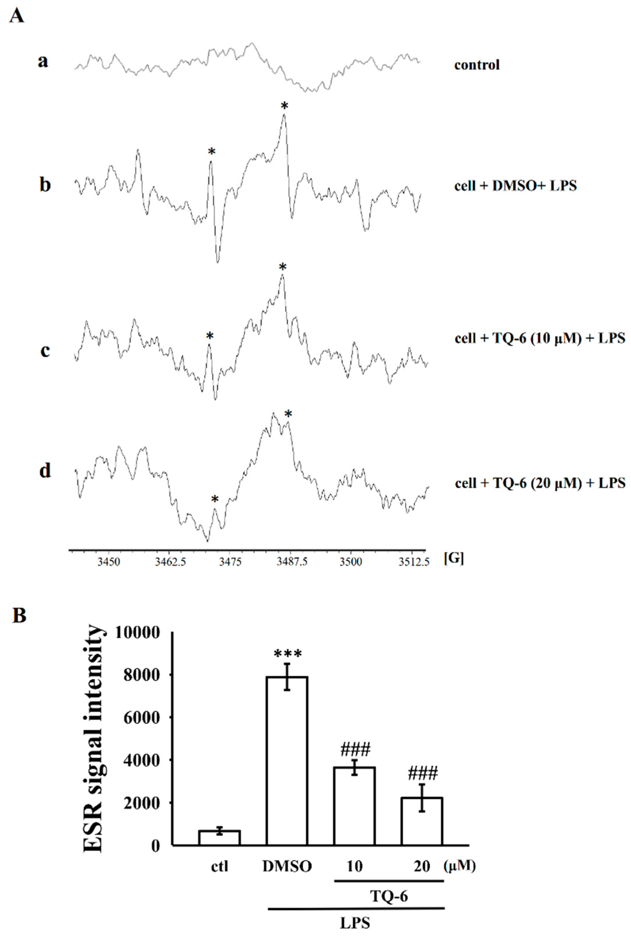
The generation of nitric oxide and superoxide anions is highly associated with cell and tissue injury and redox signaling in activated macrophages [19]. A classic ESR signal for the production of OH radical was detected in LPS-stimulated (1 μg/mL) cells (Figure 3A(a,b)). TQ-6 (10 and 20 μM) considerably attenuated LPS-triggered OH radical formation in RAW 264.7 cells (Figure 3A(c,d)). From this result, it can be proposed that the inhibitory effect of TQ-6 in LPS-induced macrophage activation may partially be suppressed through free radical generation. In addition, the total ROS level was estimated by using DCFDA, a cell-permeative ROS-sensitive dye. The results found that LPS-induced ROS formation in macrophages was significantly not affected by TQ-6 (data not shown).

3.4. TQ-6 Enhances Nrf2/HO-1 mRNA and Protein Expression

Nrf2 inhibits proinflammatory cytokine expression, inflammatory responses, and ROS production [20]; HO-1 is a major antioxidant enzyme mediated by Nrf2 activation [21,22,23]. To determine whether TQ-6 activates Nrf2 signaling, RAW264.7 cells were treated with 20 μM TQ-6 for 20 min and then LPS (1 µg/mL) was added. The Nrf2 and HO-1 mRNA expression was measured using RT-qPCR (Figure 4A,B) and their protein expression was measured by Western blotting (Figure 4C,D). Nrf2 and HO-1 TQ-6 mRNA and protein expressions were upregulated in LPS-stimulated RAW264.7 cells (Figure 4A–D). However, cells treated with TQ-6 alone did not change the expression of Nrf2 and HO-1, as they almost similarly expressed to normal cells (Figure 4E,F). This indicates that TQ-6 holds protective effects without inducing cytolysis.

3.5. Carrageenan-Induced Inflammatory Response in Rats

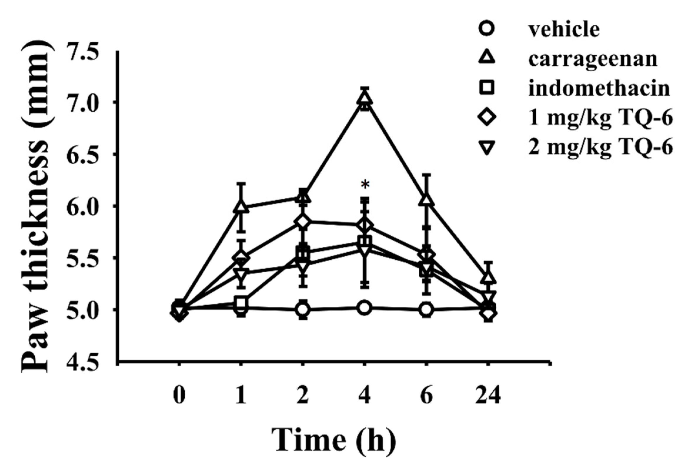
Carrageenan injection into the hind paw of rats resulted in edema, as shown by paw thickness. The paw thickness of DMSO injection was 5.02 ± 0.08 mm at 0 h and remained constant throughout the subsequent 24 h. The initial paw thickness of 5.02 ± 0.04 mm reached its maximum of 7.03 ± 0.10 mm at 4 h, and it again decreased into 5.30 ± 0.15 mm at 24 h in the carrageenan injected control group. The indomethacin treatment group showed an initial paw thickness of 5.00 ± 0.02 mm, which increased to 5.65 ± 0.39 mm at 4 h and steadily reduced to 5.38 ± 0.23 mm and 5.00 ± 0.11 mm at 6 and 24 h, respectively. Animals treated with TQ-6 (1 mg/kg) showed an initial paw thickness of 4.97 ± 0.05 mm, which increased to 5.85 ± 0.28 mm at 2 h and decreased to 5.82 ± 0.26 mm, 5.53 ± 0.25 mm, and 4.97 ± 0.05 mm at 4, 6, and 24 h, respectively. The paw thickness of animals treated with TQ-6 (2 mg/kg) at 0 h was 5.00 ± 0.03 mm and increased to 5.58 ± 0.37 mm at 4 h, which decreased to 5.42 ± 0.15 mm and 5.13 ± 0.12 mm at 6 and 24 h, respectively (Figure 5). The data showed that TQ-6 exhibited anti-inflammatory activity via reducing carrageenan-induced paw edema.

4. Discussion

Our previous studies have shown that TQ-6 potently inhibited LPS-induced inflammatory events in RAW264.7 cells [9] and thrombosis in mice [10]. Moreover, TQ-6 diminishes collagen-induced OH in platelets [10]. This study mainly focused on examining the free radical scavenging mechanism of TQ-6 to its anti-inflammatory action and found that TQ-6 scavenges DPPH, galvinoxyl, superoxide (O2), and OH radicals in cell-free systems. Furthermore, TQ-6 exhibited potent antioxidative activity through induction of the Nrf2/HO-1 signaling pathway. Additionally, TQ-6 prevented carrageenan-induced paw edema in rats. These results have demonstrated that the free radical scavenging properties of TQ-6 may play a role in its cell protective effect.
LPS can activate a local or systemic inflammatory response in both immune non-immune cells to initiate the inflammatory events. The LPS-pattern recognition receptor, the Toll-like receptor 4 (TLR4) is widely expressed including cardiomyocytes. Therefore, post- treatment of TQ-6 may not exacerbate specific targets on LPS-induced inflammatory dysfunctions such as LPS induce diversity of makers that were recurrently detected in patients with sepsis and also in animals [24]. Thus, in this study, TQ-6 was pretreated to test its anti-inflammatory function in the LPS-induced macrophage cells.
Exogenous and endogenous stimulators induce inflammatory reactions, which are reflected in vascular and cellular events. Inflammation-induced ROS production, such as O2, OH, hydrogen peroxide (H2O2), peroxy radicals (ROO), and singlet oxygen (1O2) induce oxidative stress which may lead to cancer and neurodegenerative diseases [25]. In addition, DPPH and galvinoxyl radicals are largely used as substrates to evaluate the antioxidant activity of a drug or substance. DPPH-electron paramagnetic resonance system has been widely used to detect the radical scavenging activities of hydrophilic and lipophilic antioxidants [26]. Experiments with galvinoxyl allow not only a comparison of the radical scavenging activity between samples but also the determination of the concentration of antioxidants. A previous study showed that Co(II) complexes had stronger scavenging activity against DPPH than did mefenamic acid [27]. Furthemore, Ingleson et al. [28] used galvinoxyl-radical as a model to study the radical scavenging effect of Co(II) complex and the result showed that this metal significantly inhibited galvinoxyl radicals. In this study, TQ-6 substantially suppressed DPPH free radical production, showing that TQ-6 possesses the capacity to scavenge such free radicals.
Xanthine oxidase (XO) produces free radicals in post-ischemic cells and tissues [29], and the X/XO system assay related to an enzymatic reaction which catalyzing the hydroxylation of hypoxanthine to xanthine and then to uric acid which is excreted via kidneys. However, this systemic assay is not appropriate for lipid-soluble antioxidants. You et al. [30] showed the inhibitory activity of some metal complexes against XO, of which the cadmium (II) complex was the more potent XO inhibitor than Zn(II) and Mn(II) complexes. This is the first study to demonstrate that even 10 μM TQ-6 effectively scavenges non-enzymatic, potassium superoxide (KO2) system-generated superoxide and was subsequently validated in the X/XO enzymatic system. These data strongly suggest that TQ-6 can act as a strong antioxidant in different applications. The antioxidative properties of TQ-6 could not be only due to XO inhibition but also a direct inhibition of superoxide generation.
Under pathological conditions, macrophages induced ROS through NADPH oxidase (NOX) activation could be involved in the damages of cell and tissue, killing of pathogen, and inflammatory signaling [31]. NOX-mediated free radical production is triggered by the induced p38 MAPK and NF-κB signaling [32]. Studies found that TQ-6 inhibits LPS-stimulated macrophage activation through inhibiting of NF-κB signaling pathway, such as IκBα degradation, NF-κB p65 phosphorylation and its nuclear translocation [9]. This also inhibits OH formation in activated platelets [10]. A study was found that Ru(II) complexes, such as [Ru(bpy)2(maip)]2+ and [Ru(bpy)2(paip)]2+ exhibit good antioxidant activity in terms of inhibiting OH formation [33]. Here, the free radical scavenging ability of TQ-6 was found in cell-free system and LPS-induced macrophages through ESR spectrometry, and this assay may have provided direct evidence indicating that TQ-6 has antioxidant properties. Thus, TQ-6 is capable of reducing the severity of inflammatory diseases via reduction of oxidative stress and related inflammatory signaling.
Certain anti-inflammatory signaling pathways negatively regulate inflammatory responses to maintain homeostasis. Several lines of confirmation revealed that HO-1 inhibits the production of nitric oxide (NO), prostaglandin E2 (PGE2), interleukin-1β (IL-1β), IL-6, and tumor necrosis factor-α (TNF-α) in vitro and in vivo [34,35,36]. Normally, Nrf2 attaches to its negative regulator, Kelch-like ECH-associated protein 1 (Keap1), and localizes in the cytoplasm; however, various stimuli, including ROS, induce nuclear translocation of Nrf2 and enhance HO-1 production [34]. HO-1 exhibits cytoprotective, antioxidant, and anti-inflammatory activities [37,38]. Previous study explained that TQ-6 exhibits anti-inflammatory effects via suppressing of p38 MAPK, NF-κB, iNOS and NO in LPS-stimulated macrophages [9]. A clear evidence indicated that ROS could activate Nrf2, NF-κB and MAPK [39]. Here, we found that TQ-6 significantly upregulated Nrf2 expression followed by HO-1 expression in LPS-stimulated RAW cells. Moreover, Xaus et al. [40] provide evidence that macrophage apoptosis induced by LPS is mediated by both NO and TNF-α production. A previous our paper also showed that TQ-6 dampened NO production, and TNF-α expression in LPS-induced macrophages; however, these are not increased when treating with TQ-6 alone [9]. From this finding, it assumed that TQ-6 could inhibit LPS-induced macrophage apoptosis. The absence of evaluating the direct evidence whether TQ-6 has effective in LPS-induced apoptosis in macrophages and the similar attributes between macrophages and neutrophils as new therapeutic anti-inflammatory strategies subjected to TQ-6 treatment is regarded as the limitation of this study.
The animal paw edema model has been commonly used to evaluate the anti-edematous effect of drugs. Carrageenan-induced edema model has long been appreciated as an excellent model for quick in vivo screening of NSAIDs [41]. Kale et al. [42] noticed a zinc complex reduced carrageenan-induced rat paw edema. In this study, as TQ-6 administered to rats acutely for 24 h to test its in vivo anti-inflammatory efficacy, we have chosen carrageenan injection rather than LPS, and resulted in a significant reduction of rat paw thickness, thus demonstrating its anti-inflammatory properties. Consistently, in this inflammation model, TQ-6 showed a significant decrease in rat paw thickness, thus demonstrating its anti-inflammatory response. Pharmacokinetic (PK) study is an extensive area to focus for the new drug development. The total accumulation of ruthenium in peripheral WBC had been reported in nanomoles/mg protein and quantified in 23 patients [43]. However, the lack of PK study to know the bioavailability of TQ-6 in macrophages and clinical trial for the safety of TQ-6 in humans is the limitation of this study and this needs to be performed in the near future.

5. Conclusions

This study analyzed the antioxidative role of TQ-6 in cell-free and RAW 264.7 cell model systems by using ESR in vitro and its anti-inflammatory properties in rat paw edema model in vivo. Our results demonstrated that TQ-6 scavenged DPPH, hydroxyl, galvinoxyl, and superoxide free radicals. Furthermore, we found that TQ-6 exerted anti-inflammatory action via inducing Nrf2/HO-1 pathway in vitro. Additionally, the anti-inflammatory mechanism of TQ-6 may be beneficial for treatment of oxidative stress-mediated inflammatory diseases.

Author Contributions

C.-Y.H. and C.-H.H. conceptualized the study; K.-W.H. conducted experiments and drafted the manuscript; M.V. synthesized and analyzed TQ-6; C.-C.C., T.J., C.-W.H., N.T.T.T. and D.-S.C. assisted in experiments and analyzed the data. The final version of the manuscript was approved by all the authors to submit. All authors have read and agreed to the published version of the manuscript.

Funding

This work was supported by grants from the Ministry of Science and Technology of Taiwan (MOST 107-2320-B-038-035-MY2; MOST 108-2320-B-038-021-MY2), Shin Kong Wu Ho-Su Memorial Hospital (2020SKHAND007), Yuan’s General Hospital-Taipei Medical University (107YGH-TMU-07), and the University Grants Commission, India (MRP-MAJOR-CHEM-2013-5144; 69/2014 F. No. 10-11/12UGC).

Institutional Review Board Statement

The Institutional Animal Care and Use Committee, Taipei Medical University (LAC-2015-0267) approved animal experiments and care procedures.

Informed Consent Statement

Not applicable.

Data Availability Statement

All data generated or analyzed during this study are included in this published article.

Conflicts of Interest

We have no conflict of interest.

References

  1. Lambeth, J.D. NOX enzymes and the biology of reactive oxygen. Nat. Rev. Immunol. 2004, 4, 181–189. [Google Scholar] [CrossRef]
  2. Mittal, M.; Siddiqui, M.R.; Tran, K.; Reddy, S.P.; Malik, A.B. Reactive oxygen species in inflammation and tissue injury. Antioxid. Redox Signal. 2014, 20, 1126–1167. [Google Scholar] [CrossRef] [Green Version]
  3. Crunkhorn, S. Deal watch: Abbott boosts investment in NRF2 activators for reducing oxidative stress. Nat. Rev. Drug Discov. 2012, 11, 96. [Google Scholar] [CrossRef] [PubMed]
  4. Tamion, F.; Richard, V.; Renet, S.; Thuillez, C. Intestinal preconditioning prevents inflammatory response by modulating heme oxygenase-1 expression in endotoxic shock model. Am. J. Physiol. Gastrointest. Liver Physiol. 2007, 293, 1308–1314. [Google Scholar] [CrossRef] [PubMed]
  5. Ejidike, I.P.; Ajibade, P.A. Synthesis, characterization, antioxidant, and antibacterial studies of some metal(ii) complexes of tetradentate Schiff base ligand: (4E)-4-[(2-{(E)-[1-(2,4-Dihydroxyphenyl) ethylidene]amino}ethyl)imino] pentan-2-one. Bioinorg. Chem. Appl. 2015, 2015, 890734. [Google Scholar] [CrossRef] [PubMed] [Green Version]
  6. Raman, N.; Pothiraj, K.; Baskaran, T. DNA-binding, oxidative DNA cleavage and coordination mode of later 3d transition metal complexes of a Schiff base derived from isatin as antimicrobial agents. J. Coord. Chem. 2011, 64, 3900–3917. [Google Scholar] [CrossRef]
  7. Pascu, G.I.; Hotze, A.C.G.; Sanchez-Cano, C.; Kariuki, B.M.; Hannon, M.J. Dinuclear Ruthenium(II) triple-stranded helicates: Luminescent supramolecular cylinders that bind and coil DNA and exhibit activity against cancer cell lines. Angew. Chem. Int. Ed. 2007, 46, 4374–4378. [Google Scholar] [CrossRef]
  8. Ejidike, I.P.; Ajibade, P.A. Synthesis, characterization, in vitro antioxidant and anticancer studies of ruthenium(III) complexes of symmetric and asymmetric tetradentate Schiff bases. J. Coord. Chem. 2015, 68, 2552–2564. [Google Scholar] [CrossRef]
  9. Hsia, C.H.; Velusamy, M.; Jayakumar, T.; Chen, Y.J.; Hsia, C.W.; Tsai, J.H.; Teng, R.D.; Sheu, J.R. Mechanisms of TQ-6, a Novel Ruthenium-Derivative Compound, against Lipopolysaccharide-Induced In Vitro Macrophage Activation and Liver Injury in Experimental Mice: The Crucial Role of p38 MAPK and NF-κB Signaling. Cells 2018, 7, 217. [Google Scholar] [CrossRef] [Green Version]
  10. Hsia, C.H.; Velusamy, M.; Sheu, J.R.; Khamrang, T.; Jayakumar, T.; Lu, W.J.; Lin, K.H.; Chang, C.C. A novel ruthenium (II)-derived organometallic compound, TQ-6, potently inhibits platelet aggregation: Ex vivo and in vivo studies. Sci. Rep. 2017, 7, 9556. [Google Scholar] [CrossRef] [Green Version]
  11. Fridovich, I. Superoxide radical and superoxide dismutases. Annu. Rev. Biochem. 1995, 64, 97–112. [Google Scholar] [CrossRef] [PubMed]
  12. Chou, D.S.; Hsiao, G.; Shen, M.Y.; Tsai, Y.J.; Chen, T.; Sheu, J.R. ESR spin trapping of a carbon-centered free radical from agonist-stimulated human platelets. Free Radic. Biol. Med. 2005, 39, 237–248. [Google Scholar] [CrossRef] [PubMed]
  13. Livak, K.J.; Schmittgen, T.D. Analysis of relative gene expression data using real-time quantitative PCR and the 2(-Delta Delta C(T)) method. Methods 2001, 25, 402–408. [Google Scholar] [CrossRef] [PubMed]
  14. Sheu, J.R.; Chen, Z.C.; Hsu, M.J.; Wang, S.H.; Jung, K.W.; Wu, W.F.; Pan, S.H.; Teng, R.D.; Yang, C.H.; Hsieh, C.Y. CME-1, a novel polysaccharide, suppresses iNOS expression in lipopolysaccharide-stimulated macrophages through ceramide-initiated protein phosphatase 2A activation. J. Cell. Mol. Med. 2018, 22, 999–1013. [Google Scholar] [CrossRef] [PubMed] [Green Version]
  15. Amdekar, S.; Roy, P.; Singh, V.; Kumar, A.; Singh, R.; Sharma, P. Anti-inflammatory activity of lactobacillus on carrageenan-induced paw edema in male wistar rats. Int. J. Inflam. 2012, 2012, 752015. [Google Scholar] [CrossRef] [Green Version]
  16. Shahidi, F.; Zhong, Y. Measurement of antioxidant activity. J. Funct. Foods 2015, 18, 757–781. [Google Scholar]
  17. Prior, R.L.; Wu, X.; Schaich, K. Standardized methods for the determination of antioxidant capacity and phenolics in foods and dietary supplements. J. Agric. Food Chem. 2005, 53, 4290–4302. [Google Scholar] [CrossRef]
  18. Shi, H.; Noguchi, N.; Niki, E. Galvinoxyl method for standardizing electron and proton donation activity. Methods Enzymol. 2001, 335, 157–166. [Google Scholar]
  19. Brüne, B.; Dehne, N.; Grossmann, N.; Jung, M.; Namgaladze, D.; Schmid, T.; von Knethen, A.; Weigert, A. Redox control of inflammation in macrophages. Antioxid. Redox Signal. 2013, 19, 595–637. [Google Scholar] [CrossRef] [Green Version]
  20. Guo, C.; Yang, L.; Luo, J.; Zhang, C.; Xia, Y.; Ma, T.; Kong, L. Sophoraflavanone G from Sophora alopecuroides inhibits lipopolysaccharide-induced inflammation in RAW264.7 cells by targeting PI3K/Akt, JAK/STAT and Nrf2/ HO-1 pathways. Int. Immunopharmacol. 2016, 38, 349–356. [Google Scholar] [CrossRef]
  21. Zhang, B.; Yan, L.; Zhou, P.; Dong, Z.; Feng, S.; Liu, K.; Gong, Z. CHP1002, a novel andrographolide derivative, inhibits pro-inflammatory inducible nitric oxide synthase and cyclooxygenase-2 expressions in RAW264.7 macrophages via up-regulation of heme oxygenase-1 expression. Int. Immunopharmacol. 2013, 15, 289–295. [Google Scholar] [CrossRef] [PubMed]
  22. Shie, P.H.; Wang, S.Y.; Lay, H.L.; Huang, G.J. 4, 7-Dimethoxy-5-methyl-1, 3-benzodioxole from Antrodia camphorata inhibits LPS-induced inflammation via suppression of NF-κB and induction HO-1 in RAW264.7 cells. Int. Immunopharmacol. 2016, 31, 186–194. [Google Scholar] [CrossRef] [PubMed]
  23. Wei, W.; Shurui, C.; Zipeng, Z.; Hongliang, D.; Hongyu, W.; Yuanlong, L.; Kang, Z.; Zhaoliang, S.; Yue, G.; Chang, L.; et al. Aspirin suppresses neuronal apoptosis, reduces tissue inflammation, and restrains astrocyte activation by activating the Nrf2/HO-1 signaling pathway. Neuroreport 2018, 29, 524–531. [Google Scholar] [CrossRef] [PubMed]
  24. Natanson, C.; Danner, R.L.; Elin, R.J.; Hosseini, J.M.; Peart, K.W.; Banks, S.M.; MacVittie, T.J.; Walker, R.I.; Parrillo, J.E. Role of endotoxemia in cardiovascular dysfunction and mortality. Escherichia coli and Staphylococcus aureus challenges in a canine model of human septic shock. J. Clin. Investig. 1989, 83, 243–251. [Google Scholar] [CrossRef] [PubMed] [Green Version]
  25. Sharma, P.; Jha, A.B.; Dubey, R.S.; Pessarakli, M. Reactive oxygen species, oxidative damage, and antioxidative defense mechanism in plants under stressful conditions. J. Bot. 2012, 2012, 217037. [Google Scholar] [CrossRef] [Green Version]
  26. Lin, M.H.; Chou, Y.S.; Tsai, Y.J.; Chou, D.S. Antioxidant properties of 5,7-dihydroxycoumarin derivatives in in vitro cell-free and cell-containing systems. J. Exp. Clin. Med. 2011, 3, 126–131. [Google Scholar] [CrossRef]
  27. Leung, C.H.; Lin, S.; Zhong, H.J.; Ma, D.L. Metal complexes as potential modulators of inflammatory and autoimmune responses. Chem. Sci. 2015, 6, 871–884. [Google Scholar] [CrossRef] [Green Version]
  28. Ingleson, M.J.; Pink, M.; Fan, H.; Caulton, K.G. Redox chemistry of the triplet complex (PNP)Co(I). J. Am. Chem. Soc. 2008, 130, 4262–4276. [Google Scholar] [CrossRef]
  29. Li, W.; Yang, S. Targeting oxidative stress for the treatment of ischemic stroke: Upstream and downstream therapeutic strategies. Brain Circ. 2016, 2, 153–163. [Google Scholar]
  30. You, Z.L.; Shi, D.H.; Xu, C.; Zhang, Q.; Zhu, H.L. Schiff base transition metal complexes as novel inhibitors of xanthine oxidase. Eur. J. Med. Chem. 2008, 43, 862–871. [Google Scholar] [CrossRef]
  31. Bylund, J.; Brown, K.L.; Movitz, C.; Dahlgren, C.; Karlsson, A. Intracellular generation of superoxide by the phagocyte NADPH oxidase: How, where, and what for? Free Radic. Biol. Med. 2010, 49, 1834–1845. [Google Scholar] [PubMed]
  32. Gloire, G.; Legrand-Poels, S.; Piette, J. NF-kappaB activation by reactive oxygen species: Fifteen years later. Biochem. Pharmacol. 2006, 72, 1493–1505. [Google Scholar] [CrossRef] [PubMed]
  33. Liu, Y.J.; Zeng, C.H.; Huang, H.L.; He, L.X.; Wu, F.H. Synthesis, DNA-binding, photocleavage, cytotoxicity and antioxidant activity of ruthenium (II) polypyridyl complexes. Eur. J. Med. Chem. 2010, 45, 564–571. [Google Scholar] [CrossRef] [PubMed]
  34. Paine, A.; Eiz-Vesper, B.; Blasczyk, R.; Immenschuh, S. Signaling to heme oxygenase-1 and its anti-inflammatory therapeutic potential. Biochem. Pharmacol. 2010, 80, 1895–1903. [Google Scholar] [CrossRef] [PubMed] [Green Version]
  35. Son, Y.; Chung, H.T.; Pae, H.O. Differential effects of resveratrol and its natural analogs, piceatannol and 3, 5,4′-trans-trimethoxystilbene, on anti-inflammatory heme oxigenase-1 expression in RAW264.7 macrophages. Biofactors 2014, 40, 138–145. [Google Scholar] [CrossRef]
  36. Choo, Y.Y.; Lee, S.; Nguyen, P.H.; Lee, W.; Woo, W.H.; Min, B.S.; Lee, J.H. Caffeoylglycolic acid methyl ester, a major constituent of sorghum, exhibits anti-inflammatory activity via the Nrf2/heme oxygenase-1 pathway. RSC Adv. 2015, 5, 17786–17796. [Google Scholar] [CrossRef]
  37. Li, B.Z.; Guo, B.; Zhang, H.Y.; Liu, J.; Tao, S.S.; Pan, H.F.; Ye, D.Q. Therapeutic potential of HO-1 in autoimmune diseases. Inflammation 2014, 37, 1779–1788. [Google Scholar] [CrossRef]
  38. Loboda, A.; Damulewicz, M.; Pyza, E.; Jozkowicz, A.; Dulak, J. Role of Nrf2/HO-1 system in development, oxidative stress response and diseases: An evolutionarily conserved mechanism. Cell. Mol. Life Sci. 2016, 73, 3221–3247. [Google Scholar] [CrossRef] [Green Version]
  39. Klaunig, J.E.; Kamendulis, L.M.; Hocevar, B.A. Oxidative stress and oxidative damage in carcinogenesis. Toxicol. Pathol. 2010, 38, 96–109. [Google Scholar] [CrossRef] [Green Version]
  40. Xaus, J.; Comalada, M.; Valledor, A.F.; Lloberas, J.; López-Soriano, F.; Argilés, J.M.; Bogdan, C.; Celada, A. LPS induces apoptosis in macrophages mostly through the autocrine production of TNF-α. Blood 2000, 95, 3823–3831. [Google Scholar] [CrossRef]
  41. Patil, K.R.; Mahajan, U.B.; Unger, B.S.; Goyal, S.N.; Belemkar, S.; Surana, S.J.; Ojha, S.; Patil, C.R. Animal Models of Inflammation for Screening of Anti-inflammatory Drugs: Implications for the Discovery and Development of Phytopharmaceuticals. Int. J. Mol. Sci. 2019, 20, 4367. [Google Scholar] [CrossRef] [PubMed] [Green Version]
  42. Kale, M.A.; Shelke, R.; Nawale, R.B. Zinc-aceclofenac complex: Synthesis, hydrolysis study and antiinflammatory studies. Antiinflamm. Antiallergy Agents Med. Chem. 2014, 13, 36–44. [Google Scholar] [CrossRef] [PubMed]
  43. Rademaker-Lakhai, J.M.; van den Bongard, D.; Pluim, D.; Beijnen, J.H.; Schellens, J.H.M. A phase I and pharmacological study with imidazolium-trans-DMSO-imidazole-tetrachlororuthenate, a novel ruthenium anticancer agent. Clin. Cancer Res. 2004, 10, 3717–3727. [Google Scholar] [CrossRef] [PubMed] [Green Version]
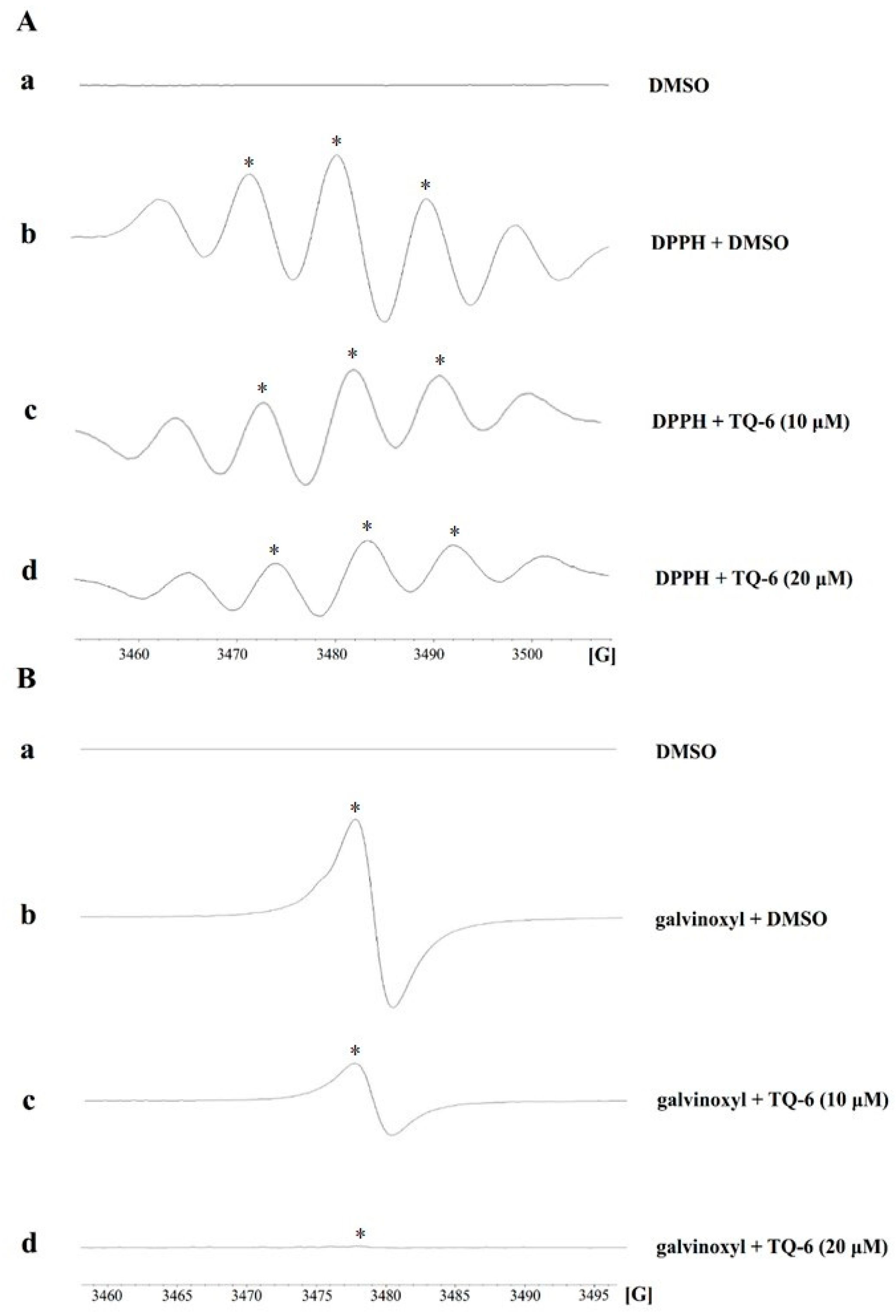
Figure 1. Scavenging effect of TQ-6 on 1,1-diphenyl-2-picrylhydrazyl (DPPH) and galvinoxyl radicals. (A) The reaction mixture contained 1.0 mM DPPH in the presence or absence of TQ-6. Line a, DMSO (0.1%) without DPPH; Line b, dimethyl sulfoxide (DMSO); Line c, 10 μM TQ-6; and Line d, 20 μM TQ-6 with DPPH. (B) TQ-6 with different concentrations were mixed with 500 µM galvinoxyl. Line a, DMSO (0.1%) without galvinoxyl; Line b, DMSO; Line c, 10 μM TQ-6; and Line d, 20 μM TQ-6. Spectra are representative examples of four similar experiments. The horizontal axis means the magnetic field [G].
Applsci 11 01008 g001
Figure 2. Effect of TQ-6 on the xanthine/xanthine oxidase system-generated 5-Diethoxyphosphoryl-5-methyl-1-pyrroline N-oxide (DEPMPO)-superoxide spin adduct and Fenton reaction solution (n = 4). (A) The ESR settings were followed as described in Figure 1; however, KO2 was absent, and 36 μM xanthine and 32 mU/mL xanthine oxidase were present. Line a, DMSO (0.1%); Line b, DMSO; Line c, 10 μM TQ-6; and Line d, 20 μM TQ-6. (B) The reaction mixture contained 1 mM Fe2+, 1.0% H2O2, and 200 mM DMPO in the presence or absence of TQ-6. The horizontal axis means the magnetic field [G].
Applsci 11 01008 g002
Figure 3. TQ-6 inhibits OH formation in LPS-stimulated RAW264.7 cells. (A) Cells were treated with (a) 0.1% DMSO or (b) DMSO or TQ-6 at (c) 10 µM or (d) 20 µM and then LPS (1 µg/mL) added to trigger OH formation. An asterisk (*) indicates the formation of OH. (B) Signal intensity was expressed as mean ± SEM (n = 4), which analyzed by using WIN-EPR, version 921201. *** p < 0.001, compared with the control group; ### p < 0.001, compared with the LPS group. Spectra are representative examples of four similar experiments. ctl: control (0.1% DMSO). The horizontal axis means the magnetic field [G].
Applsci 11 01008 g003
Figure 4. Effect of TQ-6 on Nrf2/HO-1 in LPS-stimulated RAW 264.7 cells. In TQ-6-treated cells followed with LPS exposure, the mRNA expression levels of (A) Nrf2 and (B) HO-1 were evaluated by RT-qPCR. The protein expression of (C) Nrf2 and (D) HO-1 was determined by immunoblotting. (E,F) Cells were treated with TQ-6 (10 and 20 μM) alone for 20 min, (E) Nrf2 and (F) HO-1 protein expression were determined by immunoblotting. Results are given as the means ± SEM (n = 4) and measured by the Biolight Windows Application, V2000.01 (Bio-Profil, Vilber Lourmat, France). * p < 0.05 and *** p < 0.001 compared with the LPS group.
Applsci 11 01008 g004
Figure 5. Effects of TQ-6 on carrageenan-induced rat paw edema. The right paw of Wistar rats was injected with TQ-6, 30 min before the carrageenan injection. The paw volume was measured before 0 h and after the intervals of 1, 2, 4, 6, and 24 h in carrageenan injection (○: vehicle; △: carrageenan; □: 5 mg/kg indomethacin; ◇: 1 mg/kg TQ-6; ▽: 2 mg/kg TQ-6). Data are shown as mean ± SEM (n = 6). * p < 0.05 compared with the carrageenan group.
Applsci 11 01008 g005
Table 1. Primer sequences.
GenePrimer SequenceAccession No.
Nrf2Forward 5′-AGC AGG ACA TGG AGC AAG TT-3′
Reverse 5′-TTC TTT TTC CAG CGA GGA GA-3
NM_010902.4
HO-1Forward 5′-GCA CTA TGT AAA GCG TCT CC-3′
Reverse 5′-GAC TCT GGT CTT TGT GTT CC-3’
NM_010442.2
GAPDHForward 5′-GAA CAT CAT CCC TGC ATC CA-3′
Reverse 5′-GCC AGT GAG CTT CCC GTT CA-3′
NM_001289726.1
Publisher’s Note: MDPI stays neutral with regard to jurisdictional claims in published maps and institutional affiliations.