Review

Developments in Smart Multi-Function Gait Assistive Devices for the Prevention and Treatment of Knee Osteoarthritis—A Literature Review

Institute for Health and Sport (IHES), Victoria University, Melbourne 3011, Australia
*
Author to whom correspondence should be addressed.
Appl. Sci. 2021, 11(22), 10947; https://doi.org/10.3390/app112210947
Submission received: 27 October 2021 / Revised: 17 November 2021 / Accepted: 17 November 2021 / Published: 19 November 2021
(This article belongs to the Special Issue Biomechanical and Biomedical Factors of Knee Osteoarthritis)

Abstract

:

Featured Application

Knee osteoarthritis (OA) can be prevented using orthoses with actuators controlled using real-time biofeedback from wearable motion-sensors and multi-function exoskeletons and, incorporating integrated electro-magnetic stimulators may also enhance regenerative medicine treatments for knee OA.

Abstract

Knee osteoarthritis (OA) is a degenerative condition that critically affects locomotor ability and quality of life and, the condition is particularly prevalent in the senior population. The current review presents a gait biomechanics conceptual framework for designing active knee orthoses to prevent and remediate knee OA. Constant excessive loading diminishes knee joint articular cartilage and, therefore, measures to reduce kinetic stresses due to the fact of adduction moments and joint compression are an essential target for OA prevention. A powered orthosis enables torque generation to support knee joint motions and machine-learning-driven “smart systems” can optimise the magnitude and timing of joint actuator forces. Although further research is required, recent findings raise the possibility of exoskeleton-supported, non-surgical OA interventions, increasing the treatment options for this prevalent, painful and seriously debilitating disease. Combined with advances in regenerative medicine, such as stem cell implantation and manipulation of messenger ribonucleic acid (m-RNA) transcription, active knee orthoses can be designed to incorporate electro-magnetic stimulators to promote articular cartilage resynthesis.

1. Introduction

Knee osteoarthritis (OA) is a progressive, degenerative condition that critically affects quality of life with a prevalence of 22.9% in the population above 40 years [1]. The biomechanical causes of OA are associated with thinning of articular cartilage and associated micro-fractures due to the fact of bone-on-bone contact [2]. Excessive knee joint loading following foot contact can lead to friction between the femur and tibia, suggesting that reducing knee joint compression is a fundamental intervention for OA [3]. The direction for OA prevention is, therefore, established and practical applications to manage knee joint kinetics (i.e., forces, moments) should now be the focus of our assistive technology research and development.
Wearable devices are a promising approach to treating knee OA, because biomechanical impacts on the knee are primarily associated with everyday locomotion. Wearable assistive devices (e.g., orthotics, exoskeletons) have the potential to support fundamental actions, such as walking, by correcting joint motion, absorbing excessive impact forces and reinforcing muscular work, all of which contribute to reducing OA risk [4,5,6,7]. Practical wearable devices include knee orthoses and shoe insoles [8,9]; there are a range of ergonomic orthotic designs on the market, but some may not be based on sound biomechanical principles. The first major section of the current review summarises knee joint biomechanics and the requirements for an effective assistive device capable of protecting the knee.
In addition to established passive, supportive assistive devices, more sophisticated powered systems utilising motor-driven actuators can also relieve knee joint loading by applying a supplementary rotational torque [10,11,12]. In addition to the magnitude of joint torque provided by an actuator, optimal torque application timing within the gait cycle is essential for effective knee joint support. The next generation of powered orthoses is likely to be enhanced using artificial intelligence (AI) software linked to signals from wearable sensors. Powered actuator devices will become smart systems, allowing individual customisation, continuous augmented feedback and sensor data accumulation to feed big data systems for clinical gait assessment and long-term motor control evaluation [13]. In addition to motor-driven joint actuation, using wearable sensors with AI features can be incorporated into active knee orthoses to monitor knee joint motion and provide biofeedback, but these features are in the developmental phase and not incorporated into market-ready products. Conceptual designs for active knee orthoses are, therefore, discussed in the current review supplemented by proposals for future research and development.
Knee replacement surgery is considered one of the few effective treatments for advanced symptomatic OA [14]. Progress in regenerative medicine, however, presents the opportunity for OA remediation via resynthesis of articular cartilage, using stem cell implantation and messenger ribonucleic acid (mRNA) transcriptions [15]. These approaches are promising in artificially cultivating new chondrocytes and promoting anabolic cellular conditions while regulating catabolic reactions. Electrical and magnetic stimuli can also trigger articular cartilage resynthesis, and this function is already provided via a commercially available wearable device [16]. Although few active knee orthoses are equipped to provide specific anti-arthritic stimuli, these mechanisms could be incorporated into the next generation of knee orthoses.
In this review, we first summarise the biomechanical characteristics of joint motion associated with increased OA risk and suggest how joint mechanics can be modified to help in the prevention or relief of OA symptoms. The following section introduces ergonomic designs for passive wearable assistive devices that can control OA risk. To flesh out our overview of AI-driven smart assistive devices, wearable sensors and their application for knee OA management are discussed. The review then progresses to consider more sophisticated, motor-driven, powered orthoses, designed to compensate and assist muscular work to reduce knee OA. Remedial aspects of knee OA are reviewed, including articular cartilage resynthesis and, from a joint biomechanics perspective, we consider the possibility of incorporating knee orthosis functions to complement tissue resynthesis. The review concludes by summarising the advantages of smart systems in managing online cloud data for population-based orthopaedic health promotion.

2. Biomechanical Factors for Knee Osteoarthritis

OA grades (I–IV) can be assigned depending on the extent of knee joint deformity but thinning of articular cartilage impairs protection of both the femoral and tibial heads, causing more direct collision, joint deformity (i.e., varus, valgus) and painful micro-fractures [2]. For OA prevention, biomechanical risks should be first quantified so that interventions can target minimising them. As described below, the biomechanical parameters of interest include knee adduction moment, knee compression force, knee power absorption and impact transferred to the knee joint.

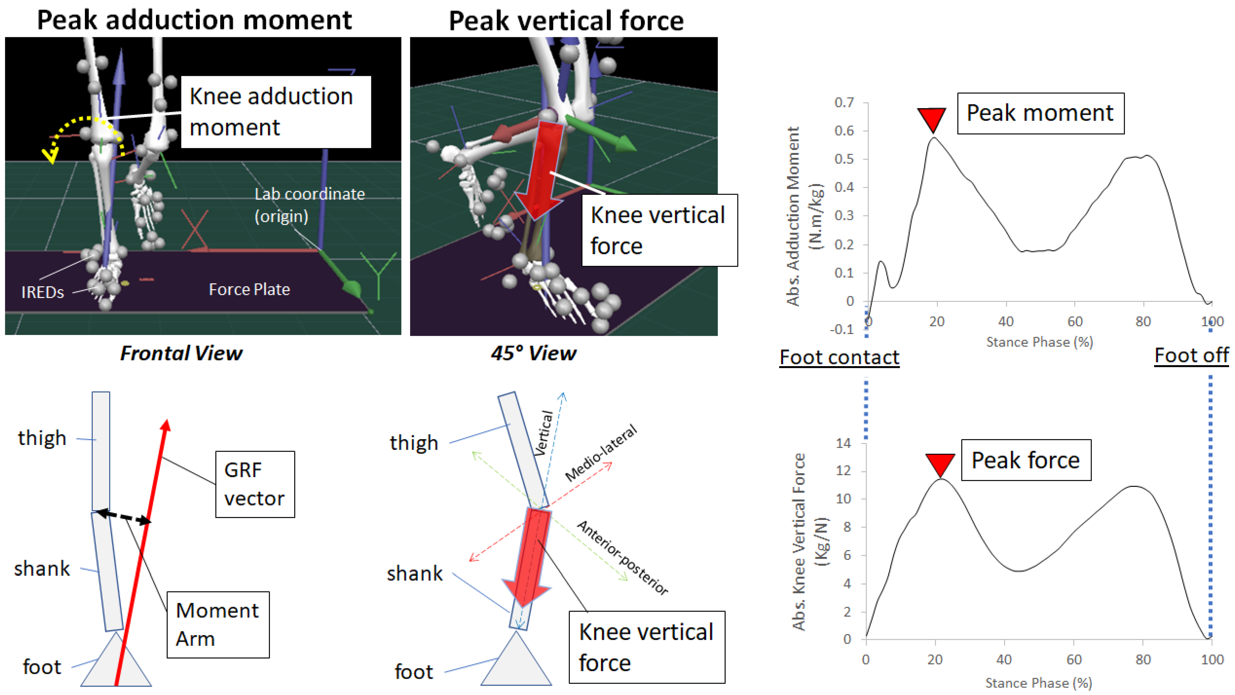
2.1. Knee Adduction Moment

Knee adduction moment is the most well-recognised biomechanical marker of knee OA, causing increased pressure on the medial compartment of the knee leading to varus deformity [17]. As seen in the top left of Figure 1, the knee adduction moment rotates the knee, and the illustration in the bottom left shows the relationship between ground reaction forces (GRFs) and moment arm, where:
Knee Adduction Moment = Moment Arm × GRF
As shown in the top right panel of Figure 1, the knee adduction moment has two peaks corresponding to the foot–ground reaction forces exerted at foot contact and push-off [18,19]. The adduction moment, described using negative values, causes tibial and femoral abduction but body weight and limb length are also determinants of joint moments and, various normalisation techniques have been proposed to account for the subject’s anthropometric contribution to adduction moments. There are, however, cautions concerning the risk of normalisation techniques washing out important information [20].

2.2. Knee Compression Forces

Of the three force components (i.e., medio-lateral, anterior–posterior and vertical), vertical peak compression force is most important, accounting for more than 80% of the total in walking [21,22]. Figure 1 shows that the maximum joint compression force is aligned with the tibia and approximates the timing of peak adduction moment [23,24], indicating that total compression force within the joint contributes significantly to the adduction moment. Compression forces are, therefore, the critical kinetic parameters associated with knee OA due to the pressures exerted on articular cartilage and their role in determining the adduction moment [24].
By recording acceleration and normalising for body mass (i.e., acceleration = force/mass), wearable sensors incorporating inertial measurement units (IMUs) can be used to estimate external forces on the knee following foot contact. The most straightforward normalisation is dividing force components by body mass, leaving acceleration as the force indicator. But it is important to note that body mass is a major contributor to joint force and associated knee OA. While sophisticated filtering techniques and high-grade IMUs enable 3D measurement, acceleration generally reflects overall “normalised force” in walking.

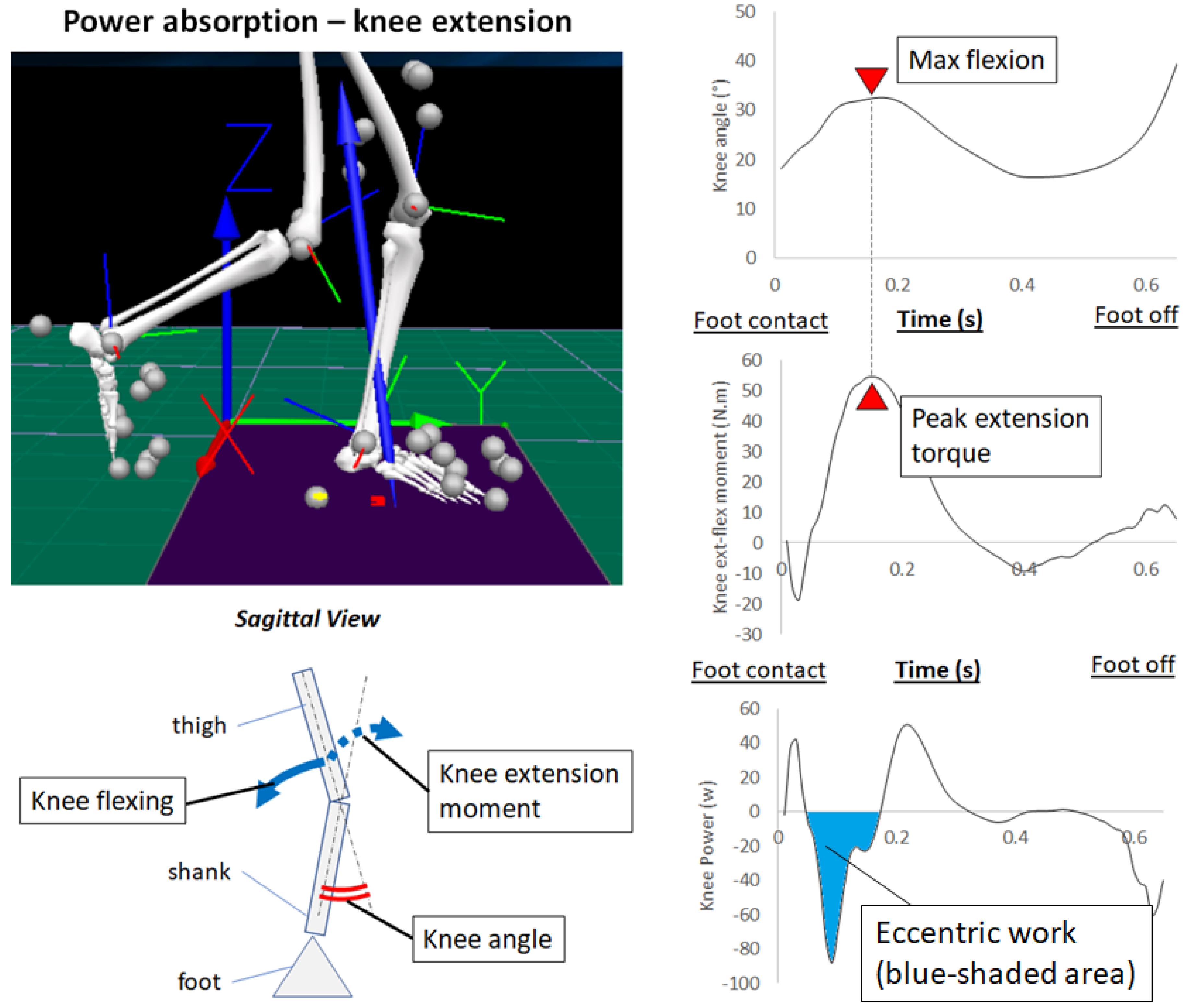
2.3. Power Absorption

As shown in Figure 2, the knee begins to flex after foot contact, and impact forces can be absorbed to reduce knee joint stresses [4,5]. During this phase of the walking cycle, eccentric muscle work is associated with power absorption, indicated by the integral of negative power in the power–time domain (blue-shaded area of the bottom-right graph) [25]. The quadriceps muscles are responsible for power absorption at foot contact performing eccentric work to absorb foot–ground impacts [4,5,26,27]. Following foot contact, the quadriceps then serve as the primary knee extensors at push off via positive work. Given these biomechanical requirements, a knee orthosis should be designed to support the quadriceps’ eccentric work, both passive and active.
Similarly, shank muscles acting as dorsiflexors (i.e., tibialis anterior) or plantar flexors (i.e., soleus, gastrocnemius) absorb power from foot contact until foot flat. Depending on the foot contact forces due to the heel or toe landing, eccentric work can be performed by either the dorsiflexors or plantar flexors [26,27,28]. In normal walking, a well-defined heel contact phase is preferred for optimal force absorption via eccentric work of the tibialis anterior while toe-landing accompanies plantar flexor work by the soleus and gastrocnemius, reported to effectively absorb foot contact impact, particularly in running [29].

2.4. Foot Contact Impact

Shock transferred to the knee can also be estimated by observing acceleration patterns due to the vibration of the motion capture system’s anatomically located markers such as passive reflective markers or active infrared emitting diodes [30,31]. As discussed further in a later section, wearable devices, such as IMUs, can also measure high acceleration events reflecting shock–impact vibration [32]. In terms of OA prevention, any intervention to reduce foot contact vibration in the knee joint is helpful. The following section provides a more extensive review of ergonomic features of passive wearable devices, such as knee braces and shoe insoles, that may assist in controlling OA.

3. Passive Mechanics of Wearable Devices for Knee OA Prevention
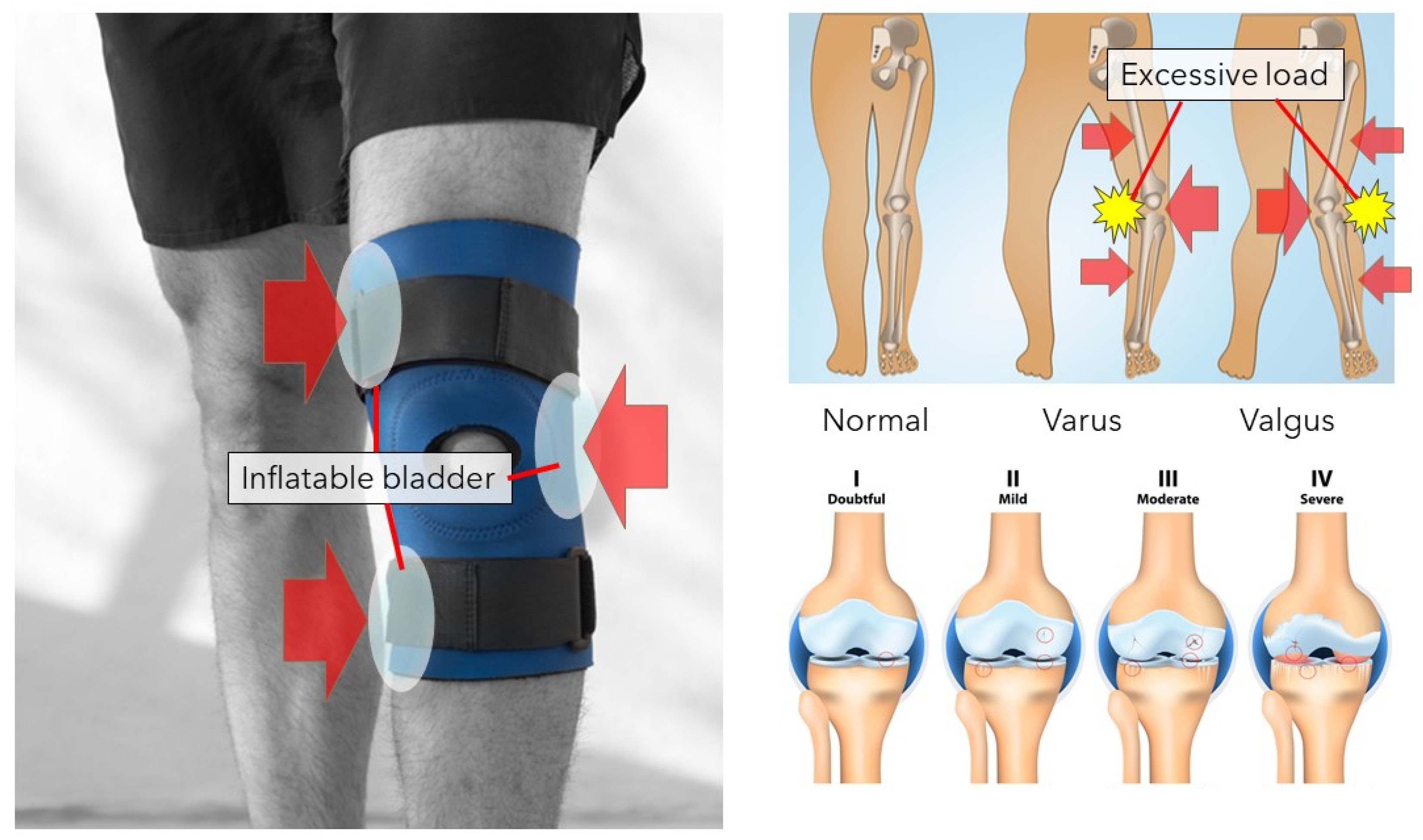
3.1. Knee Orthosis

Various knee orthoses are commercially available, but many of them only provide joint stabilisation by strapping and compression, which can, nevertheless, be useful for injury prevention and pain management [33,34]. More specialised products incorporate abduction–adduction control, for example, by using inflatable bladders to regulate knee motion [35]. Figure 3 illustrates an inflatable bladder application (left) for both varus and valgus OA (top right) and, adjustable pads and condylar pads are also commonly used to control abduction–adduction moments [36]. In general, these knee braces are effective in reducing knee adduction moment and easing OA symptoms, but Khosravi et al. [35] also reported that long-term use of a bulky knee orthosis could cause discomfort such as skin irritation.

3.2. Shoe Insole

In addition to measures designed to apply a direct intervention to the knee joint, shoe insoles are used to control knee biomechanics by correcting ankle–tibia–knee alignment. The most widely recognised is the lateral wedge insole, which everts the ankle to shorten the moment arm between the knee and resultant GRF vector as illustrated in Figure 1 [37]. A more laterally everted ankle aligns the tibia to shorten the moment arm and reduce associated knee adduction moment [38], but some studies have not observed such effects [39], suggesting that insole construction and design features, such as rigidity and dimensions, may be critical determinants of ankle joint support. It is, however, possible that reduced knee adduction moment can help in preventing OA but may not alleviate the symptoms [40].
While addition of a lateral wedge to evert the ankle and reduce the moment arm (see Figure 1) can be a useful feature, a further insole function can be to reduce GRFs, which are also an essential component of the knee adduction moment. In understanding the fundamental biomechanics of force-absorbing insole features, the following equation applies:
Force = (Mass × Velocity) / Time
Increased force application time is key to reducing GRFs using insoles and, heel cushioning materials can prolong compression time [41]. The sites for cushioning materials are, however, critical because energy absorption can cause adverse effects unless appropriately provided [42,43]. A further force-absorbing measure is to increase dorsiflexion support by prolonging the time between foot contact and foot flat [44,45]. Knee orthoses and shoe insoles are widely available, but market-available products may not be equipped with specific functions to effectively reduce OA risk factors as detailed in Section 2.2.
In addition to structural designs, the current review proposes potential attachment of wearable sensors and powered motor actuators to evolve from passive to active “smart” knee protection devices. The following section discusses how OA-related gait parameters can be monitored by incorporating wearable sensors into assistive devices.

4. Incorporation of Wearable Sensors to Measure OA Risks

Biomechanical factors associated with OA risks were described earlier in Section 2, but the technological challenge is devising methods to constantly monitor gait data in everyday settings. Traditionally, gait data have been obtained only in the laboratory, but recent advances in machine learning are making it possible to estimate real-world kinetic and kinematic motion characteristics using data from wearable systems [19,46,47,48]. In this section, concepts for gait monitoring and risk assessment utilising wearable systems are discussed.

4.1. Laboratory-Based 3D Motion Capture Systems

The gold standard for biomechanical assessments are highly reliable data obtained from marker-based 3D motion capture systems such as Optotrak, Vicon and Optitrak. More practical but less precise markerless systems, such as Kinect Azure, are available, which can provide acceptable accuracy for some applications [49]. All these motion capture systems are designed to sample raw position–time data from which information, such as knee joint angles, velocity and acceleration, can be computed from noise-filtered coordinate data. When this kinematic information is used in combination with 3D foot–ground reaction forces, the computational technique “inverse dynamics” allows for estimation of joint kinetics to compute adduction moment and compression forces [19,50]. It is difficult to obtain sufficient kinetic data, because in the usual biomechanical laboratory setup, each walkthrough trial can provide only limited GRF data depending on the number of force plates installed. Data volume can be considerably increased, using continuous sampling when treadmill walking, but there is debate as to whether it represents natural, everyday locomotion [51]. While 3D motion capture and force plates are the laboratory standard, wearable systems will be needed to assess OA effects outside the laboratory. Although the types of data obtained by wearable systems are limited (joint angle, acceleration, etc.), in principle, machine learning algorithms can be further developed to estimate knee OA-related mechanics with sufficient reliability [13,48]. Below we consider wearable systems, and the data obtainable from them with application to knee OA.

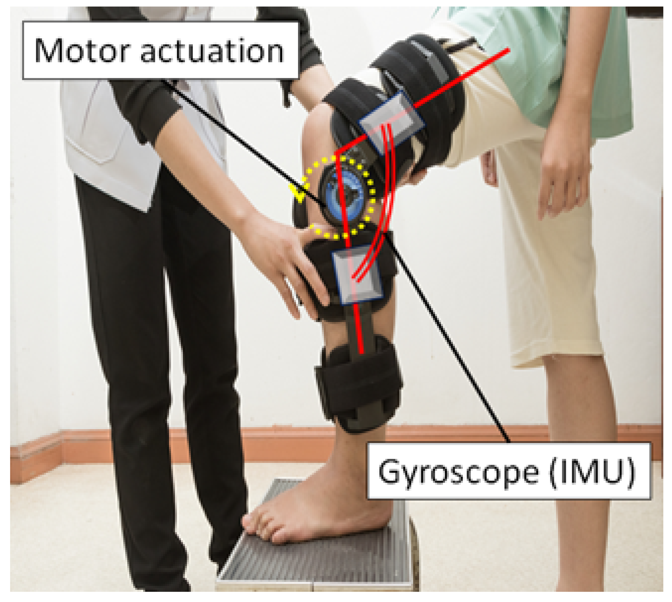
4.2. Inertial Measurement Units (IMUs)

IMUs are one of the most widely recognised, practical and affordable wearable sensors that, depending on specifications, can record acceleration and joint angles [52,53]. The simplest application is the direct attachment of IMUs to body segments, such as the shank and knee, to record vibration transferred from the foot [54]. This concept has been reported by Gruber et al. [32] in which a progressive reduction in vibration between the shank and head were considered to reflect the amount of the shock absorbed by the body.
Measurements of lower limb joint kinematics have also been used to predict highly associated kinetic information. Knee abduction–adduction angle can be indicative of adduction moment, and maximum knee flexion is related to power absorption as described in Section 2.3. Joint angles coupled with acceleration data can be used to estimate kinetic values at particular events and, as Table 1 describes, various parameters can be considered for risk assessment. As technology stands, IMUs are, therefore, the essential wearable sensors to enable an active exoskeleton system to predict knee OA risks [55].

4.3. Force Transducer

Force transducers attached to a knee orthosis are a viable option for recording forces and torques acting on the joint. Lee et al. [56] attached force transducers bilaterally on the knee. Force transducers can reveal not only the external kinetic effects on the knee joint but also, by computing the ratio between the lateral and medial, suggest which side of the knee is more susceptible to deformity (i.e., varus or valgus). External attachment is the great advantage of force transducers and, similarly, pressure sensors located directly over the device flexion site can also indicate the risk of deformity. Although continuous, automatic, calibration is required for practical use, these types of direct kinetic data collection methods can reflect OA risks.

4.4. Electromyography (EMG)

One of the most compelling developments in EMG applications for motor control dysfunction is feedforward sensing of neurotransmitters, allowing intended muscle actions and associated limb movements to be predicted. Neurotransmitters have already been used to control exoskeleton actuators [57,58] and incorporation into an active knee orthosis for OA is a promising research direction. Intention to contract muscles generates an action potential (e.g., 30–40 millivolts) [59] that can be recorded using a non-invasive system of surface electrodes attached to the muscle belly (surface-EMG). Analysis of amplified EMG signals can show which muscles are active in producing the targeted movement [60]. Despite controversy surrounding EMG noise-reduction filtering techniques, on/off is the most reliable EMG information, because the intensity of muscle contraction is difficult to assess without specialised techniques such as relative amplitude methods [61]. Wearable EMG systems, designed primarily for research, are commercially available [62], but future developments in gait monitoring systems for knee OA may be able to incorporate wearable EMG devices to record activation patterns associated with power absorption by the quadriceps, dorsiflexors and plantar flexors as described in Table 1 [63].

4.5. Foot Pressure Measurement Insole

Foot–ground impact recordings from pressure sensitive insoles (e.g., Pedar, F-scan) can be used to predict gait-related injury risks associated with foot deformities (e.g., hallux valgus, hammer toe, craw toe), inversion ankle sprains and knee joint conditions [64]. A battery pack is required for continuous data acquisition [65], but footwear-integrated insoles have the potential to estimate foot–ground contact pressures and modulate joint stresses by augmenting an active knee orthosis control system.
Wearable sensors can, therefore, supply real-time gait-related data that can be useful for OA risk assessment and monitoring. Sensor data can be stored in the cloud and made available to clinicians or exercise prescribers for more effective personalised interventions. In addition to data management applications, wearable sensor signals can be used to refine active orthosis actuation to enable protective mechanisms by setting torque motor thresholds (e.g., joint angle, acceleration, force, pressure) as considered in the next section.

5. Exoskeletons to Minimise Risk Factors

The primary function of active exoskeletons is augmenting joint actuation using torque motors to compensate for muscle weakness and to regulate excessive joint motions [9,10]. As illustrated in Table 1, increasing knee eccentric work (i.e., extension moment) when approaching maximum knee flexion and reducing peak adduction moments are practical goals to minimise damage to the knee. For active systems to support these joint actions, ideally both frontal and sagittal plane motions should be incorporated for adduction–abduction and extension–flexion, respectively. It is, therefore, important to detect the correct timing and optimum intensity of actuation to provide adequate support. Figure 4 (right) shows motor actuation in an active knee orthosis and, in this case, angular joint kinematics determine the activation threshold for the motor-driven knee extension torque. While activation mechanisms, such as pneumatic artificial muscles and spring-like systems, can also be incorporated, motor actuation is most widely used [66].
Peak adduction moment is generally identified slightly before contralateral foot-off, where adduction angle coincides with the local peak moment. For power absorption, knee extension torque is demanded until maximum knee flexion (Figure 2), a gait cycle event that can also be identified using IMUs. IMUs alone are, therefore, capable of identifying these key events, but multiple wearable systems can be combined to polish the machine learning algorithms to predict event timings more accurately by employing an extended data volume. In addition to optimising torque application timing, the magnitude of support provided by the powered motor systems should also be considered. Many wearable systems, including exoskeletons and therapeutic devices, can be user-adjusted to change the “intensity” [11], but if automation is required to determine the adequate support level, normative data from healthy individuals can be used as a reference [67,68,69]. Kinetic parameters can be user-normalised, for example, by body mass as in knee kinetics research [70,71,72].
In summary, an active knee orthosis can provide additional joint torque in the sagittal and frontal planes to assist the lower limb’s kinetic functions (Figure 4). Wearable systems, such as IMUs, can provide the support timing, which can be individualised using machine-learning-driven controllers. In addition to gait event identification, such as peak knee flexion and adduction angles, it is also necessary to determine safety thresholds for each joint motion.

6. Remedial Aspects of a Knee Orthosis

A primary focus of knee joint OA prevention is reducing mechanical stresses by regulating excessive knee kinetics and promoting power absorption. In recent years, non-surgical remediation using articular cartilage resynthesis is, however, becoming more practicable. Knee OA is usually characterised as a progressive condition, implying that the knee structures, including articular cartilages and surrounding soft tissues, are in a constant catabolic state. Unless this degenerating environment is changed, the condition can deteriorate and until recent advances in regenerative medicine, surgery or temporary relief were considered the only treatment options. For practical interventions using an active knee orthosis, the current section discusses whether electrical and magnetic stimulation can improve knee OA symptoms.
From the perspective of regenerative medicine, cartilage resynthesis is key to remediating knee OA. Stem cell intervention has advanced significantly following the discovery of induced pluripotent stem (iPS) cells, and knee articular cartilage resynthesis is becoming a viable treatment option [73]. When applied in conjunction with wearable exoskeletons, electrical and magnetic stimulation can be included to trigger cartilage regeneration, and both types of stimuli can be provided by portable devices widely available on the market [74]. Electrical and magnetic stimulation to chondrocytes has the potential to alter messenger ribonucleic acid (mRNA) expression and possibly increase cartilage compounds, including type II collagen, aggrecan, glycosaminoglycans and proteoglycans [73,74,75,76,77,78], revitalising the anabolic environment within the knee. At the same time, excessive secretion of catabolic cytokines, such as interleukin 1β, should be prevented to inhibit OA progression and successful electromagnetic stimulation effects have been reported in mouse models [75,76].
Electrical stimulation of the knee is possible via surface electrodes using electrical muscle stimulation (EMS), transcutaneous electrical nerve stimulation (TENS) and pulsed electrical stimulation (PES). Recent studies generally agree on the positive effects of electrical stimulation in relieving OA symptoms [15,16,77]. Most importantly, pain and joint dysfunction showed improvement as assessed by the visual analogue scale (VAS) or Western Ontario and McMaster Universities osteoarthritis index (WOMAC) [16]. To incorporate electrical therapeutic effects into a wearable knee orthosis, it is important to determine optimal stimulation parameters including rhythm, intensity, frequency, and duration. Bardoloi et al. [77], for example, proposed 0.2 milliseconds of acupuncture-like pulsing by TENS at 2 Hz for 20–60 min, twice daily. This is classified as “high intensity–low frequency” stimulation, but higher frequency (20–50 Hz) at lower intensity was also suggested. Despite the further research efforts required to determine the most effective stimulation regime, these treatment options appear to be promising components of new generation multi-function exoskeletons.
Similarly, magnetic fields may prevent or reverse the progression of OA by triggering anti-inflammatory effects and tissue regrowth [15]. Pulsed electromagnetic fields may regulate catabolic reactions by inhibiting interleukin 1β, the primary cytokines that break down the extracellular matrix and cause inflammation while stimulating anabolic reactions by generating type II collagen, aggrecan, glycosaminoglycans and proteoglycans [15,76,77,78]. As a result, proliferation of chondrocytes and associated cartilage thickening can be expected [78]. Implementation of electrical or magnetic systems into active exoskeletons is, therefore, a practical possibility at relatively low cost, but one concern are the electromagnetic effects on the device from external electronics. Computers are sensitive to electromagnetic waves and prone to malfunction, for real-world application, therefore, distancing the control unit or electromagnetic shielding materials may be required.

7. Conclusions

Knee osteoarthritis is a serious degenerative condition that restricts active walking, but a cure has been considered difficult. Taking advantage of rapid progress in wearable sensors, an active knee orthosis can be developed to counteract OA factors by providing precisely graded motor-driven torques at key gait cycle events with machine learning control algorithms used for individual customisation. In considering the remedial potential of orthoses, electrical and magnetic chondrocyte stimulation may be an effective OA treatment when incorporated into a multi-function knee orthosis. Further research is, however, required to confirm the remedial effects of these technologies and the potential for electromagnetic interference must be considered.

Author Contributions

H.N. was responsible for conceptualisation, resources, data curation and writing—original draft preparation; W.S. was responsible for writing—review and editing; R.B. was responsible for overall supervision. All authors have read and agreed to the published version of the manuscript.

Funding

This research received no external funding.

Conflicts of Interest

The authors declare no conflict of interest.

References

  1. Cui, A.; Li, H.; Wang, D.; Zhong, J.; Chen, Y.; Lu, H. Global, regional prevalence, incidence and risk factors of knee osteoar-thritis in population-based studies. EClinicalMedicine 2020, 29, 100587. [Google Scholar] [CrossRef] [PubMed]
  2. Cicuttini, F.M.; Wluka, A.E.; Stuckey, S.L. Tibial and femoral cartilage changes in knee osteoarthritis. Ann. Rheum. Dis. 2001, 60, 977–980. [Google Scholar] [CrossRef] [PubMed] [Green Version]
  3. Quatman, C.E.; Harris, J.D.; Hewett, T.E. Biomechanical outcomes of cartilage repair of the knee. J. Knee Surg. 2012, 25, 197–206. [Google Scholar] [CrossRef] [PubMed] [Green Version]
  4. Nagano, H.; Tatsumi, I.; Sarashina, E.; Sparrow, W.; Begg, R.K. Modelling knee flexion effects on joint power absorption and adduction moment. Knee 2015, 22, 490–493. [Google Scholar] [CrossRef] [PubMed]
  5. Nagano, H.; Levinger, P.; Downie, C.; Hayes, A.; Begg, R. Contribution of lower limb eccentric work and different step re-sponses to balance recovery among older adults. Gait Posture 2015, 42, 257–262. [Google Scholar] [CrossRef]
  6. McGibbon, C.; Sexton, A.; Jayaraman, A.; Deems-Dluhy, S.; Fabara, E.; Adans-Dester, C.; Bonato, P.; Marquis, F.; Turmel, S.; Belzile, E. Evaluation of a lower-extremity robotic exoskeleton for people with knee osteoarthritis. Assist. Technol. 2021, 1–14. [Google Scholar] [CrossRef] [PubMed]
  7. McGibbon, C.A.; Brandon, S.; Bishop, E.L.; Cowper-Smith, C.; Biden, E.N. Biomechanical Study of a Tricompartmental Unloader Brace for Patellofemoral or Multicompartment Knee Osteoarthritis. Front. Bioeng. Biotechnol. 2021, 8, 1528. [Google Scholar] [CrossRef] [PubMed]
  8. Nagano, H.; Begg, R. A shoe-insole to improve ankle joint mechanics for injury prevention among older adults. Ergonomics 2021, 64, 1271–1280. [Google Scholar] [CrossRef]
  9. Ezzibdeh, R.; Arora, P.; Amanatullah, D.F. Utilization of a pneumatic exoskeleton after total knee arthroplasty. Arthroplast. Today 2019, 5, 314–315. [Google Scholar] [CrossRef] [Green Version]
  10. Pamungkas, D.S.; Caesarendra, W.; Soebakti, H.; Analia, R.; Susanto, S. Overview: Types of Lower Limb Exoskeletons. Electronics 2019, 8, 1283. [Google Scholar] [CrossRef] [Green Version]
  11. Wang, Z.; Wu, X.; Zhang, Y.; Chen, C.; Liu, S.; Liu, Y.; Peng, A.; Ma, Y. A Semi-active Exoskeleton Based on EMGs Reduces Muscle Fatigue When Squatting. Front. Neurorobot. 2021, 15, 625479. [Google Scholar] [CrossRef] [PubMed]
  12. Vantilt, J.; Tanghe, K.; Afschrift, M.; Bruijnes, A.K.; Junius, K.; Geeroms, J.; Aertbeliën, E.; De Groote, F.; Lefeber, D.; Jonkers, I.; et al. Model-based control for exoskeletons with series elastic actuators evaluated on sit-to-stand movements. J. Neuroeng. Rehabil. 2019, 16, 65. [Google Scholar] [CrossRef]
  13. Stetter, B.J.; Krafft, F.C.; Ringhof, S.; Stein, T.; Sell, S. A Machine Learning and Wearable Sensor Based Approach to Estimate External Knee Flexion and Adduction Moments During Various Locomotion Tasks. Front. Bioeng. Biotechnol. 2020, 8, 9. [Google Scholar] [CrossRef]
  14. Rönn, K.; Reischl, N.; Gautier, E.; Jacobi, M. Current Surgical Treatment of Knee Osteoarthritis. Arthritis 2011, 2011, 454873. [Google Scholar] [CrossRef] [PubMed] [Green Version]
  15. Aini, H.; Itaka, K.; Fujisawa, A.; Uchida, H.; Uchida, S.; Fukushima, S.; Kataoka, K.; Saito, T.; Chung, U.-I.; Ohba, S. Messenger RNA delivery of a cartilage-anabolic transcription factor as a disease-modifying strategy for osteoarthritis treatment. Sci. Rep. 2016, 6, 18743. [Google Scholar] [CrossRef] [Green Version]
  16. Kim, E.-D.; Won, Y.H.; Park, S.-H.; Seo, J.-H.; Kim, D.-S.; Ko, M.-H.; Kim, G.-W. Efficacy and Safety of a Stimulator Using Low-Intensity Pulsed Ultrasound Combined with Transcutaneous Electrical Nerve Stimulation in Patients with Painful Knee Osteoarthritis. Pain Res. Manag. 2019, 2019, 7964897. [Google Scholar] [CrossRef] [PubMed]
  17. Heidari, B. Knee osteoarthritis diagnosis, treatment and associated factors of progression: Part II. Casp. J. Intern. Med. 2011, 2, 249–255. [Google Scholar]
  18. Kutzner, I.; Trepczynski, A.; Heller, M.O.; Bergmann, G. Knee adduction moment and medial contact force—Facts about their correlation during gait. PLoS ONE 2013, 8, e81036. [Google Scholar] [CrossRef]
  19. Nagano, H.; Begg, R. Ageing-related gait adaptations to knee joint kinetics: Implications for the development of knee oste-oarthritis. Appl. Sci. 2020, 10, 8881. [Google Scholar] [CrossRef]
  20. Robbins, S.M.K.; Birmingham, T.B.; Maly, M.R.; Chesworth, B.M.; Giffin, J.R. Comparative diagnostic accuracy of knee ad-duction moments in knee osteoarthritis: A case for not normalizing to body size. J. Biomech. 2011, 44, 968–971. [Google Scholar] [CrossRef]
  21. Beschorner, K.E.; Albert, D.L.; Redfern, M.S. Required coefficient of friction during level walking is predictive of slipping. Gait Posture 2016, 48, 256–260. [Google Scholar] [CrossRef] [PubMed]
  22. Kram, R.; Griffin, T.M.; Donelan, J.M.; Chang, Y.H. Force treadmill for measuring vertical and horizontal ground reaction forces. J. Appl. Physiol. 1998, 85, 764–769. [Google Scholar] [CrossRef] [PubMed]
  23. Purevsuren, T.; Kim, K.; Nha, K.W.; Kim, Y.H. Evaluation of compressive and shear joint forces on medial and lateral com-partments in knee joint during walking before and after medial open-wedge high tibial osteotomy. Int. J. Precis. Eng. Manuf. 2016, 17, 1365–1370. [Google Scholar] [CrossRef]
  24. Richards, R.; Andersen, M.; Harlaar, J.; Noort, J.V.D. Relationship between knee joint contact forces and external knee joint moments in patients with medial knee osteoarthritis: Effects of gait modifications. Osteoarthr. Cartil. 2018, 26, 1203–1214. [Google Scholar] [CrossRef] [PubMed] [Green Version]
  25. Elmer, S.J.; Madigan, M.L.; LaStayo, P.C.; Martin, J.C. Joint-specific power absorption during eccentric cycling. Clin. Biomech. 2010, 25, 154–158. [Google Scholar] [CrossRef]
  26. Hernandez, H.J.; McIntosh, V.; Leland, A.; Harris-Love, M.O. Progressive Resistance Exercise with Eccentric Loading for the Management of Knee Osteoarthritis. Front. Med. 2015, 2, 45. [Google Scholar] [CrossRef] [Green Version]
  27. Kao, P.-C.; Ferris, D.P. Motor adaptation during dorsiflexion-assisted walking with a powered orthosis. Gait Posture 2009, 29, 230–236. [Google Scholar] [CrossRef] [Green Version]
  28. Meinders, M.; Gitter, A.; Czemiecki, J.M. The role of ankle plantar flexor muscle work during walking. Scand. J. Rehabil. Med. 1998, 30, 39–46. [Google Scholar] [CrossRef] [PubMed]
  29. Ahn, A.; Brayton, C.; Bhatia, T.; Martin, P. Muscle activity and kinematics of forefoot and rearfoot strike runners. J. Sport Health Sci. 2014, 3, 102–112. [Google Scholar] [CrossRef]
  30. Beerse, M.; Lelko, M.; Wu, J. Acute effect of whole-body vibration on acceleration transmission and jumping performance in children. Clin. Biomech. 2020, 81, 105235. [Google Scholar] [CrossRef] [PubMed]
  31. Chadefaux, D.; Gueguen, N.; Thouze, A.; Rao, G. 3D propagation of the shock-induced vibrations through the whole low-er-limb during running. J. Biomech. 2019, 96, 109343. [Google Scholar] [CrossRef] [PubMed] [Green Version]
  32. Gruber, A.H.; Boyer, K.A.; Derrick, T.; Hamill, J. Impact shock frequency components and attenuation in rearfoot and forefoot running. J. Sport Health Sci. 2014, 3, 113–121. [Google Scholar] [CrossRef]
  33. Vahdatpour, B.; Taheri, P.; Asl, M.M.; Ramezanian, H. Effects of Taping on Pain and Functional Outcome of Patients with Knee Osteoarthritis: A Pilot Randomized Single-blind Clinical Trial. Adv. Biomed. Res. 2017, 6, 139. [Google Scholar] [CrossRef] [PubMed]
  34. Schmitt, L.C.; Rudolph, K.S. Muscle stabilization strategies in people with medial knee osteoarthritis: The effect of instability. J. Orthop. Res. 2008, 26, 1180–1185. [Google Scholar] [CrossRef] [PubMed] [Green Version]
  35. Khosravi, M.; Arazpour, M.; Saeedi, H.; Rezaei, M. Design evaluation in novel orthoses for patients with medial knee oste-oarthritis. J. Biomed. Phys. Eng. 2019, 9, 719–732. [Google Scholar]
  36. Ramsey, D.K.; Russell, M.E. Unloader braces for medial compartment knee osteoarthritis: Implications on mediating progression. Sports Health 2009, 1, 416–425. [Google Scholar] [CrossRef] [Green Version]
  37. Ferreira, V.; Simões, R.; Gonçalves, R.S.; Machado, L.; Roriz, P. The optimal degree of lateral wedge insoles for reducing knee joint load: A systematic review and meta-analysis. Arch. Physiother. 2019, 9, 18. [Google Scholar] [CrossRef] [PubMed]
  38. Hinman, R.S.; Bowles, K.-A.; Metcalf, B.; Wrigley, T.; Bennell, K. Lateral wedge insoles for medial knee osteoarthritis: Effects on lower limb frontal plane biomechanics. Clin. Biomech. 2012, 27, 27–33. [Google Scholar] [CrossRef]
  39. Zhang, J.; Wang, Q.; Zhang, C. Ineffectiveness of lateral-wedge insoles on the improvement of pain and function for medial knee osteoarthritis: A meta-analysis of controlled randomized trials. Arch. Orthop. Trauma Surg. 2018, 138, 1453–1462. [Google Scholar] [CrossRef] [Green Version]
  40. Parkes, M.J.; Maricar, N.; Lunt, M.; Lavalley, M.P.; Jones, R.K.; Segal, N.A.; Takahashi-Narita, K.; Felson, D.T. Lateral Wedge Insoles as a Conservative Treatment for Pain in Patients with Medial Knee Osteoarthritis: A Meta-analysis. JAMA 2013, 310, 722–730. [Google Scholar] [CrossRef] [Green Version]
  41. Chiu, H.-T.; Shiang, T.-Y. Effects of Insoles and Additional Shock Absorption Foam on the Cushioning Properties of Sport Shoes. J. Appl. Biomech. 2007, 23, 119–127. [Google Scholar] [CrossRef] [PubMed] [Green Version]
  42. Kulmala, J.-P.; Kosonen, J.; Nurminen, J.; Avela, J. Running in highly cushioned shoes increases leg stiffness and amplifies impact loading. Sci. Rep. 2018, 8, 17496. [Google Scholar] [CrossRef] [Green Version]
  43. Maropoulos, S.; Korakidis, G.; Fasnakis, D.; Papanikolaou, S.; Papagiannaki, M.; Arabazti, F. The effect of cushioning system on impact attenuation of athletic footwear. MATEC Web Conf. 2017, 112, 8018. [Google Scholar] [CrossRef] [Green Version]
  44. Chan, C.W.; Rudins, A. Foot Biomechanics During Walking and Running. Mayo Clin. Proc. 1994, 69, 448–461. [Google Scholar] [CrossRef] [Green Version]
  45. Bolgla, L.A.; Malone, T.R. Plantar fasciitis and the windlass mechanism: A biomechanical link to clinical practice. J. Athl. Train. 2004, 39, 77–82. [Google Scholar] [PubMed]
  46. Adesida, Y.; Papi, E.; McGregor, A.H. Exploring the Role of Wearable Technology in Sport Kinematics and Kinetics: A Systematic Review. Sensors 2019, 19, 1597. [Google Scholar] [CrossRef] [PubMed] [Green Version]
  47. Lizzio, V.A.; Cross, A.G.; Guo, E.W.; Makhni, E.C. Using Wearable Technology to Evaluate the Kinetics and Kinematics of the Overhead Throwing Motion in Baseball Players. Arthrosc. Tech. 2020, 9, e1429–e1431. [Google Scholar] [CrossRef]
  48. De Brabandere, A.; Emmerzaal, J.; Timmermans, A.; Jonkers, I.; Vanwanseele, B.; Davis, J. A Machine Learning Approach to Estimate Hip and Knee Joint Loading Using a Mobile Phone-Embedded IMU. Front. Bioeng. Biotechnol. 2020, 8, 320. [Google Scholar] [CrossRef] [PubMed] [Green Version]
  49. Uhlár, Á.; Ambrus, M.; Kékesi, M.; Fodor, E.; Grand, L.; Szathmáry, G.; Rácz, K.; Lacza, Z. Kinect Azure-based accurate measurement of dynamic valgus of osteoarthritis. Appl. Sci. 2021, 11, 5536. [Google Scholar] [CrossRef]
  50. Faber, H.; Van Soest, A.J.; Kistemaker, D.A. Inverse dynamics of mechanical multibody systems: An improved algorithm that ensures consistency between kinematics and external forces. PLoS ONE 2018, 13, e0204575. [Google Scholar] [CrossRef] [PubMed]
  51. Riley, P.O.; Paolini, G.; Della Croce, U.; Paylo, K.W.; Kerrigan, D.C. A kinematic and kinetic comparison of overground and treadmill walking in healthy subjects. Gait Posture 2007, 26, 17–24. [Google Scholar] [CrossRef]
  52. Liu, S.; Zhang, J.; Zhang, Y.; Zhu, R. A wearable motion capture device able to detect dynamic motion of human limbs. Nat. Commun. 2020, 11, 5615. [Google Scholar] [CrossRef] [PubMed]
  53. Bonroy, B.; Meijer, K.; Dunias, P.; Cuppens, K.; Gransier, R.; Vanrumste, B. Ambulatory Monitoring of Physical Activity Based on Knee Flexion/Extension Measured by Inductive Sensor Technology. ISRN Biomed. Eng. 2013, 2013, 908452. [Google Scholar] [CrossRef] [Green Version]
  54. Eitzen, I.; Renberg, J.; Færevik, H. The Use of Wearable Sensor Technology to Detect Shock Impacts in Sports and Occupational Settings: A Scoping Review. Sensors 2021, 21, 4962. [Google Scholar] [CrossRef] [PubMed]
  55. Iwama, Y.; Harato, K.; Kobayashi, S.; Niki, Y.; Ogihara, N.; Matsumoto, M.; Nakamura, M.; Nagura, T. Estimation of the ex-ternal knee adduction moment during gait using an inertial measurement unit in patients with knee osteoarthritis. Sensors 2021, 21, 1418. [Google Scholar] [CrossRef] [PubMed]
  56. Lee, H.; Ha, D.; Kang, Y.-S.; Park, H.-S. Biomechanical Analysis of the Effects of Bilateral Hinged Knee Bracing. Front. Bioeng. Biotechnol. 2016, 4, 50. [Google Scholar] [CrossRef] [PubMed] [Green Version]
  57. Reaz, M.B.I.; Hussain, M.S.; Mohd-Yasin, F. Techniques of EMG signal analysis: Detection, processing, classification and applications. Biol. Proced. Online 2006, 8, 11–35. [Google Scholar] [CrossRef] [PubMed] [Green Version]
  58. Kastalskiy, I.; Mironov, V.; Lobov, S.; Krilova, N.; Pimashkin, A.; Kazantsev, V. A Neuromuscular Interface for Robotic Devices Control. Comput. Math. Methods Med. 2018, 2018, 8948145. [Google Scholar] [CrossRef]
  59. Lodish, H.; Berk, A.; Zipursky, S.L.; Matsudaira, P.; Baltimore, D.; Darnell, J. 2. The action potential and conduction of electric impulses. In Molecular Cell Biology, 4th ed.; W.H. Freeman: New York, NY, USA, 2000; p. 21. [Google Scholar]
  60. Mills, K.R. The basics of electromyography. J. Neurol. Neurosurg. Psychiatry 2005, 76, ii32–ii35. [Google Scholar] [CrossRef] [Green Version]
  61. Vigotsky, A.D.; Halperin, I.; Lehman, G.J.; Trajano, G.S.; Vieira, T.M. Interpreting signal amplitudes in surface electromyography studies in sport and rehabilitation sciences. Front. Physiol. 2018, 8, 985. [Google Scholar] [CrossRef] [Green Version]
  62. Zhao, S.; Liu, J.; Gong, Z.; Lei, Y.; Ouyang, X.; Chan, C.C.; Ruan, S. Wearable Physiological Monitoring System Based on Electrocardiography and Electromyography for Upper Limb Rehabilitation Training. Sensors 2020, 20, 4861. [Google Scholar] [CrossRef] [PubMed]
  63. Giminiani, R.D.; Cardinale, M.; Ferrari, M.; Quaresima, V. Validation of fabric-based thigh-wearable EMG sensors and oxi-metry for monitoring quadriceps activity during strength and endurance exercises. Sensors 2020, 20, 4664. [Google Scholar] [CrossRef] [PubMed]
  64. Hanatsu, N.H.; Begg, R.K. Shoe-Insole Technology for Injury Prevention in Walking. Sensors 2018, 18, 1468. [Google Scholar] [CrossRef] [Green Version]
  65. Lin, F.; Wang, A.; Zhuang, Y.; Tomita, M.R.; Xu, W. Smart insole: A wearable sensor device for unobstrusive gait monitoring in daily life. IEEE Trans. Ind. Inf. 2016, 12, 2281–2291. [Google Scholar] [CrossRef]
  66. Zhao, W.; Song, A. Active Motion Control of a Knee Exoskeleton Driven by Antagonistic Pneumatic Muscle Actuators. Actuators 2020, 9, 134. [Google Scholar] [CrossRef]
  67. Baron, R. Normative data for muscle strength in relation to age, knee angle and velocity. Wien. Med. Wochenschr. 1995, 145, 600–606. [Google Scholar]
  68. Jandacka, D.; Plesek, J.; Skypala, J.; Uchytil, J.; Silvernail, J.F.; Hamill, J. Knee Joint Kinematics and Kinetics During Walking and Running After Surgical Achilles Tendon Repair. Orthop. J. Sports Med. 2018, 6, 2325967118779862. [Google Scholar] [CrossRef] [Green Version]
  69. Bennett, H.J.; Fleenor, K.; Weinhandl, J.T. A normative database of hip and knee joint biomechanics during dynamic tasks using anatomical regression prediction methods. J. Biomech. 2018, 81, 122–131. [Google Scholar] [CrossRef] [PubMed]
  70. Wannop, J.; Worobets, J.T.; Stefanyshyn, D. Normalization of Ground Reaction Forces, Joint Moments, and Free Moments in Human Locomotion. J. Appl. Biomech. 2012, 28, 665–676. [Google Scholar] [CrossRef]
  71. Meinders, E.; Booij, M.J.; van den Noort, J.C.; Harlaar, J. How to compare knee kinetics at different walking speeds? Gait Posture 2021, 88, 225–230. [Google Scholar] [CrossRef]
  72. Norcross, M.F.; Johnson, S.T.; Pollard, C.D.; Chang, E.W.; Hoffman, M.A. Normalization influences knee adduction moment results: Could it influence ACL-injury research, too? J. Sci. Med. Sport 2017, 20, 318–320. [Google Scholar] [CrossRef] [PubMed]
  73. Lietman, S.A. Induced pluripotent stem cells in cartilage repair. World J. Orthop. 2016, 7, 149–155. [Google Scholar] [CrossRef]
  74. Hiemer, B.; Krogull, M.; Bender, T.; Ziebart, J.; Krueger, S.; Bader, R.; Jonitz-Heincke, A. Effect of electric stimulation on human chondrocytes and mesenchymal stem cells under normoxia and hypoxia. Mol. Med. Rep. 2018, 18, 2133–2141. [Google Scholar] [CrossRef] [Green Version]
  75. Veronesi, F.; Fini, M.; Giavaresi, G.; Ongaro, A.; De Mattei, M.; Pellati, A.; Setti, S.; Tschon, M. Experimentally induced cartilage degeneration treated by pulsed electromagnetic field stimulation; an in vitro study on bovine cartilage. BMC Musculoskelet. Disord. 2015, 16, 308. [Google Scholar] [CrossRef] [PubMed] [Green Version]
  76. Yoon, H.J.; Kim, S.B.; Somaiya, D.; Noh, M.J.; Choi, K.; Lim, C.; Lee, H.; Lee, Y.; Yi, Y.; Lee, K.H. Type II collagen and gly-cosaminoglycan expression induction in primary human chondrocyte by TGE-β1. BMC Musculoskelet. Disord. 2015, 16, 141. [Google Scholar] [CrossRef] [PubMed] [Green Version]
  77. Gavénis, K.; Andereya, S.; Schmidt-Rohlfing, B.; Mueller-Rath, R.; Silny, J.; Schneider, U. Millicurrent stimulation of human articular chondrocytes cultivated in a collagen type-I gel and of human osteochondral explants. BMC Complement. Altern. Med. 2010, 10, 43. [Google Scholar] [CrossRef] [PubMed] [Green Version]
  78. Sakata, S.; Kunimatsu, R.; Tsuka, Y.; Nakatani, A.; Hiraki, T.; Gunji, H.; Hirose, N.; Yanoshita, M.; Putranti, N.A.R.; Tanimoto, K. High-frequency near-infrared diode laser irraditation attenuates IL-1β-induced expression of inflammatory cytokines and matrix metalloproteinases in human primary chondrocytes. J. Clin. Med. 2020, 9, 881. [Google Scholar] [CrossRef] [PubMed] [Green Version]
Figure 1. Knee joint biomechanics during loading. (Left): Two views of knee adduction moment and vertical force relative to the joint’s local coordinate system. (Right): Absolute knee adduction moment and absolute vertical force at stance phase time intervals; normalised by body mass; data taken from a single walkthrough trial of a healthy male (26 years, 1.75 m, and 70 kg).
Applsci 11 10947 g001
Figure 2. Sagittal plane knee kinetics. (Top right): Knee joint angle during stance phase. (Middle right): Knee extension torque. (Bottom right): Power absorption and eccentric work.
Applsci 11 10947 g002
Figure 3. (Left): Passive mechanics of a knee brace for varus OA. (Top right): Types of knee deformity and required kinetic support indicated by arrows. (Bottom right): Levels of knee OA micro-fracture indicated within red circles with more severe conditions showing reduced joint space and diminished articular cartilage.
Applsci 11 10947 g003
Figure 4. A motor actuated knee exoskeleton incorporating IMU gyroscope signals to determine actuation trigger thresholds based on joint angles and other kinematic data.
Applsci 11 10947 g004
Table 1. Summary of knee biomechanical parameters and potential application of active knee orthosis [18,22,23,24,25,26,27,28].
FactorProblemHow to Manage by Active Exoskeletons
1Knee adduction momentExcessiveProvide counteracting moment
2Knee extension momentInsufficientProvide additional moment
3Knee powerInsufficientSame as 2
4Knee eccentric workInsufficientProvide knee power for a prolonged time
5Maximum knee flexionInsufficientSame as 4
6Knee compression forceExcessiveProvide counteracting force, absorb foot contact impact
7Knee abduction angleExcessiveRegulate excessive joint motion
Publisher’s Note: MDPI stays neutral with regard to jurisdictional claims in published maps and institutional affiliations.

