Article

Docosahexaenoic Acid and Eicosapentaenoic Acid Intakes Modulate the Association of FADS2 Gene Polymorphism rs526126 with Plasma Free Docosahexaenoic Acid Levels in Overweight Children

1
Genetics, Genomic Medicine Research Center, Department of Microscopic Morphology, “Victor Babes” University of Medicine and Pharmacy, 300041 Timisoara, Romania
2
Department of Pediatrics, “Victor Babes” University of Medicine and Pharmacy, 300011 Timisoara, Romania
3
Faculty of Automation and Computer Science, Politehnica University of Timisoara, 300223 Timisoara, Romania
4
Department of Functional Science, “Victor Babes” University of Medicine and Pharmacy, 300041 Timisoara, Romania
5
Lupeni County Hospital, 335600 Lupeni, Romania
6
Department of Pediatric Surgery and Orthopedics, “Victor Babes” University of Medicine and Pharmacy, 300041 Timisoara, Romania
7
Department of Physical Medicine and Balneology, Family Medicine, “Victor Babes” University of Medicine and Pharmacy, 300041 Timisoara, Romania
8
Advanced Nutrigenomics, Durham, NC 27703, USA
*
Author to whom correspondence should be addressed.
The three authors contributed equally.
Appl. Sci. 2021, 11(21), 9845; https://doi.org/10.3390/app11219845
Submission received: 31 August 2021 / Revised: 12 October 2021 / Accepted: 14 October 2021 / Published: 21 October 2021

Abstract

:
Polyunsaturated fatty acids are involved in a wide variety of biological functions. Linoleic acid and alpha-linolenic acid are two essential fatty acids that the body cannot synthesize. The conversion rates in the body depend on FADS2 genetic variants. Certain variations in this gene are directly responsible for the low levels and poor conversion efficiency of the delta-6 desaturase enzyme, resulting in low circulating levels of docosahexaenoic acid. In this study, we evaluated the impact of the rs526126 FADS2 gene polymorphism on fatty acid levels in a group of two hundred children (n = 95 males, n = 105 females) aged 7–18 years, with obesity defined by BMI > +2 SD. Fatty acid quantification was performed by LC-MS/MS while genotyping for genetic variants was performed using a custom-made hotspot sequencing panel of 55 SNPs. Our results suggest that rs526126 FADS2 gene polymorphism specifically impacts the plasma levels of free n-3 polyunsaturated fatty acids. Finally, the presence of the minor allele G of rs526126 could have beneficial effects, as it was associated with higher levels of free docosahexaenoic acid in plasma, especially in children with low n-3 intakes.

1. Introduction

Obesity in children represents a public health issue, being primarily linked to dietary intake and genetic patterns. Nowadays, the young population has easy access to high-calorie and taste-appealing foods that contribute to chronic metabolic diseases when included in dietary and lifestyle choices [1]. The systemic inflammation caused by the adipose tissue through various chemokines, especially adipokines, is directly responsible for the chronic damage to the endocrine, cardiovascular, hepatobiliary, neurological, and musculoskeletal systems [2].
The persistence of obesity into adulthood is directly correlated with a greater risk for heart and autoimmune diseases, cancer, altered brain plasticity, and poorer motor skills [3,4]. Such risks are higher with increasing body mass index (BMI) of over 30 kg/m2 [5]. Although BMI is most often considered while determining the extent of obesity-induced dysfunctions, other adiposity measurements such as visceral and central adiposity should be considered [3]. Additionally, several studies have demonstrated that high visceral and central adiposity is associated with a higher risk for cardio-metabolic diseases, independent of BMI [5].
The increased intake of saturated fatty acids in children leads to impairment in physical and cognitive functions [6]. Therefore, beneficial dietary habits that recommend using polyunsaturated fatty acids (PUFAs), such as DHA, can help counter the adverse effects of obesity. In early life, due to children’s development and growth, large amounts of arachidonic acid (ARA) and docosahexaenoic acid (DHA) are mobilized for the growing tissues [7]. As such, adequate intakes of these nutrients are essential for healthy development during childhood. Data show that imbalances in the dietary intake of polyunsaturated fatty acids (PUFAs) and their metabolism contribute to the early onset of obesity and its associated comorbidities [8]. Environmental aspects also bring variable population background through genetics and dietary habits [1].
The plasma levels of fatty acids are influenced by diet, genome, and their interaction. PUFAs, mainly through their long-chain metabolites (LC-PUFAs), are involved in a wide variety of biological functions, ranging from blood clotting and blood pressure to the optimal functioning of the immune and nervous system [9,10,11]. The LC-PUFAs cannot be synthesized de novo, and their blood levels depend on nutrition and conversion pathways from the two precursors, namely n-6 linoleic acid (LA) and n-3 α–linolenic acid (ALA). The n-3 eicosapentaenoic acid (EPA) and docosahexaenoic acid (DHA) are synthesized from ALA, while n-6 arachidonic acid (ARA) is from LA [12]. Green plants, seeds, and vegetable oils represent ALA and LA intake sources, while seafood mainly provides the EPA and DHA supply. ARA is contained in a large variety of animal foods [12,13]. Studies have shown the substantial health-related benefits of the n-3 LC-PUFAs intake [14]. Maintaining the ratio between n-3 and n-6 fatty acids is extremely important. Lately, a shift has been observed in overweight children due to the western diet’s decreased n-3 intake [15]. Besides nutrition, the conversion pathway rate from ALA offers essential differences between n-3 LC-PUFAs levels. The synthesis of EPA and DHA is provided through desaturation and elongation processes, catalyzed by a group of enzymes known as desaturases. This mechanism is dependent on the expression of the fatty acid desaturase 2 gene (FADS2)-encoded delta-6 desaturase (D6D) enzyme [16].
Genetic variants of the FADS gene cluster have an essential role in lipid profiles and glucose homeostasis [15,17]. Furthermore, gene-diet interactions emphasize the role of FADS polymorphisms on health [7].
Single-nucleotide polymorphisms in FADS1 and FADS2 genes can alter their gene products and increase the risk of obesity and dyslipidemia [18]. A consistent association between FADS1 and FADS2 loci and serum lipid traits has been reported in European, East Asian, Afro-American, and Hispanic populations. Nakayama et al. noticed that the effects of FADS1/FADS2 polymorphisms on plasma lipid profiles in the Asian population are influenced by their dietary habits. Lower levels of triglycerides, total cholesterol, and LDL-C were associated with the presence of the C minor allele in the FADS2 gene (rs174617) in the Tunisian population [19,20].
Maguolo et al. indicated that BMI is higher in children carrying the major A allele of rs1535 in the FADS2 gene and minor C allele of rs2236212 in the ELOVL2 gene. The association between rs1535 and BMI was also reported in a GWAS study including 35,669 children. [21].
This study aimed to evaluate the association between FADS2 gene variants and the levels of free PUFAs in plasma in overweight children while taking into account their dietary habits.

2. Materials and Methods

2.1. Participants

This study included 200 children (95 males, 105 females) aged 7–18 years, with obesity defined by body mass index (BMI) > +2 SD. Exclusion criteria were: diagnosis of cancer or medical history of cancer; any psychiatric disorder; blood coagulation disorders; endocrine-induced obesity (Cushing syndrome, hypothyroidism, growth hormone deficit); hypothalamus-induced obesity (Babinski–Fröhlich syndrome); genetic syndromes (Prader–Willi, achondroplasia, Bardet–Biedl, Fanconi, Turner, etc.), and personal history for convulsive disorders, nephrotic syndrome, or asthma with corticoid treatment. Four individuals (females) were excluded due to incomplete data assessment.
For each participant, their parents or legal guardians were informed about the aims and methods of the study. Informed consent was obtained verbally from the participating children and in writing from their parents or legal guardians. The study was approved by the Ethics Committee of the “Victor Babes” University of Medicine and Pharmacy (6/20 June 2016), Timișoara, Romania, and conducted in accordance with the Declaration of Helsinki. The study was registered at ClinicalTrials.gov (NCT02837367).

2.2. Anthropometric Measurement

Anthropometric measurements were performed, following international guidelines as previously described [22]. Weight and height were measured using an electronic scale with a stadiometer. Measurements for height and weight were recorded to the nearest 0.5 cm and 0.5 kg, respectively. BMI was calculated as kg/m2.

2.3. Food Intake

Food intake was evaluated using five-pass 24 h dietary recalls as previously described [22]. The dietary recalls were performed four times for each participant. For younger participants, they were addressed to both a parent and the child. The declared amounts of food were converted to energy and macro- and micronutrient intakes using a web application (Nutritio, Bucharest, Romania, https://nutritioapp.com (accessed on 10 August 2021)), and average daily intakes were computed and used in subsequent analyses.

2.4. Hematological and Biochemical Tests

Biochemical analysis for determination of total cholesterol, high-density lipoprotein (HDL) cholesterol, triglycerides, total plasma concentrations of aspartate aminotransferase (AST), alanine aminotransferase (ALT), and C reactive protein (CRP) was performed on an Ortho Clinical Vitros 350 Chemistry System (Ortho Clinical Diagnostics Inc, Raritan, NJ, USA), following the manufacturer’s protocols. Homocysteine was measured by the ELISA method on an Epoch Microplate Spectrophotometer (BioTek Instruments Inc., Winooski, VT, USA).

2.5. Analysis of Genetic Variants

Genotyping was performed on a MiSeq sequencer (Illumina, San Diego, CA, USA) using a custom-made hotspot sequencing panel for 55 single nucleotide polymorphisms (SNPs) within 14 genes selected as previously being associated with increased lipids, non-alcoholic fatty liver, or cardiovascular disease [22].
Sequence alignment to the reference genome and sequence quality filtering was performed using the Illumina MiSeq Reporter v2.6 platform. The sequences were aligned with Burrows-Wheeler Aligner (BWA), and variant calling was performed with Genome Analysis Toolkit (GATK) using the human reference sequence assembly hg19/GRCh37.

2.6. Fatty Acids Quantification

N-3 and n-6 PUFAs–ALA, EPA, DHA, and LA and ARA, respectively–were measured in their free form in plasma samples using high-performance liquid chromatography coupled with tandem mass spectrometry (LC-MS/MS), following the method described by Serafim et al. [22].

2.7. Statistical Analysis

The data were analyzed using IBM-SPSS version 25 (IBM, Armonk, NY, USA). The Shapiro–Wilk test was used to determine whether the variables were normally distributed. Correlations between the quantitative values were assessed using Spearman’s rho. The Mann–Whitney U test was used to evaluate the differences between genders in all variables studied.
The Kruskal–Wallis test was performed using PUFA measurements in plasma as dependent variables and with each of 55 SNPs as predictors. Where the tests gave significant results, the Mann–Whitney test was used for pairwise evaluation of differences in PUFA levels between genotypes.
For evaluating joint variability of FADS2 genotypes and n-3 PUFA dietary intake on plasma-free PUFAs, data were grouped according to n-3 dietary intakes (coding variables). For this purpose, medians for PUFAs intakes were used to create groups coded with 1 for 50% lowest values and 2 for 50% highest values. Boxplot graphics were used to display the distribution of PUFAs between FADS2 genotypes. The GG genotype, represented by six subjects or less, was not included in the statistical analyses to avoid erroneous results (see also Discussion).

3. Results

Descriptive statistics on anthropometric data, biochemical analysis, free PUFAs concentrations in plasma, and dietary intakes are presented in Table 1.
The frequencies of FADS2 gene single nucleotide polymorphisms studied within 196 subjects are shown in Table A1.
There were significant differences in the levels of plasma free EPA and ARA between males and females (p = 0.008 and p = 0.025 respectively), with males having higher levels of these fatty acids than females.
Spearman’s rho test results are presented in Table 2, indicating the degree of correlation between plasma free PUFAs species and the corresponding estimated intake amounts. Positive correlations were found between EPA and DHA intakes, between ALA and EPA levels in plasma, between ALA and DHA levels in plasma, and between EPA and DHA in plasma.
Significant differences in the distribution of free DHA levels were noted between the rs526126 FADS2 genotypes, with heterozygotes having higher levels of this fatty acid as compared with the CC genotype (Figure 1A). Following data grouping according to DHA and EPA dietary, significant differences in DHA plasma levels were found between GC and CC genotypes, specifically for EPA and DHA dietary intakes below medians, for all participants (3.45 mg for EPA and 10.56 mg for DHA).
Boxplot representations of DHA distribution are shown in Figure 1.

4. Discussion

This study evaluated the differences in plasma free omega-3 PUFAs in relation to FADS2 gene variants in a group of overweight children, taking into account their respective dietary intakes. The results showed that the low-frequency G allele of rs526126 was associated with higher levels of plasma DHA only for DHA and EPA intakes lower than 10.56 mg and 3.45 mg, respectively.
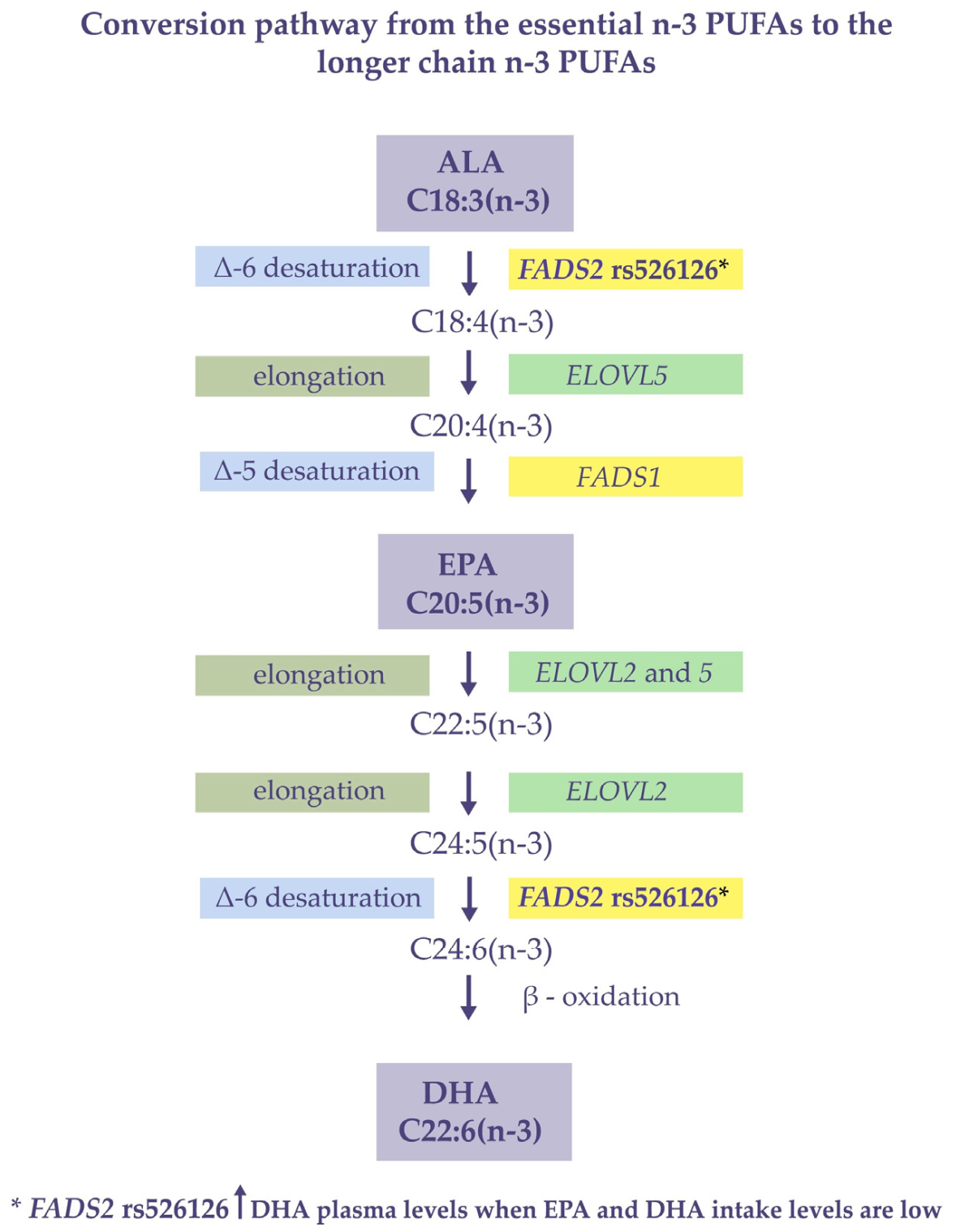
The basic steps of the n-3 LCPUFAs synthesis are shown in Figure 2.
Since only six subjects had the GG genotype overall, out of which only three participants had complete datasets, the data for GG genotype were not included in the statistical analysis for dietary intakes-grouped datasets.
Initially, data grouping according to dietary PUFAs intakes was made by quartiles, results indicating that the differences in plasma DHA levels between FADS rs526126 genotypes were found within ALA intakes Q1 and Q2 and DHA Q3. Having established that plasma DHA was influenced mainly in lower quartiles, grouping for genotype-based analysis was then performed using EPA and DHA medians as cut-off values.
In previous studies, rs526126 was found to be associated with lower D5 desaturase activity, while the other SNPs in the FADS2 gene were found to be associated with higher D6 desaturase activity [23]. However, no effect on D4 desaturase activity was observed. Until now, only in-vitro studies have demonstrated that FADS2 can regulate D4 desaturation to yield DHA [24,25]. Interestingly, neither ARA levels nor the ratio between LA and ARA was associated with FADS2 variants, suggesting that no significant D6 desaturation occurred in this case (data not shown).
In contrast to the previous reports [26], we did not find any association between rs526126 variant and C-reactive protein (CRP) levels, nor between any of the investigated free PUFAs and CRP (data not shown).
Our data indicated that DHA plasma levels were more closely associated with rs526126 than EPA plasma levels. This can be explained by the fact that DHA is the final product of the desaturation cascade and thus is more likely to be influenced by FADS2 activity. Moreover, the plasma levels of free DHA were higher than the ALA and EPA, leading to the conclusion that a significant amount of DHA is synthesized endogenously, in addition to their respective intakes. Differences were even more evident when the EPA and DHA dietary intakes were low, leading to the hypothesis that FADS2 activity is dependent on them and that the presence of rs526126 low-frequency allele (G) may result in increased desaturation activity.
Esterified FAs from plasma and red blood cells were also investigated, but no association with FADS2 variants was found (data not shown), which may indicate that FADS2 action on n-3 can only be observed over short periods of time. In addition, the fact that the blood was collected after overnight fasting may indicate that the FADS2 acts on n-3 PUFA only when the substrate competition is low. Furthermore, this study aligns with the previously reported fact that FA plasma composition can be used as an indicator of recent fat intake [27].
Even if the correlation between free n-3 PUFAs plasma levels and EPA/DHA intakes was low, it still provides reliable data on how the short-term diets impact the plasma lipid profiles and directed us to evaluate the rs526126 activity separately by grouping the subjects according to the EPA and DHA intakes.
Although no other relevant clinical outcome is associated with the presence of the low-frequency allele rs526126, its presence and the higher levels of free DHA in plasma could have long-term benefits. The ability of free DHA to readily cross the blood-brain barrier [28] and be incorporated in the cell membrane phospholipids [29] is considered an essential process for brain development in childhood and adolescence.
Additionally, studies show that despite a higher dietary PUFAs intake, obese children show lower levels of DHA than normal-weight children, possibly due to the metabolic dysfunction in the synthetic pathway of the n-3 PUFAs [30].
An essential role of DHA as a modulator in the process of inflammation response is carried by resolvins, protectins, and maresins by inhibiting the neutrophil migration, enhancing the macrophage phagocytosis of apoptotic neutrophils, and by the pro-inflammatory cytokines and chemokines suppression [31].
Furthermore, DHA is involved in several signaling processes, translating into a better brain function throughout the lifespan [32].
In this study, DHA and EPA were not found to be associated with cholesterol, triglycerides, and HOMA-IR, as previously reported [33]. This finding may be due to the measurement of FFA in plasma instead of the whole blood. Nevertheless, we can conclude that rs526126 and free n-3 PUFAs were not associated with diabetes and insulin resistance in this cohort.
In a previous study [34], we demonstrated that genetic signatures in PEMT and MTHFR genes are associated with fatty acids composition in red blood cells, including DHA. Therefore, this study adds to the knowledge that the blood’s fatty acids composition is influenced by genetic variations at different stages in lipid metabolism. To further highlight the role of genetic influences on the complex lipid metabolism and to add to the existing evidence thereof, this study included the results from a separate analysis of free FAs in plasma.
The main limitation of this study was represented by the absence of a comparison of relevant parameters in normal-weight children vs. overweight children. In addition, the underreporting in the assessment of the dietary intake using 24 h recalls could have brought limitations. Therefore, we cannot conclude that the FADS2 variation’s related changes in n-3 PUFA levels are specific to overweight children. A longitudinal analysis of the status of free PUFAs over a longer period could have provided more insight.

5. Conclusions

Results from this study indicated that the FADS2 gene polymorphism impacts the plasma levels of free n-3 PUFAs, specifically at lower EPA and DHA intakes. We found that the presence of rs526126 low frequency allele in the FADS2 gene was associated with higher plasma levels of free DHA, differences being more evident when the dietary intake of n-3 PUFAs was low. However, further research is needed to confirm the extent of this association and if rs526126 minor allele has a protective role in subjects with low intakes of n-3 PUFAs.

Author Contributions

Conceptualization, A.M., V.S., C.P. and M.D.N.; methodology, A.M., V.S., C.P., N.A. and C.L.S.; software, C.G.Z.; validation, A.M., V.S. and C.P.; formal analysis, D.-A.T. and P.T; investigation, A.M., D.-A.T., P.T., A.I., V.L.D., A.I.I. and N.A.; resources, C.P., I.V. and A.M.; data curation, D.-A.T., P.T. and C.G.Z.; writing—original draft preparation, A.M., V.S. and C.P.; writing—review and editing N.A., M.P. and M.D.N.; visualization, A.M., V.S. and N.A.; supervision, M.P. and M.D.N. All authors have read and agreed to the published version of the manuscript.

Funding

This research was funded by the POC Project “Use of nutrigenomic models for the personalized treatment with medical foods in obese people” (NutriGen) 2016–2019, SMIS: 104852, 91/9 September 2016, ID P_37-684.

Institutional Review Board Statement

The study was conducted according to the guidelines of the Declaration of Helsinki, and approved by the Institutional Ethics Committee of University of Medicine and Pharmacy, Timisoara (code 6/20 June 2016).

Informed Consent Statement

Informed consent was obtained from all subjects involved in the study.

Acknowledgments

This work was performed at The Center of Genomic Medicine, “Victor Babes” University of Medicine and Pharmacy, Timisoara.

Conflicts of Interest

The authors declare no conflict of interest.

Appendix A

Table A1. The frequencies of FADS2 gene single nucleotide polymorphisms studied within 196 subjects.
GeneRs Chr Position GRCh37AA/AB/BBAA%AB%BB%
FADS2rs2526678chr1161623793GG/GA/AA82.117.90.0
rs526126chr1161624885GG/GC/CC3.126.370.6
Legend: Gene name as per international nomenclature HUGO Gene Nomenclature Committee (HGNC); Chr, chromosome position reference genome GRCh37; AA, homozygous for one allele; AB, heterozygous; BB, homozygous for the other allele; FADS2, fatty acid desaturase 2.

References

  1. Chilton, F.H.; Murphy, R.C.; Wilson, B.A.; Sergeant, S.; Ainsworth, H.; Seeds, M.C.; Mathias, R.A. Diet-Gene Interactions and PUFA Metabolism: A Potential Contributor to Health Disparities and Human Diseases. Nutrients 2014, 6, 1993–2022. [Google Scholar] [CrossRef] [PubMed] [Green Version]
  2. Calcaterra, V.; Regalbuto, C.; Porri, D.; Pelizzo, G.; Mazzon, E.; Vinci, F.; Zuccotti, G.; Fabiano, V.; Cena, H. Inflammation in Obesity-Related Complications in Children: The Protective Effect of Diet and Its Potential Role as a Therapeutic Agent. Biomolecules 2020, 10, 1324. [Google Scholar] [CrossRef] [PubMed]
  3. Wang, C.; Chan, J.S.Y.; Ren, L.; Yan, J.H. Obesity Reduces Cognitive and Motor Functions across the Lifespan. Neural Plast. 2016. [Google Scholar] [CrossRef] [PubMed] [Green Version]
  4. Versini, M.; Jeandel, P.Y.; Rosenthal, E.; Shoenfeld, Y. Obesity in autoimmune diseases: Not a passive bystander. Autoimmun Rev. 2014, 13, 981–1000. [Google Scholar] [CrossRef]
  5. Kyrou, I.; Randeva, H.S.; Tsigos, C.; Kaltsas, G.; Weickert, M.O. Clinical Problems Caused by Obesity. In Endotext; MDText.com, Inc.: South Dartmouth, MA, USA, 2000. Available online: https://www.ncbi.nlm.nih.gov/books/NBK278973 (accessed on 10 August 2021).
  6. Baym, C.L.; Khan, N.A.; Monti, J.M.; Raine, L.B.; Drollette, E.S.; Moore, R.D.; Scudder, M.R.; Kramer, A.F.; Hillman, C.H.; Cohen, N.J. Dietary lipids are differentially associated with hippocampal-dependent relational memory in prepubescent children. Am. J. Clin. Nutr. 2014, 99, 1026–1033. [Google Scholar] [CrossRef]
  7. Koletzko, B.; Reischl, E.; Tanjung, C.; Gonzalez-Casanova, I.; Ramakrishnan, U.; Meldrum, S.; Simmer, K.; Heinrich, J.; Demmelmair, H. FADS1 and FADS2 Polymorphisms Modulate Fatty Acid Metabolism and Dietary Impact on Health. Ann. Rev. Nutr. 2019, 39, 21–44. [Google Scholar] [CrossRef]
  8. Figueiredo, P.S.; Inada, A.C.; Marcelino, G.; Lopes Cardozo, C.M.; de Cássia Freitas, K.; de Cássia Avellaneda Guimarães, R.; Pereira de Castro, A.; Aragão do Nascimento, V.; Aiko Hiane, P. Fatty Acids Consumption: The Role Metabolic Aspects Involved in Obesity and Its Associated Disorders. Nutrients 2017, 9, 1158. [Google Scholar] [CrossRef] [Green Version]
  9. O’Sullivan, T.A.; Bremner, A.P.; Beilin, L.J.; Ambrosini, G.L.; Mori, T.A.; Huang, R.C.; Oddy, W.H. Polyunsaturated fatty acid intake and blood pressure in adolescents. J. Hum. Hypertens. 2012, 26, 178–187. [Google Scholar] [CrossRef] [Green Version]
  10. Anderson, B.M.; Ma, D.W. Are all n-3 polyunsaturated fatty acids created equal? Lipid Health Dis. 2009, 8, 33. [Google Scholar] [CrossRef] [Green Version]
  11. Calder, P.; Grimble, R. Polyunsaturated fatty acids, inflammation and immunity. Eur. J. Clin. Nut. 2002, 56 (Suppl. 3), S14–S19. [Google Scholar] [CrossRef] [Green Version]
  12. Simopoulos, A. An Increase in the Omega-6/Omega-3 Fatty Acid Ratio Increases the Risk for Obesity. Nutrients 2016, 8, 128. [Google Scholar] [CrossRef] [Green Version]
  13. Kawashima, H. Intake of arachidonic acid-containing lipids in adult humans: Dietary surveys and clinical trials. Lipids Health Dis. 2019, 18, 101. [Google Scholar] [CrossRef] [Green Version]
  14. Lee, J.M.; Lee, H.; Kang, S.; Park, W.J. Fatty Acid Desaturases, Polyunsaturated Fatty Acid Regulation, and Bio-technological Advances. Nutrients 2016, 8, 23. [Google Scholar] [CrossRef] [Green Version]
  15. Mathias, R.A.; Pani, V.; Chilton, F.H. Genetic Variants in the FADS Gene: Implications for Dietary Recommendations for Fatty Acid Intake. Curr. Nutr. Rep. 2014, 3, 139–148. [Google Scholar] [CrossRef] [Green Version]
  16. Xie, L.; Innis, S.M. Genetic Variants of the FADS1 FADS2 Gene Cluster Are Associated with Altered (n-6) and (n-3) Essential Fatty Acids in Plasma and Erythrocyte Phospholipids in Women during Pregnancy and in Breast Milk during Lactation. J. Nutr. 2008, 138, 2222–2228. [Google Scholar] [CrossRef] [Green Version]
  17. Huang, M.C.; Chang, W.T.; Chang, H.Y.; Chung, H.F.; Chen, F.P.; Huang, Y.F.; Hsu, C.C.; Hwang, S.J. FADS Gene Polymorphisms, Fatty Acid Desaturase Activities, and HDL-C in Type 2 Diabetes. Int. J. Environ. Res. Public Health 2017, 14, 572. [Google Scholar] [CrossRef] [Green Version]
  18. Khodarahmi, M.; Nikniaz, L.; Abbasalizad Farhangi, M. The Interaction Between Fatty Acid Desaturase-2 (FADS2) rs174583 Genetic Variant and Dietary Quality Indices (DASH and MDS) Constructs Different Metabolic Phenotypes Among Obese Individuals. Front Nutr. 2021, 8, 669207. [Google Scholar] [CrossRef]
  19. Nakayama, K.; Bayasgalan, T.; Tazoe, F.; Yanagisawa, Y.; Gotoh, T.; Yamanaka, K.; Ogawa, A.; Munkhtulga, L.; Chimedregze, U.; Kagawa, Y.; et al. A single nucleotide polymorphism in the FADS1/FADS2 gene is associated with plasma lipid profiles in two genetically similar Asian ethnic groups with distinctive differences in lifestyle. Hum. Genet. 2010, 127, 685–690. [Google Scholar] [CrossRef]
  20. Khamlaoui, W.; Mehri, S.; Hammami, S.; Hammouda, S.; Chraeif, I.; Elosua, R.; Hammami, M. Association Between Genetic Variants in FADS1-FADS2 and ELOVL2 and Obesity, Lipid Traits, and Fatty Acids in Tunisian Population. Clin. Appl. Thromb. Hemost. 2020, 26. [Google Scholar] [CrossRef]
  21. Maguolo, A.; Zusi, C.; Giontella, A.; Miraglia Del Giudice, E.; Tagetti, A.; Fava, C.; Morandi, A.; Maffeis, C. Influence of genetic variants in FADS2 and ELOVL2 genes on BMI and PUFAs homeostasis in children and adolescents with obesity. Int. J. Obes. 2021, 45, 56–65. [Google Scholar] [CrossRef]
  22. Serafim, V.; Tiugan, D.A.; Andreescu, N.; Mihailescu, A.; Paul, C.; Velea, I.; Puiu, M.; Niculescu, M.D. Development and Validation of a LC–MS/MS-Based Assay for Quantification of Free and Total Omega 3 and 6 Fatty Acids from Human Plasma. Molecules 2019, 24, 360. [Google Scholar] [CrossRef] [Green Version]
  23. Bokor, S.; Dumont, J.; Spinneker, A.; Gonzalez-Gross, M.; Nova, E.; Widhalm, K.; Moschonis, G.; Stehle, P.; Amouyel, P.; De Henauw, S.; et al. Single nucleotide polymorphisms in the FADS gene cluster are associated with delta-5 and delta-6 desaturase activities estimated by serum fatty acid ratios. J. Lipid. Res. 2010, 51, 2325–2333. [Google Scholar] [CrossRef] [Green Version]
  24. Park, H.G.; Park, W.J.; Kothapalli, K.S.D.; Brenna, J.T. The fatty acid desaturase 2 (FADS2) gene product catalyzes Δ4 desaturation to yield n-3 docosahexaenoic acid and n-6 docosapentaenoic acid in human cells. FASEB J. 2015, 29, 3911–3919. [Google Scholar] [CrossRef] [Green Version]
  25. Park, H.G.; Kothapalli, K.S.D.; Park, W.J.; DeAllie, C.; Liu, L.; Liang, A.; Lawrence, P.; Brenna, J.T. Palmitic acid (16:0) competes with omega-6 linoleic and omega-3 α-linolenic acids for FADS2 mediated Δ6-desaturation. Biochim. Biophys. Acta—Mol. Cell Biol. Lipids 2016, 1861, 91–97. [Google Scholar] [CrossRef] [Green Version]
  26. Roke, K.; Ralston, J.C.; Abdelmagid, S.; Nielsen, D.E.; Badawi, A.; El-Sohemy, A.; Ma, D.V.L.; Mutch, D.M. Variation in the FADS1/2 gene cluster alters plasma n-6 PUFA and is weakly associated with hsCRP levels in healthy young adults. Prostaglandins Leukot. Essent. Fat. Acids 2013, 89, 257–263. [Google Scholar] [CrossRef]
  27. Risé, P.; Eligini, S.; Ghezzi, S.; Colli, S.; Galli, C. Fatty acid composition of plasma, blood cells and whole blood: Relevance for the assessment of the fatty acid status in humans. Prostaglandins Leukot. Essent. Fat. Acids 2007, 76, 363–369. [Google Scholar] [CrossRef]
  28. Ouellet, M.; Emond, V.; Chen, C.T.; Julien, C.; Bourasset, F.; Oddo, S.; LaFerla, F.; Bazinet, R.P.; Calon, F. Diffusion of docosahexaenoic and eicosapentaenoic acids through the blood–brain barrier: An in situ cerebral perfusion study. Neurochem. Int. 2009, 55, 476–482. [Google Scholar] [CrossRef]
  29. Chen, C.T.; Kitson, A.P.; Hopperton, K.E.; Domenichiello, A.F.; Trépanier, M.O.; Lin, L.E.; Ermini, L.; Post, M.; Thies, F.; Bazinet, R.P. Plasma non-esterified docosahexaenoic acid is the major pool supplying the brain. Sci. Rep. 2015, 5, 15791. [Google Scholar] [CrossRef] [Green Version]
  30. Lassandro, C.; Banderali, G.; Radaelli, G.; Borghi, E.; Moretti, F.; Verduci, E. Docosahexaenoic Acid Levels in Blood and Metabolic Syndrome in Obese Children: Is There a Link? Int. J. Mol. Sci. 2015, 16, 19989–20000. [Google Scholar] [CrossRef] [Green Version]
  31. Duvall, M.G.; Levy, B.D. DHA- and EPA-derived resolvins, protectins, and maresins in airway inflammation. Eur. J. Pharmacol. 2016, 785, 144–155. [Google Scholar] [CrossRef] [Green Version]
  32. Lewis, M.D.; Bailes, J. Neuroprotection for the Warrior: Dietary Supplementation With Omega-3 Fatty Acids. Mil. Med. 2011, 176, 1120–1127. [Google Scholar] [CrossRef] [PubMed]
  33. Damsgaard, C.T.; Eidner, M.B.; Stark, K.D.; Hjorth, M.F.; Sjödin, A.; Andersen, M.R.; Andersen, R.; Tetens, I.; Astrup, A.; Michaelsen, K.F.; et al. Eicosapentaenoic acid and docosahexaenoic acid in whole blood are differentially and sex-specifically associated with cardiometabolic risk markers in 8-11-year-old danish children. PLoS ONE. 2014, 9, e109368. [Google Scholar] [CrossRef] [PubMed] [Green Version]
  34. Serafim, V.; Chirita-Emandi, A.; Andreescu, N.; Tiugan, D.A.; Tutac, P.; Paul, C.; Velea, I.; Mihailescu, A.; Șerban, C.L.; Zimbru, C.G.; et al. Single nucleotide polymorphisms in PEMT and MTHFR genes are associated with omega 3 and 6 fatty acid levels in the red blood cells of children with obesity. Nutrients 2019, 11, 2600. [Google Scholar] [CrossRef] [PubMed] [Green Version]
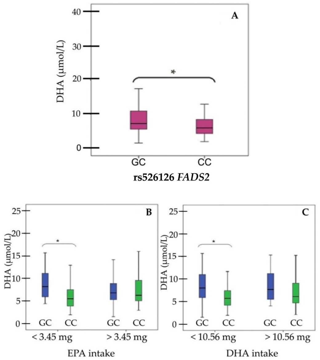
Figure 1. Distribution of plasma-free DHA levels grouped by FADS2 rs526126 for the entire group (A), grouped according to EPA (B) and DHA (C) dietary intakes. Mann–Whitney test was used to assess statistical significance between groups, the brackets marked with * indicating the two groups for which these differences were identified. Horizontal lines within each boxplot indicate minimum, first quartile (Q1), median, third quartile (Q3), and maximum. Legend: DHA, docosahexaenoic acid; EPA, eicosapentaenoic acid; FADS2, fatty acid desaturase 2 gene.
Applsci 11 09845 g001
Figure 2. ALA, alpha–linolenic acid; EPA, eicosapentaenoic acid; DHA, docosahexaenoic acid; FADS2, fatty acid desaturase 2 gene; FADS1, fatty acid desaturase 1 gene; ELOVL2, fatty acid elongase 2; ELOVL5, fatty acid elongase 5.
Applsci 11 09845 g002
Table 1. Descriptive statistics for anthropometric data, biochemical analysis, measurements of free PUFA in plasma, and dietary intakes.
VariablesAll n = 196Females
n = 101
Males
n = 95
p Value * (Males vs. Females)
MedianIQRMedianIQRMedianIQR
Anthropometric data
Age (years)12512.005.012.004.750.787
zBMI3.131.202.831.213.441.370.001 *
Biochemical analysis
TC (mg/dL)174.0054.00166.0052.00182.5055.500.109
TG (mg/dL)129.0086.50125.0088.00133.0099.750.237
HDL (mg/dL)46.0018.0043.0019.0048.006.750.085
AST (U/L)29.0014.0029.0014.0029.5015.500.127
ALT (U/L)32.0015.0031.0016.0033.5014.750.167
CRP (mg/dL)4.707.904.808.704.757.720.315
Homocysteine (µmoL/L)14.788.7814.168.0815.339.280.398
Measurements of free PUFA in plasma
ALA (µmol/L)3.724.273.243.954.094.540.145
EPA (µmol/L)0.230.140.20.130.260.14 0.008 *
DHA (µmol/L)6.264.825.884.016.766.090.217
LA (µmol/L)137.2122.12126.39105.35159.08124.620.215
ARA (µmol/L)7.154.446.84.057.784.82 0.025 *
Selected daily nutrient intakes evaluated in 24 h dietary recalls
Kilocalories1201.59494.721201.49440.111205.77580.320.889
Protein (g)66.6022.1166.6323.0066.4823.800.832
Lipids (g)44.5925.0844.1722.9945.6728.420.781
Carbohydrates (g)135.9358.15138.5251.27129.9361.290.266
Water (g)21.96829.492146.43749.582280.80912.780.207
Cholesterol (mg)260.54157.94255.68129.83274.79158.470.853
ALA (mg)77.16217.2157.29196.8587272.440.280
DHA (mg)10.632410.1619.4710.9524.740.547
EPA (mg)3.45.993.095.814.006.200.971
LA (mg)943.082306.07953.402072.57943.082334.760.527
Legend: PUFA, polyunsaturated fatty acid; IQR, interquartile range (IQR = Q3–Q1); zBMI, standardized body mass index (BMI) to account for age and gender; TC, total cholesterol; TG, triglycerides, HDL; high density cholesterol; g, grams; mg, milligrams; µmol/L, micromoles/liter; AST, aspartate aminotransferase; ALT, alanine aminotransferase; CRP, C reactive protein; ALA, alpha-linolenic acid; ARA, arachidonic acid; DHA, docosahexaenoic acid; EPA, eicosapentaenoic acid; LA, linoleic acid. * p value < 0.05.
Table 2. Spearman’s rho. results. Plasma fatty acids were quantified in their free form using LC-MS/MS. Food intake was evaluated using five-pass 24 h dietary recalls and converted into micronutrients based on USDA) Food and Nutrient Database.
EPA
Plasma
DHA
Plasma
ALA IntakeEPA IntakeDHA IntakeAge
ALA plasma rho0.519 **0.523 **0.0930.159 *0.069−0.237 **
p-value<0.001<0.0010.2270.0370.370.001
EPA plasmarho 0.682 **0.1080.130.056−0.217 **
p-value <0.0010.1580.0890.4690.002
DHA plasmarho −0.0030.182 *0.159 *−0.195 **
p-value 0.9730.0170.0380.007
ALA intakerho 0.195 **0.117−0.074
p-value 0.010.1260.337
EPA intakerho 0.769 **−0.122
p-value <0.0010.109
* Correlation is significant at ≤0.05 (2-tailed), ** Correlation is significant at ≤0.01 level (2-tailed). Legend: ALA, alpha-linolenic acid; DHA, docosahexaenoic acid; EPA, eicosapentaenoic acid, rho–Spearman coefficient.
Publisher’s Note: MDPI stays neutral with regard to jurisdictional claims in published maps and institutional affiliations.